(NCALD) as a protective modifier in mouse models of Spinal Muscular
Atrophy (SMA)
Inaugural-Dissertation zur
Erlangung des Doktorgrades
der Mathematisch-Naturwissenschaftlichen Fakultät der Universität zu Köln
vorgelegt von Anna Kaczmarek aus Sucha Beskidzka, Polen
Köln
2016
Cologne (CMMC) of the University of Cologne from December 2010 to October 2016.
Berichterstatter: Prof. Dr. rer. nat. Brunhilde Wirth Prof. Dr. rer. nat. Aleksandra Trifunovic
Tag der mündlichen Prüfung: 28.11.2016
who is actually in the arena, whose face is marred by dust and sweat and blood; who strives valiantly; who errs, who comes short again and again, because there is no effort without error and shortcoming; but who does actually strive to do the deeds; who knows great enthusiasms, the great devotions; who spends himself in a worthy cause; who at the best knows in the end the triumph of high achievement, and who at the worst, if he fails, at least fails while daring greatly, so that his place shall never be with those cold and timid souls who neither know victory nor defeat.
Theodore Roosevelt, “Citizenship In A Republic"
Sorbonne, Paris, France on 23
rdApril 1910
Mojej kochanej Rodzinie:
Mamulce, Tatulkowi,
Madziuni i Bartkowi
Z podziękowaniem za nieustanne ogromne wsparcie i wiarę
Table of content
List of figures ...IV List of tables...VI Abbreviations ...VII Summary...IX Zusammenfassung...XI
1 Introduction ... 1
1.1 SMA: clinical pathology, subtypes and genetics ... 1
1.2 SMN protein ... 5
1.3 Animal models of SMA ... 6
1.4 SMA therapy ... 9
1.4.1 SMN replacement therapy ... 9
1.4.2 Compounds increasing SMN by enhancing protein stability or SMN2 transcription... 10
1.4.3 Correcting SMN2 splicing using antisense oligonucleotides (ASOs) ... 11
1.4.4 SMN-independent therapeutics: neuroprotective agents... 12
1.5 Modifier genes in SMA ... 14
1.5.1 Identification of SMA modifiers in model organisms and biochemical screens ... 14
1.5.2 Identification of SMA modifiers in asymptomatic SMN1 -deleted relatives of SMA patients ... 15
1.5.3 Neurocalcin delta – a novel potential modifier for SMA in humans... 16
2 Preliminary results... 18
2.1 NCALD is downregulated in asymptomatic SMN1 -deleted individuals... 18
2.2 NCALD reduction rescues SMA phenotype in vitro and in vivo across species 19 3 Aim of the study ... 21
4 Materials and methods... 23
4.1 Materials ... 23
4.1.1 Laboratory equipment... 23
4.1.2 Mouse work equipment... 24
4.1.3 Chemicals... 24
4.1.4 Reagents ... 25
4.1.5 Kits ... 26
4.1.6 Enzymes... 27
4.1.7 Antibodies ...27
4.1.8 Secondary antibodies ...27
4.1.9 Solutions and media ...27
4.1.10 Primers and oligonucleotides...31
4.1.11 Plasmids...33
4.1.12 Software packages and internet databases ...34
4.2 Methods...34
4.2.1 Working with nucleic acids ...34
4.2.2 Working with bacteria ...41
4.2.3 Working with proteins ...42
4.2.4 Working with cells...44
4.2.5 Working with mice and mouse tissues...45
4.2.6 Microscopic image acquisition and analysis ...56
4.2.7 Statistical analysis ...57
5 Results...58
5.1 SMA mouse model with NCALD reduction – a transgene shRNA approach...58
5.1.1 Selection of the efficient shRNA sequences against Ncald ...60
5.1.2 Stable integration of the shRNA cassette in ES cells ...63
5.2 Severe SMA mouse model with NCALD reduction – a transgene knock-out approach ...66
5.2.1 Analysis of NCALD expression in wildtype, Ncald
ko/wtand Ncald
ko/komice...66
5.2.2 Crossing the Ncald
koallele in the severe SMA mouse model...68
5.2.3 Phenotypic in vivo analysis of SMA- Ncald
ko/wtmice: survival and weight...69
5.2.4 Phenotypic in vivo analysis of SMA- Ncald
ko/wtmice: motoric skills ...71
5.2.5 NCALD reduction in motor neurons from SMA mice restored axonal outgrowth in vitro ...73
5.2.6 Neuromuscular junctions of SMA- Ncald
ko/wtmice show larger AChR clusters ...75
5.2.7 Number of glutamatergic inputs on spinal cord motor neurons is increased in SMA- Ncald
ko/wtmice...77
5.2.8 Muscle fiber size is not changed in the EDL of SMA- Ncald
ko/wtmice ...80
5.2.9 SMA- Ncald
ko/wtmice suffer from impairment of peripheral organs outside of CNS ...81
5.3 Intermediate SMA mouse model with NCALD reduction – an SMN-ASO approach
...83
5.3.1 SMN-ASO injection increased SMN levels in the liver and spinal cord in
SMA+ASO mice ... 83
5.3.2 Phenotypic analysis of SMA- Ncald
ko/wt+ASO mice: survival and weight ... 85
5.3.3 Phenotypic analysis of SMA- Ncald
ko/wt+ASO mice: motoric tests ... 86
5.3.4 Neuromuscular junctions of SMA- Ncald
ko/wt+ASO mice are more mature and have a larger area of AChR clusters ... 87
5.3.5 The number of glutamatergic inputs on MN soma and the MN cell size are increased in SMA- Ncald
ko/wt+ASO mice ... 89
5.3.6 Muscle fiber size is increased in the EDL muscle of PND21 SMA- Ncald
ko/wt+ASO mice... 91
5.3.7 The impairment of the gastrointestinal tract is not rescued by reduced NCALD levels ... 93
5.4 SMA mouse model with NCALD reduction – an Ncald -ASO approach ... 94
6 Discussion... 100
6.1 Different strategies to reduce NCALD in vivo in SMA mice... 100
6.2 Effect of NCALD reduction on the motor neurons of SMA animals ... 102
6.3 Effect of NCALD reduction on the NMJs of SMA animals... 104
6.4 Why did NCALD reduction have no effect on the lifespan of SMA mice? ... 106
6.5 Effect of NCALD reduction on HET animals ... 109
6.6 Clinical relevance of neuronal calcium sensors ... 110
6.7 Two independently identified SMA modifiers: NCALD and PLS3 act on endocytosis as a common pathway which is impaired in SMA ... 112
6.8 Future outlook ... 114
7 References... 116
8 Appendix... 135
9 Publications, talks, poster presentations and scholarships... 176
Acknowledgements...IX
Erklärung ...X
Lebenslauf ...XI
List of figures
Fig. 1: The schematic overview of SMA pathology. ... 2
Fig. 2: The genetic landscape of SMA. ... 4
Fig. 3. The mechanism of action of SMN-ASO. ... 12
Fig. 4: SMA therapy strategies with their specific target site in an exemplary motor neuron. ... 13
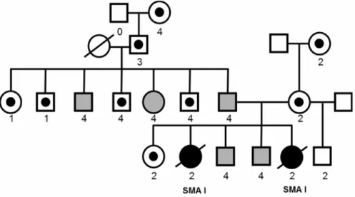
Fig. 5: Pedigree of the family with NCALD reduction in asymptomatic individuals. ... 18
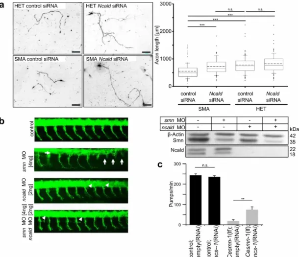
Fig. 6: NCALD reduction ameliorated neuronal outgrowth defects mediated by SMN deficiency. ... 20
Fig. 7: Annealing of shRNA oligonucleotides ... 37
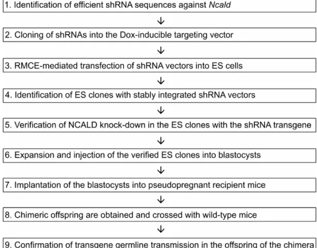
Fig. 8: The necessary steps of a mouse generation strategy using an inducible shRNA targeting vector. ... 46
Fig. 9: The breeding scheme to obtain SMA and HET animals with reduced NCALD levels ... 51
Fig. 10: The tube test positions corresponding to the respective values of the hind limb score ... 53
Fig. 11: The shRNA mouse generation strategy ... 59
Fig. 12: Induction of shRNA expression upon Doxycycline induction. ... 60
Fig. 13: The longest transcript (3733 bp) of mouse Ncald (NM_134094) ... 61
Fig. 14: Analysis of Ncald knock-down on protein and mRNA in NSC34 cells transfected with shRNAs ... 61
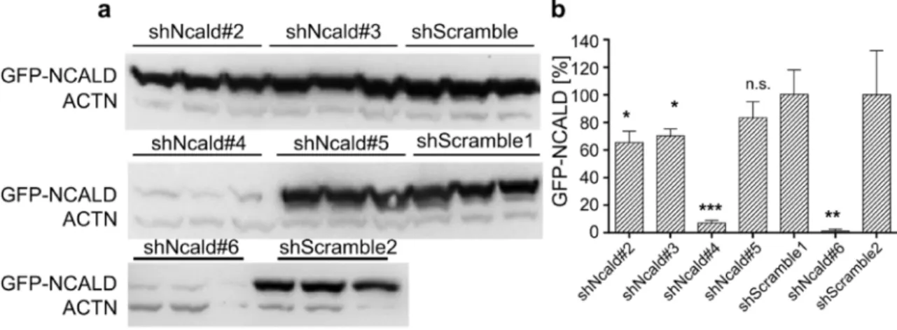
Fig. 15: Identification of efficient shRNA sequences in an Ncald-GFP overexpression set-up ... 62
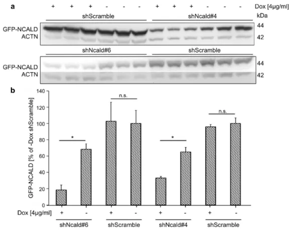
Fig. 16: Efficient knock-down of GFP-NCALD upon Dox-mediated shRNA expression ... 63
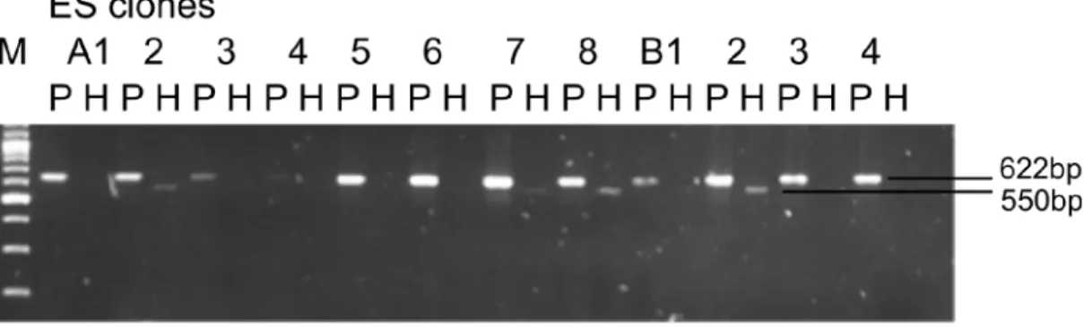
Fig. 17: Validation of the successful recombination in individual ES clones. ... 64
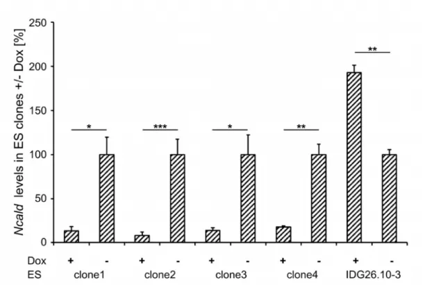
Fig. 18: Quantification of Ncald in ES clones upon Doxycycline induction ... 65
Fig. 19: Analysis of NCALD expression in wildtype mouse tissue on PND10 ... 67
Fig. 20: Analysis of NCALD expression in mutant mouse tissue on PND10 ... 68
Fig. 21. Survival and weight studies of SMA-Ncald
ko/wtmice ... 71
Fig. 22. Evaluation of motoric skills of SMA-Ncald
ko/wtmice ... 72
Fig. 23: Motor neurons from SMA-Ncald
ko/wtmice show longer axons and increased branching than SMA mice ... 74
Fig. 24: Analysis of NMJ size in the TVA muscle of PND10 mice ... 76
Fig. 25: Analysis of NMJ size in the TVA muscle of PND5 and PND13 mice ... 77
Fig. 26: Evaluation of glutamatergic inputs on spinal motor neuron soma in SMA-
Ncald
ko/wtmice ... 79
Fig. 27: Analysis of the muscle fiber size in the EDL of PND10 mice ... 81
Fig. 28: The intestinal impairment in the severe SMA mouse model ... 82
Fig. 29: SMN-ASO mode of action ... 83
Fig. 30: SMN-ASO injection increased SMN levels in liver and spinal cord ... 84
Fig. 31: Survival and weight studies of SMA+ASO and SMA-Ncald
ko/wt+ASO mice 85 Fig. 32: Evaluation of motoric skills of SMA-Ncald
ko/wt+ASO mice ... 87
Fig. 33: NMJ size and maturation are improved in SMA-Ncald
ko/wt+ASO animals .. 89
Fig. 34: Number of VGlut1+ inputs and motor neuron size are increased in SMA- Ncald
ko/wt+ASO animals ... 91
Fig. 35: Analysis of muscle fiber size in the EDL muscle of PND21 mice ... 92
Fig. 36: Intestine impairment is still visible in SMA+ASO mice. ... 93
Fig. 37: A screen of different ASO sequences against Ncald performed by Ionis Pharmaceuticals ... 95
Fig. 38: NCALD knock-down by i.c.v. Ncald-ASO injection in wildtype and SMA mice ... 96
Fig. 39: Efficient NCALD knock-down by combined i.c.v. and s.c. injection of ASO2 against Ncald in SMA mice ... 97
Fig. 40: Increased mortality following the injection of Ncald-ASOs ... 99
Fig. 41: A model of restored neuromuscular homeostasis under concomitant depletion of SMN and NCALD ... 110
Fig. 42: A map of known interaction partners of NCS proteins ... 113
Fig. 43: Proposed mode of NCALD acting on the endocytosis at the synaptic
membrane ... 114
List of tables
Table 1: Primers for cloning Ncald / qRT-PCR / genotyping ... 32
Table 2: Oligonucleotides for cloning shRNA into the pEx-H1-tetO-CAG-teR vector ... 32
Table 3: Antisense oligonucleotides for in vivo injection ... 33
Table 4: List of used and produced plasmids ... 33
Table 5: A standard 20 µl PCR composition ... 36
Table 6: A standard thermocycler PCR program ... 36
Table 7: The composition and conditions of an oligonucleotide annealing reaction ... 38
Table 8: The composition of a standard digestion using restriction enzyme ... 38
Table 9: The composition of a typical ligation reaction (10 µl) ... 39
Table 10: The composition of a typical reverse transcription reaction (10µl) ... 40
Table 11: The composition of a typical qRT-PCR (10µl) ... 41
Table 12: Primary and secondary antibodies used for protein detection ... 44
Table 13: The summary of the characteristics of SMA-Ncald
ko/wtand HET-Ncald
ko/wtanalyzed in the severe and intermediate SMA mouse model. ... 94
Abbreviations
A adenine
AChR acetylcholine receptor ALS amyloid lateral sclerosis
APS ammonium persulfate
ASO antisense oligonucleotide BBB blood-brain barrier
bp base pairs
BSA bovine serum albumin
BTX bungarotoxin
C cytosine
CNS central nervous system
cDNA coding DNA
DMEM Dulbecco’s modified Eagle medium DMSO dimethyl sulfoxide
DNA deoxyribonucleic acid
Dox Doxycycline
E embryonic day
EDL Extensor digitorum longus EDTA ethylene diamine tetraacetic acid e.g. exempli gratia
EGTA ethylene glycol tetraacetic acid ESC embryonic stem cells
et al. et alii
FCS fetal calf serum
FDA Food & Drug Administration
FL full length
fwd forward
g gravitational force
G guanine
GFP green fluorescent protein
h hours
HDAC histone deacetylase i.c.v. intracerebroventricular
kb kilobases
kDa kilodalton
ko knock-out
L liter
m mili
µ micro
M molar
min minutes
MN motor neuron
MOE methoxyethoxy
mRNA messenger RNA
n number
NCS neuronal calcium sensor
NF neurofilament
NMJ neuromuscular junction
NT neurotransmitter
n.s. not significant
OMIM Online Mendelian Inheritance in Man
o.n. overnight
P probability
PAA polyacrylamide
PBS phosphate buffered saline PCR polymerase chain reaction
PD Parkinson’s disease
PFA paraformaldehyde
pH power of hydrogen
pmol picomol
PND postnatal day
PS phosphorothioate
RNA ribonucleic acid
rev reverse
s.c. subcutaneous
scAAV self-complementary adeno-associated virus
SD standard deviation
SDS sodium dodecyl sulfate
sec seconds
SEM standard error of means shRNA short hairpin RNA siRNA small interfering RNA SMA spinal muscular atrophy
snRNP small nuclear ribonucleoprotein
T tyrosine
TVA Transversus abdominis
TEMED N,N,N’,N’-tetramethylethylenediamine UTR untranslated region
wt wildtype
Summary
Spinal muscular atrophy (SMA) is a common and devastating genetic disease characterized by degeneration of spinal alpha motor neurons and muscle atrophy. SMA is caused by homozygous deletions or rarely other mutations of the SMN1 gene, resulting in a functional loss of the Survival of Motor Neuron (SMN) protein. SMA severity is determined by a nearly identical copy gene, SMN2 , which encodes an identical SMN protein but produces only ~10% correctly spliced transcripts. Rarely, individuals with SMN1 deletion are fully asymptomatic, suggesting a protection by other modifying genes.
Identification of these modifiers would allow us to better understand the cellular pathways affected by SMN deficiency and to develop new therapies.
Here, we report a novel modifier of SMA, Neurocalcin delta ( NCALD ), that showed a reduced expression in SMN1 -deleted asymptomatic individuals as compared to other affected family members or unrelated SMA patients. NCALD is a neuronal calcium sensor mainly expressed in CNS. Further preliminary studies in vitro and in non-mammalian SMA animal models ( C. elegans , zebrafish) have shown that NCALD knock-down ameliorates SMA-related symptoms. Therefore, we proceeded to study the protective effect of NCALD suppression in SMA mouse models. First, we followed a strategy of generating an inducible Ncald knock-down mouse. We identified efficient shRNA sequences that suppressed NCALD expression and cloned them into the targeting vector, which was integrated into mouse embryonic stem cells by electroporation. This approach was discontinued when an Ncald
ko/komouse line became available at the Jackson Laboratory.
Next, we crossed the Ncald
koallele heterozygously with a severely affected SMA mouse model and performed extensive phenotypic analysis. Reduced NCALD expression improved many characteristics related to SMN deficiency, such as axonal length of cultured motor neurons, size of acetylcholine receptor (AChR) clusters at the neuromuscular junction and spinal motor neuron circuitry as assessed by the number of glutamatergic inputs on motor neuron soma. However, due to very low SMN levels, other internal organs were impaired, which resulted in early death of the SMA mice irrespective of their NCALD levels. Therefore, we hypothesized that increasing peripheral SMN levels is needed for the modifier to fully exert its function. Indeed, in SMA- Ncald
ko/wtmice injected with a suboptimal dose of SMN-ASO (antisense oligonucleotides), all characteristics listed above were reproducibly improved; additionally, also the muscle fiber size was increased.
Unexpectedly, also in this intermediate model the survival of the SMA- Ncald
ko/wt+ASO mice
was not rescued, presumably because NCALD is mainly expressed in neurons and unable
to counteract SMN deficiency in other inner organs. Currently, we are performing further
studies of NCALD suppression in an even milder SMA model, which shows only a motoric
impairment but without a shortened lifespan; this model resembles better the phenotype
of asymptomatic individuals, who all carry four SMN2 copies and would hypothetically develop a milder SMA type without compromised survival. Indeed, first results show that mild SMA mice have an improved motoric behavior upon reduced NCALD levels.
Finally, we anticipated that NCALD suppression would be eligible for a combinatorial therapy using a mix of ASOs to increase SMN and simultaneously downregulate NCALD.
In collaboration with Ionis Pharmaceuticals (USA), a walk along the Ncald gene was performed to identify the most efficient Ncald -ASOs. In our laboratory two ASOs were subsequently tested by intracerebroventricular injections in neonatal mice. While we achieved a satisfying NCALD knock-down, we encountered some toxicity issues;
therefore, the therapeutic potential of NCALD suppression requires further examination
considering its biosafety and clinical efficiency.
Zusammenfassung
Spinale Muskelatrophie (SMA) ist eine häufige verheerende genetische Erkrankung, die durch die Degenerierung von Alpha-Motoneuronen im Rückenmark sowie die Atrophie von Muskeln gekennzeichnet ist. Die Ursache von SMA sind homozygote Deletionen oder in seltenen Fällen andere Mutationen vom SMN1 -Gen, die zu einem Funktionsverlust des Survival of Motor Neuron (SMN) Proteins führen. Das Schweregrad der Erkrankung wird durch ein beinahe identisches Kopiegen bestimmt, SMN2 , jedoch werden nur etwa 10%
der SMN2 Transkripte korrekt gespleißt, die dann ein mit SMN identisches Protein kodieren. Selten zeigen Menschen mit einer SMN1 -Deletion keine SMA Symptome, was einen Schutzmechanismus durch andere modifizierenden Gene vermuten lässt. Die Identifizierung solcher Modifiergene würde ein besseres Verständnis der zellulären Prozesse ermöglichen, die durch den SMN-Mangel beeinträchtigt sind, sowie die Entwicklung neuer Therapieansätze fördern.
Wir berichten hier von einem neuen SMA-Modifiergen, Neurocalcin delta ( NCALD ), das
eine reduzierte Expression in SMN1 -deletierten asymptomatischen Probanden im
Vergleich zu den SMA-kranken Familienmitgliedern sowie nicht verwandten SMA
Patienten zeigte. Weitere vorläufige Studien in zellulären Systemen und einfacheren
Tiermodellen ( C. elegans , Zebrafisch) haben gezeigt, dass die Reduktion von NCALD die
Symptome von SMA mildert. Daher haben wir weitere Studien zum protektiven Effekt der
NCALD-Reduktion in SMA Mausmodellen unternommen. Als Erstes verfolgten wir eine
Strategie zur Generierung einer induzierbaren Ncald -Knockdown-Mauslinie. Dafür haben
wir effiziente shRNA-Sequenzen gegen Ncald identiziert und in einen Zielvektor kloniert,
der in embryonale Mausstammzellen mittels Elektroporation integriert wurde. Da jedoch
eine Ncald
ko/koMauslinie bei dem Jackson Laboratory verfügbar wurde, haben wir diese
Strategie eingestellt. Als Nächstes haben wir dann das Ncald
ko-Allel heterozygot mit dem
etablierten schwer betroffenen SMA Mausmodell gekreuzt und eine umfangreiche
Phänotypanalyse durchgeführt. Die Herunterregulierung von NCALD hat mehrere
Merkmale des SMN-Mangels verbessert, z. B. die Axonlänge von kultivierten
Motoneuronen, die Größe der Acetylcholinrezeptoren (AChR) Cluster an der
neuromuskulären Endplatte sowie die spinalen Motoneuronennetzwerke, evaluiert durch
die Zahl der glutamatergischen Inputs auf die Zellkörper der Motoneurone. Die geringe
SMN-Menge führte jedoch zu Defekten in verschiedenen inneren Organe der SMA-
Mäuse, wodurch unabhängig von der NCALD-Expression das frühe Sterben der Mäuse
verursacht wurde. Wir vermuteten daher, dass die Erhöhung der allgemeinen SMN-Menge
nötig wäre, um die vollständige Wirkung des Modifiers zu erlauben. Tatsächlich wurden
alle oben aufgeführten Eigenschaften beständig verbessert, wenn die SMA- Ncald
ko/wtMäuse mit einer suboptimalen Dosis vom SMN-ASO (Antisense Oligonukleotid) injiziert wurden; zusätzlich ist auch die Größe der Muskelfaser gewachsen. Überraschend hat die Verminderung von NCALD auch in dem intermediären SMA-Mausmodell keine Verlängerung der Lebenserwartung der SMA- Ncald
ko/wt+ASO Mäuse erzielt. Wir vermuten, dass die durch den SMN-Mangel bedingten Defekte in anderen inneren Organen durch das überwiegend in Neuronen exprimierte NCALD Protein korrigiert werden. Es werden zur Zeit Studien von NCALD-Reduktion in einem noch milderen SMA- Modell durchgeführt, das ausschließlich motorische Defekte, aber keine Beeinträchtigung der Lebenserwartung zeigt. Möglicherweise spiegelt dieses Modell besser den Phänotyp der asymptomatischen Probanden wieder, die alle das SMN2 -Gen in vier Kopien tragen.
Hypothetisch würde solch ein genetisches Profil im Menschen zu einem milden SMA Typ ohne Einschränkung der Lebenserwartung führen. Die ersten Ergebnisse zeigen, dass die NCALD-Reduktion das motorische Verhalten der mild betroffenen SMA-Tiere verbessert.
Letztlich vermuteten wir, dass die Herunterregulierung von NCALD für eine
kombinatorische Therapie geeignet wäre, in der eine Kombination von ASOs einerseits
die Menge von SMN erhöhen und andererseits die von NCALD vermindern würde. In einer
Kollaboration mit Ionis Pharmaceuticals (USA) wurde ein Screen von dem Ncald -Gen
durchgeführt, um die effizientesten Ncald -ASO Sequenzen zu identifizieren. In unserem
Labor wurde dann zwei Ncald -ASOs mittels einer intrazerebroventrikulären Injektion in
neogeborene Mäuse getestet. Während das Ausmaß der Herunterregulierung von NCALD
zufriedenstellend war, erwies sich die Verträglichkeit der ASOs als problematisch. Daher
werden weitere Tests benötigt, um das Therapiepotenzial der Herunterregulierung von
NCALD unter dem Aspekt der Sicherheit und Effizienz zu untersuchen.
1 Introduction
1.1 SMA: clinical pathology, subtypes and genetics
The proximal spinal muscular atrophy (SMA) is after cystic fibrosis the second most frequent autosomal recessive disease with the carrier frequency of about 1 in 35-40 and estimated incidence of 1 in 6,000-10,000 (Wirth et al. 2013). First cases of SMA were described in 1890s by an Austrian physician, Guido Werdnig, and a German physician, Johann Hoffmann (Werdnig 1891, Hoffmann 1893). The disease pathology is characterized by progressive neurodegeneration, where the large alpha motor neurons are lost from the ventral horns of the spinal cord. The remaining motor neurons can show swollen appearance as effect of chromatolysis and contain phosphorylated neurofilament, ribosomes or vesicles (Harding et al. 2015).
Neurogenic atrophy of skeletal muscle is another hallmark of the disease that affects proximal muscles more than the distal ones, the lower limbs more than the upper ones and intercostal and axial muscle groups more than the diaphragm (Dubowitz 2009).
Muscles of severely affected SMA patients have small, atrophic fibers interspersed with large, hypertrophic ones; some atrophic fibers are immature with central nuclei. Muscles fiber of more mildly affected SMA patients show evidence of repeated de- and re- innervation (Monani and De Vivo 2014).
Muscle wasting was long assumed to be the consequence of motor neuron death, however, many findings originated from autopsies from severe SMA patients and reflected more the endpoint stage of the disease, making it difficult to differentiate between the primary cause and secondary defects. Much insight in the early pathophysiology of SMA was granted by the studies of SMA animal models, particularly in mice (see 1.3). The observation that muscle wasting precedes the loss of motor neuron bodies in the spinal cord led to the discovery of distal defects at the neuromuscular junctions (NMJ) as one of the earliest effects of low SMN (Kariya et al. 2008). These encompass both morphological abnormalities: frequently reported accumulation of phosphorylated neurofilament (Cifuentes-Diaz et al. 2002, Murray et al. 2008), delayed maturation (Kariya et al. 2014), signs of denervation (Ling et al. 2012), as well as functional changes: impaired neurotransmitter release (Kong et al. 2009) and altered calcium homeostasis (Ruiz et al.
2010). This distal phenotype was later confirmed in human samples and established SMA
as an axonopathy, where defects begin at the synapse and presumably by a dying-back
mechanism eventually lead to cell death in the spinal cord (Harding et al. 2015). Mouse
studies unraveled also defects of the glutamatergic synapses that harbour on the motor neurons in the spinal cord, hinting to an impairment of the entire motor neuron circuitry (Ling et al. 2010, Mentis et al. 2011). These defects are depicted in Fig. 1.
Fig. 1: The schematic overview of SMA pathology.
In the spinal cord, the SMA hallmarks are loss of spinal motor neurons in the anterior ventral horns, occasional motor neuron mispositioning (heterotropy) into ventral white matter and reduced number of inputs from proprioceptive neurons. The abnormalities at the neuromuscular junction involve neurofilament aggregation, impaired neurotransmitter release and maturation delay. The skeletal muscle of SMA individuals is affected by muscle fiber atrophy (adapted from (Fallini et al. 2012, Monani and De Vivo 2014).
The clinical presentation of SMA can be highly variable: historically patients were classified into four classes (types) according to the age of onset and motoric milestones achieved (Lunn and Wang 2008). These types will be briefly described herein; however, SMA is nowadays increasingly acknowledged as a clinical continuum, ranging from a very severe form with onset in utero (Dubowitz 1999) to the very mild form with middle and late onset.
The most severe form affecting ~60% of SMA patients is type 1 (MIM #253300), known
also as Werdnig-Hoffman disease after the doctors who first described it. It is also the most
common genetic cause of infant death (Melki et al. 1994). The patients can be described
as non-sitters, as the majority does not acquire the ability to sit unaided. The first
symptoms usually observed in the first 6 months of life are poor head control, low muscle
tone (hypotonia) resulting in a clinical picture of a “floppy infant”, “frog-leg” position and
bell-shaped chest due to the weak intercoastal muscles and areflexia. The muscle weakness is generalized and affects the limb muscles, as well as muscles important for swallowing and respiration. Unfortunately, the progressive nature of the disease results in most SMA type 1 patients in a complete loss of movement (paralysis). Previously, the expected survival did not exceed 2 years, with most patients succumbing to respiratory dysfunction. However, nowadays the standards of care of SMA type 1 patients have prolonged the expected survival due to ventilation and nutritional assistance (Oskoui et al.
2007). Still, the clinical trials set the time of a complete dependency on assisted ventilation as an endpoint equal with the actual death of a patient (Finkel et al. 2014).
In the intermediate type 2 (MIM #253550), affecting ~27% of SMA patients, first symptoms manifest between 6 and 18 months of age. These patients learn to sit unaided but not to stand or walk (sitters), and frequently suffer from scoliosis as a result of weak trunk muscles. They usually reach adulthood but the life expectancy is still reduced (Dubowitz 1964).
Type 3 (MIM #253300), present in ~12% of SMA patients, is a milder form diagnosed >18 months of age. Patients learn to walk independently, though this ability is lost as the disease progresses and they become wheelchair-bound; however, the life expectancy is unaffected (Kugelberg and Welander 1956).
Very rarely (in ~1% of patients), SMA shows adult onset in the fourth to sixth decade, following a normal active early life (Zerres et al. 1995). These individuals are classified as type 4 (MIM #271150); as all types described above, they suffer from progressive muscle weakness and can be in need of a wheelchair, but with normal life expectancy.
The high variability of SMA is a direct result of the genetic background of this disorder. The disease causing gene was mapped to the longer arm of chromosome 5 (Brzustowicz et al.
1990, Gilliam et al. 1990, Melki et al. 1990), and in 1995 the SMN (survival of motor
neuron) gene was identified as absent or mutated in >98% SMA patients from the study
cohort (Lefebvre et al. 1995). Further studies of this genomic region unraveled the
presence of a copy number variant (CNV), which is constituted of two ~500kb repeat units
arranged in an inverted manner; the number of the repeat units may vary from 0-4 on each
chromosome and the entire region is prone to rearrangement and gene conversion. The
telomeric repeat unit contains the evolutionary older SMN1 gene and the centromeric one
– its duplication, SMN2 gene. These genes both encode the same protein, SMN (survival
of motor neuron), and are almost identical except for five silent nucleotides located in the
3’ end of the genes: one in exon 7 (c.840C>T), one in exon 8 (nt 27869 G>A), one in intron
6 (nt 27092 G>A) and two in intron 7 (nt 27289 A>G and 27404 A>G) (Burglen et al. 1996).
Out of those, the critical change is the c.840C>T transition at position 6 in exon 7 which disrupts an exonic splicing enhancer (ESE) and abolishes the binding of SF2/ASF (Lorson et al. 1999); instead, it creates a novel binding site for hnRNPA1 and thus an exonic splicing silencer (ESS) (Kashima and Manley 2003). As a result, most of the SMN2 transcripts are alternatively spliced so that they lack exon 7 (Lorson et al. 1999). This ∆7 SMN2 mRNA is translated to a truncated SMN protein which does not oligomerize properly and is rapidly degraded (Lorson and Androphy 2000). Only a small proportion of SMN2 mRNA escapes this defective splicing and gives rise to a functional full-length SMN protein. The scheme of SMN1 and SMN2 splicing and translation is depicted in Fig. 2.
Fig. 2: The genetic landscape of SMA.
In SMA patients,SMN1gene is absent or mutated, however, all of them retain one or more copies of theSMN2 gene.SMN2differs fromSMN1by a C>T transition (in exon 7) that leads to a frequent exon 7 skipping during splicing ofSMN2pre-mRNA. As a result, most transcripts lack exon 7 and encode a truncated, unstable protein that is rapidly degraded. Only small proportion ofSMN2transcript carries exon 7 and encode the full-length SMN protein. Both genes are subject of complex regulation by manycisandtransfactors: Two exonic splicing enhancers within exon 7 are shaded in light grey. Factors promoting exon 7 inclusion are dark grey boxes, while factors favouring exon 7 skipping are red boxes. The intronic splicing silencer ISS-N1, which is bound by hnRNP-A1, is shaded red in intron 7, and the antisense oligo preventing hnRNP-A1 binding is highlighted in yellow. (adapted from (Wirth et al. 2013).
For healthy individuals, who carry at least one SMN1 copy, the majority of their SMN
protein comes from SMN1 and the amount produced from SMN2 is negligible; indeed, 10-
15% of the general population carry no SMN2 gene at all. Classic SMA patients, however,
who lack functional SMN1 , are solely dependent on the SMN protein produced from SMN2
gene. Predictably, the SMN2 copy number is inversely correlated with SMA severity as
are the levels of SMN protein (Feldkotter et al. 2002). Still, this correlation is not sufficient to predict the clinical severity exclusively from the number of SMN2 copies, especially for the intermediate types, e.g. three SMN2 copies have been reported for all three types (Feldkotter et al. 2002). The cause for this variability is first, presence of SMN2 gene variants that produce more full-length SMN protein (Prior et al. 2009, Vezain et al. 2010), and second, action of SMN-independent modifiers, as haploidentical siblings of SMA patients (with the same SMN2 copy number) can show a phenotype of a different severity, reaching even full rescue (Cobben et al. 1995, Hahnen et al. 1995, Oprea et al. 2008).
1.2 SMN protein
SMN1 and SMN2 genes both encode an identical survival of motor neuron (SMN) protein which has the length of 294 aminoacids and the molecular weight of 38 kDa. SMN is ubiquitously expressed and localizes both to the nucleus, where it is frequently found in structures called Gems, and to the cytoplasm, commonly as part of a large multiprotein complex together with Gemin2-8 und Unrip2 (Pellizzoni 2007). The housekeeping function of the SMN complex is the assembly of Sm proteins onto small nuclear ribonucleoproteins (snRNPs), crucial factors in pre-mRNA splicing (Pellizzoni et al. 1998). Impairment of snRNP assembly has been shown in SMA mice in correlation to the severity of the phenotype; furthermore, it was suggested that transcripts necessary for motor neurons activity are preferentially affected by deficient snRNP assembly (Gabanella et al. 2007).
Numerous attempts have been undertaken to identify these transcripts in order to explain why motor neurons seem particularly susceptible to low SMN level and thus to prove the so called “snRNP theory” of SMA, which deems motor neurons as more sensitive to a global splicing defect: yet, although many splicing changes were detected, it could not be excluded that these are secondary effects from cellular stress (Zhang et al. 2008).
Alternatively, low SMN could affect the splicing of one or a subset of important motor
neuron-specific genes and thus specifically compromise this cell type: recently, Stasimon
was identified as one such potential neuronal transcript which shows specific splicing
defect when SMN is low and in vivo experiments in Drosophila and zebrafish underlined
the critical role of Stasimon for motor circuit function (Lotti et al. 2012). Recent
technological advances in cell-specific transcriptome analyses combining laser
microdissection with deep RNA sequencing (RNA-seq) revealed dysregulation of many
neuronal genes, such as complete exon skipping in agrin, which is critical for NMJ
maintenance, upregulation of C1, a complement factor promoting synapse pruning, and
downregulation of Etv1/ER81, a transcription factor required for establishing sensory-
motor, in motor neurons from PND1 SMA mice. These early defects of motor neuron
specific synaptogenesis genes connect the SMN depletion to the hallmark SMA pathology (Zhang et al. 2013). Another comprehensive RNA-seq study covering multiple tissues in an SMA mouse model could identify splicing changes in several Ca
2+channel genes, which might explain disrupted Ca
2+homeostasis observed in SMA (Doktor et al. 2016).
Also many genes associated with mitochondrial bioenergetics were reported to by dysregulated in SMN deficient motor neurons, which corresponded to increased oxidative stress, impaired mitochondrial mobility and enhanced fragmentation of mitochondrial network in these neurons under SMA disease conditions; intriguingly, these deficiencies occur presymptomatically and may therefore have a role in SMA initiation (Miller et al.
2016).
Other studies found that cytoplasmic SMN localises to axons and growth cones of motor neurons with some components of the nuclear SMN complex, but in the absence of Sm proteins, therefore a non-canonical, motor neuron-specific function of SMN was hypothesized, the “axonal theory”. Evidence for a role of SMN in axons has come from studies in cultured spinal motor neurons from SMA mice and in zebrafish which both showed axonal truncation; moreover, reduced amount of ß-actin mRNA and altered distribution of calcium channels were observed in the growth cones of SMA motor neurons (McWhorter et al. 2003, Rossoll et al. 2003, Jablonka et al. 2007). Further studies found reduced axonal localization of other neuronal transcripts in SMA motor neurons ( cpg15 , GAP43 ) and some suggested a general defect of protein synthesis within neuronal growth cones (Akten et al. 2011, Fallini et al. 2016). In the proposed model SMN is necessary for the assembly of ribonucleoproteins (mRNP) responsible for the transport and local translation of axonal mRNAs which are important for axon outgrowth and growth cone dynamics. SMN deficiency would thus contribute to axon generation and in a dying-back mode also to muscle denervation and motor neuron cell death (Fallini et al. 2012).
1.3 Animal models of SMA
Studies of animal models contributed greatly to the knowledge of SMA pathomechanism, although obviously not all characteristics are common between species, even mammals.
When first attempts were undertaken to model SMA as constitutive deficiency of the SMN
protein, soon the human specificity of the disease was revealed: most other species
(except for primates) carry only one Smn gene indispensable for survival as homozygous
whole body knock-out turned out to be embryonic lethal in fly, nematode, zebrafish or
mouse (Schrank et al. 1997, Schmid and DiDonato 2007). These models were made
available for SMA research by careful reduction of SMN protein, e.g. using RNA
interference or conditional genetic ablation; however, most relevant SMA models for human studies, particularly for testing of potential therapeutics, are transgenic mouse lines. A large number of SMA mouse models have been generated in order to accommodate the variability observed in patients; the most widely used ones will be briefly described herein. Still, interpretation of animal disease models needs to be performed with great care and caution, as the severe SMA phenotype in mice is increasingly acknowledged as a multi-systemic disorder, with involvement of multiple organs, e.g.
brain, heart, liver, pancreas (Hamilton and Gillingwater 2013). At the same time, these defects have been only sporadically observed in the most severe SMA patients; it is possible, however, that the full scope of the disease, particularly in the severe type 1 patients, is only beginning to be understood (Shababi et al. 2014).
The mouse genome harbours only a single Smn gene and its complete deletion results in early embryonic lethality (E8.5), underlining the housekeeping function of SMN protein (Schrank 2003). Heterozygous Smn
ko/wtanimals do not show gross abnormalities (unchanged life expectancy), however some studies used these mice as a mild SMA model of SMA as some neuromuscular changes occur in older animals (Jablonka et al.
2000). To mimic the SMN deficiency in mice, in most models variable numbers of human SMN2 gene were integrated: two SMN2 copies rescued the embryonic lethality of Smn
ko/koand generated a mouse model phenotypically resembling human severe SMA type 1 ( Smn
ko/ko; hSMN2
tg/tg) with a reduced survival of 5 days, while eight SMN2 copies fully rescued the phenotype (Monani et al. 2000). When an additional transgene, SMN∆7 , which lacks exon 7 and therefore encodes exclusively the misspliced transcript, was included, the survival was prolonged to 13-15 days, enabling more detailed phenotypic analysis as well as screening of therapeutics (Le et al. 2005).
Another severe mouse model of SMA (Hsieh-Li et al. 2000) is the so called “Taiwanese”
or Hung mice, as it was generated by the research group of professor Hung Li in Taiwan.
This model combines the knock-out of the endogenous murine Smn gene with a 115 kb
transgene that carries the entire coding region and the flanking sequence of the two
tandem copies of the human SMN2 gene. Importantly, the SMN2 containing transgene is
present in tandem as two copies on one allele. Depending on how many copies of the
transgene are present in Smn
ko/koanimals, mouse lines of different severity could be
obtained, somehow mimicking the SMA spectrum in humans: the most severe line does
not develop hairy fur and dies before PND10, the intermediate shows variable phenotype
and survives between 2 to 4 weeks and the mildest line reaches adulthood and is fertile,
however it has shortened and later in life also necrotic tail. Of note, this SMA mouse model
was initially studied on a mixed genetic background and therefore the offspring showed a large variability in survival.
Our group has established a breeding scheme crossing the mildest line of the Hung mice Smn
ko/ko; hSMN2
tg/tg(importantly, each hSMN2
tgallele carries two tandem copies of the gene, therefore these mice have four SMN2 copies in total), with heterozygous Smn
ko/wtanimals (Riessland et al. 2010). All offspring carry one allele with two SMN2 copies, and can be either homozygous (SMA) or heterozygous (HET) for the Smn
koallele. The SMA animals recapitulate many hallmarks observed in humans, such as diminished NMJs, loss of spinal motor neurons and muscle atrophy. Additionally, numerous non-neuronal organs show anatomical abnormalities: heart, lungs and intestine (Schreml et al. 2013). The mean survival is strongly dependent of the genetic background: while SMA mice on FVB background survive on average 9.9 days, on C57BL/6 background it is prolonged to 15.5 days (Riessland et al. 2010, Ackermann et al. 2013). It has been shown that genetic heterogeneity mitigates the severity of the disease phenotype, as crossing mice from two distinct strains (FVB x C57BL/6N) leads to SMA mice with mixed background which survive for 19.2 days, which is longer that the mean survival observed at any pure background (Ackermann et al. 2013).
Being a house-keeping protein involved in a key cellular process, SMN plays an important role in many cell types, not only in the motor neurons, therefore the question remains, which tissues need to be targeted therapeutically. As numerous Cre lines have become available allowing a tissue-specific expression of transgenes, it is now possible to test the need of SMN independently in various tissues have become possible (Nagy et al. 2009).
It has been shown in mouse models that selective depletion of SMN either in spinal cord motor neurons or in the skeletal muscle gives rise to SMA symptoms (Cifuentes-Diaz et al. 2002, Park et al. 2010). Interestingly, a motor neuron-specific ablation of SMN induces a milder phenotype than a constitutive one, indicating that also other organs contribute to the overall SMA phenotype. Indeed, also a liver specific SMN deficiency lead to severe developmental defects that resulted in late embryonic lethality (Vitte et al. 2004).
As SMA presents a significant clinical variability in humans, it would be very helpful to represent this with a range of SMA mouse models with variable severity of symptoms.
Therefore, numerous attempts to generate genetic intermediate SMA mouse models have
been undertaken: when one allele with the human SMN2 carrying a A2G missense
mutation in exon 1 (identified in SMA patients) was crossed onto the severe Monani model
( Smn
ko/ko; hSMN2
tg/tg; hSMN2
A2G/0), the survival was prolonged to ~8 months (Monani et
al. 2003). Another model helped to determine the critical threshold of SMN levels to
prevent the disease symptoms: when an Smn
2Ballele with a 3 bp substitution in the exon
7 exonic splicing enhancer (ESE) and 15% of wildtype SMN expression was combined with the Smn
koallele, resulting mice survived for ~4 weeks, while the homozygous Smn
2B/2Bwere phenotypically normal (Bowerman et al. 2011). Recently another mouse model was generated that after a survival of ~ 60 days succumbed to sudden cardiac failure, proving the susceptibility of other organs to SMN deficiency (Bogdanik et al. 2015).
Another approach to obtain an intermediate SMA model would be by administration of suboptimal doses of compounds that increase SMN levels but do not achieve a complete rescue: one example of this strategy could prove that the genetic modifier PLS3 is capable of extending lifespan of SMA mice, provided that sufficient SMN levels are present to ameliorate severe defects of peripheral organs, such as heart, lung and intestine (Hosseinibarkooie et al. 2016).
1.4 SMA therapy
There is still no approved therapy for SMA, however, since SMN1 and SMN2 were identified as the disease causing and modifying genes for SMA, the researchers have not only learned much about SMA biology but also made great advancements towards finding a cure. As in SMA the SMN protein is missing, to elevate SMN levels would be the straightforward therapeutic approach. This requires, however, the establishment of safe and efficient ways of SMN delivery. This has been achieved by development of new viral vectors with clinical potential.
As all SMA patients carry the SMN2 gene, which produces low levels of SMN protein, another promising approach would be to identify pharmacological compounds which enhance full-length SMN expression from SMN2 , either by upregulating transcription or correcting splicing towards greater proportion of full-length transcript. Many screens of chemical agents were performed aiming at identification of compounds capable of increasing SMN. Also neuroprotective agents and muscle function enhancers were speculated as potential drugs for SMA. In the following, these four classes will be briefly described; for more detailed review, see (Kaczmarek et al. 2015) .
1.4.1 SMN replacement therapy
In gene therapy, a DNA sequence encoding the full-length SMN protein is delivered to the
SMA patients packaged into modified viral vectors (so called self-complementary adeno-
associated vectors, scAAV) that can reach the target tissues when administered
systemically, e.g. intravenously. A caveat of this method is the necessity of the vectors to
cross the blood-brain barrier (BBB), therefore, great advantage came from a serotype
scAAV9 that penetrated BBB and transduced effectively spinal motor neurons (Duque et al. 2009). The first proof-of-concept study showed that exogenous administration of scAAV9-SMN results in an almost complete rescue of a severe SMA mouse model;
furthermore, the therapeutic window of this intervention was characterized: while the gene delivery on PND1 rescued neuromuscular pathologies and the survival, delaying the intervention till PND5 resulted in a partial phenotype correction, and little improvement was observed when SMN-scAAV9 was administered on PND10 (Foust et al. 2010). This therapeutic approach proceeded fast to clinical trials (ClinicalTrials Identifier:
NCT02122952).
1.4.2 Compounds increasing SMN by enhancing protein stability or SMN2 transcription First class of drugs extensively studied for SMA therapy were histone deacetylase inhibitors (HDACIs). The acetylation status of histones, core chromatin proteins, is crucial for epigenetic regulation of gene expression: inhibition of histone deacetylation can increase in a non-specific manner the transcription of many genes, also SMN2 . Numerous HDACIs have been reported to increase full-length SMN2 transcripts in fibroblasts from SMA patients and SMA mouse models: short-chain fatty acids VPA, sodium butyrate and phenylbutyrate, as well as hydroxamic acids LBH589, SAHA, TSA, JNJ-26481585 and the benzamide M344 (Sumner et al. 2003, Riessland et al. 2006, Avila et al. 2007, Garbes et al. 2009, Riessland et al. 2010, Schreml et al. 2013). Some HDACIs showed motor function and survival improvement in SMA mouse model, however, the transition to human trials was not very successful, as frequently no or at best moderate improvement could be observed (Mercuri et al. 2007, Swoboda et al. 2009, Swoboda et al. 2010). A potential cause for these apparently disappointing results could be variable responsiveness of the treated patients to the HDACIs as those who were classified as responders indeed showed positive impact of the treatment (Garbes et al. 2009).
SMN levels could also be enhanced by increasing the protein stability: as SMN is degraded
by ubiquitin proteasome system, its inhibition could potentially increase SMN in SMA
patients. Indeed, bortezomib, an FDA-approved proteasome inhibitor, ameliorated motor
phenotype in SMA mice (Burnett et al. 2009). Also aminoglycosides, antibiotics that
promote the read-through of the stop codon in exon 8 of SMN2 , were reported to stabilize
SMN protein in patients’ fibroblasts and moderately improved the phenotype of SMA mice
(Mattis et al. 2009). Other non-HDACI compounds that were shown to increase SMN levels
were: hydroxyurea, beta-adrenergic agonists albuterol and salbutamol tested in patients
cell lines, and quinazoline, prolactin, activators of p38 (celecoxib, BAY 55-9837) and
NMDA receptor activators tested in SMA mouse models (Grzeschik et al. 2005, Angelozzi
et al. 2008, Biondi et al. 2010, Farooq et al. 2011, Farooq et al. 2013, Hadwen et al. 2014).
Particularly noteworthy are small-molecule splicing modifiers SMN-C1, -C2 and -C3, developed by PTC Therapeutics, that led to an impressive survival rescue in severe SMA mice from 14 days to >6 months (Naryshkin et al. 2014); however, the clinical trials of these substances were suspended as some long-term safety concerns came up in animal studies.
1.4.3 Correcting SMN2 splicing using antisense oligonucleotides (ASOs)
A strategy that is currently likely the most advanced towards an SMA-specific, FDA approved therapy is splicing correction of SMN2 by antisense oligonucleotides. These active compounds benefited greatly from the elucidation of the complex cis and trans regulation of exon 7 inclusion or skipping in SMN1/2 genes as depicted in Fig. 2. This allowed the identification of a tandem motifs hnRNP A1/A2 in intron 7, which constitute a potent intronic splicing silencer (ISS). Blocking this ISS by sequence-complementary ASOs resulted in enhanced production of full-length SMN in patients’ cell lines and in mice carrying the SMN2 transgene (Hua et al. 2008, Singh et al. 2009). The mechanism of action of these SMN-ASO in order to promote exon 7 inclusion is depicted in Fig. 3.
Once the suitable locus for SMN-ASO treatment was identified, many following studies in vivo attempted to optimize the ASO chemistry and delivery route (Porensky et al. 2012, Zhou et al. 2013). Unexpectedly, the greatest survival rescue of a severe SMA mouse model was achieved by systemic administration of SMN-ASO while the positive impact of a CNS-specific intracerebroventricular ASO injection was significantly smaller, suggesting the importance of peripheral SMN restoration for a long-term survival rescue (Hua et al.
2011, Passini et al. 2011). The company Ionis Pharmaceuticals who cooperated with the
researchers in the former study launched Phase I (NCT01494701, completed) and
numerous Phase II clinical trials of the ASO currently under the name nusinersen
(previously ISIS-SMN
Rx) which brought very promising results, so that Phase III trials soon
followed. Very recently, Ionis Pharmaceuticals and their partner Biogen announced that a
Phase III trial (NCT02193074) has shown an acceptable safety profile and significant
improvement of treatment in type 1 SMA infants and is ready for filing FDA approval, which
would make nusinersen the first drug ever approved and developed specifically for SMA
(Biogen and Ionis Pharmaceuticals 2016). Understandably, this announcement raised
hope and excitement in the community of both researchers and patients.
Fig. 3. The mechanism of action of SMN-ASO.
Single-stranded ASOs interact with proteins at the cell membrane and are taken up by endocytosis. In the cytoplasm they escape the endosome and enter the nucleus, where they bind to theSMN2pre-mRNA and disable the binding of hnRNP, which normally suppresses exon 7 splicing. In the presence of ASO, exon 7 inclusion in theSMN2transcript is promoted and subsequently the production of full-length SMN protein is enhanced (courtesy of Frank Bennett, Ionis Pharmaceuticals, Carlsbad, California).
1.4.4 SMN-independent therapeutics: neuroprotective agents
Increasing SMN levels has not been the only therapeutic strategy to counteract SMA:
numerous compounds with a neuroprotective mode of action have been tested, building on the research of other neurodegenerative disease, e.g. amyotrophic lateral sclerosis.
Olesoxime (TRO01922) has been identified in a screen of rat motor neurons as a compound that most potently promoted the motor neuron survival; the neuroprotective function is presumably mediated by inhibition of mitochondrial permeability transition pore complex (mPTP) at the mitochondrial membrane (Bordet et al. 2007). The positive effect of olesoxime could be recapitulated in vivo in a model of motor neuron degeneration and also a Phase II clinical trials in SMA patients (NCT01302600) has shown positive results.
Another neuroprotectant riluzole restored the outgrowth defects in SMN deficient cells, likely through activation of small conductance Ca
+activated K
+channels; furthermore, it improved the phenotype in SMN-deficient C.elegans and mouse models (Haddad et al.
2003, Dimitriadi et al. 2013).
Actin cytoskeleton dynamics has emerged as a pathway impaired by SMN deficiency
(Oprea et al. 2008, Bowerman et al. 2009, Nolle et al. 2012). One of its hallmark is an
aberrant expression profile of the actin stabilizer profilin IIa and an overactivation of the
small GTPase protein RhoA, observed in SMN deficient cell lines and in the intermediate
Smn
2B/KOmice (Bowerman et al. 2007). Therefore, compounds that would inhibit RhoA kinase (ROCK) and thus prevent the RhoA overactivation were speculated as SMA drugs:
Y-27632 and Fasudil increased the lifespan, improved NMJ maturation and muscle fiber size in the Smn
2B/KOmice (Bowerman et al. 2010, Bowerman et al. 2012). Notably, both compounds also act systemically and could improve not only motor neuron phenotype, but also heart physiology and glucose metabolism (Coque et al. 2014).
All therapeutic strategies for SMA discussed above are depicted in Fig. 4.
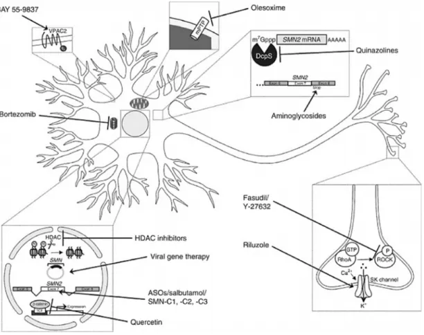
Fig. 4: SMA therapy strategies with their specific target site in an exemplary motor neuron.
BAY 55-9837, an agonist of the VPAC2 receptor in the plasma membrane, activates the p38 pathway.
Olesoximeinhibits mPTP opening in mitochondria and acts neuroprotectively.QuinazolinespreventSMN2 mRNA decapping by inhibiting the scavenger enzyme DcpS.Aminoglycosidesincrease the probability to read through a stop codon in exon 8 ofSMN2.ROCK inhibitors FasudilandY-27632have a neuroprotective effect.Riluzoleacts neuroprotectively by activating SK (small conductance Ca+-activated K+) channels in the cell membrane. HDAC inhibitorsincrease FL-SMN2levels.Viral gene therapies(particularly scAAV9) are able to deliverSMN1 DNA in the cell nucleus. ASOs,salbutamol andPTC SMA C1-C3 act as splicing modulators promoting FL-SMN expression. Quercetin prevents accumulation of β-catenin. Bortezomib increases SMN levels by inhibiting proteasomal degradation (adapted from (Kaczmarek et al. 2015).
![Table 7: The composition and conditions of an oligonucleotide annealing reaction Components Volume [µl] oligonucleotide A 2.5 oligonucleotide B 2.5 10x annealing buffer 5 ddH 2 O 45](https://thumb-eu.123doks.com/thumbv2/1library_info/3760827.1510907/56.918.243.647.126.282/composition-conditions-oligonucleotide-annealing-components-oligonucleotide-oligonucleotide-annealing.webp)