The function of the TGF-β and Toll signalling pathways in Tribolium dorsoventral patterning
Inaugural-Dissertation
zur
Erlangung des Doktorgrades
der Matematisch-Naturwissenchaftlichen Fakultät
der Universität zu Köln
vorgelegt von
Rodrigo Nunes da Fonseca aus Rio de Janeiro, Brasilien
Köln, 2008.
Berichterstatter: Prof. Dr. Siegfried Roth Prof. Dr. Einhard Schierenberg
Tag der letzten mündlichen Prüfung: 30. Juni 2008
“The developmental mechanics of organisms is divided in two parts, one
ontogenetical, the other phylogenetical. In a distant future, both will be essential
components of a more exact concept of common descent based on them. To
begin with, and for a long time, attention will have to be restricted to the
ontogenetical part alone. Once this is developed quite well, some enlightening
shimmer of the causes of phylogeny will emerge from it, and thereby phylogeny
will gain some fundament, albeit still be a very hypothetical one” (Wilhelm
Roux, 1892) .
Contents:
Summary ... 5
Zusammenfassung... 6
Chapter 1 - General Background, specific objectives and methods ... 9
1.1 - The emergence of a concept: Evolution and development ... 9
1.2 - Insect embryology and key questions in insect Evo-Devo ... 13
1.2.1 - Oogenesis modes and their correlation with germ types ... 13
1.2.2 - From classical embryology to the molecular era ... 17
1.3 - Drosophila melanogaster molecular findings and their consequences for understanding the evolution of insect embryogenesis... 20
1.3.1 - Understanding the dorsoventral (DV) system: the establishment of polarity during oogenesis... 21
1.3.2 – Downstream events inside the embryo... 23
1.3.3 - Dorsal has a central role in the Gene Regulatory Network (GRN) responsible for DV axis formation ... 25
1.3.4 - Decapentaplegic (BMP/Dpp) and its modulators constitute a conserved network involved in DV patterning ... 28
1.4 - Establishing a comparative approach: the beetle Tribolium castaneum and its DV axis formation... 30
2 - Specific Objectives ... 35
3 - Methods... 36
3.1 - Cloning Tribolium castaneum and Nasonia vitripennis genes and phylogenetic analysis ... 36
3.2 – RNA Probe preparation, in situ hybridization and immunohistochemistry ... 37
3.3 - dsRNA synthesis, egg, pupae and beetle injections... 38
3.4 - Confocal sections ... 39
Chapter 2 - Evolution of TGF-β modulators in insects ... 40
Introduction: ... 40
Results and Discussion:... 42
TGF-β ligands and receptors ... 43
TGF-β extracellular modulators ... 46
Activin or BMP: TGF-β signalling in Tribolium embryos... 48
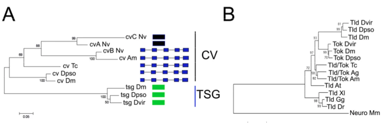
Chapter 3 - Evolution of BMP/Dpp extracelllular modulators in insects - the study case of twisted-gastrulation/crossveinless and tolloid-like molecules .. 51
Introduction: ... 51
Results: ... 54
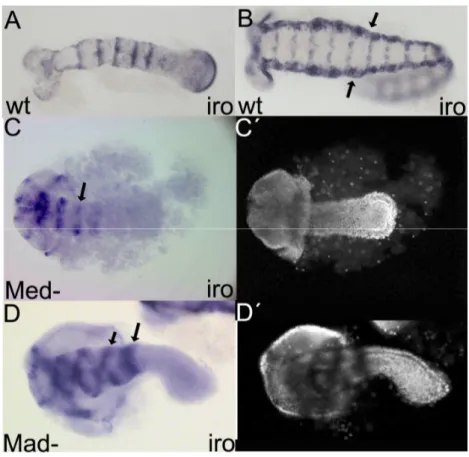
Only one Twisted gastrulation/Crossveinless and one Tolloid-like molecule exist in the beetle genome ... 54
Understanding how gastrulation, DV axis and Dpp activity are established in the Tribolium WT enables a proper analysis of Tc-cv and Tc-tld knock-down phenotypes ... 57
Tc-cv and Tc-tld expression suggest a role in early embryo patterning ... 60
Tc-tld and Tc-cv loss-of-function affect Dpp activity leading to changes in early fate map of Tribolium blastoderm ... 63
Gastrulation is severely affected after Tc-cv and Tc-tld knock-downs ... 65
Tc-tld and Tc-cv knock-down germ-band embryos are ventralized ... 68
Injection of two different dsRNA enables the establishment of an epistatic hierarchy
among dpp, cv(tsg), sog and tld... 70
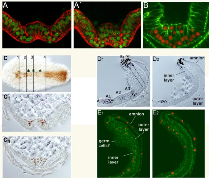
Cells from the inner-layer (IL) of the growth zone migrate over the outer-layer (OL)
after Tc-cv or Tc-dpp RNAi... 73
Cv/Tsg-related proteins also have a major role in Nasonia vitripennis Dpp modulation ... 76
Discussion: ... 79
Duplications in Cv/Tsg and Tld-related molecules: a possible road to fast development ... 79
Tc-cv and Tc-tld are essential for the establishment of Dpp activity and pattern the dorsal regions of Tribolium embryos ... 80
Tc-cv major role in Tribolium implies that Dpp modulation has changed during insect evolution ... 81
Chapter 4 - Self-regulatory circuits in dorsoventral axis formation of the short- germ beetle Tribolium castaneum... 84
Introduction ... 84
Results ... 85
The Toll pathway is required for DV axis establishment in Tribolium... 85
Feedback loops regulate the Tc-Dorsal gradient ... 92
The later development of Tc-Toll RNAi embryos provides insights into growth zone function... 97
Disruption of Dorsal/Toll pathway leads to a periodic pattern of DV cell fates along the AP axis... 101
Discussion: ... 104
Feedback control involving Toll signalling components and Tc-twi ... 104
Feedback control at the level of Dpp signalling... 106
The DV polarity of the growth zone ... 106
The evolution of Toll signalling ... 107
Chapter 5 – General Discussion... 109
Self-activation coupled to a long-range inhibition: a common strategy for patterning in biology ? ... 109
Genome content coupled to functional studies highlights macroevolutionary trends 114 Changes in the DV GRN involves not only cis-regulatory evolution, but also gene duplications, gene losses and arising of new protein domains... 115
DV pattern of the growth zone: forward induction mechanism and its evolutionary implications ... 119
Pleiotropy, immune function and DV patterning: a single event in insect evolution?120 Conclusions... 122
References ... 123
Erklärung... 136
Curriculum vitae ... 137
Summary
Dorsoventral (DV) patterning in Drosophila melanogaster is one of the most well-known gene regulatory networks (GRN) in biology. To investigate if this GRN is conserved during insect evolution, functional analysis of TGF-β and Toll pathways in the short-germ beetle Tribolium castaneum was performed.
In the first part, the function of several BMP/Dpp extracellular modulators, including the products of Tolloid (Tld) and Twisted-gastrulation/Crossveinless (Tsg-Cv), was investigated in Tribolium via parental RNAi (pRNAi). While Tc-tld pRNAi knock-down decreases embryonic BMP activity, Tc-tsg(cv) knock-down completely abolishes it. These observations are strikingly different from those in Drosophila, where tsg is only required for a subset of Dpp activity. These results suggest that Tsg/Cv-like proteins are essential for BMP signalling in Tribolium. Since duplicated copies of tsg(cv)- and tld-related genes are present in the Drosophila melanogaster genome, duplication followed by sub-functionalization of these modulators might have changed the BMP/Dpp gradient during evolution of the dipteran Drosophila lineage.
In the second part, a functional analysis of the Toll pathway was performed. This analysis addressed the question of why the Tribolium Dorsal nuclear gradient is not stable, but rapidly shrinks and disappears, in contrast to the stable Drosophila gradient. Negative feedback accounts for this dynamic behavior: Tc-Dorsal and one of its target genes (Tc-Twist) activate transcription of the I-κB homolog Tc-cactus, which in turn terminates Dorsal function.
Despite its transient role, Tc-Dorsal is strictly required to initiate DV polarity, as in
Drosophila. However, unlike Drosophila, embryos lacking Tc-Dorsal display a periodic
pattern of DV cell fates along the AP axis, indicating that a self-organizing ectodermal
patterning system operates independently of mesoderm or maternal DV polarity cues. The
presence of self-organizing patterning systems in short-germ insects like Tribolium is in
agreement with a regulative type of embryogenesis proposed by classical fragmentation
studies on hemimetabolous insects. These results also elucidate how extraembryonic tissues
are organized in short-germ embryos, and how patterning information is transmitted from the
early embryo to the growth zone. Altogether, the functional analysis of the TGF-β and Toll
pathways in Tribolium dorsoventral patterning suggests that extensive changes in this GRN
have occurred during insect evolution.
Zusammenfassung
Die dorsoventrale (DV) Musterbildung in Drosophila melanogaster (D.m.) gehört zu den am besten verstandenen Gen-regulatorischen Netzwerken (GRN) in der Biologie. Um zu untersuchen, ob dieses GRN während der Insektenevolution konserviert ist, wurden funktionale Analysen der TGF-β und Toll-Signaltransduktionwege im Kurzkeiminsekt Tribolium castaneum durchgeführt.
Im ersten Teil dieser Arbeit wurden verschiedene extrazelluläre BMP/Dpp-Modulatoren, wie z.B. Tolloid (Tld) und Twisted-gastrulation/Crossveinless (Tsg-Cv), mittels parentaler RNAi (pRNAi) in Tribolium analysiert. Während Tc-tld pRNAi zu einer reduzierten BMP-Aktivität im Embryo führt, verhindert Tc-tsg(cv) pRNAi diese vollständig. Diese Beobachtungen zeigen deutliche Veränderungen zu den Mechanismen in Drosophila, wo Dm-tsg nur teilweise für die Dpp-Aktivität benötigt wird. Die Ergebnisse deuten auf eine essentiellere Funktion Tsg/Cv-ähnlicher Proteine im BMP-Signalweg in Tribolium hin. Im Drosophilagenom befinden sich duplizierte Kopien von tsg(cv)- und tld-verwandten Genen. Die Duplikation gefolgt von einer Sub-Funktionalisierung dieser Mediatoren könnte im Laufe der Evolution zu Veränderungen des BMP/Dpp-Gradienten geführt haben.
Im zweiten Teil dieser Arbeit wurde die funktionale Analyse des Toll- Signaltransduktionsweges durchgeführt. Dabei sollte die Frage beantwortet werden, weshalb der Dorsal-Gradient in Tribolium nicht stabil ist und schnell verschwindet, wohingegen Dorsal in Drosophila einen stabilen Gradienten bildet. In Tribolium ist ein negativer Rückkopplungsmechanismus für das dynamische Verhalten des Dorsal-Gradienten verantwortlich: Tc-Dorsal aktiviert zusammen mit einem seiner Zielgene (Tc-twist) die Transkription des I-κB Homolog Tc-cactus. Tc-cactus wiederum ist für die Termination der Dorsal Funktion verantwortlich. Anders als in Drosophila führt der Verlust von Tc-Dorsal zu einer periodischen DV-Musterbildung entlang der AP-Achse. Dies deutet auf ein selbstorganisierendes ektodermales Musterbildungssystem hin, welches unabhängig vom Mesoderm und maternalen DV-Polaritätsinformationen agiert.
Die Präsenz selbstorganisierender Musterbildungssysteme in Kurzkeiminsekten wie
Tribolium castaneum stimmt mit dem regulativen Entwicklungstypus hemimetaboler
Insekten überein, welcher in klassischen Fragmentierungs Studien vorgeschlagen wurde.
Die gewonnen Ergebnisse erklären außerdem die Organisation extraembryonalen Gewebes in Kurzkeiminsekten und machen deutlich wie Musterbildungsinformation vom frühen Embryo auf die Wachstumszone übertragen wird.
Zusammenfassend führte die funktionale Analyse der TGF-β und Toll-
Signaltransduktionswege in Tribolium zu der Annahme, dass während der Insektenevolution
intensive Veränderungen im DV-GRN stattgefunden haben.
Chapter 1 - General Background, specific objectives and methods
1.1 - The emergence of a concept: Evolution and development
In 1859, in the book “Origin of Species” Charles Darwin declared on p. 449 that common descent is “the hidden bond of connection which naturalists have been seeking under the term of the natural system….the structure of the embryo is even more important for classification than that of the adult. For the embryo is the animal in its less modified state; and in so far it reveals the structure of its progenitor” (Darwin 1859). From the middle till the end of the 19
thcentury, the link between embryology and evolution were propagated mainly by Ernst Haeckel. For Haeckel not only all animals would share a common ancestor, but also embryological events in extant animals would reflect their evolutionary path (recapitulation theory). In other words, animals from distinct groups would appear very similar at a specific embryological stage, the so called phylotypic state; and would then progressively differ.
Specific morphological traits of each group would only appear after the phylotypic stage (Haeckel 1866). Although the great similarity of vertebrate embryos at the phylotypic stage has been questioned (Richardson et al. 1997), it´s widely assumed that at the germ-band stage, insect embryos show a remarkable morphological similarity, a clear phylotypic stage (reviewed in Sander and Schmidt-Ott 2004).
In the beginning of the 20
thcentury genetics and embryology had split in different
disciplines. First, geneticists led by Thomas Hunt Morgan focused on the mechanisms
involved in heredity. Although Morgan was initially sceptical about Mendel´s laws, he
showed later that the chromosomes carry the genetic information. Morgan is considered the
father of genetics and his research initiated the Drosophila melanogaster escalade as the
golden bug of genetics. A second group, the experimental embryologists led by Wilhelm
Roux and Hans Driesch, started the developmental mechanics studies
(“Entwicklungsmechanik”), followed by Hans Spemann and Mangold in their seminal
transplant experiments (Spemann and Mangold 1923). These are considered the precursor
studies of the modern developmental biology. By that time, the ultimate goal of these
developmental studies was to explain their results based on “the simple components of
physics and chemistry” (reviewed in Gould 1977). Haeckel recapitulation theory was by that
time rejected by the Thomas Morgan School of Geneticists and by the developmental
mechanicists. These two areas stayed largely separate till the end of 80s, with some notable
exceptions in the works of Waddington and De Beer (De Beer 1958; Waddington 1966).
Stephen Jay Gould in this seminal Ontogeny and Phylogeny (Gould 1977) provided the theoretical basis for the recent fusion of genetics and developmental mechanics.
Intriguingly, the first framework for comparative studies was obtained from the characterization of embryonic mutants in Drosophila (Lewis 1978). The observation that molecules with a similar structural domain, the HOX genes, are involved in anteroposterior patterning in flies and vertebrates is probably the starting point of this new era: The Evolution of Development (Carroll et al. 2001).
Following studies led to the astonishing observations that the same set of molecules e.g. transcription factors and secreted molecules are involved in pattern formation in distantly related organisms, such as flies and mammals. For instance, Bone-Morphogenetic Protein (BMP) in vertebrates and its respective homolog Decapentaplegic (Dpp) in flies are secreted molecules that act in dorsoventral pattern formation in both groups, and probably in all animals with bilateral symmetry (Bilaterians) (Ferguson 1996). Several other examples of conserved sets of transcription factors and signalling molecules have been shown to act in pattern formation on several groups over the last two decades (Carroll et al. 2001).
During late 80s and 90s Evo-Devo started to emerge as a discipline divided in two sub-areas: the micro and macro-evolutionary studies. The first tackles how genotypic changes (nucleotide sequences), in related species or different populations, lead to phenotypic (trait) differences. Examples of these kinds of studies are the analysis of the evolution of bristle patterns or pigmentation among related Drosophillid species (Sucena et al. 2003, Jeong et al.
2008) or the genetic basis of pelvic reduction in threespine stickleback fishes (Shapiro et al.
2004).
The second major area of Evo-Devo involves comparisons of distantly related groups, the so called macroevolution. For instance, recently comparisons among the extant choanoflagellates plants, fungi and animals (Metazoa) provided convincing molecular evidence that choanoflagellates are more related to Metazoa than to fungi (a sister group to all Metazoa) (Philippe et al. 2004). Macroevolutionary as well as microevolutionary studies are based on phylogenetic trees, which are hypothesis about relationships among groups. The knowledge of the last common ancestor among related groups or species is fundamental for the interpretation of Evo-Devo studies (Jenner and Wills 2007).
In the pre-genome era these phylogenetic hypothesis were based on very scarce
information such as only one or fewer genes per group of organisms studied, but this has
changed in the past few years. The use of several concatenated genes to build a phylogenetic
tree is called a phylogenomic approach (Telford 2007). This latter approach has recently
strengthened Evo-Devo, because 1) One can determine the direction in which developmental features are evolving; 2) the knowledge of the divergence times of branches in a tree, allows evolution rates to be inferred; and 3) Phylogenetic trees allow homologies to be inferred or, conversely, show that apparently homologous features are not so (Raff 2000; Raff 2007). The three points above are important to discuss the results in an Evo-Devo framework, as well as influencing the choice of new model organisms (Jenner and Wills 2007).
Studies using ribosomal RNA and phylogenomic approaches have changed the topology of the Metazoan Tree of Life. Before the end of 90s, the Bilateria Tree of Life was based on the view that animal evolution occured from simple to complex forms through gradual steps. This view was mainly based on the possession of a body cavity, the coelom. In this traditional view, acoelomates (e.g. Platyhelminthes), Pseudo-coelomates (e.g. Nematodes) and Coelomates (arthropods and chordates) appeared successively after the split from the last common Bilaterian ancestor, the UrBilateria. In contrast, in the new Metazoan Tree of Life Nematodes and Arthropods are grouped together in the Ecdysozoan clade, animals characterized by the presence of moulting cuticle. Annelids, Molluscs and acoel flatworms belong to another group, the Lophotrochozoans (Adoutte et al. 1999; de Rosa et al. 1999).
Lophotrochozoans share a spiral mode of cleavage and ciliated larval forms that may be considered as modified trochophores. Both groups are protostomes (the first opening forms the mouth) while a third group contains the deuterostomes (the first opening during gastrulation forms the anus and mouth is formed from a second opening). In this new phylogeny hemichordates, echinoderms, cephalochordates, urochordates and vertebrates constititute the monophyletic group of deuterostomes. What has this new Tree of Life changed in Evo-Devo? Basically the two major invertebrate genetic model systems, C.
elegans and D. melanogaster, belong to the same group, the Ecdysozoans and no Lophotrochozoa model system has been established so far. This situation is starting to change with the establishment of the the polychaete annelid Platynereis as a model system (Arendt et al. 2008). Additionally this new tree of Life also indicates that the deuterostomes, including chordates, are closer to the putative Urbilaterian organism, than previously believed.
Important to mention is that although this new Tree of Animal Life is widely propagated, some controversy still exist (see e.g. Rokas and Carroll 2006).
The example above is just one among many of how the availability of sequenced
genomes and ESTs of several species has changed remarkably our understanding of Metazoa
evolution and the relationships among their groups. Recently the genomes of twelve
Drosophillids species have been sequenced (Stark et al. 2007), and more are expected to come.
A striking observation from the genome sequencing and developmental studies in D.
melanogaster and vertebrates is that the same basic set of molecules is involved in similar pattern mechanisms of all animals investigated so far (Carroll et al. 2001). An immediate question then came up: How can complex vertebrates like us be different from a fruit fly, if both groups share a comparable amount of developmental genes in their genomes? Again studies on the developmental genetics of D. melanogaster helped Evo-Devo researchers to build a “working hypothesis”, centred on gene regulation.
In the beginning of 90s, studies on gene regulation of even-skipped (eve), a gene expressed in seven stripes in the D. melanogaster early embryo, provided the first hint that gene regulation would play a major role in these differences (Small et al. 1992; Arnosti et al.
1996). Mike Levine and colleagues discovered that the expression of every eve stripe in the D.
melanogaster embryo is regulated by a different stretch of DNA (Module) present in the non- coding part of the DNA. From this example, it was inferred that every developmentally regulated gene contains independent DNA modules (cis-regulatory modules- CRMs), typically around 500 base pairs, that control its expression during development. From the combinatorial action of several CRMs, one can observe the seven stripes of eve. This example highlights the modulatory nature of the control of developmental genes. The employment and variation among different CRMs has been invoked to explain the disparate morphological variation among species. At least two pieces of evidence favor that view: 1) developmentally regulated genes, i.e., genes involved in several processes of development such as transcription factors or signalling molecules were shown to contain larger intergenic regions comparatively to structural genes in D. melanogaster and C. elegans (Nelson et al. 2004a). Structural genes, by Sean Carroll´s definition are primarily ubiquitous or 'housekeeping' genes, general transcription factors, ribosomal constituents or genes acting in metabolism (Carroll 2005b).
This larger amount of non-coding DNA (intergenic) region in developmentally regulated
genes would be related to the existence of several CRMs. This allows the same gene to act
independently in several developmental contexts due to its several independent CRMs. A
direct consequence from these results is that the modular nature of the CRMs may trigger
evolutionary innovations, since mutations or appearance of new CRMs do not affect the
coding region of a given gene. Coding regions of genes important for embryonic development
are normally under negative selection due to its several roles in different processes
(pleiotropy) (reviewed in Prud'homme et al. 2007). 2) The ratio of non-coding/coding DNA in
the human genome is much higher than in Drosophila, so it was proposed that most of the differences among these two groups rely on the non-coding part of the DNA (Carroll 2005a).
This concept of regulatory evolution is a central theme in Evo-Devo studies, although only recently clear examples of phenotypic variation due to CRM changes have been demonstrated (Jeong et al. 2008; Marcellini and Simpson 2006; McGregor et al. 2007). A global model to understand how changes in CRM may contribute to evolution is being generated by the analysis of entire gene regulatory networks (GRNs). Examples of well understood GRNs are the endomesodermal specification in sea urchin (Davidson et al. 2002), the Ciona intestinalis (Urochordate) heart differentiation (Satou and Satoh 2006), the C- Lineage Specification in C. elegans (Maduro et al. 2001; Baugh et al. 2005) and the dorsoventral (DV) axis formation in Drosophila melanogaster (Stathopoulos and Levine 2005) .
Altogether, Evo-Devo is a new discipline that aims to bring together knowledge from cell, molecular and developmental biology in an evolutionary framework. Consequently, as any evolutionary hypothesis, Evo-Devo is based on the evolutionary relationships among organisms (phylogenetic trees). The current thesis focuses on the macroevolutionary area of Evo-Devo, by studying the dorsoventral axis formation in Tribolium castaneum (beetle, Coleoptera) in comparison with the genetic model Drosophila melanogaster (fly, Diptera).
Beetles and flies share a last common ancestor about 274-285 million years ago (Savard et al. 2006b, Zdobnov and Bork 2007). The vast knowledge of the dorsoventral GRN in D. melanogaster (described below), enables a direct comparison of these two genetic networks. As the whole Evo-Devo area is now in the “genome sequencing era”, this thesis has largely profited from the genome sequencing of Tribolium castaneum. Before the description of any molecular comparative analysis, it is important to describe the major embryological events occurring in insects.
1.2 - Insect embryology and key questions in insect Evo-Devo 1.2.1 - Oogenesis modes and their correlation with germ types
Insect embryogenesis is highly variable among the different groups. On the other hand
a few general trends can be observed. Among the winged insects the holometabolous insects,
containing larval and pupal stages, have generally faster embryonic development than the
hemimetabolous, which hatch as nymphs (mini-adults). This embryonic “acceleration” is correlated with changes in oogenesis mode during evolution.
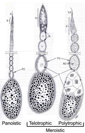
The panoistic oogenesis mode found in many hemimetabolous insects is believed to be the ancestral type (Bier 1970). This oogenesis type has oocytes lacking nurse cells, a common situation in the Metazoans. Irrespective of its oogenesis type, each ovary consists of several ovarioles. Each ovariole contains a germarium located anteriorly (or proximally) followed by the vittelarium where an array of oocytes in progressively stages of development can be observed (Figure 1.1).
Telotrophic meroistic and polytrophic meroistic ovaries are found mainly in holometabolous insects. In both types nurse cells exist, which are absent in panoistic ovarioles. In telotrophic meroistic ovarioles the nurse cells do not accompany the oocytes on their way through the vitellarium. This type of ovary is found for instance in beetles such as Tribolium castaneum (Trauner and Buning 2007). In contrast, polytrophic meroistic ovarioles contain a cluster of nurse cells, derived from the germline, and a unique posteriorly located oocyte, which migrate together along the vittelarium, surrounded by a somatic monolayer epithelium, the follicle cells. Nurse cells are responsible for synthesis of RNAs, ribossomes and other molecules, which are required during embryonic development. Polytrophic ovaries are found in hymenopteras, such as Nasonia vitripennis (Olesnicky and Desplan 2007), lepidopterans and dipterans such as Drosophila melanogaster (Roth 2003). In general, insects containing polytrophic meroistic ovaries have a faster embryonic development when compared to insects with panoistic ovaries, due to the “pre-fabrication” of organelles performed by the nurse cells.
Importantly, a somatic follicle cell layer surrounding the oocyte is a common feature
of insect oogenesis. In addition, in all insect species observed so far, the oocyte nucleus
moves to an asymmetric position in later stages of oogenesis (Roth 2003). In Drosophila this
final asymmetric position of the nucleus defines the dorso-anterior position of the future
embryo. This nuclear migration is important for the establishment of the dorsoventral polarity
in Drosophila, and may be a conserved feature of insect embryogenesis (reviewed in Roth
2003).
Bier, in a seminal paper (Bier 1970), was the first to propose a correlation among the type of oogenesis and the size of the germ anlage, the region of the egg that gives rise to the embryo. Before establishing this correlation, it is important to explain the differences in germ anlagen among insects. Thirty years before Bier, Gerhard Krause divided insects in short, semi-long (intermediate) and long-germ type according to the size of the germ anlage (Krause 1939). In one extreme, the short-germ type, where the germ anlage contains essentially the procephalon (head) and the remaining metameric part of the body emerges from the budding zone (growth zone) by a secondary process. In the other extreme, the long-germ type, where the different body regions in the germ anlage are represented proportionally to their relative position in the germ band. Intermediate germ types lie in between, since not only head but
Figure 1.1: The three ovary types present in insects.
Panoistic ovaries are found in the cricket Gryllus bimaculatus, meroistic telotrophic, the beetle
Tribolium castaneum, and meroistic polytrophic in the fly Drosophila melanogaster.Adapted
from Anderson 1972. (TF) Terminal filament, (FC) Follicle cells, (NC) nurse cells, (G)
germarium, (NC) nurse cells, (N) nutritive cords, (O) oocyte.
also gnathal segments are present before gastrulation, but the abdominal segments are generated later during development from the growth zone.
Nowadays, two different approaches are used in the distinction among insects with short, intermediate or long-germ-types of development. The first approach is based on the timing of segment specification relative to ventral furrow formation. Segment specification would be, in this first view, defined by the appearance of engrailed expression, which marks the irreversible segment determination in Drosophila (Davis and Patel 2002). Since engrailed expression is variable in relation to gastrulation movements in many Diptera species, which are presumably all long-germ type, this approach has been recently questioned (Roth, 2004).
The alternative approach proposed by Roth, 2004 involves fate-map establishment after local irradiation of small parts of the germ anlage. Using this latter approach, no convincing evidence of germ anlage containing only the head segments (short-germ type) has been so far observed (Roth, 2004). In this thesis the term short-germ development will be adopted for all insects which generate segments from the posterior growth zone and long-germ type of development for embryos where all presumptive segments are specificied early along the AP axis and no secondary growth zone exists. Examples of long-germ type are Apis mellifera (Hymenoptera) and Drosophila melanogaster (Diptera), and of short-germ the beetles Tribolium castaneum and Atrachya menetriesi. In addition, beetles also contain species undergoing long-germ development such as Bruchidius villosus.
Long-germ type of development occurs mainly in holometabolous insects, which present meroistic type of oogenesis. Apparently, a “pre-fabrication” of organelles by the nurse cells in meroistic types can provide the oocyte with molecules required for early embryogenesis, leading to faster development. Until recently it was widely accepted by morphological and embryological evidence that the emergence of polytrotrophic ovaries in Hymenopterans and Dipterans would have a common origin (Figure 2A). In this “traditional”
version of insect phylogeny, beetles (Coleoptera) including Tribolium, would occupy a basal position in relation to Hymenoptera and Diptera. Recently, this traditional phylogenetic tree has been questioned by a phylogenomic approach (Figure 2B, Savard et al. 2006b; Zdobnov and Bork 2007). According to this new tree Hymenoptera would be basal to Coleoptera and Diptera. What are the possible implications of this new phylogenetic tree? It could imply that the meroistic polytrophic ovaries and the long-germ development of Hymenoptera and Diptera have emerged twice independently. Either in the “modern” or in the “classical”
phylogenetic trees, the Eumetabola ancestor is believed to be a short-germ type insect. On the
other hand, in the “modern” phylogeny, the holometabolous ancestor might have been a long-
germ insect (Figure 2B). So, although widely propagated as an example of a primitive short- germ insect, Tribolium short-germ type of development might have arisen secondarily from a long-germ ancestor.
1.2.2 - From classical embryology to the molecular era
Classical embryological experiments involving transplantions, centrifugation and UV or X-ray irradiation have been extensively performed by several researchers until the end of the first-half of the 20
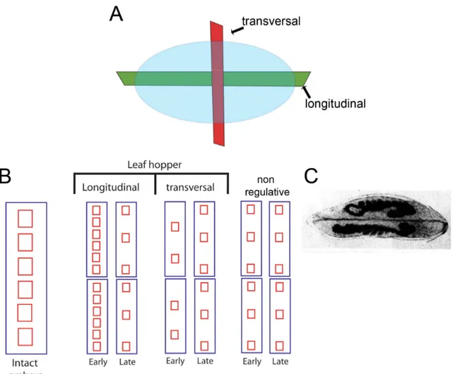
thcentury. These studies suggested the existence of two separate systems involved in patterning the embryo (reviewed in Sander 1976). The longitudinal axis, nowadays called anteroposterior (rostro-caudal), and the transversal axis, the current dorsoventral axis, were described much before any molecular study was carried out.
Although a complete review on these topics is out of the scope of the present thesis (see for instance Sander 1976), it is important to mention the conceptual framework which arose from those experiments.
Eggs of the leaf hopper Eucelis plebejus were fragmented along the longitudinal and transversal axis (Fig 1.3A) during early cleavage stage, and observed during several successive days (Sander 1971). Interestingly, eggs fragmented along the longitudinal axis, which disrupts the dorsoventral axis, displayed a remarkable capacity of recovery (Fig 1.3B).
Figure 1.2: Schematic drawings of classical phylogenetic relationships among insect groups
(A) and the new molecular phylogeny (B). L - long-germ type of development, S-short-germ
type of development, I - intermediate-germ type of development. No species belonging to the
Neuroptera order was used for the molecular phylogeny, so this group is omitted from B.
Complete twinning was observed in several cases (Figure 1.3C), suggesting that “Embryonic
Regulation” occurred. “Embryonic Regulation” was previously defined by Hans Driesch
(1909) as “an event or group of events which occur in the living organism after disturbances
and result in restitution of the normal functional state or lead toward this restitution” (Driesch
1909). This type of complete germ-band regeneration was never observed after transversal
sectioning (Sander 1959; Sander 1971). After transversal sectioning, which disturbs the
anteroposterior axis, the fragments observed after recovery contained just parts of germ bands
(Fig 1.3B). Normally, these partial germ-bands generated fewer segments than expected from
their location in the egg before the fragmentation. In conclusion, these experiments clearly
indicated that two different systems operate in the embryonic pattern of the leaf hopper, one
of them clearly displays “Embryonic Regulation” (dorsoventral), while the other does not
(anteroposterior).
Figure 1.3: Summary of experimental results obtained by classical fragmentation studies (A) Using leaf hopper eggs, Klaus Sander performed transverse and longitudinal fragmentation studies (Sander 1959; Sander 1971). Longitudinal sections disrupt the dorsoventral axis while transversal sections disrupt the anteroposterior axis. (B) Summary of the results obtained with leafhoppers and with non-regulative (originally called “mosaic”) embryos (e.g. Drosophila). The height of the frames represents the size of the system or fragment. After early longitudinal fragmentation, two separate embryos containing a complete pattern can be generated. So, more pattern elements than expected are generated. If the same longitudinal fragmentation is performed later in development, this phenomenon does not occur. After transversal fragmentation, the fragments produce less pattern elements than expected. Non-regulative embryos after longitudinal fragmentation generate pattern elements according to their fates, so the expected amounts of pattern elements are generated in early or late fragmentation. Taken together, these results suggest more flexibility in the mechanism responsible for dorsoventral axis (longitudinal sections) than in anteroposterior axis patterning (transversal sections) in the leaf hopper. (C) An example of complete twinning formation after longitudinal fragmentation.
The existence of extensive embryonic regulation along the dorsoventral axis has also
been reported in the beetle Atrachya menetriesi, where up to four embryos may be formed
from one egg after cold-shock treatment (Miya and Kobayashi 1974). Occurrence of two
embryos in a single egg (dorsoventral duplication) is also observed in spiders, basal
arthropods under natural conditions (Holm 1952, Oda and Akiyama-Oda 2008).
Similar experiments using long-germ embryos such as Drosophila melanogaster did not provide similar results in relation to regeneration. After sectioning, UV irradiation or transplantion almost no embryonic regulation takes place (reviewed in Sander 1976) . For this reason these embryos are called non-regulative (mosaic in Sander, 1971) embryos, where their cells are determined early in development and cannot acquire a new function after injury (Sander 1971). It is noteworthy to mention that transplantion and centrifugation experiments also provided evidence for the existence of an anterior determinant (Sander 1976, Kalthoff 1983) before its molecular nature was uncovered (Driever and Nusslein-Volhard 1988; St Johnston and Nusslein-Volhard 1992). Importantly, regulation of the dorsoventral axis after injury as described above in short-germ embryos does not occur in long-germ embryos like Drosophila. This indicates that changes on the events responsible for the DV axis patterning axis must have occurred during insect evolution.
1.3 - Drosophila melanogaster molecular findings and their consequences for understanding the evolution of insect embryogenesis
The molecular characterization of numerous mutants affecting early patterning has been carried out during the last 25 years. These studies led to the conclusion that four maternal genetic systems, containing several genes each, are involved in setting up the polarity along the anteroposterior and the dorsoventral axis. First, the anterior system is involved in the anterior localization of bicoid, a transcription factor that activates zygotic downstream genes involved in the anteroposterior pattern. Second, posterior group genes (e.g.
oskar, nanos, vasa) are involved in establishing the pole plasm, which is required for germ cell development, besides their requirement in posterior pattern. Third, the terminal class genes ensure that the receptor Torso is activated both at anterior and posterior poles of the embryo. Fourth, the DV system sets up the dorsoventral polarity of the egg (St Johnston and Nusslein-Volhard 1992). Downstream of these maternal systems zygotic genes such as gap, pair-rules, segment polarity and Hox genes are involved in the establishment of the anteroposterior (AP) axis. The fact that Hox genes are expressed in similar regions in several insect species during the phylotypic stage, the late germ band in insects, suggested that a conserved molecular mechanism would act at that stage (reviewed in Sander and Schmidt-Ott 2004).
On the other hand an overall variation in the role of these four maternal systems has
been observed in insects. Duplications followed by subfunctionalization of key factors played
an important role in evolutionary changes. A classic example of an important duplication during insect evolution was described for the AP patterning system. bicoid, a core gene in this system, has arisen by a duplication event from a derived Hox3 gene (zerknüllt, zen) in the common ancestor of the cyclorrhaphan flies (Stauber et al. 1999; Stauber et al. 2002). Since bicoid activates most of the anterior zygotic genes such as gap and pair-rule genes, the emergence of this gene was probably a key evolutionary innovation for anterior patterning. In beetles and wasps, orthodenticle (otd) and hunchback (hb), two bicoid target genes in flies, are responsible for anterior pattern, constituting the putative ancestral system in insect evolution (Schroder 2003; Lynch et al. 2006a).
The role of terminal genes was also investigated in other insects. Interestingly, the function of the terminal system, e.g. torso signalling, is apparently conserved in both poles among short-germ insects like Tribolium and long-germ Drosophila (Schoppmeier and Schroder 2005). Conversely, the function and regulation of tailless, a transcription factor involved in the establishment of the terminal regions in the fly seems not to be conserved in the wasp Nasonia vitripennis (Lynch et al. 2006b).
The role of posterior genes such as vasa and nanos in specifying germ cell development is apparently conserved not only in insects (Lall et al. 2003), but also in Lophotrochozoans and non-bilaterian animals (Extavour et al. 2005, Tsuda et al. 2003; Sato et al. 2006). On the other hand, oskar which is also involved in the determination of germ cell fate, besides the determination of posterior polarity, is not conserved at the sequence level outside Drosophillids.
1.3.1 - Understanding the dorsoventral (DV) system: the establishment of polarity during oogenesis
The last maternal system, the DV system is the main topic of the current thesis. 17
maternally provided factors were identified by genetic screenings and are involved in the
formation of a ventralizing signal in the fly embryo. This ventralizing signal and embryonic
dorsoventral polarity are dependent on events taking place during oogenesis (reviewed in
Roth 2003). The Drosophila melanogaster oocyte at mid-oogenesis (stage 8-10) is surrounded
by a somatic layer of follicle cells (Fig 1.4). At stage 9 the first visible sign of symmetry
breaking along the dorsoventral axis occurs. The oocyte nucleus migrates from a posterior
position towards an anterior asymmetric position along the DV axis. The oocyte nucleus final
destination determines the dorso-anterior location of the oocyte (Micklem et al. 1997; Peri and
Roth 2000). At that region, the oocyte nucleus acts as a signalling center, since the mRNA of the secreted TGF-α-like ligand Gurken (Grk) accumulates around it in an anterior-dorsal cap (Neuman-Silberberg and Schupbach 1993). This localization of Grk mRNA in the anterior- dorsal cap is required for the correct orientation along the future DV axis, but not for the establishment of the different cell fates. This conclusion was obtained from the analysis of mutants where Grk mRNA is mis-localized, which display all DV fates along the AP axis (Roth and Schupbach 1994).
The secretion of Grk activates the Drosophila Epidermal Growth Factor receptor (Egfr) present in the overlying dorsal follicle cells, generating DV polarity in the follicle epithelium. This Egfr activation in the dorsal region leads to the restriction of pipe expression in ventral follicle cells by an unknown mechanism (Nilson and Schupbach 1998). pipe encodes a heparan sulphate 2-O-sulphotransferase, so it is generally assumed that pipe is involved in the modification of an unknown glycoprotein or glycolipid-associated carbohydrate in the perivitelline space. Recent evidence have discarded all glycosaminoglicans carbohydrates (heparin sulfate, heparin, chondroitin sulfate and dermatan sulfate) as pipe substrates (Sen et al. 1998; Sen et al. 2000; Zhu et al. 2005; Zhu et al. 2007), so the glycoprotein which acts as a substrate for Pipe remains unknown. The pipe domain extends around 40% of the ventral embryonic circumference, a similar width as the final Dorsal nuclear gradient (figure 1.4).
Figure 1.4: Major events during dorsoventral axis formation in Drosophila.
(A) Gurken (green), a TGFα protein emanating from a dorsal source in the oocyte, represses pipe (orange)
in the follicular epithelium, restricting its expression to a ventral stripe. (B) Pipe leads to the modification
of an unknown ECM component (red) which is secreted by the follicle cells and maintained within the
extracellular space surrounding the late oocyte and early embryo. (C) Subsequently, an extracellular signal
is generated within the region defined by the modified ECM component. This signal induces the nuclear
Importantly, this pipe expression domain cannot explain why the graded nuclear Dorsal gradient is achieved, since the ventral most 15% region of the embryonic circumference contain the highest amounts of nuclear Dorsal. Apparently, the pipe expression domain exerts a permissive role, but downstream events in the perivitelline space are required for the establishment of the correct graded nuclear Dorsal gradient inside the embryo. These downstream events are mediated in the perivitelline space by a cascade of four proteases (gastrulation defective, snake, easter, nudel), the serine protease inhibitor serpin27A and spätzle, a Nerve Growth Factor-like (NGF-like) molecule, which is the ligand of the receptor Toll (reviewed in Moussian and Roth 2005). Pairs of activators and inhibitors act in the perivitelline space, showing that extensive feedback regulation occurs in this region. The carboxy- and amino-terminal fragments of gastrulation defective (GD), the protease Easter and its inhibitor Serpin27A, and the carboxy and amino-terminal fragments of Spätzle (reviewed in Moussian and Roth 2005) are these pairs of activators-inhibitors, which are required for the formation of a unique peak of Dorsal activity in the ventral embryonic region.
Spätzle mRNA injection may generate embryos with duplicated axis, as judged by two opposite twist expression domains. Such axis duplication phenomenon also occurs in egfr and grk mutant eggs, where an expanded pipe expression domain in the follicle cells is observed (Morisato 2001). These axis duplications might arise from perturbation of the balance between the ligand (Spätzle) and its inhibitor (Spätzle amino-terminal fragment). Higher concentrations of unprocessed Spätzle or an expansion of the ventral domain where Spätzle is processed (in egfr or grk mutants) are likely to cause increased inhibitor concentrations at ventralmost positions. It is clear that the molecular events responsible for axis duplication occurs outside the embryo, since embryos with uniform high amounts of Toll signalling, show twist expression along the whole dorsoventral axis, but do not exhibit a duplicated axis (Roth et al. 1989; Roth and Schupbach 1994, Morisato 2001).
1.3.2 – Downstream events inside the embryo
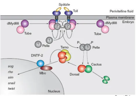
Upon Spätzle signalling in the ventral region, a cascade of downstream events takes place inside the embryo. A complex involving Toll, the adaptor molecules Tube and Krapfen/Myd88, and the serine/threonine kinase Pelle is formed upon signalling. This leads to a local increase of Pelle concentration, resulting in autophosphorylation of this kinase.
Remarkably, an activated form of Pelle is able to generate all known Toll-Dorsal patterning
thresholds when ectopically expressed along the AP axis. This implies that all required functions downstream of the Toll pathway can be mediated by the kinase Pelle (Stathopoulos and Levine 2002b).
This kinase phosphorylates Toll and Tube, thereby being released from the receptor complex (Fig 1.5). Pelle can then act directly on Cactus and Dorsal. The phosphorylation of Cactus, followed by its ubiquitination and degradation by the proteasome, releases Dorsal in the cytoplasm. Phosphorylated Dorsal then dimerises and enters the nucleus (Fig 1.5). Tamo, Drosophila Nuclear Transport Factor-2 (DNTF-2) and Members-only (Mbo), a nuclear pore protein, also interacts specifically with Dorsal and are required for nuclear transport (Fig 1.5).
This nuclear transport is independent of Toll signalling. Importantly, Cactus degradation only occurs in the ventral regions where Spätzle binds to Toll, so the dorsal region of the Drosophila egg is free of nuclear Dorsal (reviewed in Moussian and Roth, 2005).
Figure 1.5: The cytoplasmic events downstream of the Toll receptor.
The activated Toll receptor recruits the membrane-localised dMyd88–Tube heterodimer to the intracellular domain of the activated Toll receptor. Subsequently, the kinase Pelle is recruited to the heterotrimeric Toll–dMyd88–Tube complex and undergoes autophosphorylation, thereby enhancing its kinase activity. By phosphorylation of Toll and Tube, Pelle is released from the heterotetrameric complex. At this step, signalling is disrupted, and the Toll–Spätzle complex is presumably internalised.
Next, Pelle transduces the signal to downstream factors, resulting in the degradation of Cactus. This
allows binding of Dorsal to Tamo which regulates nuclear entry of Dorsal through a nuclear pore
containing DNTF-2 and Mbo. From Moussian and Roth, 2005 Curr Biol.
1.3.3 - Dorsal has a central role in the Gene Regulatory Network (GRN) responsible for DV axis formation
Once Dorsal is transported to the nuclei after Cactus degradation in ventral regions, this transcription factor activates several genes along the Drosophila DV axis. High amounts of Dorsal activate snail (sna) and twist (twi) in the prospective mesoderm, progressive lower amounts activate short-gastrulation (sog), brinker (brk) and rhomboid (rho) in the neurogenic ectoderm and even lower amounts repress zerknüllt (zen) and decappentaplegic (dpp) from the non-neurogenic ectoderm (dorsal ectoderm) (Fig 1.6, Stathopoulos and Levine 2002a).
The above described genes are just examples of Dorsal target genes that were discovered by classical genetic screening (around 30 direct targets).
cDNA microarrays uncovered additional 20-40 direct Dorsal target genes (Stathopoulos et al. 2002). Recently, tilling array screenings (Biemar et al. 2006) and chromatin immunoprecipitation (ChIP)-on-chip (Sandmann et al. 2007; Zeitlinger et al. 2007) approaches added more 30 genes to this GRN. Since this latter approach covers the vast majority of the Drosophila melanogaster genome, 80-100 Dorsal target genes might be closer
Figure 1.6: Positional information generated by the Toll pathway.
(A) A schematic half cross-section of an early Drosophila embryo; the nuclear Dorsal gradient
leads to the activation of target genes in different regions along the dorsoventral axis. (B)
Lateral views with the exception of sim which is a ventral view. Genes are activated in the
presumptive mesoderm (Mes2 in B), mesectoderm (single-minded - sim in B) and
neuroectoderm (sog in B). Genes such as dpp are repressed from the non-neurogenic (dorsal
ectoderm) region.
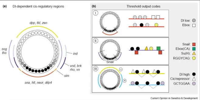
to the actual number of Dorsal targets (Fig 1.7b). As can be observed in Figure 1.7a, not only Dorsal has a central role in this GRN, but also Dorsal target genes can be separated in different categories (colors in the picture) depending on the region of the embryo they are expressed. In blue one can observe genes expressed in the mesoderm, in yellow genes expressed in the neurogenic ectoderm and in green genes expressed in non-neurogenic ectoderm and in the amnioserosa (Fig 1.7a). Importantly, these genes are expressed in different embryonic territories due to the different characteristics of their DV enhancers (Fig 1.8).
Genes responding to the Dorsal gradient can be divided in at least three different types, I, II and III. This distinction is based on the individual binding sites present on their enhancers and the region along the DV axis where the gene is expressed (Fig 1.8, reviewed in Stathopoulos and Levine 2004). Genes expressed in the ventralmost region, the presumptive mesoderm, contain the so called type I Dorsal responsive enhancer. Correlated to the highest amounts of Dorsal and Twist present in the mesoderm, enhancers of genes expressed in this Figure 1.7: The Gene Regulatory Network responsible for DV axis formation in Drosophila.
(A) Dorsal directly regulates several enhancers in the mesoderm (light blue), in the mesectoderm (dark
yellow), in the neurogenic ectoderm (light yellow), and in the dorsal ectoderm (light green). So, Dorsal is a
central molecule in this GRN. (B) Between 80-100 enhancers are Dorsal direct targets in Drosophila
genome, as judged by whole genome studies like, tilling arrays.
region, e.g. twist, snail, mir-1, htl, contain binding sites with the lowest affinity for the transcription factor Dorsal (Fig 1.8). In addition these enhancers also contain binding sites for Twist (Ebox). Genes containing type II enhancers such as brk, ventral neuroblasts defective (vnd), rho, and vein (vn) contain binding sites for the repressor Snail, so they are excluded from the mesoderm (Fig 1.8, reviewed in Stathopoulos and Levine 2004). These genes also contain binding sites for the transcription factor involved in the Notch signalling pathway, Suppressor of Hairless (SuH), as well as Twist and Dorsal. Interestingly, although Twist is known to be a mesodermal determinant, Twist antibody stainings have shown that the Twist domain extends around 5 cells onto the prospective neurogenic ectoderm (Kosman et al. 1991, Leptin 1991), so the aforementioned neurogenic ectodermal genes depend on Twist expression and contain high affinity binding sites for Twist (Papatsenko and Levine 2005).
Genes containing type III enhancers contain optimal binding sites for Dorsal and are expressed in regions where the amount of this transcription factor is the lowest. sog and thisbe (ths) are activated by Dorsal, while other genes such as tolloid (tld), dpp and zen are repressed by it and by the co-repressors Groucho and CtBP (reviewed in Stathopoulos and Levine 2004). Taken together, the different amounts of Dorsal along the DV axis correlate to distinct enhancer organizations along this axis. So, Dorsal can be considered as a bona fide morphogen in Drosophila (Wolpert 1969).
Figure 1.8: Dorsal-dependent cis-regulatory regions in Drosophila DV patterning
(A) Genes regulated by Dorsal along the Drosophila DV axis. (B) The known Dorsal-target gene enhancers can be categorized into three different classes. Type I enhancers contain low-affinity dorsal binding sites and may also contain E-box sites, or binding sites for Twist. Type II enhancers have binding sites for Dorsal, Twist (E-boxes with a CA core), Snail, Su(H), and a transcription factor that recognizes the RGGYCAG motif, possibly Dip3. Type III enhancers support expression in a broad lateral domain and contain high-affinity Dorsal binding sites that are closely spaced. Adapted from Stathopoulos and
1.3.4 - Decapentaplegic (BMP/Dpp) and its modulators constitute a conserved network involved in DV patterning
dpp is directly repressed by Dorsal in fly embryos, so its expression is confined to the 40% most dorsal region. In addition, two other mechanisms repress Dpp activity in ventral regions, one involving sog and the other brk, which are described in detail in Chapter 3. Dpp itself is a morphogen since high amounts of Dpp activate genes in the amnioserosa and progressive lower amounts control non-neurogenic ectodermal genes (Ashe et al. 2000).
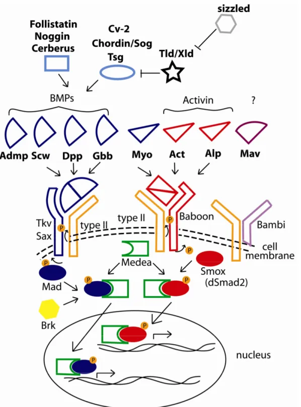
The role of BMP/Dpp4 and its modulators, e.g Sog/Chordin, Twisted-gastrulation (Tsg), Tolloid (Tld), in DV patterning is conserved during Bilateria evolution (Ferguson 1996, De Robertis 2008, Chapter 3). Studies in Xenopus laevis and D. melanogaster have shown that a similar set of molecules is involved in this process. Briefly, the conserved role of these molecules is the major argument favoring the revival of the old hypothesis of the inversion of the DV axis between protostomes and deuterostomes. A detailed historical description of this topic is available in Arendt 2004.
Although much attention has been drawn to the conserved aspects of the BMP/Dpp4 in DV axis formation, several aspects differ between BMP/Dpp4 modulation in Drosophila and Xenopus. First, a subset of BMP/Dpp4 modulators is not present in Drosophila melanogaster genome, although present in vertebrate genomes. This point will be addressed in detail in the Chapter 2 (Umulis et al. 2006; De Robertis 2008).
Second, the control of BMP/Dpp4 antagonists by maternal NF-κB related molecules might be be restricted to insects (reviewed in Bier 2000). Two BMP/Dpp4 antagonists, sog and brk, are directly regulated by the NF-κB related molecule, Dorsal, in Drosophila, while this regulation might be absent in vertebrates. Since it is not clear if Dorsal/NF-κB-related molecules participate in early DV polarity during vertebrate embryogenesis (Armstrong et al.
1998; Prothmann et al. 2006), it is possible that the major maternal role of Dorsal/NF-κB in Drosophila early patterning is a late acquisition in insect evolution. This hypothesis is analysed in detail on Chapter 4.
Third, while in Drosophila the molecular system involved in DV and AP axis
formation is considered completely independent one from each other, this is not the case for
vertebrates. It has been long realized that zebrafish and mouse mutants affecting the DV axis
formation are also affected along their AP axis (reviewed in Bier, 2000), but molecular links
between these molecular systems have only recently been uncovered in Xenopus (Fuentealba
et al. 2007, Fig 1.9). It was recently shown that Xenopus DV and AP axis converge at the
level of Smad1,5,8 phosphorylation. While the Wnt gradient, responsible for AP axis in
vertebrates, is responsible for the duration of Smad1,5,8 signal, the BMP4 gradient provides the intensity of this signal (Fuentealba et al. 2007, De Robertis 2008, Fig 1.9). This mechanism was suggested to explain the overlapping mechanisms between AP and DV axis.
In the long-germ development of Drosophila AP and DV axis patterning are largely
independent. In insects of short-germ type, which generate segments from a posterior
secondary process, similarly to the somite generation during vertebrate segmentation (Tautz
2004), it is not clear if AP and DV axis are independently patterned. To sum up, insects with
short-germ development might be suitable to answer at least three important questions related
to the evolution of DV axis formation: 1) Did the loss of several Dpp modulators occur early
in insect evolution, or did it occur later, e.g. in higher Diptera like Drosophila (analysed in
Chapter 2 and 3) ?; 2) Is the major role of NF-κB related molecules controlling DV axis an
specific feature of Drosophila, or is it a general insect feature, being present in more primitive
insects (analysed in Chapter 4)?; 3) Does the AP and DV crosstalk, as demonstrated for
vertebrates, occur in primitive insects and was lost in higher Diptera like Drosophila
(analysed in Chapter 4)? In order to answer these questions a suitable insect model system
with short-germ development should be used for a comparative approach.
1.4 - Establishing a comparative approach: the beetle Tribolium castaneum and its DV axis formation
Tribolium castaneum (Tenebrionidae, Rust Red Flour Beetle) is an organism that has been used for more than 50 years in laboratory studies (Sokoloff 1975). In the past few years Tribolium research has been boosted by the development of several molecular techniques such as gene knock-down by systemic RNAi and transgenesis (Bucher et al. 2002; Klingler 2004).
Lethal and enhancer trap screenings are being currently performed (Richards and Genome Figure 1.9: The Conserved Chordin-BMP Signalling Network and AP-DV integration at the level of
Smad phosphorylationThe Chordin-BMP signalling network is conserved between Drosophila and Xenopus, (A) In Xenopus, Chordin is expressed on the dorsal side and BMP4 at the opposite ventral pole. (B) In Drosophila, dpp is dorsal (blue) and sog is ventral (in brown) in the ectoderm. (C) A network of conserved secreted proteins mediates DV axis patterning in Xenopus and Drosophila, although some molecules do not act in both organisms. (D) AP (Wnt) and DV (BMP) gradients are integrated at the level of phosphorylation of the transcription factors Smads 1, 5, and 8. Note that in this self-regulating model the BMP gradient provides the intensity, but the Wnt gradient controls the duration of the Smad 1, 5, 8 signal. Direct protein-protein interactions are shown in black, and transcriptional activity of Smads 1, 5, and 8 are in blue. Adapted from De Robertis 2008.