The role of prohibitin-2 in podocytes – mitochondrial function and beyond
Inaugural-Dissertation zur
Erlangung des Doktorgrades
der Mathematisch-Naturwissenschaftlichen Fakultät der Universität zu Köln
vorgelegt von
Christina Ising aus Steinfurt
Berichterstatter: Prof. Dr. Thomas Langer (Gutachter)
Prof. Dr. Thomas Benzing
Tag der mündlichen Prüfung: 20. Januar 2014
I Table of contents
Table of contents ... I List of figures ... V List of tables ... VI Abbreviations ... VII
1 Abstract ... 1
2 Zusammenfassung ... 2
3 Introduction ... 4
3.1 Podocytes in glomerular diseases ... 4
3.2 SPFH domain-containing protein family ... 5
3.2.1 General information ... 5
3.2.2 Podocin ... 6
3.2.3 Prohibitins ... 6
3.3 Regulation of metabolism by mTOR signaling ... 8
3.3.1 The mTOR pathway ... 8
3.3.2 Role of mTOR signaling in podocytes ... 10
4 Thesis aims ... 12
5 Material and methods ... 14
5.1 Material ... 14
5.1.1 Chemicals, reagents and solutions ... 14
5.1.2 Assays/Kits ... 19
5.1.3 Buffers and solutions ... 20
5.1.4 Oligonucleotides ... 27
5.1.5 Plasmids ... 29
5.1.6 Antibodies ... 30
5.1.7 Enzymes ... 32
5.1.8 Materials ... 32
5.1.9 Equipment ... 34
5.1.10 Software ... 37
5.2 Methods ... 38
5.2.1 Working with nucleic acids ... 38
5.2.1.1 Polymerase chain reaction (PCR) ... 38 5.2.1.2 Restriction and purification of plasmids, vectors and PCR products38
II
5.2.1.4 Recombination ... 38
5.2.1.5 RNA extraction and cDNA synthesis ... 39
5.2.1.6 TaqMan® assay ... 39
5.2.1.7 DNA sequencing ... 39
5.2.1.8 Dual-luciferase® reporter assay ... 39
5.2.2 Bacteria ... 40
5.2.2.1 Chemical transformation of E.coli ... 40
5.2.2.2 Isolation of plasmid DNA and diagnostic digest ... 40
5.2.3 Cell culture ... 40
5.2.3.1 Cells ... 40
5.2.3.2 Freezing and thawing of cells ... 40
5.2.3.3 Passaging of cells ... 41
5.2.3.4 Doxycycline-treatment of mouse podocytes ... 41
5.2.3.5 Transient expression of plasmids in 293T HEK cells ... 41
5.2.3.6 Virus production ... 41
5.2.3.7 Infection of cells with virus and selection ... 42
5.2.3.8 Immunofluorescence on cells ... 42
5.2.4 Protein biochemistry ... 42
5.2.4.1 Protein extraction ... 42
5.2.4.2 Protein measurement ... 43
5.2.4.3 Co-immunoprecipitation ... 43
5.2.4.4 SDS-polyacrylamide-gelelectrophoresis ... 44
5.2.4.5 Western Blot ... 44
5.2.4.6 Colloidal coomassie staining ... 45
5.2.5 Antibody production ... 45
5.2.5.1 Purification of recombinant protein ... 45
5.2.5.2 Immunization of mice and hybridoma generation ... 47
5.2.5.3 ELISA ... 47
5.2.6 Worm experiments ... 48
5.2.6.1 Worm strains ... 48
5.2.6.2 RNA interference ... 48
5.2.6.3 Touch assay ... 49
III
5.2.6.4 Microinjection ... 49
5.2.6.5 Immunofluorescence on worms ... 49
5.2.7 Mouse experiments ... 50
5.2.7.1 Mouse strains and animal care ... 50
5.2.7.2 Serum analysis ... 51
5.2.7.3 Extraction of DNA from mouse tissue ... 51
5.2.7.4 Polymerase-chain reaction (PCR) for genotyping purposes ... 51
5.2.7.5 Albumin ELISA ... 52
5.2.7.6 Creatinine Assay ... 52
5.2.7.7 PAS staining ... 52
5.2.7.8 Specific immunohistochemistry ... 53
5.2.7.9 Electron microscopy and immunogold labeling ... 54
5.2.7.10 Glomeruli isolation ... 54
5.2.7.11 Tamoxifen diet ... 55
5.2.7.12 Rapamycin injection ... 55
5.2.8 Human tissue ... 55
5.2.8.1 Immunofluorescence on human tissue ... 55
5.2.9 Statistical analysis ... 56
6 Results ... 57
6.1 Loss of PHB2 leads to severe glomerular disease ... 57
6.1.1 Podocyte-specific Phb2-knockout mice (Phb2pko) develop albuminuria and die prematurely ... 57
6.1.2 Phb2pko mice develop glomerulosclerosis ... 59
6.1.3 Phb2het mice present with changes in mitochondrial ultrastructure ... 60
6.1.4 Glomeruli of Phb2pko mice display no increased rate of apoptosis at day 21 ... 61
6.1.5 PHB2 is required to maintain structural integrity of podocyte foot processes ... 62
6.1.6 Inducible depletion of Phb2 results in loss of slit diaphragm function ... 63
6.2 PHB2 as a novel slit diaphragm protein ... 65
6.2.1 PHB2 localizes to the slit diaphragm ... 65
6.2.2 PHB1 and PHB2 co-immunoprecipitate with podocin ... 67
IV
touch sensation in C. elegans ... 69
6.3 PHB2 in metabolic signaling ... 72
6.3.1 Podocyte-specific knockout of the insulin receptor (Insr) and IGF-1 receptor (Igf1r) prolongs survival of Phb2pko mice ... 72
6.3.2 Inhibition of mTOR signaling increases lifespan of Phb2pko mice ... 74
7 Discussion ... 79
7.1 PHB2 – a slit diaphragm protein? ... 79
7.1.1 The phenotype of Phb2pko mice is similar to slit diaphragm protein-deficient mice ... 79
7.1.2 PHB2 is necessary for the function of the slit diaphragm complex ... 81
7.1.3 Phb2-deficiency in podocytes affects mitochondrial and non-mitochondrial functions ... 82
7.2 PHB2 is linked to the regulation of cell metabolism ... 84
7.2.1 Phb2-deficiency influences metabolic signaling via the insulin and IGF-1 receptor ... 84
7.2.2 Dysregulated mTOR signaling leads to glomerular diseases ... 86
7.2.3 Hypothesis: Loss of PHB2 sensitizes the insulin receptor ... 88
8 Conclusion ... 91
9 Publications ... 93
9.1 Publications in academic journals ... 93
9.2 Publications in international academic conferences ... 93
10 References ... 94
11 Danksagung ... 111
12 Erklärung ... 112
13 Curriculum vitae ... 113
V List of figures
Figure 1: The three layers of the glomerular filtration barrier ... 4
Figure 2: Multimeric ring complexes of prohibitins ... 7
Figure 3: mTOR pathway ... 10
Figure 4. Podocyte-specific Phb2-knockout mice (Phb2pko) develop albuminuria and die prematurely ... 58
Figure 5. Phb2pko mice develop glomerulosclerosis ... 60
Figure 6. Phb2het mice present with changes in mitochondrial ultrastructure ... 61
Figure 7. Glomeruli of Phb2pko mice display no increased rate of apoptosis at day 21 ... 62
Figure 8. PHB2 is required to maintain structural integrity of podocyte foot processes ... 63
Figure 9. Inducible depletion of Phb2 results in loss of slit diaphragm function ... 64
Figure 10. PHB2 localizes to the slit diaphragm ... 66
Figure 11. PHB1 and PHB2 co-immunoprecipitate with podocin ... 68
Figure 12. PHB-2 co-localizes with MEC-2 at touch receptor punctae and regulates touch sensation in C. elegans ... 71
Figure 13: Loss of the insulin receptor but not IGF-1 receptor prolongs lifespan of Phb2pko mice ... 73
Figure 14: Podocyte-specific knockout of the insulin receptor (Insr) and IGF-1 receptor (Igf1r) prolongs survival of Phb2pko mice ... 74
Figure 15. Generation and validation of an inducible Phb2 shRNA podocyte cell line ... 76
Figure 16. Inhibition of mTOR signaling increases lifespan of Phb2pko mice ... 78
Figure 17. Hypothesis: Loss of PHB2 sensitizes the insulin receptor ... 89
Figure 18. Why does loss of PHB2 lead to glomerular disease? ... 91
VI
Table 1. List of chemicals, reagents and solutions ... 14
Table 2. List of assays and kits ... 19
Table 3. Composition of buffers and solutions ... 20
Table 4. List of oligonucleotides used for cloning ... 27
Table 5. List of oligonucleotides used for genotyping ... 27
Table 6. List of oligonucleotides for sequencing ... 28
Table 7. List of shRNAs ... 28
Table 8. List of TaqMan® probes ... 29
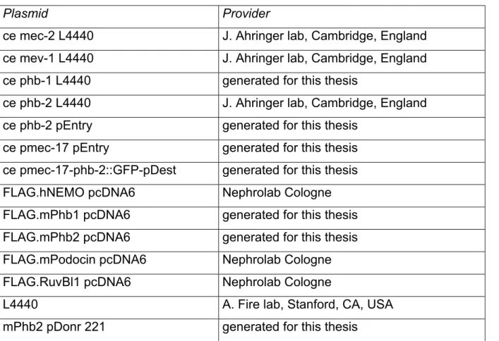
Table 9. List of plasmids ... 29
Table 10. List of primary antibodies ... 30
Table 11. List of secondary antibodies ... 31
Table 12. List of enzymes ... 32
Table 13. List of materials ... 32
Table 14. List of equipment ... 34
Table 15. List of software... 37
Table 16. List of online software ... 37
VII Abbreviations
µl microliter
4E-BP1 eukaryotic initiation factor 4-binding protein AMPK AMP-activated protein kinase
APS ammonium persulfate b base
BSA bovine serum albumin CaCl2 calcium chloride cDNA complementary DNA CKD chronic kidney disease DAB 3,3′-diaminobenzamidine
DAPI 4',6-diamidino-2-phenylindole DEG/eNaC degenerin/epithelial Na+ channels DNA deoxyribonucleic acid dNTP deoxyribonucleotide triphosphate DRM detergent resistant membrane dsRNA double stranded RNA
DTT dithiothreitol
EC endothelial cell
ECL enhanced chemoluminescence EDTA ethylenediaminetetraacetic acid
eIF4E eukaryotic initiation factor 4E ETC electron transport chain FBS fetal bovine serum fl/fl flox/flox
FOXO1 forkhead box O1 (mammalian protein)
FP foot process
FSGS focal segmental glomerulosclerosis
G standard gravity
GBM glomerular basement membrane GFP green fluorescent protein
H human
HEBS HEPES-buffered saline solution HEK human embryonic kidney
het heterozygous His histidine-tag
HPRT1 hypoxanthine phosphoribosyltransferase 1 HRP horse radish peroxidase
IF immunofluorescence IGF-1 insulin-like growth factor 1
Igf1r insulin-like growth factor 1 receptor (mammalian gene)
IgG immunoglobulin G
IHC immunohistochemistry Insr insulinreceptor (mammalian gene) IP immunoprecipitation
VIII IRS Insulin receptor substrate
kb kilobase
KCl potassium chloride
kDa kilodalton KO knockout l liter
LB lysogeny broth
m mouse
MEF mouse embryonic fibroblast MgSO4 magnesium sulfate ml milliliter
mRNA messenger RNA
mTOR mammalian target for rapamycin (mammalian protein) mTORC1 mammalian target for rapamycin complex 1
mTORC2 mammalian target for rapamycin complex 2 mtUPR mitochondrial unfolded protein response NaCl sodium chloride
NCBI National Center for Biotechnology Information NDS normal donkey serum
NEB New England Biolabs NGM nematode growth medium Nphs2 podocin (mammalian gene) OPA1 optical atrophy 1
OXPHOS oxidative phosphorylation system PAGE polyacrylamide gel electrophoresi PAN puromycin aminonucleoside PAS periodic acid schiff
PBS phosphate-buffered saline PCR polymerase chain reaction
PFA paraformaldehyde
PHB1 prohibitin-1 (mammallian protein) Phb1 prohibitin-1 (mammalian gene) phb-1 prohibitin-1 (C. elegans gene) PHB-1 prohibitin-1 (C. elegans protein) PHB2 prohibitin-2 (mammalian protein) Phb2 prohibitin-2 (mammalian gene) phb-2 prohibitin-2 (C. elegans gene) PHB-2 prohibitin-2 (C. elegans protein) PI3K phosphoinositide 3-kinase pko podocyte-specific KO PMSF phenylmethylsulfonyl fluoride pS6RP phospho S6 ribosomal protein PVDF polyvinylidene difluoride RNA ribonucleic acid RNAi RNA interference
IX ROS reactive oxygen species
RT room temperature
S6RP S6 ribosomal protein SDS sodium dodecyl sulfate
SE standard error
shRNA short hairpin RNA
SEM standard error of the mean SOC super optimal broth medium
SPFH stomatins, prohibitins, flotillins and HflK/C TAE tris-acetate-EDTA
TBS tris-buffered saline
TEM transmission electron microscope
TEMED N,N,N´,N´-tetramethylethylenediamine
Tm melting temperature
Tris tris(hydroxymethyl)aminomethane TRP transient recepor potential
TSC1 tuberous sclerosis complex 1 (mammalian protein) Tsc1 tuberous sclerosis complex 1 (mammalian gene) TSC2 sclerosis complex 2 (mammalian protein)
UV ultraviolet V volts
WB Western blot
WT wildtype
1 1 Abstract
Diseases of the kidney filtration barrier are a major cause of renal failure and cardiovascular mortality. Podocytes maintain the glomerular filtration barrier and podocyte dysfunction leads to the development of glomerulosclerosis, i.e. glomerular scarring. Mutations in the SPFH domain-containing protein podocin, which is localized to the specialized cell-cell contact of podocytes, the slit diaphragm, can cause one of the most frequent glomerulopathies, FSGS. Podocin is one of the most extensively studied proteins in podocytes but nothing is known about other SPFH domain-containing proteins in podocytes so far. Since it has been speculated that mitochondrial dysfunction may contribute to podocyte injury in glomerular diseases this thesis work investigated the podocyte-specific function of a mitochondrially localized SPFH domain-containing protein, prohibitin-2 (PHB2). PHB2 is important for maintaining normal cristae structures and proper mitochondrial function.
Podocyte-specific loss of PHB2 in mice resulted in the development of progressive albuminuria, glomerulosclerosis and endstage renal failure. Unexpectedly, immunofluorescence stainings and immunogold labeling detected PHB2 not only in mitochondria but also at the slit diaphragm. PHB2 co-precipitated with podocin, thereby suggesting an extramitochondrial role of PHB2 at the slit diaphragm.
Supporting these results, the ortholog of PHB2 in C. elegans was also not restricted to mitochondria but associated with a mechanosensory complex containing the podocin ortholog MEC-2. Given the high similarity of the mechanosensory complex in worms and the slit diaphragm complex in mammals, functional assays of the mechanosensor were performed. Knockdown of phb-2 as well as loss of mec-2 in the mechanosensitive neurons resulted in impaired touch sensitivity, showing a functional impact of PHB2 on this conserved protein-lipid supercomplex.
Furthermore, it was shown before that loss of insulin signaling increases lifespan of Phb2/phb-2-deficient yeast and worms. Therefore, apart from the findings at the slit diaphragm, the impact of PHB2 on podocyte metabolism was investigated.
Phb2-deficiency in podocytes led to increased activity of mTORC1. Treatment of these animals with rapamycin or additional knockout of the insulin and IGF-1 receptor prolonged survival despite progressive albuminuria. Collectively, these data indicate that loss of PHB2 at the slit diaphragm resulted in the development of albuminuria but loss of podocytes was dependent on metabolic dysregulation.
2 2 Zusammenfassung
Erkrankungen des Nierenfilters stellen eine der Hauptursachen für Nierenversagen und kardiovaskuläre Mortalität dar. Podozyten sind ein wichtiger Bestandteil der glomerulären Filtrationsbarriere und eine podozytäre Dysfunktion führt zur Proteinurie und Glomerulosklerose, d.h. glomerulären Vernarbung. Mutationen in dem SPFH- Domänen-Protein Podocin, das an den spezialisierten Zell-Zell-Kontakt von Podozyten, die Schlitzmembran, lokalisiert, führen zu einer der häufigsten Glomerulopathien, der FSGS. Podocin gehört zu den am meisten untersuchten Proteinen im Podozyten, jedoch ist über weitere SPFH-Domänen-Proteine in diesem Zelltyp bisher nichts bekannt. Da spekuliert wird, dass eine mitochondriale Dysfunktion zum Podozytenschaden in glomerulären Erkrankungen beitragen könnte, beschäftigt sich diese Doktorarbeit mit der podozyten-spezifischen Funktion eines mitochondrial-lokalisierten SPFH-Domänen-Proteins, Prohibitin-2 (PHB2).
PHB2 ist unter anderem wichtig für den Aufbau und Erhalt der Cristae-Strukturen sowie normale mitochondriale Funktionen.
Der Podozyten-spezifische Verlust von PHB2 führt in Mäusen zur Entwicklung einer fortschreitenden Albuminurie, Glomerulosklerose und terminalem Nierenversagen.
Dabei konnten Immunfluoreszenz- und Immunogoldfärbungen PHB2 nicht nur in Mitochondrien, sondern auch an der Schlitzmembran nachweisen. PHB2 ko-präzipitierte mit Podocin, was eine extramitochondriale Rolle von PHB2 an der Schlitzmembran nahelegt. Darüber hinaus fand sich das PHB2-Ortholog in C. elegans nicht nur in Mitochondrien, sondern auch im mechanosensorischen Komplex, der das Podocin-Ortholog MEC-2 enthält. In Anbetracht der hohen Vergleichbarkeit des mechanosensorischen Komplexes in Würmern und dem Schlitzmembrankomplex in Säugern wurden funktionelle Assays des Mechanosensors durchgeführt. Sowohl eine verringerte phb-2-Expression als auch der Verlust von mec-2 in den mechanosensitiven Neuronen führte zu einer verringerten Berührungssensitivität. Dies deutet auf einen funktionellen Einfluss von PHB2 auf diesen konservierten Protein-Lipid-Superkomplex hin.
Ferner wurde bereits gezeigt, dass der Verlust des Insulinsignalweges die Lebensspanne von Phb2/phb-2-defizienten Hefen und Würmern verlängert. Deshalb wurde – unabhängig von den Erkenntnissen an der Schlitzmembran – der Einfluss von PHB2 auf den Metabolismus von Podozyten untersucht. Eine Phb2-Defizienz in
3 Tiere mit Rapamycin sowie der zusätzliche Verlust des Insulin- und IGF-1-Rezeptors verlängerten das Überleben der Tiere trotz fortschreitender Albuminurie.
Zusammengefasst bedeutet dies, dass das Fehlen von PHB2 an der Schlitzmembran zur Entstehung der Albuminurie beiträgt, während der Verlust der Podozyten vermutlich auf eine metabolische Dysregulierung in Folge einer Dysregulation des Insulin/mTOR-Signalweges zurückzuführen ist.
4 3 Introduction
3.1 Podocytes in glomerular diseases
Glomerular diseases represent a major cause of chronic kidney disease (CKD) affecting more than 5% of all human beings world-wide. They share common features e.g. loss of plasma proteins into the urine (albuminuria and proteinuria) and glomerular degeneration, fibrosis and scarring (glomerulosclerosis). Both, proteinuria and chronic kidney disease are independently associated with increased risk for endstage renal failure and cardiovascular diseases [1,2].
Each human kidney contains about one million small filtering units, the glomeruli.
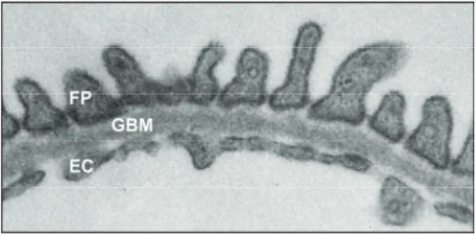
Blood plasma has to pass the glomerular filtration barrier, which is responsible for the size- and charge-selectivity of the kidney filter and produces an almost protein-free ultrafiltrate. The glomerular filtration barrier is composed of three layers: fenestrated endothelium, glomerular basement membrane and podocytes [3,4] (Figure 1).
Figure 1: The three layers of the glomerular filtration barrier
The glomerular filtration barrier consists of three layers that are important for filtration of blood plasma: podocytes with their foot processes (FP), glomerular basement membrane (GBM) and fenestrated endothelial cells (EC).
Podocytes are the visceral epithelial cells of the kidney glomerulus. They elaborate long interdigitated foot processes that completely enwrap the glomerular capillaries in a complex network. The intercellular slit inbetween neighbouring foot processes is bridged by a continous membrane-like cell junction, the slit diaphragm [3].
Genetic studies of the last decade showed that glomerular podocytes are essential for the function and integrity of the kidney filter and critically involved in the development of proteinuria [5,6]. Podocyte injury in renal diseases is reflected by foot process effacement, i.e. retraction of the membrane extensions, and ultimately cell
5 inevitably leads to a reduced cell number which is negatively correlated with renal survival [7,8]. Not only inflammation, toxins and mechanical stress but also gene mutations can be the cause of podocyte injury [9]. Numerous studies within the last decades on hereditary glomerular diseases identified mutations in genes encoding for proteins that localize to the podocyte slit diaphragm, e.g. cytoskeleton-associated CD2AP [10,11], the immunoglobulin family member nephrin [12,13], the ion channel TRPC6 [14,15] and the SPFH domain-containing protein podocin [16–19].
3.2 SPFH domain-containing protein family 3.2.1 General information
The SPFH domain-containing protein family was named after the initials of the proteins in this family: stomatins, prohibitins, flotillins and HflK/C. Initially, the SPFH domain was identified in stomatins and database searches revealed that other proteins like prohibitins contain a strikingly similar domain (also called PHB domain) [20]. The SPFH domain is not only conserved among different proteins but also across species [20–22]. Among others, erlins and podocin as well as the podocin homolog MEC-2 in C. elegans are prominent members of this protein family [3,23,24]. Many SPFH domain-containing proteins form homo-oligomers by interacting via the SPFH domain [25–27]. However, also hetero-oligomers exist as shown for the complexes that are made up of prohibitin-1 and prohibitin-2 [28]. Most of the SPFH domain-containing proteins have been shown to be enriched in detergent resistant membranes (DRM) [21,29–34], but so far no common function could be attributed to all members of this family. The SPFH domain is suggested to be either a protein-binding or a lipid-binding motif but no protein-binding partner was identified which is universal for all family members [21]. Since many of the SPFH domain-containing proteins are associated with DRMs - which are defined by their specific lipid composition [35] – it seems to be very probable that this domain is necessary for specific protein-lipid interactions [21]. This has already been shown for podocin/MEC-2, which bind to cholesterol in the plasma membrane [24], but still needs to be proven for other members of the SPFH domain-containing protein family.
6 3.2.2 Podocin
Podocin is an integral membrane protein encoded by NPHS2. It consists of one transmembrane domain and a carboxy-terminal cytoplasmic tail. The protein sequence shows strong homology to the corresponding regions of stomatin family proteins [16,36]. During glomerular development podocin is expressed in
“mesonephric podocytes from the S-shaped body and, later, in the metanephric kidney, in the future podocytes at the late S-shaped body stage” [36]. In the mature kidney podocin expression is exclusively detectable in podocytes where the protein is localized at the slit diaphragm [36]. NPHS2 mutations are involved in the development of a familial form of early-onset steroid-resistant nephrotic syndrome progressing towards focal segmental glomerulosclerosis (FSGS) as well as in sporadic cases of the disease and late-onset inherited FSGS [16,17,37–41].
NPHS2-knockout mice develop normally but show massive proteinuria shortly after birth due to severe podocyte foot process effacement resulting in end-stage renal failure and premature death within the first weeks of life [18].
In the nematode C. elegans MEC-2 - the worm homolog of podocin – is located along regular touch punctae along the six touch receptor neuron processes. Here, MEC-2 is part of a multiprotein-channel complex that is needed to convert mechanical stimuli into electrical signals [42,43]. MEC-2 regulates the function of MEC-4/MEC-10, two degenerin/epithelial Na+ channels (DEG/ENaC) that transduce gentle body touch sensation [42,44,45]. In mammals, the ion channel TRPC6 - a member of the transient receptor potential (TRP) superfamily - is associated with the slit diaphragm [14,15], where it interacts with other slit diaphragm proteins [14,24]. TRP channels are mechanically gated ion channels [46] which can be regulated by podocin [24].
This renders the slit diaphragm a possible mechanosensitive structure which can more easily be studied in C. elegans [47].
3.2.3 Prohibitins
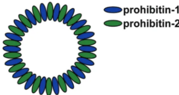
Prohibitin-1 (PHB1) and prohibitin-2 (PHB2) are two closely related proteins that reside within the inner mitochondrial membrane. Unconventional sorting sequences target both proteins to mitochondria, where they are inserted into the inner membrane by TIM23-translocase activity. Protein structure analysis revealed that they are anchored to the membrane by a hydrophobic stretch located at the amino terminal end while the large carboxy terminal end, which consists of a PHB-domain
7 PHB2 directly interact by forming multimeric ring complexes (Figure 2) and stabilize each other [28,49,50].
Figure 2: Multimeric ring complexes of prohibitins
Prohibitin-1 and prohibitin-2 are assembled in heteromeric ring structures and thereby stabilize each other. (Figure adapted from Osman et al., 2009 [51])
Prohibitins are involved in numerous functions within the cell. Not only are they needed for the maintenance of the mitochondrial membrane and cristae structure but also serve a scaffolding function during biogenesis of oxidative phosphorylation system (OXPHOS) complexes [52,53]. Moreover, prohibitins seem to be important for the regulation of apoptosis [53–56] and can reside within the nucleus where they modulate transcriptional activity [54,57–60]. Nevertheless, it is generally conceived that the majority of prohibitins is localized within the mitochondrial compartment.
Prohibitins are upregulated in various types of cancers e.g. breast cancer [61] and gastric cancer [62] but seem to have protective roles in many normal tissues. For example PHBs are required to prevent ethanol-mediated damage of pancreatic β-cells [55,63] and can stop the progression of TGF-β1-induced renal tubulointerstitial lesions [64].
In 2008, Merkwirth et al. [53] showed that cellular depletion of PHB2 in mouse embryonic fibroblasts (MEFs) led to decreased levels of PHB1, markedly reduced proliferation and an increased susceptibility to apoptotic stimuli. The cells exhibited disorganized and swollen mitochondrial cristae structures. However, mitochondrial membrane potential and enzymatic activities of respiratory complexes were unaffected. An impaired processing of the dynamin-like GTPase OPA1 (optic atrophy 1) was identified as the primary cellular defect in the absence of prohibitins [53]. Correct cleavage of OPA1 into five different short and long isoforms is required
8 for mitochondrial fusion, thereby promoting cristae maintenance and remodeling [65–
68].
Universal knockout of Phb2 in a mouse model resulted in no viable offspring [53,59].
Recently, loss of regular mitochondrial cristae structures and impaired OPA1 cleavage was demonstrated in a neuronal-specific Phb2-knockout mouse model in vivo [69], thereby confirming the phenotype that was observed in isolated Phb2-deficient MEFs [53]. Phb2-deficiency in C. elegans led to increased fat utilization, increased mitochondrial proliferation, consequent cellular damage and an ultimately reduced lifespan. However, loss of PHB-2 increased lifespan of already long-lived insulin receptor/daf-2-mutant worms and partially restored the metabolic defects observed in Phb2-deficient worms, thereby linking prohibitins to the metabolic signaling pathways [70]. Furthermore, a recent study in prohibitin-deficient yeast cells revealed that loss of prohibitins leads to increased mitochondrial proteotoxic stress, which activates the mitochondrial untranslated protein response (mtUPR). This effect can be reversed under dietary restriction i.e. less nutrient signaling, probably via decreasing cytoplasmic mRNA translation [71]. In conclusion, experiments in yeast and worm link PHB2 to the action of insulin.
3.3 Regulation of metabolism by mTOR signaling 3.3.1 The mTOR pathway
The nutrient-sensing Ser/Thr kinase mammalian target of rapamycin (mTOR) is one of the central regulators for metabolism in cells and organisms. mTOR can assemble with other proteins into two distinct complexes, mTORC1 and mTORC2. In the rapamycin-sensitive mTORC1 complex mTOR associates with raptor and regulates temporal aspects of cell growth whereas the rapamycin-insensitive mTORC2 complex contains rictor and controls spatial aspects of cell growth [72–76]. To exert its function mTORC1 signaling increases e.g. transcription and protein synthesis but inhibits autophagy. The mTORC2 complex mainly regulates actin organization [77].
The activity of mTOR complexes is regulated by different upstream signals that either downregulate or upregulate mTOR activity. On the one hand, stress signals like hypoxia as well as low cellular energy states downregulate mTOR signaling to arrest cell growth [78–81]. Hypoxia increases the HIF1-mediated expression of REDD,
9 and TSC2. TSC1-TSC2 negatively regulate mTOR activity via Rheb [82–84]. Low cellular energy leads to activation of AMP-activated protein kinase (AMPK), which activates TSC2, resulting in the inhibition of mTORC1 activity [80,85]. On the other hand, amino acids and growth factors increase mTOR signaling. So far, it is not clear how the amino acid status is communicated to the mTOR complex, but one hypothesis is that it works independent of the TSC complex by direct activation of Rheb, which in turn activates the mTOR complex. Two of the most important activators of the mTOR pathway are the growth factors insulin and insulin-like growth factor 1 (IGF-1). Each binds to its specific receptor at the plasma membrane and inhibits TSC1-TSC2 via the phosphoinositide 3-kinase (PI3K) pathway, thereby promoting activation of mTOR and cell growth [86–90] (Figure 3).
Two of the best understood downstream mediators of mTOR function are the ribosomal S6 kinase S6K1 and the eukaryotic initiation factor 4-binding protein 4E-BP1. mTORC1 activation leads to phosphorylation of S6K1, which then phosphorylates the 40S ribosomal protein S6, ultimately leading to translation of proteins necessary for ribosomal biogenesis [91]. Moreover, mTORC1 signaling promotes phosphorylation of 4E-BP1, leading to the release of eukaryotic initiation factor 4E (eIF4E) [91,92]. Both downstream effectors initiate protein translation and can be used as in vitro and in vivo readout for mTORC1 activity [93].
10 Figure 3: mTOR pathway
The mTOR pathway can be activated by different signals. Insulin and IGF-1 are acting via growth factor receptors and the PI3K pathway to inhibit the TSC1-TSC2 complex. Hypoxia mediates inhibition of TSC1-TSC2 via increasing expression of REDD. Low energy states are sensed by AMPK, which also acts on the TSC complex. Amino acids most likely act downstream of the TSC complex by activating Rheb. The two mTOR-complexes contain either mTOR and raptor (mTORC1) or mTOR and rictor (mTORC2). The mTORC1 complex controls temporal aspects of cell growth by regulation of e.g. translation, transcription and autophagy whereas the mTORC2 complex regulates spatial aspects of cell growth via actin organization.
(Figure adapted from Wullschleger et al., 2006 [77])
3.3.2 Role of mTOR signaling in podocytes
Several studies revealed that patients with glomerular diseases like FSGS [94] and diabetic nephropathy [95–99] may benefit from mTORC1 inhibition by rapamycin treatment. However, rapamycin treatment also frequently leads to the development of de novo proteinuria, especially in transplant patients [100–102]. To further understand the role of mTOR in podocytes Gödel et al. [95] generated a podocyte- specific raptor-knockout mouse to specifically abrogate function of the mTORC1 complex. These mice developed albuminuria at the age of four weeks and later
11 month of age. By using an inducible mouse model they also showed that mTORC1 function is not only important for the development but also for the maintenance of the glomerulus during adulthood. Ablation of mTORC2 function by podocyte-specific deletion of rictor did not lead to any overt phenotype under normal conditions, but these mice developed albuminuria when they were exposed to stress cues. However, a combination of raptor- and rictor-knockout led to a much faster progressing and more severe phenotype compared to the knockout of raptor alone [95]. In line with these findings, podocyte-specific depletion of the insulin receptor led to the development of proteinuria and first subtle changes in foot process structures at the age of five weeks. Histologically, these mice showed features of diabetic nephropathy [103]. Another study used a podocyte-specific TSC1-knockout mouse, thereby increasing mTORC1 activity. These mice developed albuminuria already at the age of two weeks and foot process effacement at four weeks of age. In addition, this phenotype was inducible in adult animals as well and the effects could be reversed by treatment with rapamycin [104]. These studies clearly demonstrate that a balance in mTORC1 activity is crucial for a healthy glomerular metabolism, but further work is needed to better understand the effects of different mTOR activity states in glomerular health and disease [105].
12 4 Thesis aims
The SPFH domain-containing protein podocin, a component of the protein-lipid supercomplex at the slit diaphragm of podocytes, is one of the best studied proteins in podocyte biology. Mutations in NPHS2, the gene encoding for podocin, are causing frequent glomerulopathies in humans. So far, little is known about the role of other SPFH domain-containing proteins in podocytes. Therefore, this PhD work investigated the role of prohibitin-2, another SPFH domain-containing protein with primarily mitochondrial function, in podocytes. Furthermore, recent reports from worms and yeast suggest a link between PHB2 and insulin signaling [70,71]. Since mTOR activity downstream of insulin signaling seems to be dysregulated in glomerulosclerosis [95,104,105], the connection between PHB2 and insulin/mTOR signaling was explored further.
Therefore, the major aims for this thesis were:
1. Generation and characterization of podocyte-specific Phb2-knockout mice Conditional Phb2 flox/flox mice were mated to podocyte-specific Cre mice (NPHS2.cre mice) to generate podocyte-specific Phb2-knockout mice (Phb2pko mice). The development of glomerular disease in these mice was analyzed at day 14, 21 and 28 after birth by measuring urinary albumin-to- creatinine ratio with a combination of an ELISA and a colorimetric assay.
Moreover, PAS staining, specific immunohistochemistry and electron microscopy were used to further characterize these mice.
2. Determination of the localization of PHB2 in podocytes and investigation of a possible interaction with podocin
To assess localization of PHB2 in podocytes two different techniques, immunofluorescence and immunogold labeling, were applied. To analyze a possible interaction with podocin co-immunoprecipitation experiments were performed from overexpressed and endogenous samples.
3. Analysis of a functional impact of PHB2 on the protein-lipid supercomplex Immunofluorescence stainings were used to confirm the conserved localization of PHB-2 to the protein-lipid supercomplex in C. elegans. Gentle
13 sensation.
4. Analysis of an impact of the insulin and IGF-1 receptor on the development of glomerular disease in Phb2pko mice
Conditional Phb2 flox/flox mice were mated to Insr flox/flox / Igf1r flox/flox mice and NPHS2.cre mice to generate Phb2pko/Insrpko/Igf1rpko mice. Lifespan of these animals was recorded and albuminuria at different time points assessed in coomassie gels.
5. Determination of a role of mTOR activation in the development of glomerular disease in Phb2pko mice
Kidney sections from Phb2pko were stained by specific immunohistochemistry for mTORC1 activity. Phb2pko mice were treated with the mTORC1 inhibitor rapamycin and albuminuria by means of a coomassie gel as well as survival was assessed.
14 5 Material and methods
5.1 Material
5.1.1 Chemicals, reagents and solutions Table 1. List of chemicals, reagents and solutions
Chemicals/Reagents/Solutions Product no. Provider
2-Mercaptoethanol M3148 Sigma
Agarose A9539-500G Sigma
Ammonium persulfate (APS) A0834 Applichem
Ammonium sulfate 3746 Carl Roth
Ampicillin Sodium Salt K029 Carl Roth
ANTI-FLAG® M2 Affinity Gel A2220 Sigma
Bacto™ yeast extract 212750 BD Biosciences
Blasticidin S ant-bl-1 InvivoGen
Boric acid 100165 Merck
Bovine Serum Albumin (BSA) A7030 Sigma
Bovine Serum Albumin (BSA), 10 mg/ml B9001 New England BioLabs Bovine Serum Albumin (BSA), fraction V 1066 Gerbu
Bromphenol Blue A512 Carl Roth
Cacodylic acid sodium salt trihydrate 5169 Carl Roth
Calcium chloride dihydrate HN04 Carl Roth
Cholesterol C8667 Sigma
Citric acid monohydrate 27490 Sigma
Collagen I, bovine A1064401 Invitrogen
Collagenase Type 1A C9891 Sigma
Coomassie brilliant blue G-250 161-0406 BioRad
Coumeric Acid C9008 Sigma
D(+)-Glucose monohydrate 1040740 Merck
DAB substrate (10x) 1855920 Thermo Scientific
Deoxyribonuclease I, bovine (DNase) D5319 Sigma
15 Deoxyribonucleotide triphosphate (dNTP)-mix 200415 Stratagene
Dimethyl sulfoxide (DMSO) Hybri-Max D2650 Sigma
Dithiothreitol (DTT) 6908 Carl Roth
Doxycycline hyclate A2951 Applichem
Dulbecco’s Modified Eagle Medium (DMEM) D6429 Sigma
Ethanol 96% + 1% Methyl ethyl ketone WAL641.5000 Th.Geyer Group Ethanol 99% + 1% Methyl ethyl ketone A5007 Applichem
Ethanol absolut 9065 Carl Roth
Ethidiumbromide solution (1%) 2218 Carl Roth Ethylene glycol-bis(2-aminoethylether)-
N,N,N′,N′-tetraacetic acid (EGTA)
E3889 Sigma
Ethylenediaminetetraacetic acid disodium salt dihydrate (EDTA)
E5134 Sigma
Fetal Bovine Serum (FBS) S 0115 Biochrom AG
Formaldehyde solution 37% 2137.1011 Th.Geyer Group
Freund's adjuvant, complete F5581 Sigma
Freund's adjuvant, incomplete F5506 Sigma
GeneRuler 1kb DNA Ladder SM0311 Fermentas
GeneRuler 50bp DNA Ladder SM0373 Fermentas
Glycerol 3783 Carl Roth
Glycine 3908 Carl Roth
HAT supplement 100x 21060-017 Gibco
Heparin-Rotexmedica 3862340 Rotexmedica
HEPES Buffer 1M H0887 Sigma
Histo-Clear 905006 Biozym
Histomount HS-103 National
Diagnostics
Hydrochlorid acid 2 N T134 Carl Roth
Hydrogen Peroxide 30% 107209 Merck
Igepal® CA-630 I8896 Sigma
Imidazole X998 Carl Roth
16 Chemicals/Reagents/Solutions Product no. Provider
Incidin Plus 3011520 Ecolab
Interferon gamma, murine 1476960100 provitro
Isopropanol 5752.3 Carl Roth
Isopropyl-thio-β-d-galactoside (IPTG) A1800 Applichem
Kanamycin sulfate K4000 Sigma
Ketavet® 100 mg/ml Pfizer
LB-Agar X965 Carl Roth
LB-Medium X964 Carl Roth
L-Glutamine 200mM 25030-081 Gibco
Lipofectamine-2000 11668-027 Invitrogen
Loading Dye Solution (6X) R0611 Thermo Scientific
Luminol 9253 Fluka
Lysozyme A3711 Applichem
Magnesium chloride hexahydrate 105833 Merck
Magnesium sulfate heptahydrate P027 Carl Roth
Mayer's hematoxylin MHS16 Sigma
MEM non-essential amino acids (MEM NEAA)
100x 11140-050 Gibco
Methanol 4627 Carl Roth
MitoTracker® Red FM M22425 Invitrogen
N,N,N´,N´-tetramethylethylenediamine (TEMED)
2367 Carl Roth
Ni-NTA agarose 30210 Qiagen
NEBuffer 3 B7003 New England
BioLabs
Ni-NTA agarose R901-01 Qiagen
Normal Donkey Serum 017-000-121 Dianova
Opti-MEM® 31985-047 Invitrogen
Osmium tetroxide 8371 Carl Roth
PageRuler Plus Prestained Protein Ladder 26620 Fermentas
Paraformaldehyde (PFA) P6148 Sigma
17
Passive Lysis Buffer 5x E1941 Promega
Penicillin-Streptomycin 10,000 U/mL 15140-122 Gibco Peptone from casein, enzymatic digest 82393 Sigma
Periodic acid 99% 3257 Carl Roth
Phenylmethylsulfonyl fluoride (PMSF) P7626 Sigma
Phosphoric acid 85% 79617 Sigma
PIPES disodium salt P3768
Polyacrylamide T802 Carl Roth
Polyethylenglykol 400 0144 Carl Roth
Ponceau S P7170 Sigma
Potassium chloride 6781 Carl Roth
Potassium dihydrogen phosphate P-018 Carl Roth ProLong Gold antifade reagent with DAPI P-36931 Invitrogen
Protein A Sepharose 10-1041 Invitrogen
Protein G Sepharose GEHE17-
0618-01
VWR
Proteinase K 82456 Sigma
Pure acetic acid 99% - 100% 7332 Carl Roth
Puromycin ant-pr-1 InvivoGen
Qiazol 79306 Qiagen
Rapamycin R-5000 LC Labs
RNase-free water Ultra Pure 10977-035 Invitrogen
Rompun® 2% Bayer HealthCare
RPMI-1640 medium 31870025 Gibco
RPMI-1640 medium R8758 Sigma
Schiff's reagent 109033 Merck
Sodim citrate dihydrate 194868 MP Biomedicals
Sodium acetate 6268 Merck
Sodium azide S2002 Sigma
Sodium chloride 3957 Carl Roth
Sodium chloride solution 0,9%, isotonic 3563293 AlleMan Pharma
18 Chemicals/Reagents/Solutions Product no. Provider
Sodium deoxycholate D6750 Sigma
Sodium dodecyl sulfate (SDS) pellets CN30 Carl Roth
Sodium fluoride S-1504 Sigma
Sodium hydrogen carbonate 106329 Merck
Sodium hydroxide 402 J.T. Baker
Sodium hydroxide solution 2 N T135 Carl Roth Sodium hypochlorite solution 12% 9026 Carl Roth
Sodium orthovanadate S6508 Sigma
Sodium pyruvate 100 mM 11360-070 Gibco
Sodium pyruvate 100 mM S8636 Sigma
Sodium(di-) hydrogen phosphate heptahydrate 106574 Merck Sodium(tetra-) diphosphate decahydrate 106591 Merck Spectinomycin dihydrochloride pentahydrate 567570 Merck
Spermidine HCl S2501 Sigma
TE buffer 60191 Invitrogen
Tetramethylbenzidine (TMB) T2885 Sigma
Tissue-Tek® O.C.T.™ compound 4583 Sakura
Tris Hydrochlorid (HCl) 9090 Carl Roth
Triton X-100 108603 Merck
Trizma Base T1503 Sigma
Trypsin-EDTA Solution (1x) T3924 Sigma
Tryptone 1010817 MP Biomedicals
Tween®20 3472 Caesar&Lorentz
Tween®80 9139 Carl Roth
Uranyl acetate 19481 Ted Pella Inc.
Uranyl acetate dihydrate 73943 Sigma
Vectastain® R.T.U. Elite™ ABC reagent PK-7100
Vector Laboratories
Zeocin ant-zn-1 InvivoGen
19 Table 2. List of assays and kits
Assay/Kit Product no. Provider
Avidin/Biotin Blocking Kit SP-2001 Vector Laboratories BCA Protein Assay Kit
23227
Pierce/Thermo Scientific Big Dye® Terminator v3.1 Cycle Sequencing
Kit
4337455 Applied Biosystems Creatinie (urinary) Assay Kit 500701 Cayman
DAB Substrate Kit 34002 Pierce/Thermo
Scientific Direct-zol™ RNA Mini Prep Kit R2052 ZymoResearch Dual-Luciferase® Assay System E1910 Promega
Epoxy Embedding Medium Kit 45359 Sigma
GeneJet™ PCR Purification Kit K0702 Fermentas
GeneJet™ Gel Extraction Kit K0692 Fermentas
GeneJet™ Plasmid Miniprep Kit K0503 Fermentas
GoTaq® Flexi DNA Polymerase M8301 Promega
High Capacity cDNA Reverse Transcription 4368814 Applied Biosystems KOD Hot Start DNA Polymerase Kit 71086 Merck/Novagen
Lowicryl® K4M Polar Kit 15923 Polysciences
Mouse Albumin ELISA Kit
E99-134
Bethyl Laboratories NucleoBond® Xtra Midi Prep Kit 740410 Macherey-Nagel REDtaq® ReadyMix™ PCR Reaction Mix R2523 Sigma
SuperSignal West Femto Chemiluminescent
Substrate 34095 Pierce/Thermo
Scientific
TaqMan® Universal Master Mix II
4440040 Applied Biosystems Venor® GeM Mycoplasma Detection Kit 11-1050 Minerva Biolabs
20
Assay/Kit Product no. Provider
Zenon® Tricolor Rabbit IgG Labeling Kit Z25360 Molecular Probes
5.1.3 Buffers and solutions
Table 3. Composition of buffers and solutions
Buffer/Solution Composition Anesthesia
6.8 ml 0.9% NaCl
1 ml 100 mg/ml Ketavet®
0.4 ml Rompun®
Antibody Buffer A
1% (w/v) BSA
0.5% (v/v) Triton X-100 0.05% (w/v) NaN3 1 mM EDTA in 1x PBS Antibody Buffer B
0.2% (w/v) BSA in Antibody Buffer A Base Solution
0.025 N NaOH 0.2 mM EDTA pH 12
Blocking Solution
5% (v/v) NDS
0.1% (v/v) Triton-X100 in 1x PBS
Borate Buffer (40x)
1 M boric acid 500 mM NaOH pH 9.2
Cell Culture Medium (HEK 293T)
10% (v/v) FBS in DMEM Cell Culture Medium (Hybridoma)
10% (v/v) FBS
100 U/ml Penicillin-Streptomycin 2 mM L-Glutamin
0.5 mM Sodium pyruvate (Gibco) 1x HAT supplement
21
1x MEM NEAA
in RPMI-1640 (Gibco) Cell Culture Medium (Mouse Podocytes)
10% (v/v) FBS 10 mM HEPES
1 mM Sodium pyruvate (Sigma) in RPMI-1640 (Sigma)
Cell Freezing Solution
45% (v/v) FBS
45% (v/v) DMEM or RPMI-1640 10% (v/v) DMSO
Citrate Buffer (10mM)
1.26 mM Citric acid 8.74 mM Sodium citrate pH 6
Colloidal Coomassie Stock Solution
755 mM (NH4)2SO4
2.55% (v/v) Phosphoric acid
0.1% (w/v) Coomassie brilliant blue G- 250
Colloidal Coomassie Solution
80% Colloidal Coomassie Stock Solution
20% (v/v) Methanol Developer Solution
48 mM Sodium acetate 13.4% (v/v) H2O2 98 µg/ml TMB pH 5.2
ECL Detection Solution
100 mM Tris 1,25 mM Luminol 0,2 mM Coumaric acid 0,75% (v/v) H2O2 pH 8.5
Electron Microscopy Antibody Buffer
1% BSA
0.5% Tween®20 0.1% Triton X-100
22 Buffer/Solution Composition
0.1 M Tris
Electron Microscopy Fixation Buffer
4% (v/v) PFA
2% (v/v) Glutaraldehyde 0.1 M Cacodylate Elution Buffer
20 mM Tris 150 mM NaCl 300 mM Imidazole pH 8
Fixing Solution for Worms
1% (v/v) formaldehyde in 1x MRWB
Fixing Solution for Coomassie
25% (v/v) isopropanol 10% (v/v) acetic acid HBSS (1x)
1x HBSS solution A 1x HBSS solution B HBSS Solution A (10x)
5.4 mM KCl 0.3 mM Na2HPO4
0.4 mM KH2PO4
4.2 mM NaHCO3
137 mM NaCl 5.6 mM D-glucose pH 7.4
HBSS Solution B (10x)
1.3 mM CaCl2
0.5 mM MgCl2
0.6 mM MgSO4 HEBS (2X)
50 mM HEPES 280 mM NaCl 10 mM KCl
1.5 mM Na2HPO4 pH 7.08
23 His-Buffer
50 mM Tris 150 mM NaCl 20 mM Imidazole pH 7.5
IP Buffer
20 mM Tris
1% (v/v) Triton X-100 50 mM NaCl
15 mM Na4P2O7
50 mM NaF pH 7.5 Laemmli Sample Buffer (2X)
100 mM Tris 4% (w/v) SDS 20% (v/v) Glycerol Bromphenol Blue 100 mM DTT pH 6.8 Laemmli Sample Buffer (5X)
250 mM Tris 10% (w/v) SDS 50% (v/v) Glycerol Bromphenol Blue 250 mM DTT pH 6.8 Ligation Buffer (10X)
400 mM Tris 100 mM MgCl2
100 mM DTT 5 mM ATP pH 7.8 M9 Buffer
22 mM KH2PO4
42 mM Na2HPO4
85.5 mM NaCl 1 mM MgSO4
24 Buffer/Solution Composition modified RIPA Buffer
50 mM Tris-HCl
1% (v/v) Igepal® CA-630
0.25% (v/v) Sodium deoxycholate 150 mM NaCl
1 mM EDTA 1 mM NaF pH 7.4 modified Ruvkun's Witches Brew (MRWB)
(2x)
160 mM KCl 40 mM NaCl 20 mM EGTA 10 mM Spermidine 30 mM PIPES 50% Methanol Nematode Growth Medium (NGM)
51 mM NaCl 59 mM Agar Peptone 1 mM CaCl2
1 mM Mg2SO4
25 mM KH2PO4
5 µg/ml Cholsterol Neutralization Solution
40 mM Tris-HCl pH 5
Phosphate Buffered Saline (PBS)
137 mM NaCl 2.7 mM KCl 10 mM Na2HPO4
2 mM KH2PO4 PBS-Tween
0.05% (w/w) Tween®20 in 1x PBS
Proteinase K Solution
20 µg/ml Proteinase K 50 mM Tris-HCl
pH 7.8
25 Rapamycin Solution for injection
0.3 mg/ml Rapamycin 5% Tween®80
5% PEG400
sterile filtered with 0.22 µm filter Rapamycin Stock Solution
6 mg/ml Rapamycin in 100% Ethanol Resolving Gel
750 mM Tris 10% (v/v) PAA 0.2% (w/v) SDS pH 8.8
Running Buffer
25 mM Tris 192 mM Glycine 0.1% (w/v) SDS Stacking Gel
250 mM Tris 5% (v/v) PAA 0.2% (w/v) SDS pH 6.8
Staining PBS
1 mM CaCl2
0,5 mM MgCl2
in 1x PBS SOC Medium
2% (w/v) Tryptone
0.5% (w/v) Yeast Extract 8.6 mM NaCl
2.5 mM KCl 20 mM MgSO4
20 mM Glucose TAE (1X)
40 mM Tris
20 mM Acetic Acid 1mM EDTA
pH 8.5
26 Buffer/Solution Composition TBS (1x)
15 mM Tris-HCl 4.5 mM Tris 150 mM NaCl pH 7.6
TBS-Tween (1x) 15 mM Tris-HCL
4.5 mM Tris 150 mM NaCl pH 7.6
0.025% (v/v) Tween20 Transfer Buffer
25 mM Tris 188 mM Glycine 0.1% (w/v) SDS
Tris-EDTA-Tween 10 mM Tris
1 mM EDTA pH 9.0
0.05% (v/v) Tween20 Tris-Triton Buffer (TTB)
100 mM Tris-HCl 1% (v/v) Triton X-100 1 mM EDTA
pH 7.4 Vehicle Solution for injection
5% (v/v) Ethanol 5% (v/v) Tween®80 5% (v/v) PEG400 Wash Buffer
30 mM Tris 300 mM NaCl 0.3% (v/v) Tween20 pH 7.5
2x YTA
85 mM NaCl 16 g/l Tryptone 10 g/l Yeast extract
27 Table 4. List of oligonucleotides used for cloning
Cloning primers Sequence (5’ 3’)
ce phb-1 fp CAATGTTGATGGAGGTCAACG
ce phb-1 rp GGTGACATTCTTGTTCTTGGC
ce phb-2 ORF pEntry fp
GGGGACAAGTTTGTACAAAAAAGCAGGCTTGGCGAAA CAAGGGCAAGAAGC
ce phb-2 ORF pEntry rp
GGGGACCACTTTGTACAAGAAAGCTGGGTAGCGTCTT TTGTCGGTCAC
ce pmec-17 fp
GGGGACAACTTTGTATAGAAAAGTTGATTTTCTGAAATT ACTATTA
ce pmec-17 rp
GGGGACTGCTTTTTTGTACAAACTTGTCATGATCGAAT CGTCTCACAACT
mPhb1 Mlu1 fp GCTGCCACGCGTACCATGGCTGCCAAAGTG mPhb1 Not1 rp ATGCACGCGGCCGCTCACTGGGGAAGCTGG mPhb2 Mlu1 fp GCTGCCACGCGTACCATGGCCCAGAACTTG mPhb2 Not1 rp ATGCACGCGGCCGCTCATTTCTTACCCTTAATG
Table 5. List of oligonucleotides used for genotyping Genotyping primers Sequence (5' 3')
β-globin fp TGCTCACACAGGATAGAGAGGGCAGG β-globin rp GGCTGTCCAAGTGATTCAGGCCATCG
Cre fp GGACATGTTCAGGGATCGCCAGGCG
Cre rp GCATAACCAGTGAAACAGCATTGCTG
Dicer fp CCTGACAGTGACGGTCCAAAG
Dicer rp CATGACTCTTCAACTCAAACT
iCreER(T2) fp TCAACATGCTGCACAGGAGAT iCreER(T2) rp ACCATAGATCAGGCGGTGGGT
Igf1r fp TCCCTCAGGCTTCATCCGCAA
Igf1r rp CTTCAGCTTTGCAGGTGCACG
Insr fp GATGTGCACCCCATGTCTG
Insr rp CTGAATAGCTGAGACCACAG
28 Genotyping primers Sequence (5' 3')
Phb2 fp ATCGTATTGGTGGCGTGCAGCA
Phb2 rp1 CGAGGTCTGGCCCGAATGTCAT
Phb2 rp2 AGGGAGGCTTGGTTTGAGGGGA
Table 6. List of oligonucleotides for sequencing Sequencing primers Sequence (5’ 3’) ce phb-2 ORF
pos. 1262 fp CATCAATTTGTTATTGTTTTG ce phb-2 ORF
pos. 169 rp CAATTAATTTGATCGTTATAC ce phb-2 ORF
pos. 251 fp CACTTCAGAATCCCATGGTTC ce phb-2 ORF
pos. 658 fp GAAAATTATGGGTTATGATC ce phb-2 ORF
pos. 963 fp GAAGAACGATCCAGGATTTTTG
emGFP fp GGCATGGACGAGCTGTACAA
mPhb1 pos. 428 fp GAGAGCTGGTCTCCAG mPhb1 pos. 601 rp CCACCACAAATCTGGC mPhb2 pos. 416 fp GAGGTGCTCAAGAGTG mPhb2 pos. 548 rp CTGTGTACTCTCGGC
pcDNA6 fp CGTGTACGGTGGGAGGTCTA
pcDNA6 rp AGGAAAGGACAGTGGGAGTG
pmec-17 pos. 216 rp TCTCTCCAACAAACTTATTGT
Table 7. List of shRNAs
shRNAs Sequence (5’ 3’)/Clone ID mPhb2 shRNA 1
bottom strand
CCTGAGCTAAGTCCTAAGTTCTGGTCAGTCAGTGGCC AAAACCAGAACTTGAAGGACTTAGCTC
mPhb2 shRNA 1 top strand
TGCTGAGCTAAGTCCTTCAAGTTCTGGTTTTGGCCACT GACTGACCAGAACTTAGGACTTAGCT
mPhb2 shRNA 2 CCTGTAACAATGGACCAGCACTCGTCAGTCAGTGGCC
29 bottom strand AAAACGAGTGCTGCCGTCCATTGTTAC
mPhb2 shRNA 2 top strand
TGCTGTAACAATGGACGGCAGCACTCGTTTTGGCCAC TGACTGACGAGTGCTGGTCCATTGTTA
scrambled shRNA bottom strand
CCTGAAATGTACTGCGTGGAGACGTCAGTCAGTGGCC AAAACGTCTCCACGCGCAGTACATTTC
scrambled shRNA top strand
TGCTGAAATGTACTGCGCGTGGAGACGTTTTGGCCAC TGACTGACGTCTCCACGCAGTACATTT
Table 8. List of TaqMan® probes
TaqMan® probe Assay ID Provider
mHPRT Mm01545399_m1 Applied Biosystems
mPhb1 Mm01627033_g1 Applied Biosystems
mPhb1 Mm00476104_m1 Applied Biosystems
5.1.5 Plasmids Table 9. List of plasmids
Plasmid Provider ce mec-2 L4440 J. Ahringer lab, Cambridge, England ce mev-1 L4440 J. Ahringer lab, Cambridge, England ce phb-1 L4440 generated for this thesis
ce phb-2 L4440 J. Ahringer lab, Cambridge, England ce phb-2 pEntry generated for this thesis
ce pmec-17 pEntry generated for this thesis ce pmec-17-phb-2::GFP-pDest generated for this thesis FLAG.hNEMO pcDNA6 Nephrolab Cologne FLAG.mPhb1 pcDNA6 generated for this thesis FLAG.mPhb2 pcDNA6 generated for this thesis
FLAG.mPodocin pcDNA6 Nephrolab Cologne
FLAG.RuvBl1 pcDNA6 Nephrolab Cologne
L4440 A. Fire lab, Stanford, CA, USA
mPhb2 pDonr 221 generated for this thesis
30 Plasmid Provider
mPhb2 psiCheck Dest LR generated for this thesis mPhb2 shRNA1+2 pcDNA6.2 generated for this thesis mPhb2 shRNA1+2 pLenti4/TO/V5-
Dest generated for this thesis
pLentiTTR Invitrogen
pMD2 VSV-G Nephrolab Cologne
pMDL g/p Nephrolab Cologne
pRSV Nephrolab Cologne
p∆R8.74 D. Trono lab, Lausanne, Switzerland scrambled shRNA pcDNA6.2 Nephrolab Cologne
scrambled shRNA pLenti4/TO/V5-
Dest generated for this thesis
scrambled shRNA TRIPZ Thermo Scientific
V5.mPhb1 pcDNA6 generated for this thesis V5.mPhb2 pcDNA6 generated for this thesis
5.1.6 Antibodies
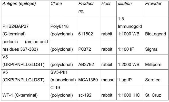
Table 10. List of primary antibodies
Antigen (epitope) Clone Product no.
Host dilution Provider
cleaved caspase-3 (polyclonal) 9661 rabbit 1:200 IHC Cell Signaling
Flag (DYKDDDDK) (polyclonal) F7425 rabbit
1:10,000
WB Sigma
Flag (DYKDDDDK)
M2
(monoclonal) F3165 mouse 1 µg IP Sigma
mec-2 rabbit 1:100 IF M. Chalfie
PHB1 (C-terminal)
Poly6031
(polyclonal) 603101 rabbit 1:1000 WB BioLegend
PHB1/PHB2 mixed (monoclonal) mouse undiluted IF
newly generated
31 no.
PHB2/BAP37 (C-terminal)
Poly6118
(polyclonal) 611802 rabbit 1:5
Immunogold
1:1000 WB BioLegend podocin (amino-acid
residues 367-383) (polyclonal) P0372 rabbit 1:100 IF Sigma V5
(GKPIPNPLLGLDST) (polyclonal) AB3792 rabbit 1:2000 WB Millipore V5
(GKPIPNPLLGLDST)
SV5-Pk1
(monoclonal) MCA1360 mouse 1 µg IP Serotec
WT-1 (C-terminal)
C-19
(polyclonal) sc-192 rabbit 1:1000 IHC St. Cruz
Table 11. List of secondary antibodies
Antigen (epitope) Product no Dilution Provider
Cy3-AffiniPure Donkey α-Rabbit IgG
(H+L) 711-165-152 1:400
Jackson ImmunoRe search F(ab')2 fragment anti-rabbit IgG
(H+L)-Biotin 711-066-152 1:500 Dianova
gold-labeled goat anti-rabbit IgG EM.GAR15 1:10
BBInternati onal Polyclonal goat anti-mouse
immunoglobulins/HRP P0447 1:30,000 Dako
Polyclonal goat anti-rabbit
immunoglobulins/HRP P0448 1:30,000 Dako
32 5.1.7 Enzymes
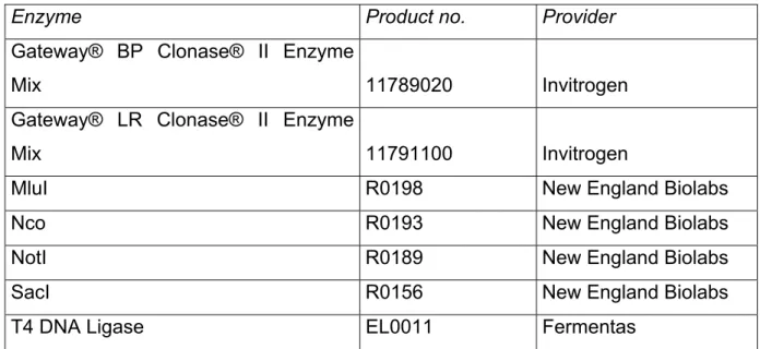
Table 12. List of enzymes
Enzyme Product no. Provider
Gateway® BP Clonase® II Enzyme
Mix 11789020 Invitrogen
Gateway® LR Clonase® II Enzyme
Mix 11791100 Invitrogen
MluI R0198 New England Biolabs
Nco R0193 New England Biolabs
NotI R0189 New England Biolabs
SacI R0156 New England Biolabs
T4 DNA Ligase EL0011 Fermentas
5.1.8 Materials Table 13. List of materials
Material Product no. Provider
8-Lid chain, flat 65.989.002 Sarstedt
10 cm dish for Agar Plates 82.1473 Sarstedt BD Microlance™ 3 27 G 3/4'' cannula 302200 BD Medical BD Plastipak™ syringe, 1 ml 300013 BD Medical BD Primaria™ cell culture dish 353803 BD Biosciences Blotting paper (Type BF4, 580 x 580) FT-2-521-
580580G
VWR
Cell culture dishes (12-well) 3513 Corning Cell culture dishes (6-well) 3516 Corning Cell culture dishes (10-cm) 430167 Corning
Cell culture dish (96-well F-Form) 655180 Greiner BioOne
Cell strainer, 100 µm 352360 BD Biosciences
Combs (10 well, 1 mm) for acrylamide gels
NC3010 Invitrogen
Combs (15 well, 1 mm) for acrylamide gels
NC3015 Invitrogen
33 Cover glass (round 18 mm no. 1.5) 631-0153 VWR
Cover glass (square18 mm no. 1.5) 631-0125 VWR
Crymold® standard 4557 Sakura
Discofix®-3 4098102 Braun
Dounce glass-glass homogenizer Wheaton Dynabeads® MPC®-S (magnetic
particle concentrator)
A13346 Invitrogen
Fast Optical 96-well plates 4346907 Applied Biosystems Flask rectangular 25 cm2 431463 Corning
Formvar/Carbon-coated nickel grids S162N Plano
Gel cassette (1 mm) NC2010 Invitrogen
Glass cuvette WINZER
Laborglastechnik
Histosette® I M499-11 Simport
Intrafix® SafeSet 4063000 Braun
Polypropylene conical tube (15 ml) 188271 Greiner BioOne Polypropylene conical tube (50 ml) 227261 Greiner BioOne
MicroAmp® Optical Adhesive Film 4311971 Applied Biosystem
Micro tubes (1.5 ml) 72.690.001 Sarstedt
Micro tubes (2 ml) 72.691 Sarstedt
Millipore Immobilon-P Transfer Membranes
T831.1 Carl Roth
Multiple well cluster plate, 96-well 3596 Corning Multiply®-µStrip 0.2 ml chain 72.985.002 Sarstedt
Nunc Cryotube™ 368632 Nunc
Nunc-immuno frame 460348 Nunc
Nunc-immuno module F8 maxisorb loose
469949 Nunc
Omnifix® Solo syringe, 10 ml 4617100V Braun PCR Soft-tubes 0.2 ml assorted
colours
711088 Biozym
34
Material Product no. Provider
PCR Soft-tubes 0.2 ml clear 710920 Biozym PCR Soft-tubes 0.2 ml 8 Tubes/Flat
Caps
710970 Biozym
Pipette tips (200 µl yellow) 70.760.002 Sarstedt Pipette tips (1000 µl blue) 70.762 Sarstedt
Polypropylene column 34964 Qiagen
Rotilabo®-syringe filters, 0.22 µm P666.1 Carl Roth Rotilabo®-syringe filters, 0.45 µm P667.1 Carl Roth Safe Lock 1.5 ml Eppendorf tubes 211-2130 VWR
Stripettes (5 ml) 4051 Corning
Stripettes (10 ml) 4101 Corning
Stripettes (25 ml) 4251 Corning
SuperFrost®/Plus microscope slides H867.1 Th.Geyer Group Syringe (PlastipakTM 1 ml) 7392/2007 BD
TipOne (0.1-10 µl XL), sterile S1110-3810-c Starlab TipOne (1-200 µl beveled), sterile S1111-1816-c Starlab TipOne (101-1000 µl graduated),
sterile
S1111-2831-c Starlab
Venofix® Safety 21 G 3/4'' 4056520-01 Braun
Weighing tray 140 x 140 mm 2159.1 Carl Roth Weighing tray 89 x 89 mm 2150.1 Carl Roth
5.1.9 Equipment Table 14. List of equipment
Equipment Model/Product no. Provider
Analogue tube roller SRT6 Stuart
Autoclave V-150 Systec
Avanti centrifuge J-301 Beckman
AxioCam ICc 1 Zeiss
AxioCam MRm Zeiss
Axiovert microscope 200M Carl Zeiss MicroImaging
35 Centrifuge (refrigerated) 5810 R Eppendorf
Confocal microscope LSM/Axiobserver Z1
LSM 710 Zeiss
Cryostat CM1850 UV Leica
Dumont #5 forceps 14098 WPI
Dumont #55 forceps 14099 WPI
Eppendorf Research® Multipipette (10-100 µl)
3122000043 Eppendorf
Fast Real-Time PCR System 7900HT ABI Fusion Solo chemiluminometer 60-FU-SOLO PeqLab Hamilton syringe (50 µl Type 705) 549-1155 VWR
HERAcell Incubator 240 Heraeus
HERAcell Incubator 150 Heraeus
High-speed centrifuge (refrigerated) RC-5C plus Sorvall Horizontal electrophoresis system 1582 - 030305 Dan-Kar
Incubator (Agarose) T 6030 Heraeus
Innova Incubator Shaker 4400 New Brunswick Scientific
Inverted microscope CK2 Olympus
JuLiTM Smart Fluorescence Cell Imager
DBJ01B Bulldog Bio
Laminar Flow Cabinet HS12 Heraeus
MacsMix Tube Rotator MX100 Miltenyi Biotech
Microcentrifuge 5424 Eppendorf
Microcentrifuge (refrigerated) 5417R Eppendorf Microcentrifuge (refrigerated) 5415 R Eppendorf
Microtome RM2235 Leica
Mithras multimode microplate reader LB 940 Berthold Technologies
Multifuge 4KR Heraues
Nanodrop Spectrophotometer 1000 PeqLab
Operating Scissor 501754 WPI
Pipetboy acu 155 015 Integra Biosciences AG
36
Equipment Model/Product no. Provider
Pipetman Pipette set (P2, P10, P100) F167500 Gilson Pipetman Pipette set (P20, P200,
P1000)
F167300 Gilson
Power supply (for Dan-Kar system) EPS200 Pharmacia Biotech Powerpac 200 Power supply 1655052 Bio-Rad
Powerpac 3000 Power supply 1655057 Bio-Rad
Shaker KS 260 IKA
Suction Pump MD 4C Vacuubrand
TEM CCD camera Megaview III Olympus
Thermal cycler (MJ mini) PTC-1148 Bio-Rad Thermomixer Comfort 1.5 ml
Thermoblock
5360 000.011 Eppendorf
Thermomixer Comfort shaker &
heating plate
5355 000.011 Eppendorf
Transmission electron microscope EM 902 Zeiss Transmission electron microscope JEM 1200 JEOL
Ultra-centrifuge (Optima) TLX-120 Beckman
Ultra-centrifuge rotor TLA-55 Beckman
Ultracut UCT ultramicrotome EM FCS Leica Ultrasound homogenisator
SONOPLUS
HD2070 Bandelin
Ultrasound water bath 2200 Branson
UV Transilluminator system MW312 Intas
Vannas Scissors 500086 WPI
Vortex Mixer MS525-20 Heidolph (Reax)
Water bath WNB 22 Memmert
Water bath (digital heating bath) HBR4 IKA Water bath (for paraffin sections) HI1210 Leica XCell SureLockTM Mini-Cell
electrophoresis system
100601-1408 Invitrogen
37 Table 15. List of software
Software Version Provider
Adobe Illustrator CS4 14.0.0 Adobe
Adobe Photoshop 11.0.0.0 Adobe
Axiovision 4.8 Carl Zeiss MicroImaging
Edit-Seq 5.06 DNASTAR
FinchTV 01.04.2000 Geospiza Inc.
Fusion-CAPT 15.16 Vilber Lourmat
Graphpad Prism 5 for Windows 5.2 GraphPad Software Inc.
ImageJ/Fiji 1.46j Wayne Rasband
Microsoft Office Suite 2003 Microsoft
MikroWin 2000 Berthold Technologies
Nanodrop 1000 3.7 Thermo Scientific
SDS Software 2.4 Applied Biosystems
ZEN Software 2009 Zeiss
Table 16. List of online software
Software (online) Website
BLOCK-iT™ RNAi Designer http://rnaidesigner.invitrogen.com/rnaiexpress/
Ensembl.org http://www.ensembl.org/
NCBI Primerblast software http://www.ncbi.nlm.nih.gov/tools/primer-blast/
NCBI Pubmed http://www.ncbi.nlm.nih.gov/pubmed/
NEB Double Digest Finder http://www.neb.com/nebecomm/doubledigestcalculator.
asp