Analysis of the arachidonyl-CoA synthetase ACSL4a as a potential regulator of BMP expression and
of the role of BMPs in timing of cell commitment along the dorsoventral axis of the gastrulating zebrafish embryo
I n a u g u r a l - D i s s e r t a t i o n zur
Erlangung des Doktorgrades
der Mathematisch-Naturwissenschaftlichen Fakultät
der Universität zu Köln
vorgelegt von
Björn Renisch
aus Bad Soden a. Ts.
Köln, 2009
Berichterstatter: Prof. Dr. Matthias Hammerschmidt
Berichterstatter: Prof. Dr. Siegfried Roth
Tag der mündlichen Prüfung: 27. November 2009
1. Summary ... 6
1.2 Zusammenfassung ... 8
2. General Introduction ... 10
2.1. Early zebrafish development ... 10
2.2 The establishment of the dorsal-ventral axis in zebrafish ... 12
2.3. The role of Bone Morphogenetic Proteins during gastrulation ... 14
2.4 The role of Bone Morphogenetic Proteins in stem cell biology ... 18
2.5. Aim of the work ... 20
3. Material and Methods ... 21
3.1. Zebrafish lines and husbandary ... 21
3.2. Embryological methods ... 21
3.3. Molecular methods ... 25
3.4. Materials ... 28
4. The role of acyl-coA-synthetase longchain family member 4 in dorsoventral patterning during zebrafish gastrulation ... 31
4.1. Introduction ... 31
4.1.1 The arachidonic acid pathway ... 31
4.1.2 The cyclooxygenase pathway of prostanoid production ... 32
4.1.3 The leukotriene and other pathway ... 35
4.1.4 The enzyme acyl-coA-synthetase longchain family member 4 (ACSL4) ... 35
4.1.5 Aim of the project ... 36
4.2. Results ... 38
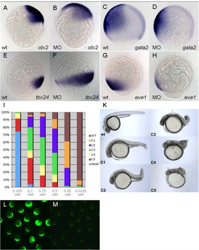
4.2.1. Knock-down with morpholino SP3005a causes dorsalisation of the zebrafish embryo ... 38
4.2.2. ACSL4a seems to be required for the maintenance of BMP signaling ... 41
4.2.3. During gastrulation, acsl4a is mainly expressed and required in the Yolk Syncytial Layer ... 44
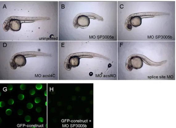
4.2.4. Further morpholinos targeting acsl4a do not lead to dorsalisation ... 47
4.2.5. Injection of acsl4a RNA does not lead to a rescue of the dorsalisation caused by morpholino SP3005a ... 48
4.2.6. Knockdown of acsl4a shows no specific defects during gastrulation comparable to loss of PGE2 signaling ... 52
4.2.7. Morpholinos targeting genes downstream of acsl4a show no phenotype during early development ... 53
4.2.8. Treatments with agonist and antagonist of the arachidonic acid pathway ... 55
4.3. Discussion ... 58
4.3.1. Knockdown of acsl4a create a new role for arachidonic acid metabolism during early embryonic development ... 58
4.3.2 acsl4a is expressed in the yolk syncytial layer of the gastrulating embryo, from where it might regulate dorsoventral patterning of the blastoderm ... 60
4.3.3 The phenotype of SP3005a morphants might be caused by off-target effects ... 62
4.3.4 Further components of the arachidonic acid cascade can most likely be excluded as regulators of dorsoventral patterning ... 63
4.3.5 Outlook and perspectives ... 66
5. Studies for cell commitment during early gastrulation stages ... 67
5.1. Introduction ... 67
5.1.1. During gastrulation cells are specified towards their final fate in a region-and time- dependent manner ... 67
5.1.2. Aim of the project ... 71
5.2. Results ... 73
5.2.1. Dorsal and ventral cells do not show significant commitment differences after heterotopic transplantations at shield stage ... 73
5.2.2. Dorsal and ventral cells do not show significant commitment differences after heterotopic transplantations at the 60-70% epiboly stage ... 78
5.2.3. Heterochronic transplantations of dorsal ectodermal cells from 60% epiboly stage donors into ventralised blastula stage embryos ... 81
5.2.4. Heterochronic transplantations of ventral ectodermal cells from 60% epiboly stage donors into dorsalised blastula stage embryos ... 83
5.2.4 Heterochronic transplantations of dorsal and ventral ectodermal cells of 80% epiboly stage embryos ... 86
5.3. Discussion ... 89
5.3.1. After heterotopic transplantations, both dorsal and ventral cells do not maintain their initial fate and can integrate into the new tissues ... 89
5.3.2. After heterochronic transplantations, both dorsal and ventral cells show an increased tendency to lack the neural as well as the epidermal marker ... 93
5.3.3. Outlook and further perspective ... 96
6. Large-scale screen using morpholino antisense nucleotides to identify new genes involved in early development, pituitary or skin development ... 98
6.1. Introduction ... 98
6.2. Results ... 98
6.3. Discussion ... 109
7. References ... 110
8. Appendix ... 130
Abbreviations ... 131
9. Acknowledgements ... 132
1. Summary
Signaling by Bone Morphogenetic proteins (BMPs) plays a pivotal role during early dorsoventral (D-V) patterning of fish and frog embryos, determining both differential cell fates as well as the direction of cell movements along the D-V axis during gastrulation.
Several forward genetics screens with ENU-mutagenised zebrafish have unravelled the indispensable function of several components of the BMP signaling pathway, including the BMP-ligands themselves, BMP receptors, the BMP-regulated transcription factor Smad5 and the extracellular BMP inhibitor Chordin. The BMP signaling pathway interacts with that of other signaling molecules, like Fibroblast Growth Factors or Wnts. However, there are still new genes to be discovered involved in early patterning processes; also the exact role of the BMP gradient in cell fate determination along the D-V axis of the gastrulating zebrafish embryo is not absolutely clarified.
As starting point of my thesis work I participated in a reverse genetics screen based on antisense morpholino oligonucleotides for specific gene knock-downs in zebrafish. As part of a consortium of several labs, I designed and screened a collection of morpholinos for phenotypes in early patterning, pituitary development and skin development. A morpholino targeting the gene acyl-CoA Synthetase longchain family member 4a (acsl4a), whose product is involved in the metabolism of the longchain fatty acid arachidonic acid, caused defects in D-V patterning.
In the first part of this thesis I characterise the phenotype of embryos after acsl4a knock- down, pointing to an essential role of Acsl4a in maintaining the BMP signaling gradient during gastrulation. However, the phenotype could only be obtained by one of several tested morpholinos. Also, I failed to rescue the defects by concomitant forced expression of acsl4a that is not targeted by the morpholino. Furthermore, I tested other components of the arachidonic acid metabolism, none of which interfered with early patterning of the zebrafish embryo. In conclusion, the role of acsl4a, and its connection to BMP signaling during D-V patterning of the zebrafish embryo remains elusive.
In the second part of the thesis I address the question of cell fate commitment, thus the timing of cell determination, along the D-V axis and its dependence on BMP signaling. According to the morphogen concept, the BMP gradient with from dorsal-to-ventral progressively increasing BMP signaling determines differential cell fates along the D-V axis. In the ectoderm, high BMP levels are thought to induce epidermal fates, while blocking neural development, so that ventral cells will give rise to skin, whereas neurons are formed from the
dorsal ectoderm, where BMP levels are lowest. In other instances, BMPs have been described as factors that maintain the pluripotency of stem cells, thus acting as a principle inhibitor of cell specification processes. Interestingly, the BMP gradient is highly dynamic over time, with a rather broad expression early, which becomes progressively restricted to the ventral side of the embryo during the course of gastrulation. Therefore, I wondered whether BMPs might keep the ventral ectoderm in a more pluripotent state, while dorsal cells become specified and committed to their final fate significantly earlier, consistent with the earlier loss of BMP expression. To test this notion, I carried out differential cell commitment studies by heterotopic and heterochronic transplantations of dorsal versus ventral ectodermal cells of the zebrafish embryo during different stages of gastrulation. Surprisingly, neither ventral nor dorsal cells were found to be committed to their initial fates, indicated by the loss of their respective marker genes expression upon transplantation into the ectopic environment.
However, only ventral cells were able to adopt the fate of their new environment (dorsal), whereas dorsal cells in an ectopic ventral environment lacked both dorsal and ventral marker gene expression. This indicates that ventral ectodermal cells do indeed maintain their pluripotency longer than dorsal cells. Further experiments with mutant embryos have to show whether this effect requires BMP signaling.
1.2 Zusammenfassung
Signale durch Bone Morphogenetic Proteins (BMPs) spielen im Zebrafish und Frosch Embryo eine entscheidende Rolle bei der Musterbildung entlang der dorsoventralen Achse, dabei bestimmen sie sowohl unterschiedliche Zellschicksale als auch die gerichtete Zellbewegung entlang der D/V-Achse während der Gastrulation.
Mehrere soegannte „forward genetic“ Screens mit ENU-mutagenisierten Zebrafischen haben die unentbehrliche Funktion von mehreren Komponenten des BMP-Signal-Wegs aufgezeigt, darunter die BMP-Liganden selbst, die Rezeptoren von BMPs, der durch BMPs regulierte Transkriptionsfaktor Smad5 und der extrazelluläre BMP-Inhibitor Chordin. Der BMP- Signalweg interagiert mit denen anderer Signalwege, wie Fibroblast Growth Factors oder Wnts. Jedoch gibt es immer noch neue Gene zu entdecken, die in Prozessen der frühen Musterbildung beteiligt sind; weiterhin ist die genaue Rolle des BMP Gradienten in der Festlegung von Zellschicksalen entlang der D-V Achse eines gastrulierenden Zebrafisch Embryos nicht vollkommen geklärt.
Als Ausgangspunkt meiner Doktorarbeit habe ich an einem rückwärts gerichteten genetischen Screen, basierend auf Antisense Morpholino Oligonukleotiden für spezifischen Knock-Down von Genen im Zebrafisch, teilgenommen. Als Teil eines Konsortiums mehrerer Arbeitsgruppen habe ich eine Kollektion von Morpholinos erstellt und auf Phänotypen in der Frühentwicklung, Entwicklung der Hypophyse und der Haut untersucht. Ein Morpholino, gerichtet gegen das Gen acyl-CoA Synthetase longchain family member 4a (acsl4a), dessen Produkt im Metabolismus der langkettigen Fettsäure Arachidonsäure beteiligt ist, verursachte Defekte in der D-V Musterbildung.
Im ersten Teil dieser Arbeit beschreibe ich den Phänotyp von Embryonen nach dem Knock- down von acsl4a, der auf eine essentielle Rolle von Acsl4a in der Aufrechterhaltung des Gradienten des BMP Signals während der Gastrulation hindeutet. Jedoch konnte der Phänotyp nur mit einem von mehreren untersuchten Morpholinos erreicht werden. Ebenfalls ist es mir missglückt die Defekte durch gleichzeitige Überexpression von acsl4a, welches nicht von dem Morpholino erkannt wird, zu retten. Ausserdem habe ich weitere Komponenten des Archidonsäure Metabolismus untersucht, jedoch keine der getesteten Komponenten interferierte mit der Frühentwicklung des Zebrafisch Embryos. Zusammengefasst bleibt die Rolle von acsl4a und seine Verbindung zum BMP-Signalweg während der D-V Musterbildung des Zebrafisch Embryos ungeklärt.
Im zweiten Teil dieser Arbeit befasse ich mit der Frage der Festlegung von Zellschicksalen, genauer der zeitlichen Folge der zellulären Bestimmung, entlang der D-V Achse und ihrer Abhängigkeit von BMP Signalen. Entsprechen dem Konzept von Morphogenen bestimmt der stufenweise von dorsal nach ventral ansteigende BMP Gradient die verschiedenen Zellschicksale entlang der D-V Achse. Im Ektoderm wird angenommen, dass ein hoher Grad von BMP Signal epidermale Schicksale induziert, bei gleichzeitigem blockieren der neuralen Entwicklung, so dass ventrale Zellen zu Haut werden, während Neuronen sich aus dem dorsalen Ektoderm, wo der Grad der BMP Signale am niedrigsten ist, entstehen. In anderen Zusammenhängen wurden BMPs als Faktoren beschrieben die die Pluripotenz von Stammzellen aufrechterhalten, dabei arbeiten sie als prinzipieller Inhibitor von Zellspezifikationsprozessen. Interessanterweise ist der Gradient von BMPs sehr dynamisch über die Zeit, mit einer sehr breiten Expression zu einem frühen Zeitpunkt, die während der Gastrulation schrittweise auf die ventrale Seite des Embryos beschränkt wird. Deswegen habe ich mich gefragt, ob BMPs das ventrale Ektoderm in einem mehr pluripotenten Zustand halten, während dorsale Zellen zu einem wesentlich früheren Zeitpunkt spezifiziert und auf ihr endgültiges Zellschicksal festgelegt werden, passend zu dem früheren Verlust der Expression von BMPs. Um diesen Gedanken zu überprüfen habe ich Studien zur zellulären Festlegung mit heterotopischen und heterochronischen Transplantationen von dorsalen gegen ventralen ektodermalen Zellen des Zebrafisch Embryos während verschiedener Zeitpunkte der Gastrulation durchgeführt. Überraschenderweise, konnte ich weder für ventrale noch dorsale Zellen zeigen, dass sie auf ihr ursprüngliches Schicksal festgelegt sind, da sie die Expression ihrer entsprechenden Markergene nach der Transplantation in eine ektopische Umgebung verlieren. Jedoch, nur ventrale Zellen waren fähig das Schicksal ihrer neuen Umgebung (dorsal) anzunehmen, wobei dorsalen Zellen in einer ektopischen ventralen Umgebung sowohl die Expression dorsaler als auch ventraler Markergene fehlt. Dies zeigt, dass ventrale ektodermale Zellen tatsächlich ihre Pluripotenz länger als dorsale Zellen behalten. Weitere Experimente mit mutanten Embryonen haben zu zeigen ob dieser Effekt Signale durch BMPs benötigt.
2. General Introduction
Higher animals develop from a fertilised egg to a three-dimensional, highly structured organismn consisting of multiple different cell types and tissues which - in the case of bilateria – are organized along at least two main body axes: the anterior-posterior (A-P) and the dorsal-ventral (D-V) axis. After fertilisation of the egg by a sperm cell, the first developmental process called cleavage is initiated, during which the zygote divides by several rounds of mitosis and (total or partial) cytokinesis into the so-called blastomeres. During these rapid cell divisions, the cell cycle just consists of an M- (mitosis) and an S- (DNA synthesis) phase, development is driven by maternally provided gene products, and cells become progressively smaller. However, once a certain ratio of DNA and cytoplasm is reached, G1 and G2 phases are established, cell divisions slow down, and zygotic transcription starts (mid- blastula-transition). After the blastula stages, the process of gastrulation starts, during which the three germlayers, ectoderm, mesoderm and endoderm, are formed. The ectoderm will later give rise to the epidermis, the nervous system and the neural crest, the mesoderm will form notochord, muscle, parts of the body skeleton, the blood, as well as vasculature and excretion tissues, and the endoderm will form the gastrointestinal system. In addition, the gastrulating embryo changes its overall shape and acquires a much more complex architecture, driven by the so-called morphogenetic cell movements during which mesoderm and endoderm become internalized. In addition, the body axes are set up and the general body plan is acquired.
Gastrulation is followed by neurulation, during which the neural tube of the central nervous system is formed, and by organogenesis, during which the organs of the animal develop (Gilbert, 2003).
All of these developmental processes are under the control of a complex system of signaling pathways and transcription factors. These factors act in a spatially and temporally strictly regulated and coordinated fashion. While some of them have redundant roles, others are absolutely indispensable, so that a single loss of their function would cause major changes in the embryonic body plan.
2.1. Early zebrafish development
In 1981, George Steisinger introduced the zebrafish (Danio rerio) as a genetic model system for vertebrate development (Streisinger et al., 1981). The large number of progeny, the transparency of embryos and larvae, and the extracorporal development makes the zebrafish
highly suitable for large scale forward genetics screens and embryological methods like cell transplantations, treatments with chemical compounds and microinjection to overexpress and knock-down specific genes (reverse genetics). In the following, I will give an introduction into the early patterning processes during gastrulation of zebrafish, focussing on the establishment of the dorsal-ventral axis.
The anteriorposterior axis of the zebrafish embryo already become apparent shortly after fertilization, when the cytoplasm accumulates on one side of the zygote (the animal pole) and the yolk granules on the other (the vegetal pole). This animal-vegetal axis corresponds to the later anterior-posterior axis, with the head forming at the animal pole and the tailbud at the vegetal pole. The first cleavage of the zebrafish zygote occurs around 45 minutes after fertilisation, followed by meroblastic cleavages every 15 minutes until the 128-cell-stage.
Between two hours and five hours post fertilisation (hpf), the blastula stages, the embryo, also called blastoderm, sits like a cap on the huge yolk cell, which does not cleave. At the 512- cell-stage (2.75 hpf), the embryo enters mid-blastula-transition (MBT; see above) and the first two cell lineages segregate from each other, the deep cells, which will form the proper embryo, and the Yolk Syncitial Layer (YSL), which does not contribute to the later animal.
During cleavage, the cytoplasm of the most marginal blastomere has remained in contact with the yolk cell and the YSL is formed when these blastomeres fuse to the yolk cell, thereby releasing their nuclei into the cytoplasma of the yolk cell, where they form a syncytial layer (Kimmel and Law, 1985b). Shortly after the MBT a third cell lineage is formed, the enveloping layer (EVL). It derives from superficial blastomeres that flatten out to form an embryonic skin, also called the periderm (Kimmel and Law, 1985c). It is currently unclear whether the periderm is maintained throughout the entire life of the fish, or eventuall shed off and replaced by derivatives of basal keratinocytes from the ventral ectoderm (see below).
Gastrulation starts at approximately 4.5 hpf with the morphogenetic movement of epiboly, which leads to a progressive covering of the yolk cell by the blastoderm (Kimmel et al., 1995;
Kimmel and Law, 1985a). Accordingly, gastrula stages are named in percentage of yolk coverage. The 30% epiboly stage is reached shortly after the onset of gastrulation, the 50%
epiboly stage at 5.5 hpf, the 70% epiboly stage (mid-gastrulation) and the 100% epiboly stage (end of gastrulation) at 10 hpf. In addition to epiboly, two other main morphogenetic movements of gastrulation can be distinguished: emboly (by some authors also called involution) and convergence and extension (C&E), both of which start at the 50% epiboly stage (Warga et al., 1990; Solnica-Krezel, 2005). During emboly, the mesoderm and the endoderm, which are induced in marginal cells facing the yolk cell and by signals emanating
from the YSL (Schier and Talbot, 2005), are brought into the interior of the embryo, whereas cells of the ectoderm remains outside. During convergence (Myers et al., 2002), lateral cells of both the ectoderm and the mesendoderm move towards the dorsal side to form the body axis. The dorsal side thickens accordingly. This thickening becomes first apparent at 6 hpf as a structure called the embryonic shield, which marks the dorsal side and which for the first time allows to morphologically recognize the dorsovental axis. Within this shield, the dorsal organizer, also called the Spemann-Mangold organizer, is located (see below). Driven by extension movements, the dorsal tissue further extends along the animal-vegetal / anterior- posterior axis of the embryo to form the body axis.
2.2 The establishment of the dorsal-ventral axis in zebrafish
Fate mapping experiments have shown that the future fate of an embryonic cell depends on its position within the late blastula and early blastula embryo (Kimmel et al., 1990). As outlined above, marginal cells facing the yolk will give rise to mesodermal and endodermal derivatives, while animal cells form to ectoderm. Of particular importance is also the position along the dorsoventral axis. Within the mesoderm, dorsal-most cells will form prechordal plate and notochord, dorsolateral cells somatic muscle, ventrolateral cells blood and ventral- most cells kidney and anus. Similarly, within the ectoderm, dorsal cells give rise to the neuroectoderm, intermediate cells to neural crest and placodes, and ventral-most cells to the epidermis. Initially organized within one plane along the dorsoventral circumference of the embryo, all of these cells types will move dorsally, so that eventually, the neuroectoderm will also be internalized and covered by skin. Over the past ten to fifteen years, enourmous progress has been made in eludidating the genetic and molecular network regulating dorso- ventral axis formation during early zebrafish development (Schier and Talbot, 2005). Two major steps can be distinguished; first, the formation of the Spemann-Mangold-organizer as the initial step of dorsoventral pattern formation (Nojima et al., 2004; Pelegri and Maischein, 1998), and second, the function of the organizer to further pattern the dorsoventral axis (Hammerschmidt and Mullins, 2002). Similar to the effects obtained by Mangold and Spemann with amphibian embryos, transplantation of the shield of zebrafish embryos into ventral regions leads to secondary body axis formation by an inducing ectopic muscle and neural development in adjacent tissue, while the organizer itself gives rise to prechordal plate, notochord and some fore- and midbrain tissues (Saúde et al., 2000). On the molecular level, organizer formation can be first recognized by the presence of ß-catenin protein in the nuclei
of the dorsal marginal cells and the YSL (Schneider et al., 1996). ß-catenin is an intracellular transducer of canonical Wnt (named because of D.melanogaster Wingless and vertebrate Integrated) signaling. In the absence of Wnt signaling, ß-catenin is phosphorylated by a complex of APC, Axin and the kinase GSK3, and thereby targeted for proteasomal degradation. However, upon binding of Wnt ligands to their Frizzled transmembrane receptors and the co-receptor LRP, this destruction complex is inactivated by Dishevelled and GBP, and ß-catenin can accumulate in the cytoplasm and translocate into the nucleus, where it participates as a partner of LEF/TCF transcription factors in transcriptional regulation (Logan and Nusse, 2004). While the nature of the Wnt ligand remains elusive, zebrafish mutants in ß- catenin-2 display compromised organizer formation and severe hyper-ventralization (Bellipanni et al., 2006). Among the first genes activated by ß-catenin is bozozok, a homeobox containing gene, which acts as repressor of ventralising genes like bmp2b , encoding a Bone morpogentic protein (BMP; see below) and vox/vent, encoding transcription factors mediating BMP signaling (Fekany et al., 1999; Imai et al., 2001; Leung et al., 2003; Shimizu et al., 2002). Other targets of ß-catenin are Nodal-like factors expressed in the YSL, mainly involved in the induction of the mesendoderm, but also required for the formation of the dorsal organizer (Dougan et al., 2003; Feldman et al., 1998). A second wave of ß-catenin dependent gene activation results in the expression of Fibroblast Growth Factors (FGFs) (Maegawa et al., 2006).
Fibroblast Growth Factors are involved in mesoderm formation, neural induction, dorsoventral and anterior-posterior patterning. FGFs signal via receptor-tyrosine kinases and the Ras/ MAPK pathway. During late blastula, fgf3, fgf8 and fgf24 expression is initiated in dorsal marginal cells, the precursors of the Spemann-Mangold organizer, where they promote expression of the BMP inhibitor Chordin (see also below), thereby restricting BMP activity and regulating dorsoventral patterning (Maegawa et al., 2006; Draper et al., 2003; Furthauer et al., 1999; Furthauer et al., 1997; Kiefer et al., 1996). Later in gastrulation, FGFs induce posterior neuroectodermal cell fates, but independent of the BMP signaling or organizer function (Rentzsch et al., 2004).
Another gene activated by ß-catenin and Nodal signals is dickkopf (dkk), constituting a negative feedback and temporal restriction of active Wnt signaling in the organizer domain.
dkk1 is expressed at the dorsal margin and later in the prechordal plate. Dkk protein binds the LRP subunit of the Wnt-receptor, thereby preventing the action of canonical Wnt signals and promoting the specification of anterior neural fates and axial mesendoderm, probably by blocking Wnt signals (Hashimoto et al., 2000; Shinya et al., 2000). Thus, while being
essential for early steps, canonical Wnt signaling needs to be actively blocked to allow later steps of dorsal development during mid- and late gastrulation. At the same time, active Wnt signaling, mainly via the ligands Wnt8 and Wnt3a, becomes indispensable for the specification of ventral and posterior cell fates (Erter et al., 2001; Lekven et al., 2001;
Shimizu et al., 2005), This switch form an early pro-dorsal to a later pro-ventral and anti- dorsal effect, while still quite puzzling, has also been observed for canonicalWnt signaling during amphibian development (Moon et al., 1997). Thus, it seems to be a more common phenomenon, which might point to a primary role of Wnt signaling in the timing of cell specification, rather than or in addition to cell fate determination per se (see also below).
2.3. The role of Bone Morphogenetic Proteins during gastrulation
Like the aforementioned Nodals, Bone Morphogenetic Proteins are members of the large family of TGFß signaling molecules. Zygotic expression of bmp2b and bmp7 in zebrafish starts right after mid-blastula transition. However, transcripts encoding Bmp4, Bmp7 and Radar, a homologue of the mammalian TGFß-family member Growth Differentiation Factor 6 (GDF6), are also maternally provided (Goutel et al., 2000; Kramer et al., 2002; Sidi et al., 2003). GDF6 is driving the expression of bmp2b and bmp7, thereby most likely acting via the BMP type I receptor Alk8 and the BMP-regulated transcription factor Smad5 (see also below) (Kramer et al., 2002; Sidi et al., 2003).. During late blastula and gastrula stages three bmps are prominently expressed, bmp2b, bmp7 and bmp4 , all of which are required for dorsoventral patterning and the establishment of ventrolateral fates (Dick et al., 2000; Kishimoto et al., 1997; Nguyen et al., 1998; Schmid et al., 2000; Stickney et al., 2007). Thus, bmp2b and bmp7 mutants are severely hyper-dorsalised, with an extension of muscle and neuroectodermal precursors into ventral most regions of the embryo, whereas ventral cell fates like epidermal, kidney or blood precursors are completely or largely absent. Compared to bmp2b and bmp7, however, the role of bmp4 during dorsoventral patterning is minor, whereas it is more important in specifing left-right asymmetry (Chen et al., 1997; Chocron et al., 2007; Hwang et al., 1997; Martinez-Barbera et al., 1997; Stickney et al., 2007)
The Bmp-ligands act as dimers. During dorsoventral patterning, Bmp2b and Bmp7 most likely work as heterodimer (Schmid et al., 2000; Little and Mullins, 2009). They bind to type a complex of type I and type II transmembrane receptor Ser/Thr kinases. To date five type I receptors are identified in zebrafish, alk3a and alk3b (Nikaido et al., 1999; Little and Mullins, 2009), alk6a and alk6b (Nikaido et al., 1999b; Little and Mullins, 2009) and alk8, which is
most similar to mammalian Alk2 (Bauer et al., 2001; Mintzer et al., 2001). The Bmp2b/7 heterodimers signal through heteromeric type I receptors consisting of Alk8, which is indispensable, and either of the four Alk3/6 molecules, which can replace each other in a redundant manner (Little and Mullins, 2009). Similar to Bmp4, the zebrafish orthologues of the BMP type II receptors have been found to be largely involved mediating BMP signaling during in left-right patterning (Monteiro et al., 2008), whereas during dorsoventral patterning, ActRII, a type II receptor shared with TGFß-like Nodal and Activin factors, seems to be used to mediate BMP signaling (Nagaso et al., 1999). Upon ligand binding the receptor cross- phosphorylate each other by their Ser/Thr kinase domain, followed by the phosphorylation of the receptor-regulated Smad (R-Smad) proteins (named after C.elegans Sma (Savage et al., 1996)and D.melanogaster Mad (Sekelsky et al., 1995) via the type I receptors. Upon phosphorylation, R-Smad disaggregate from the cytoplasmic part of the BMP receptors and associate with so-called Co-Smad proteins, with which they are translocated into the nucleus where they work as transcriptional regulators (Massague, 1996; Massague and Weis-Garcia, 1996; Raftery and Sutherland, 1999) (Fig.2-1). R-Smads specifically involved in BMP signal transduction are Smad1, Smad5 and Smad8, the most common Co-Smad shared by BMP and Nodal/Activin signaling is Smad4, while for zebrafish dorsoventral patterning, only Smad5 could thus far be shown to play an indispensable role (Hild et al., 1999; Kramer et al., 2002;
McReynolds et al., 2007). Several direct or indirect transcriptional target of Bmp/Smad target genes have been described, some of which in turn encode for regulators or components of the BMP signal transduction pathway, thereby constituting positive or negative feedback loops involved in the fine-tuning of BMP signaling. Examples are the bmp2b/4/7 genes themselves, constituting a positive feedback (Kishimoto et al., 1997), or the the smad7 gene, which encodes an inhibitory Smad protein, thereby constituting a negative feedback (Pogoda and Meyer, 2002).
In blastula stages bmp2b and bmp7 are broadly expressed throughout the embryo, sparing just the dorsal-most regions of the future Spemann-Mangold organizer, where their expression is repressed by Boz (Fekany et al., 1999; Leung et al., 2003). In addition, BMP inhibitors start to be expressed on the dorsal side, attenuating the positive feedback of BMP signaling on bmp2b/7 gene expression, and thereby progressively restricting BMP activity and bmp expression to the ventral side of the embryo (Hammerschmidt and Mullins, 2002).
The most relevant BMP inhibitor is the protein Chordin, which is secreted from organizer cells and has long-range signaling effects to attenuate BMP signaling in dorsolateral cells of both the ectoderm and the mesenderm (Schulte-Merker et al., 1997; Hammerschmidt et al.,
1996). Accordingly, chordin mutants are hyper-ventralised, with a reduction in the number of neuroectodermal and muscle precursor cells, while derivatives of the ventral ectoderm and mesoderm are made in excess. The organizer and adjacent dorsal tissues also make generate other extracellular Bmp inhibitors such as Noggin-1 or Follistatin-like 2. Compared to Chordin, however, they are less important (Bauer et al., 1998; Dal-Pra et al., 2006; Fainsod et al., 1997; Furthauer et al., 1999; Zimmerman et al., 1996). The activity of Chordin itself is also under tight control. Thus, it is cleaved by metalloproteases Tolloid/Bmp1, which are most active on the ventral side of the embryo (Blader et al., 1997; Connors et al., 1999;
Muraoka et al., 2006). Tolloid, in turn, is inactived upon binding to Sizzled, a secreted protein promoting Chordin activity, but in contrast to Chordin made on the ventral side in response to Bmp signaling (Muraoka et al., 2006; Yabe et al., 2003). Further secreted and BMP-binding factors like Twisted gastrulation (Little and Mullins, 2004; Ross et al., 2001; Xi and Fisher, 2005) and Crossveinless-2 (Rentzsch et al., 2006; Zhang et al., 2008) can regulate BMP signaling in a context-dependent positive or negative manner.
The BMP activity gradient is important for the establishment of the dorsal-ventral axis, as BMP play important roles in cell fate determination and differentiation. Among the target genes of BMP signaling is deltaNp63 that is directly activated by BMP/Smad5 signaling in the ventral ectoderm and involved in the development of basal keratinocytes of the epidermis (Lee and Kimelman, 2002; Rentzsch et al., 2003). Similarly, although no direct target genes have been identified via DNA binding and transactivation as yet, BMP signaling is supposed to activate the expression of blood or kidney-specifying genes in the ventral mesoderm.
Furthermore, according to the morphogen concept, it should with lower thresholds activate ectodermal genes involved in the specification of neural crest and placodal ectoderm.
In addition to cell fate specification along the dorsoventral axis, the BMP gradient has been shown to govern cell migrations along the dorsoventral axis during dorsal convergence movements, establishing a reverse gradient of cadherin-dependent cell-cell adhesivness (von der Hardt et al., 2007).
An additional aspect of BMP signaling similar to that alluded to above for canoncial Wnt signaling could be concerned with the timing of cell specification, rather than or in addition to determining the exact fate specification cells will require. Such a role would be consistent with the stem cell-maintaining effect of BMP signaling in other developmental contexts.
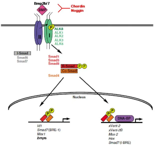
Figure 2-1: The signaling pathway of Bone Morphogenetic Proteins (modified from Varga and Wrana, 2005)
During dorsoventral patterning in zebrafish, the ligands Bmp2b and Bmp7 bind as heterodimer to a heteromeric complex of consisting of the type I receptors Alk8 and Alk3/6, and most likely the type II receptor ActRII. The intracellular signal transducers Smad1, 5, 8 become phosphorylated after receptor activation, associate with Smad4 and transloctate to the nucleus where they participate in transcriptional regulation. Inhibitors of BMP signaling like chordin and noggin bind to the ligands and prevent activation of the receptor. Among the target genes are the regulators and components of the BMP signal transducting pathway, thereby constituting positive and negative feedback loops.
2.4 The role of Bone Morphogenetic Proteins in stem cell biology
Stem cells are undifferentiated cells that have the ability of renewal while remaining capable of differentiating into a specialised cell (Chambers and Smith, 2004). The differentiation potentials can vary, depending on the stem cell type. A fertilized egg is totipotent, and can give rise to any cell type including extraembryonic tissues. Pluripotent stem cell is the term used for embryonic stem cells that can differentiate into any cell type of the embryo proper.
Multipotent stem cells are usually stem cells that are restricted to a tissue or cell lineage, for example hematopoietic stem cell, the progenitors of all blood cells. Even more restricted cells are often called oligopotent. And cells that are still self-renewing but only differentiate into one cell type by asynchronous cell division are called unipotent stem cell, for example the satellite cell of adult muscles (Scholer, 2004).
Several intrinsic and extrinsic factors are known to regulate and maintain the fate of a stem cell and to prevent it from differentiating, like the POU-domain transcription factor Oct4 (Nichols et al., 1998a), homeodomain protein Nanog (Chambers et al., 2003; Mitsui et al., 2003), the cytokine Leukemia Inhibitory Factor (LIF) (Matsuda et al., 1999), the prostaglandin PGE2 (North et al., 2007), Wnts (Nusse, 2008; Sato et al., 2004), FGFs (Dvorak et al., 2006) and BMPs (Varga and Wrana, 2005; Ying et al., 2003; Zhang and Li, 2005).
The effects of BMPs on stem cells can vary up to being seemingly contrary, depending on the cell types and the context. Embryonic stem cells of mouse (mES) cultured on a stromal feeder layer and treated with serum differentiate into neurons unless treated with BMPs, which will drive them towards epidermal differentiation (Kawasaki et al., 2000), consistent with the aforementioned positive effects of BMP signaling on the developmental of ventral (skin) versus dorsal (neural) cell types in the ectoderm of the zebrafish embryo. However, when mES cells are incubated in a less complex medium (supplemented with LIF and FGF2), BMP will also exhibit their a neural-blocking effect. However, rather than promoting their epidermal differentation, they will keep the mES in an undifferentiated state, as for instance indicated by the expression of the stem cell-specific marker Oct4 (Munoz-Sanjuan and Brivanlou, 2002; Tropepe et al., 2001; (Ying et al., 2003). This effect is most likely achieved by interfering with LIF function and suppressing the transcription of differentiation genes via blocking MAPK pathways (Qi et al., 2004). Along these lines, it is tempting to speculate that the primary function of BMP signaling might be concerned with blocking neural development, consistent with the neural default model (Stern, 2005), while for epidermal
specification, additional factors are necessary. Similarly, neural specification most likely requires other factors in addition to BMP inhibitors. Thus, Chordin and Noggin fail to induce neural fate in the absence of FGF signaling (Launay et al., 1996; Sasai et al., 1996). In addition, Wnts have been shown to be required for neural induction (Baker et al., 1999).
However, it remains a matter of debate to which extent FGFs and Wnts act as direct neural inducers, and to which extent by interfering with and blocking BMP signaling (Furthauer et al., 1997; Furthauer et al., 2004; Rentzsch et al., 2004). FGF signaling for instance can block BMP signal transduction via its downstream effector MAPK, which phosphoralytes Smad1 at a specific site of the linker region., thereby preventing its translocation into the nucleus (Pera et al., 2003). Similarly, Wnts can crosstalk to BMPs via phosphorylation of Smads through GSK3 (Fuentealba et al., 2007). In conclusion, the role of BMP signaling in stem cell biology has to be seen in relation to other signaling pathways.
A specification-blocking effect as observed in mES cells would also be consistent with the reported roles of Bmp2, Bmp4 and Bmp8b and their Drosophila orthologue Decapentaplegic to maintain germ line stem cells in the mammalian testis and the fly ovary, respectively (Xie et al., 1998; (Funk et al., 2002). However, experiments with human embryonic stem cells (hES) indicate an opposite role for BMPs. Here, BMP-2 promotes the differentiation of hES, mainly into extraembryonic endoderm, accompanied by a loss of stem cell marker expression, whereas the BMP inhibitor Noggin blocks this differentiation (Pera et al., 2004).
For dorsovental patterning of the zebrafish embryo, there are different data sets in line with a possible differentiation -blocking and/or stem cell-maintaining role of BMP signaling. One comes form the analysis of mutants in Pou2, the zebrafish orthologue of the stem cell maintenance factor Oct4 (Belting et al., 2001; Reim and Brand, 2002). Mutants lacking both maternal and zygotic pou2 gene products display loss of bmp expression and a hyper- dorsalization similar to that of bmp mutants. Futhermore, the dorsalised phenotype of pou2 mutants can be rescued by re-introduction of bmp transcripts (Reim and Brand, 2006), suggesting that Pou2 acts via BMPs, and that the dorsalised phenotype might result from precocious specification of ventral cells. Consistent with this notion, experiments with transgene-driven temporally controlled BMP inhibition revealed that compared to dorsal cell types, deriviates of ventral cells require sustained BMP signaling (Pyati et al., 2005, 2006).
2.5. Aim of the work
The genetic network controlling dorsoventral patterning and axis formation of the vertebrate embryo, and the pivotal role of BMP signaling within this control system, are rather well understood. However, several questions remain open. To identify new genes involved in developmental processes I joined a consortium for a large-scale reverse genetic screen using antisense morpholino oligonucleotides (Pickart et al., 2006). During the screen I found acyl- CoA synthetase longchain family member 4a (acsl4a) to be required for dorsal-ventral patterning of the zebrafish gastrula. Acsl4a catalyzes the synthesis of a range of arachachidonic acid derivates, such as prostaglandins. Interestingly, the prostaglandin PGE2 has recently been shown to be required for the maintainence of hematopoietic stem cells of zebrafish larvae (North et al., 2007), raising the possibility that it might fulfil a similar function to allow proper ventral development during dorsoventral patterning of the early zebrafish embryo. This would be consistent with the stem cell maintaining roles of Pou2 and BMP signaling described above, and was subject of the first part of my thesis work.
If BMPs and their partners do indeed pattern the dorsoventral axis by attenuating cell specification, ventral cells, which receive high and longer BMP signaling, should become specified later than dorsal cells, in which BMP signaling is weaker and less persistent. In the second part of my thesis, I tested this notion, carrying out cell commitment studies via heterotopic and heterochronic transplantations of ectodermal cells.
3. Material and Methods
3.1. Zebrafish lines and husbandary
Wild-type embryos were obtained from crosses of TL/EK or AB zebrafish. gsc::GFP transgenic embryos were obtained from outcross of heterozygous fish with wildtype strains.
Embryos were raised and staged according to Kimmel et al. (1995). Dechorionation was performed with Watchmakers forceps, and embryos older than 15 hpf, which were to be manipulated were anaesthetized by addition of 0.2% v/v Tricaine to embryo medium.
Dechorionated embryos were kept in petridishes coated with 1% agarose.
3.2. Embryological methods
Microinjection of zebrafish embryos
Morpholino oligonucleotides (MOs) were either purchased from Gene Tools and a stock of 1mM was prepared, or the screen consortium sent aliquots of 25µg/µl stocks.
Synthetic mRNA, MOs, or linage tracers were diluted to working concentrations in Danieau- buffered solution (Nasevicius and Ekker, 2000) containing 10% phenol red (Sigma). A volume of 2 nl was injected into the yolk cell of 1-2 cell stage embryos with a fine glas needle connected to a pressure-driven P420 picopump (World Precision Instruments) and an M3310 manual micromanipulator (Science Products).
For the injection of MOs and dyes into the yolk cell, embryos were dechorionated and injected through a very narrow glass capillary with a M3310 Manual micromanipulator at high stage (3-3,5hpf).
The working concentrations are specified in the text; used RNA constructs and MO sequences are given below.
Cell transplantation experiments
Donor embryos were injected at 1-cell stage with fluorescent lineage tracers like fluorescein dextran (1mg/ml) or rhodamin dextran (1mg/ml). For the heterochronic transplantations the Host embryos were injected with in vitro synthesised RNA of truncated BMP receptor Ia or constitutive active alk8, three hours after the donor embryos were injected. Donor and Host embryos were manually dechorionated and placed into 1% methylcellulose on groove-slides.
Depending on the experiments around 10 cells were transplanted with a glass capillary from the dorsal or ventral ectoderm embryo to the ventral or dorsal ectoderm, respectively, of the
host embryos in case of heterotopic transplantation; or into the animal pole of ventralised or dorsalised Host embryos in case of heterochronic transplantations; for a detailed describtion of the experiments performed see chapter 5. After the transplantation procedure the embryos were transferred into a coated petridish.
Treatment of embryos with chemical compounds
The chemical compounds were diluted in DMSO, MeOH or water to prepare stock solutions according to the instruction manuals. The working solutions were prepared just prior to use by the desired dilution of the stock solution in Embryo medium, in case of acidic solution an embryo medium with citrate-buffer was used. Embryos to be treated with agonist and antagonist of the arachidonic acid cascade were dechorionated and transferred into 20ml glass vials, maximum 20 embryos per vial and covered with 1-2ml Embryo medium, so that the embryos are just covered with water. The medium was replaced with working solutions; the control clutch was treated with embryo medium containing the appropriate solvent. Embryos were kept in the dark and slightly shaked during incubation. The working solutions were replaced by a fresh one every two hours until end of the treatment.
in vitro transcription of sense RNA (RNA synthesis for micro-injection)
For in vitro transcription of the different sense RNAs 10µg of the respective plasmids were linearized with 2µl restriction enzyme and 10µl Buffermix in a total volume of 100µl for 2h at appropiate temperature; followed by a Phenol/ Chloroform extraction. Linearised plasmids were checked on a 0,8% agarose gel and quantified by photometry.
For in vitro transcription the mMessage mMachine Kit was used. 2 µg linearized plasmid DNA was mixed with 2 µL 10x transcription buffer, 2 µL NTP Mix, 2 µL enzyme mix in a 20 µL reaction and incubated at 37°C for 2 hours. 1 µL DNase was added and the mixture was incubated at 37°C for another 20 minutes to remove the template DNA. The reaction was stopped and purified by addition of 15µl ammonium acetate, 115 µL water and 150µl of Phenol/ Chloroform Mixture (Roth), followed by centrifugation at 14000 rpm for 3 minutes.
The liquid phase was transferred into a new microreaction tube and mixed with 150µl Isopropanol, incubated for 20 minutes at -20°C and followed by centrifugation at 14000 rpm at 4°C for 15 minutes. The pellet was resuspend in 90µl ddH2O, 10µl NH4Ac and 100µl Isopropanol, incubated for 20 minutes at -20°C and followed by centrifugation at 14000 rpm at 4°C for 15 minutes again. The pellet was rinsed in 500 µL 70% ethanol and resuspended in 20 µL ddH2O. The RNA was checked on a 1% agarose gel and stored at -20°C.
Whole-mount in situ-hybridizations
Synthesis of labeled anti-sense RNA (in situ probe synthesis)
Linearisation and purification of Plasmid were done as described for sense RNA synthesis.
For in vitro transcription 2 µg linearized plasmid DNA was mixed with 2 µL 10x transcription buffer, 2 µL Dig-RNA labeling Mix or FITC-RNA labeling Mix (Roche), 1 µL RNasin (Promega), 2 µL of the appropiate polymerase (T7, T3, Sp6) in a 20 µL reaction and incubated at 37°C for 2 hours. 2 µL of 10x transcription buffer, 17 µL ddH2O and 1 µL DNase (RNase free, Roche) were added and the mixture was incubated at 37°C for another 30 minutes to remove the template DNA. Free nucleotides were removed by addition of 20 µL 7.8 M ammonium acetate and 120 µL ethanol and incubation at room temperature for 50 minutes, followed by centrifugation at 14000 rpm for 20 minutes. The pellet was rinsed in 300 µL 70% ethanol and resuspended in 20 µL ddH2O. 20 µL formamide (Fluka) was added and RNA was stored at -20°C.
Whole-mount in situ hybridization
Embryos for in situ-hybridization were fixed in 4% PFA/PBS overnight at 4°C or 4 hours at room temperature, washed in PBST, dechorionated, transferred though a methanol series and stored in 100% methanol at -20°C.
For in situ-hybridization embryos were transferred from MeOH to PBST and depending on their developmental stage digested with 1 mg/ml proteinase K (24 hpf: 8 minutes; 36 hpf: 10 minutes; 48 hpf: 15 minutes; 72 hpf: 20 minutes) at room temperature and re-fixed in 4%
PFA/PBS for 20 minutes. From PBST embryos were rinsed in Hyb- and incubated in Hyb+ at 70°C for a minimum of 4 hours. RNA-probes were diluted in Hyb+ to working concentration and incubated with embryos at 70°C overnight. On the next day embryos were washed at 70°C with the following buffers: 100% Hyb-, 25% 2xSSCT/ 75% Hyb-, 50% 2xSSCT/ 50%
Hyb-, 75% 2xSSCT/25% Hyb-, 2x SSCT for 15 minutes each and 0.2x SSCT for 30 minutes two times. Then embryos were transferred into PBS buffered conditions at room temperature (75% 0.2xSSCT/ 25% PBST; 50% 0.2x SSCT/ 50% PBST; 25% 0.2x SSCT/ 75% PBST;
PBST for 10 minutes each). Embryos were blocked against unspecific binding for at least 2 hours in 2% sheep serum/ 2% BSA/ PBST. For probe detection embryos were incubated with alkaline phosphatase-coupled antibody overnight at 4°C. Embryos were intensively washed with PBST, transferred into staining buffer (5 minutes for three times) followed by detection of alkaline phosphatase activity by addition of 1:50 NBT/BCIP (blue) in staining buffer. For fluorescent in situs embryos were transferred into 0,1M Tris-HCl buffer adjusted to pH 8.0 (three times for 5 minutes) followed by addition of 1 tablet/ ml of FastRed and Tris buffer
(Sigma). The color reaction was stopped by rinsing the embryos with PBST and fixing in 4%
PFA/PBS overnight. Stained embryos were either transferred though a methanol series into benzylbezoat/benzylalcohol (2:1) for microscopy or transferred into 80% glycerol in PBS.
PBST: PBS (D8662, Sigma), 0.1% Tween20
20xSSC: 175.3 g NaCl, 88.2 g Sodiumcitrate dehydrate (C6H5Na3O7 x 2H2O), pH 7.0; adjust to 1 L with distilled water.
Stain: 100 mM Tris (pH 9.5), 50 mM MgCl2, 100 mM NaCl, 0.1% Tween20
Immunohistochemistry/ whole-mount antibody staining
After fixation and reconstitution as described in 7.2.2, embryos were incubated for 1-2 hours at room temperature in antibody blocking solution. This was replaced by primary antibody in blocking solution and incubated overnight at 4°C. The following embryos were washed six times in PBST for 15 minutes, blocked for 1 hour before appropriate secondary antibody in blocking solution was added for 2 hours at room temperature or overnight at 4°C. The secondary antibody was discarded and embryos were washed six times in PBST.
Fluorescently stained specimens were fixed in 4% PFA/PBS and transferred to 60%
glycerol/40% PBS.
For DAB staining, the signal was amplified using the Vectastain Elite ABC Kit (Vector Laboratories). This system consists of avidine and biotin-labeled horseradish peroxidase, the two of which form multi-molecule complexes of avidine. The peroxidase is recruited to the secondary antibody by free biotin-binding sites in the avidine molecules. After incubation in AB solution (<24 hpf for 45 minutes; 24 hpf for 1 hour; 2-3 dpf 3-4 hours; >3 dpf for 4 hours) embryos were rinsed and washed six times for 30 minutes in PBST and incubated 30 minutes in DAB solution. The detection reaction was started by addition of 1 µL 0.3% H2O2 per 1 ml DAB solution. Staining reactions were stopped by rinsing in PBST, followed by fixation in 4% PFA/PBS. Stained embryos were transferred through a methanol series and stored and analyzed in benzylbenzoat/benzylalcohol (2:1).
Blocking solution: 10% fetal calf serum; 1% DMSO; 0.1% Tween 20; in PBS
DAB solution: 1 DAB tablet (3, 3-Diaminobenzidine tetrahydrochloride, Sigma, D5905) was dissolved in 15 mL PBST and filtered through a 0.4 µm micro filter prior to use.
Microscopy
Stained embryos were analysed in either benzylbenzoat/benzylalcohol (2:1) mixture or in 80%Glycerol/20%PBS; live embryos were mounted in 1% methylcellulose or 1% low melting agarose. Documentation was done using a Zeiss AxioImager with an AxioCam MRc for light microscopy and a Zeiss LSM 510/ 710 for confocal microscopy.
3.3. Molecular methods
Bacterial growth and plasmid preparation
Available bacterial clones were ordered from Imagenes.
Glycerol stocks or agar stabs of bacterial cell stocks were propagated by streaking on LB-agar plates containing 50 µg/mL ampicillin or 25 µg/mL kanamycin, and incubated overnight at 37°C. After inoculation with single colonies, bacterial cultures were grown in LB-medium supplemented with appropriate selective antibiotics in a shaking 37°C-incubator.
Small yield plasmid preparations from 3 mL bactertial cultures (Mini-preparations) were done with the QIAprepSpin Miniprep Kit (Qiagen 27106) as per manufacturer instructions, to obtain high quality DNA for subsequent sequencing and cloning procedures.
Medium yield plasmid preparations from 50 mL cultures (Midi-preparations) were made with the QIAfilter plasmid Midi Kit (Qiagen 12243), for cloning and preparing stocks of plasmid DNA.
Isolation of total RNA from embryos
50 embryos were mixed with 500 µL E3-medium and 1.5 mL Trizol LS Reagent (Gibco, BRL) and homogenized using a seringe. 400 µL chloroform was added after 5 minutes incubation, followed by vortexing and 10 minutes incubation at room temperature.
Afterwards the cell debris was spun down at 13000 rpm and the supernatant, containing DNA and RNA, transferred into a fresh tube. For the precipitation 1mL isopropanol was added.
After centrifugation at 14000 rpm at 4°C the pellet was washed with 70% ethanol, dried and taken up in RNAse-free ddH2O.
Genomic DNA was removed by a DNase (Boehringer) treatment, followed by a phenol/chloroform (Roth) extraction. The concentration of the purified RNA was measured at 540nm using Standard photometer.
cDNA synthesis
In a 10.6 µL reaction, 2 µg of total RNA together with 1 µL oligo (dT)15 (100pmol, Roche) was denatured at 70°C for 5 minutes then stored on ice. Reverse transcriptase buffer, 2 mM DTT, 10 mM dNTP and 200 U SuperScript II reverse transcriptase (Gibco, BRL) were added to give a final volume of 20 µL. After incubation at 42°C for 1.5 hours 20µl Tricine EDTA buffer (Clontech RACE) was added and the cDNA stored at -80°C.
Restriction digests
Digestion of DNA by restriction enzymes was performed using the conditions recommended by the manual (New England Biollabs).
Electrophoretic separation of DNA
Size fractionation of DNA fragments was performed by electrophoresis on 0.8-2% (w/v) agarose (Roth) gels in 1xTAE containing 100 ng/mL ethidium bromide, submerged in 1xTAE running buffer. Samples were mixed with loading buffer and run at 100-150 V. The DNA was visualized and photographed under UV light (λ=312 nm).
50x TAE (1L): 242 g Tris base, 57.1 mL glacial acetic acid, 100 mL 0.5 M EDTA (pH 8.0)
Purification of DNA fragments
For recovery of DNA from agarose gels, bands of specific sizes were cut from the gel on a transilluminator and purified with the QIAquick SpinColumn Gel Elution as per manufacturer instructions.
DNA concentration was determined by UV spectrophotometry at a wavelength of 260 nm.
Polymerase Chain Reaction (PCR)
PCR reactions were usually carried out at a final volume of 25 µL, containing 1xPCR-buffer, 0.2 mM dNTP (PeqLab), 1 µM forward and reverse primers (MWG or Sigma), 2.5 U Taq DNA polymerase (Genaxxon), or proof reading polymerase Pfu or advantage Taq (Roche), and template DNA.
Amplification was done in DNAengine Thermocycler (Applied Biosystems), using programs with 35-40 cycles of denaturation, annealing and elongation.
For temporal expression profiling, RT-PCR was performed using 1 µL cDNA (described in 7.1.5) in above described PCR mixtures. cDNA samples were adjusted to equivalent relative
mRNA amounts via control PCR reactions with primers specific for the house-keeping gene ef1α (Nordnes et al., 1994).
Sequencing
DNA sequencing was done using the chain termination method, which uses fluorophore labeled didesoxynucleotide phosphate to terminate strand elongation. Sequencing reactions were performed using 400-600 ng plasmid-DNA or 10-50 ng PCR product purified with the appropriate Qiagen Kits mentioned above. The DNA, 3 pmol primer and 1 µL BigDyeTerminator Mix (Applied Biosystems) were mixed to a 12 µL reaction. The amplification program for plasmid sequencing started with an initial template denaturation at 96°C for 2 minutes, followed by 25 cycles consisting of denaturation at 96°C for 10 seconds, primer annealing at 53°C (unless otherwise required with gene specific primers) for 5 seconds and extension at 60°C for 4 minutes. After addition of 8 µL ddH20, samples were purified and column chromatography was performed at the in-house sequencing facility with an ABI DNA sequencer module.
Cloning of DNA fragments
For cloning of coding sequences from cDNA, PCR products were amplified using proofreading polymerases (Pfu, Fermentas). For further subcloning, primers with restrictions sites at the 5’end were designed; the restriction enzymes have be non-cutters in the cloned sequence. After purification, PCR products were cloned by non-directional cloning into pGem-T Easy vector (Promega) or pCRII Topo vector (Invitrogen) following manufators instructions.
To clone the DNA fragments into the destination vector, pGem-T easy or Topo vector containing the correct insert, proven by sequencing, were completely digested with the choosen restriction enzymes. After complete DNA restriction und purification of the insert DNA, ligations were performed using 50-100 ng vector DNA and a three molar excess of insert DNA, catalysed by 10 U T4-DNA-ligase in a 10 µL reaction. When required, dephosphorylation of DNA ends prior to ligation was achieved by addition of 1 U Shrimp Alkaline Phosphatase in 10 µL, incubation at 37°C for 30 minutes and inactivation of the phosphatase at 65°C for 15 minutes. Ligations were incubated at 16°C overnight or at room temperature for 2 hours.
Plasmids were transformed into chemo-competent E. coli strains (DH5α or TOP10) via heat- shock at 42°C for 45 seconds, followed by 2 minutes cooling on ice.