and blood vessels in the development of the zebrafish thyroid gland
Inaugural-Dissertation zur Erlangung des Doktorgrades
Der Mathematisch-Naturwissenschaftlichen Fakultät Der Universität zu köln
Vorgelegt von Osama Ahmed Elsalini
Aus Benghazi, Libya
Köln 2006
Page CONTENTS ………...
ACKNOWLEDGMENTS ………..
LIST OF FIGURES ………
LIST OF TABLES ………..
1. INTRODUCTION ………..
1.1. Zebrafish as a model organism………
1.2. Zebrafish embryonic development………
1.3. Thyroid gland………
1.4. Thyroid gland in Zebrafish………
1.5. Development of the thyroid gland………
1.6. Development of Endoderm………
1.7. Development of the vascular system………
1.8. Hedgehog………
1.9. Aim of the work………
. 2. MATERIALS AND METHODS……….
.
. 2.1. Used chemicals and materials………
. 2.2. Methods………
. 2.3. Molecular biology techniques………
. 2.4. Histological methods………
. 2.5. Cyclopamine treatments………
3. RESULTS
……….3.1. Thyroid differentiation………
3.1.1. Nodal signalling and subsequent steps of endoderm specification are required for thyroid development……….
3.1.2. hhex is required for differentiation of thyroid follicles but not for formation or migration of a thyroid primordium………
3.2. Thyroid Morphogenesis………
3.2.1. Hedgehog signalling is required for guidance of thyroid follicles along the midline……….
A C D F
1 1 1 4 6 8 9 12 15 18
19 19 22 24 25 29
30 30
30
33 41
41
A
3.2..3 Endothelial cells are required for directing thyroid growth along the pharyngeal midline in zebra fish………
4. DISCUSSION………..
4.1. Thyroid differentiation………
4.1.1. Establishment of endoderm by Nodal signalling and its
downstream effectors is a prerequisite for thyroid development………
4.1.2. A late role of hhex in zebrafish thyroid development………
4.2. Thyroid Morphogenesis………
4.2.1. Members of both the Hedgehog and the Vegf pathway might be involved in the pathogenesis of localisation defects in human thyroid development………
4.2.2. Endothelial cells are able to influence thyroid morphology in zebrafish……….
5. CONCLUSION ……….
6. BSTRACT………..
7. Zusammenfassung………
8. REFERENCES ……….
9. ABBRAVIATION………
10. Erklärung...
11. Lebenslauf………..
49
52 52
52 52 56
56
57
59 60 61 63 72 73 74
B
My sincere thanks are due to the Department of Developmental Biology, University of Cologne for giving me the opportunity to complete my higher study, and to the Libyan High Ministry of Education and Garyounis University for the financial support.
I should express my grateful and thanks to Dr. Klaus Rohr for his supervision on my thesis and for the valuable advice and suggestions. Also my great thanks to Prof. Dr. Siegfried Roth and Prof. Dr. Einhard Schierenberg.
My thanks should goes to the following people for their unlimited help during the practical part of the thesis: Burkhard Alt, Thomas Wendl, Dejan Adzic, Julia von Gartzen and Natalia Feitosa,.
My thanks are also due to Mr. Masanari Takamiya for his discussion and support.
My great thanks should go to my wife for her unlimited help during my study. Also, great thanks to my family (Father, Mother, Sisters and Brothers) in Benghazi– Libya, for their support.
C
Figure 1: Zebrafish developmental series……… 2 Figure 2: The thyroid gland in human……… 5 Figure 3: (A) the normal position and distribution of thyroid gland in adult zebrafish and (B) the structure of thyroid follicular cell in zebrafish……….. 7 Figure 4: The thyroid follicular cells derive from the endodermal tissue in the pharynx in zebrafish……… 9 Figure 5: Zebrafish fate maps……… 11 Figure 6: Blood and endothelial cell development……….. 13 Figure 7: Schematic diagram of early blood and endothelial development in the trunk of zebrafish embryos……… 14 Figure 8: The Hedgehog signal transduction pathway……… 17 Figure 9: Reduced endoderm coincides with a smaller thyroid primordium in cyc-/-………. 31 Figure 10: Thyroid follicles are absent in b16-/- larvae but present in cyc m294-/- larvae… 32 Figure 11: A thyroid is induced in both b16-/- and m294-/- embryos………... 33 Figure 12: Thyroid follicles are absent in b16-/- embryos but present in cyc m294-/-
embryos………... 35 Figure 13: Abrogation of hhex function causes absence of thyroid follicles……… 35 Figure 14: Abrogation of hhex function causes absence of thyroid primoridum………. 35 Figure 15: Migration of thyroid primordium occurs in b16-/-, and noi tb21-/- embryos……... 37 Figure 16: Overexpression of hhex leads to late expansion of the thyroid primordium……. 38 Figure 17: Abrogation of nk2.1a function causes failure of thyroid differentiation………….. 40 Figure 18: Hedgehog signalling is required for development of the ventral aorta and for dispersal of thyroid follicles along the anterior-posterior axis in developing
zebrafish……… 42 Figure 19: Shh signalling is required for proper localisation of thyroid follicles along
the ant.-post. axis in zebrafish……….. 44 Figure 20: Ectopic shh or vegf expression causes abnormal distribution of endothelial
cells in the pharyngeal area.……….. 45 Figure 21: Vegf and its receptor Flk1 expression in the developmental ventral aorta and its surrounding mesenchym……… 46 Figure 22: Mutants with defects in ventral aorta development show correlating thyroid
abnormalities……… 48 Figure 23: Ubiquitous vegf mRNA expression causes the same effect on thyroid
development as shh does……….. 48
D
correlating thyroid abnormalities………51 Figure 26: Comparison between thyroid development in zebrafish and mice………..54
E
Table 1: The plasmid and the enzymes used to prepare the in situ probe or mRNA
injections………..24 Table 2: The concentration of Proteinase K and the time of incubation for different
zebrafish stages………. 28 Table 3: hhex morphants fail to develop a functional thyroid gland, but coinjection of
hhex mRNA restores follicle development in part of the morphants………...36 Table 4: A functional thyroid is missing in nk2.1a morphants……….39
F
Berichterstatter: PD Dr. Klaus Rohr Prof. Dr. Siegfried Roth
Tag der mündlichen Prüfung: 22.02.2007
To my Father
1. Introduction
1.1. Zebrafish as a model organism
The zebrafish is a teleost, the generic name of which is Danio rerio, a name designated since the 1993 Zebrafish Meeting at the Cold Spring Harbour Laboratory rather than Brachydanio rerio. It is a member of the family Cyprinidae and originated from the Ganges River, India.
Zebrafish are simple, rapidly developing animals that are amenable to detailed developmental analyses. Despite their relatively simple morphology, they share many developmental features with other commonly-studied vertebrates, such as frogs, chickens, and mice. Thus, studies of zebrafish will teach us about the development of other organisms, including humans. Several features make zebrafish an excellent animal to use in studies of genetics and development.
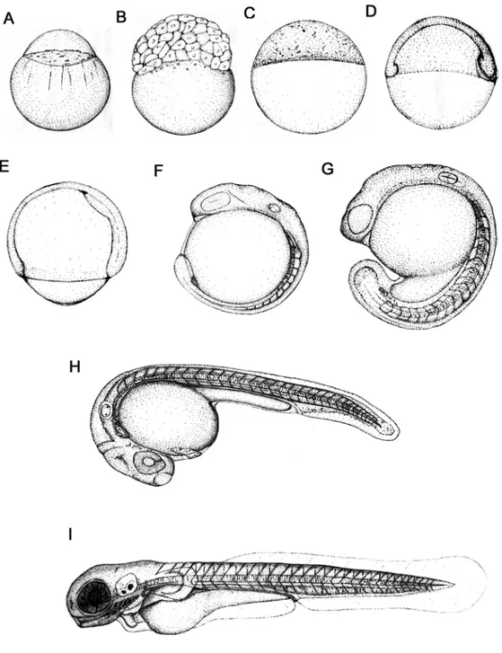
First, zebrafish adults reach sexual maturity quickly and are very fecund. Thus, large numbers of embryos can be collected for study. Second, zebrafish embryos develop externally. Eggs that are laid into the surrounding water by adult females are immediately fertilized by sperm from adult males. Embryos are thus easily accessible for observation and experimentation. Third, zebrafish embryos are optically clear. Under a microscope, one can visualize tissues that lie deep within the embryo, follow individual cells during development, and recognize developmental mutants. Fourth, zebrafish embryos develop rapidly, as illustrated.
1.2. Zebrafish embryonic development
The newly fertilized egg or zygote (0 – ¾ h) (Fig.1A), is about 0.7mm in diameter
at the time of fertilization. The embryo develops within a transparent eggshell,
called the chorion. In the zygote, cytoplasmic streaming separates the zygote
into two visibly different parts: a clear blastodisc at the animal pole and a yolky
cytoplasm at the vegetal pole.
Figure 1: Zebrafish developmental series. The animal pole is to the top for the early stages(A- E), anterior is up (F) or to the left (G-I) at later stages. (From: Kimmel et al.,1995. developmental Dynamics 203:253-310).
The blastodisc is the part of the zygote that undergoes cleavage and gives rise to blastomeres, the cells that later form the embryo. Cleavage period (¾ – 2 ¼ h) (Fig. 1B) includes the first six regular, rapid, and synchronous divisions of the embryo. At the end of the cleavage period, the blastomeres sit as a mound of cells on top of the large yolk. Up to midblastula stages the embryo has utilized proteins and RNA that the mother deposited in the egg (Amacher, 2001).
During midblastula stages, the embryo begins to transcribe its own genes.
Another major event that begins during the blastula period is epiboly, which describes the thinning and spreading of the cellular blastodisc over the yolk. Near the end of this period, the blastomeres are referred to collectively as the blastoderm, a sheet-like array of cells that sits like a cap on top of the large yolk.
Epiboly continues during the gastrula period (5¼ –10 h) (Fig. 1D, E). During this period, cell movements dramatically reorganize the embryo, so that by the end of this period, three embryonic axes are clearly visible. That is, the anterior head of the embryo is clearly distinguished from the posterior tail bud, dorsal tissues are distinct from ventral ones, and medial tissues are easily discernible from lateral ones. Many embryonic genes are expressed in region-specific patterns. Cell movements during gastrulation also produce the germ layers (ectoderm, mesoderm and endoderm) of the embryo. Finally, near the end of the gastrula period, the neural plate forms; this is the first morphological sign of central nervous system development.
The embryo elongates considerably during the segmentation period (10–24 h,
Fig. 1F-H). A complete complement of about 30 bilateral somite pairs forms in an
anterior to posterior sequence. The rudiments of primary organs, such as
notochord, kidney, and blood, become visible. The central nervous system
undergoes dramatic changes; the neural plate forms a solid neural keel, which
then hollows out to form a neural tube. Additionally, the brain becomes
regionalized, with forebrain, midbrain and hindbrain subdivisions becoming
distinct from one another. From around 24 hpf, muscular contractions occur, and
the embryo moves within the chorion. In the pharyngula period (24–48 h, Fig. 1H) the seven pharyngeal arches rapidly form. Pharyngeal arch development is common to all vertebrates, and in zebrafish, pharyngeal arches later essentially form the jaw and gills. Also during the second day, the head straightens, fins form, pigment cells differentiate, the circulatory system forms, the heart starts to beat, and coordinated swimming movements begin. In addition, embryos become responsive to touch, revealing the development of sensory-motor reflexive circuits.
Embryos hatch from their chorions during hatching period (48–72 h, Fig. 1I). By this time, many of the organ rudiments have formed; however, the gut and its associated organs are still developing. In addition, this period is marked by rapid development of pectoral fins, jaws and gills. During the two days after hatching (72–120 h), the zebrafish juvenile starts to swim and feed. The embryonic yolk, which has sustained the embryo until this time, is almost depleted. The zebrafish embryo has rapidly become a small version of the adult.
1.3. Thyroid gland
The thyroid gland is a member of the endocrine system. The English medical term derives from the Greek words “thyreos” meaning “shield”, and “eidos”
meaning “form”. In humans, the thyroid gland is located at the front of the neck (Fig.2 A). It is slightly heavier in women than in men and enlarges in pregnancy.
The thyroid gland is responsible for producing thyroid hormone in all vertebrates.
Although thyroid hormone is best known for its role in regulating metabolism in the adult organism, it is also required during development for many processes.
The thyroid gland in humans is a brownish-red organ, in most cases having two
lobes connected by an isthmus; it normally weighs about 28g and consists of
cuboidal cells arranged to form epithelial follicles (Fig.2 B). These follicles are
supported by connective tissue that forms a framework for the entire gland. In the normal thyroid gland, the follicles are usually filled with a colloid substance containing the protein thyroglobulin in combination with the main thyroid hormone thyroxine, tetraiodothyronine (T
4), and a secondary metabolistic form, triiodothyronine (T
3). These hormones are composed of the amino acid tyrosine, containing four and three iodine atoms, respectively. Thyroxin is an iodine- containing compound. In fact, the thyroid gland itself has a high content of iodine, which is necessary for optimal thyroid health and function. Thyroid hormone production starts with the synthesis of thyroglobulin. Thyroglobulin is then secreted into the colloidal lumen of the follicle where tyrosine residues are iodinated and where it is condensed to produce tri- (T3) and tetra-iodinated thyronine (T4, thyroxine) (Frieden and Lipner, 1971). T3 and T4 remain covalently bound to thyroglobulin as long as they are stored in the colloid. The bound forms of T3 and T4 are eventually taken up by the follicular cells and prototypically separated from the thyroglobulin. Free T3 and T4 are then released and act as thyroid hormone.
Figure 2: The thyroid gland in human. (A) Drawing showing the thyroid position and related structures in the neck region. (B) Cross section in the thyroid showing the structure of the thyroid follicles. (From University of Michigan webpage, http://www.um-endocrine-surgery.org).
Although the thyroid gland constitutes about 0.5 percent of the total human body weight, it holds about 25 percent of the total iodine in the body, which is obtained from food and water in the diet. Iodine usually circulates in the blood as an inorganic iodide and is concentrated in the thyroid to as much as 500 times the iodide level of the blood.
The amount of T4 and T3 secreted by the thyroid is controlled by the thyroid- stimulating hormone (TSH) of the pituitary gland. TSH, in turn, is regulated by a substance called thyroid-stimulating hormone releasing factor (TRF), which is secreted by the hypothalamus.
The functions of the thyroid gland include: regulation of normal body growth in infancy and childhood, regulation of metabolism, regulation of body temperature, maintenance of skeletal maturation and regulation of protein, fat and carbohydrate metabolism. These functions are dependent upon serum T4 levels.
Any disorder of the thyroid that leads to reduced thyroxine production at birth is called Congenital Hypothyroidism (CH) (Macchia, 2000). On the one hand, defects in genes responsible for thyroxine production can account for congenital hypothyroidism. On the other hand, such a phenotype can also be the result of earlier defects in development, such as mis-specification or mis-localisation of thyroid primordial cells.
1.4. Thyroid gland in zebrafish
The thyroid of teleost fishes is organised as thyroid follicles as in other
vertebrates, and these follicles are presumed to produce thyroid hormone in the
same way (Leatherland, 1994; Rauther, 1940). In contrast to higher vertebrates,
however, the thyroid of many teleosts is not a compact single organ. Most teleost
species that have been investigated have thyroid follicles loosely distributed
within the mesenchyme of the ventral head area, mainly in the vicinity of the anterior aorta. Nevertheless, comparative data are scarce and there is wide variation in teleost thyroid morphology from one set of bilateral nuclei of follicles in medaka (Raine et al., 2001) to dense groups of follicles throughout the gill region in trout (Raine and Leatherland, 2000).
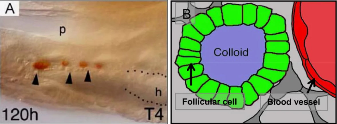
Follicular cell Blood vessel
B
Figure 3: (A) The normal position and distribution of thyroid gland in adult zebrafish and (B) the structure of thyroid follicular cell in zebrafish. P. pharynx, h. heart, and the arrow head indicate the ventral aorta. (A) lateral veiw +T4 immunostaining in 120h zebrafish embryo.
The thyroid gland in adult zebrafish is not a compact structure encapsulated in connective tissue; it’s rather a loose aggregation of follicles, close to the ventral aorta, distributed between the first pair of gills and the heart (Fig.3A) (Wendl, et al., 2002). Colloid filled follicles are detected along the ventral aorta, in the ventral midline of the gill chamber (Fig.3A). The follicles appear alone or in loose aggregations of two or three embedded in connective tissue (Wendl, et al. 2002).
Their shape is irregular and varies in diameter from 14 mm to 140 mm. In
summary, the zebrafish thyroid gland is composed of follicles that are dispersed
as described in other teleosts. In zebrafish, all follicles are found close to the
ventral aorta, from the first gill arch to the bulbus arteriosus. These cells form
colloid-filled follicles (Fig.3B), produce thyroid hormone, store it in the colloid-
filled lumen of the follicle and control the release of the hormone from the colloid
into the blood stream (Leatherland, 1994). In zebrafish development,
immunostaining of thyroid hormone reveals a first follicle at around 60 hour’s
postfertilization (hpf), when the embryo hatches from the egg shell. An increasing
number of further follicles is generated throughout larval life (Wendl et al., 2002;
Elsalini et al., 2003).
1.5. Development of the thyroid gland
In humans and mice, the thyroid gland develops from two types of primordia (De Felice and Di Lauro, 2004). One primordium, the so called midline diverticulum, evaginates from the midline of the pharyngeal floor, on the level between the first and second pharyngeal pouch. This midline diverticulum relocates and reaches a position deep in the cervical mesenchyme. During relocation it fuses with a second type of paired, bilateral primordial, the ultimobranchial bodies. After fusion of both types of primordia, the gland’s cells differentiate into follicles and C-cells (Kusakabe et al., 2005; Fagman et al., 2006). The endoderm-derived midline diverticulum gives rise to follicle cells and the bilateral ultimobranchial bodies to C-cells. C-cells produce the peptide hormone calcitonin-related polypeptide alpha (Calca), also known as calcitonin. Calca is thought to be involved in calcium regulation, however, its detailed role in humans is unclear (Inzerillo et al., 2002).
In non-mammalian vertebrates such as fish, amphibia, and birds, thyroid follicle cells and C-cells are found in separate organs. Thyroid follicle cells form the thyroid gland and do not fuse with the calcitonin producing ultimobranchial bodies that can be found as distinct glands elsewhere in the body (Le Douarin et al., 1974; Le Lievre and Le Douarin, 1975; Noden, 1991; Walker and Liem, 1994).
In zebrafish, the thyroid develops from the same tissue as the thyroid in
mammals, the endodermal tissue in the pharynx (Wendl et al. 2002; Alt et al.,
2006a), indicating they are homologous structures. Thyroid development is
classically subdivided into a few distinguishable steps, primarily based on
observations in mammals: first, a group of cells buds off the floor of the primitive
pharynx. Second, these cells reposition dorsocaudally to reach the anterior wall of the trachea. Third, the precursor cells proliferate and, fourth, differentiate into thyroid follicular cells (Macchia, 2000) (Fig.4).
Figure 4: The thyroid follicular cells derive from the endodermal tissue in the pharynx in zebrafish. (A+A’) Induction, (B+B’) Relocalisation and (C+C’) Growth/Differentiation of thyroid primodia. The first three pictures are: lateral views in three different stages (indicated bottom left) of wildtype zebrafish embryos with thyroid marker nk2.1a in situ staining.
1.6. Development of endoderm
Gastrulating vertebrate embryos generate the three germ layers, known as
ectoderm, mesoderm and endoderm, during early development. The induction
and differentiation of endodermal cells and the formation of organs derived from
the gut tube have been poorly analyzed in comparison to ectoderm or
mesoderm. Zebrafish fate map studies began in the 1990s and these fate maps
showed that both endoderm and mesoderm originate from a common progenitor
(Fig.5). Both germ layers derive from cells near the blastoderm margin and both
involute into the forming hypoblast during gastrulation (Warga and Kimmel,
1990). At 40% epiboly, just prior to gastrulation, endoderm progenitor cells are
located in a narrow field of cells along the margin. Furthermore, there is asymmetric distribution of the endoderm progenitor in the margin, and more endoderm progenitor is found dorsally than ventrally (Warga and Nüsslein- Volhard, 1999) (reviewed in Fukuda and Kikuchi, 2005).
Detailed fate mapping studies have shown that at the late blastula stage the position of the endodermal progenitors in the marginal zone resembles a topographic arrangement of the presumptive digestive system. The position a cell occupies along the dorsoventral axis before gastrulation reflects its future location with respect to the anteroposterior (AP) axis: dorsal-most cells will give rise to the most anterior endoderm, the pharynx; lateral cells will give rise to the digestive organs, and ventral cells to the posterior part of the alimentary canal (Warga and Nüsslein-Volhard, 1999).
Analyses of zebrafish mutants as well as overexpression studies have identified key components of the pathway leading to endoderm formation in zebrafish. At the top of this pathway lies Nodal signalling. The first evidence that members of the Nodal family were essential for endoderm formation in zebrafish came from the analysis of mutants deficient for two Nodal-related factors, Cyclops (Cyc) and Squint (Sqt), which lack endoderm and the majority of mesoderm (Feldman et al., 1998; Ober et al., 2003).
Nodal signals are received by EGF-CFC coreceptors and type I and II Activin
receptors, which function as serine/threonine kinases. Receptor activation leads
to phosphorylation of the transcription factors Smad2 and Smad3. This results in
their binding to Smad4, nuclear translocation, and association with additional
transcription factors such as FoxH1 and Mixer to regulate target genes. Nodal
signalling is antagonized by feedback inhibitors such as Lefty proteins, which are
divergent members of the TGFβ family and block EGF-CFC coreceptors, and
Dapper2, which enhances the degradation of type I Activin receptors (Schier and
Talbot, 2005).
Figure 5: Zebrafish fate maps. (a) Fate map at 50% epiboly stage, the onset of gastrulation.
Lateral view, dorsal to the right, animal pole to the top. Future germ layers are arranged along the animal-vegetal axis. Different mesodermal and ectodermal fates are arranged along the dorsal- ventral axis. (b) Fate map of ectoderm at 90% epiboly. Lateral view, dorsal to the right, animal pole and anterior to the top. (c) Model fate map of mesoderm at early somite stage. Lateral view, dorsal to the right, animal pole and anterior to the top. Regions shown here are approximations derived in part from the expression patterns of marker genes (ZFIN.org). The posterior region of the tail bud will continue to extend and give rise to different mesodermal and ectodermal fates.
(From: Schier and Talbot, 2005).
Mutant screens in zebrafish have identified several components of the Nodal signalling pathway. These include the Nodal signals Cyclops (Cyc) and Sqt, the EGF-CFC coreceptor One-eyed pinhead, and FoxH1 (schmalspur (sur)) and Mixer (bonnie and clyde (bon)) (See: Schier and Talbot, 2005).
Absence of Nodal signaling in cyc;sqt double mutants or maternal-zygotic one-
eyed pinhead mutants results in embryos that lack all endoderm and mesoderm,
with the exception of a few somites in the tail. Mutants also lack trunk spinal cord,
but develop forebrain, midbrain, hindbrain, and tail spinal cord (Schier et al.,
1996). Conversely, increasing Nodal signaling by loss of Lefty1 and Lefty2 or
overexpression of Cyc or Sqt results in the fate transformation of ectodermal cells into mesoderm or endoderm. Several genes have been identified that are regulated by Nodal signalling and mediate its endoderm-inducing activity, including the Sox gene casanova, the GATA gene faust, and the homeobox genes bonnie, clyde and mezzo (reviewed in Schier and Talbot, 2005). These four genes encode transcription factors and appear to be direct targets of the Nodal signalling pathway.
1.7. Development of the vascular system
The formation of a functional vascular system is essential for the proper development of vertebrate embryos, as well as for the survival of adults. The vascular system provides oxygen, carries away metabolic waste products, serves as the conduit for hormones and provides space for the immune response.
During embryogenesis, the vascular system is constructed by two distinct processes. The first is vasculogenesis, which forms a primary capillary network from hemangioblasts, which are putative precursors specified from mesoderm.
The mature vascular structures are formed by a second process, called angiogenesis, which is a remodeling of the endothelial cells from the existing capillary network into mature blood vessels (Yamada et al., 2000). During embryogenesis, blood and endothelial cell development are closely associated with each other. In mammalian embryos, the blood islands on the yolk sac consist of both endothelial and blood cells. Both cell types are thought to emerge from a common precursor, the hemangioblast (Fig.6) (Choi et al., 1998).
The zebrafish has recently emerged as an advantageous model organism to
study how the network of vertebrate blood vessels arises during development. In
zebrafish embryos, both endothelial and blood cell precursors originate in the
lateral plate mesoderm (Fig.7) and during the middle and late somitogenesis
stages reside in the region known as the intermediate cell mass (ICM) (Fig.7).
During vasculogenesis, the endothelial cell progenitors (angioblasts) give rise to the major blood vessels of the trunk, the dorsal aorta, and axial vein, as well as the endocardium of the heart (Fig.6).
Figure 6:Blood and endothelial cell development. During vasculogenesis, the endothelial cell progenitors (angioblasts) give rise to the major blood vessels of the trunk, the dorsal aorta, and axial vein, as well as the endocardium of the heart. (From Gilbert, 2000).
During subsequent angiogenesis, the axial vessels sprout to form secondary vessels in the trunk region of a zebrafish embryo (for a review, see Sumanas et al., 2005). The molecular mechanisms of development of the vascular system in vertebrate embryos remain relatively unexplored. Several signaling molecules and transcription factor genes have been implicated in the development of the vertebrate vasculature: vegf, vegfr1, vegfr2, vegfr3, tie1, tie2, angiopoietin1, angiopoietin2, ephrinB2, ephB4, scl, fli1, ets1, runx, semaphorins and plexins have been analyzed in zebrafish, amphibians, birds and mammals, and found to display similar temporal and spatial expression patterns (for a review, see Jin et al., 2005).
During the last decade, the molecular mechanisms of blood and blood vessel formation started to be elucidated. The transcription factor Scl/tal1 functions at the level of the hemangioblast, affecting both hematopoietic and vascular development in both mouse and zebrafish (Liao et al.,1998; Liao et al., 2000).
Transcription factors including Gata2, Gata1, C-myb, and transcription factor–
interacting proteins such as Lmo2 gradually restrict the subpopulation of blood-
cell precursors to adopt the erythroid fate (for a review, see Sumanas et al., 2005).
Figure 7: Schematic diagram of early blood and endothelial development in the trunk of zebrafish embryos. Both endothelial and blood cell precursors originate in the lateral plate mesoderm and during the middle and late somitogenesis stages reside in the region known as the intermediate cell mass (ICM). E, endoderm; HC, hypochord; ICM, intermediate cell mass; NC, notochord; NP, neural plate; PCV, posterior cardinal vein; PLM, posterior lateral mesoderm; SPM, somitic paraxial mesoderm; DA, dorsal aorta. (From: Gering and Patient, 2005).
Vascular endothelial growth factor (Vegf) is required for vascular endothelial
development in both mouse and zebrafish and functions through tyrosine kinase
receptors Vegfr1/Flt1 and Vegfr2/Kdr (Carmeliet et al., 1996). Angiopoietin
receptor tyrosine kinase Tie2 is important in angiogenesis, while Tie1 is critical
for endothelial cell integrity and survival (Puri et al., 1995). Zebrafish homologs of
tie1 and tie2 are also expressed in vasculature (Lyons et al., 1998). Vegf
regulates the migration and survival of endothelial cells. Mice lacking a functional
Vegf signal show various developmental defects, including a reduced number of
endothelial cells and a failure to form a functional vasculature (Carmeliet et al.,
1996). Similar phenotypes have been reported in Xenopus and chick embryos
with compromised Vegf signaling (Cleaver and Krieg, 1998). In zebrafish, Vegf
appears to be crucial for angioblast formation as well as for the subsequent differentiation into arterial endothelial cells (Lawson et al., 2002).
Another example of functional conservation in vascular development is Hh. In zebrafish embryos, Shh in the notochord appears to regulate vegf expression in the somites, which in turn regulates vascular development (Lawson et al., 2002).
In chick and mice, Hh signaling also appears to be important in vascular development (Vokes et al., 2004). However, complete signal transduction pathways involved in hematopoiesis, vasculogenesis, and angiogenesis are still unknown.
1.8. Hedgehog (Hh)
During animal development, gradients of signaling molecules play an important role in cell type specification. Members of the Hedgehog (Hh) family of intercellular signaling molecules control a variety of developmental processes, ranging from segment patterning in Drosophila to organogenesis, left-right asymmetry and dorsoventral patterning of the spinal cord and forebrain in vertebrates (reviewed by Ingham and McMahon, 2001). Aberrant regulation of Hh signaling in humans causes developmental defects such as holoprosencephaly (HPE) and postaxial polydactyly, and can also lead to various types of cancers, including basal cell carcinoma and medulloblastoma (for a review, see Sekimizu et al., 2004).
Because Hh signaling plays such a central role in development and disease, the Hh signaling pathway has been investigated in considerable detail (Fig.8).
Genetic and in vitro studies in Drosophila have revealed that Hh signals are
transduced by binding of Hh ligands to the Patched (Ptc) cell-surface receptor,
resulting in the activation of the transmembrane protein Smoothened (Smo). In
Drosophila, the intracellular regulation of Hh signaling is mediated by post-
translational modifications of Cubitus interruptus (Ci), a zinc-finger-containing
transcription factor of the Gli family that can be both an activator and a repressor of Hh target genes. In the absence of Hh signal, proteolytic cleavage converts Ci to a transcriptional repressor. In the presence of Hh signals, cleavage of Ci is inhibited and a full-length activator isoform predominates. In vertebrates, at least three Gli genes, Gli1, Gli2 and Gli3, mediate the transcriptional response to Hh signals. The functions of these different Gli genes have been analyzed in mouse, Xenopus, zebrafish and cultured cells (reviewed by Ingham and McMahon.
2001).Thus, in both vertebrates and invertebrates, Hh signaling controls the expression of target genes by modulating the activity of the downstream Gli/Ci transcription factors. Studies in Drosophila have identified a large number of proteins that are involved in the regulation of this Gli/Ci activity (for a review, see:
Sekimizu et al., 2004).
Genetic studies of Hh signaling in zebrafish complement the analyses in fly and other vertebrate species, and provide an approach to look into the regulation of Hh signaling in vertebrates. In vertebrates, Sonic hedgehog (Shh) is expressed in the notochord and floor plate of the neural tube (Echelard et al., 1993; Krauss et al., 1993; Roelink et al., 1994; Ekker et al., 1995), and is essential for the induction of floor plate, motoneurons and a class of ventral interneurons in the neural tube (Chiang et al., 1996; Ericson et al., 1996). Shh signaling is also required for the induction of muscle and sclerotome cell types in somites (reviewed by Bumcrot and McMahon, 1995). A large number of zebrafish mutations, collectively called the midline mutants, have been identified that lead to ventral neural tube defects, absence of an optic chiasm and defects in slow muscle fiber formation (Brand et al., 1996; Chen et al., 1996; Karlstrom et al., 1996; van Eeden et al., 1996). Many of these midline mutants have now been shown to encode components of the Hh signal cascade.
shh is disrupted in sonic-you (syu) mutants (Schauerte et al., 1998), smo is
disrupted in slow muscleomitted (smu) (Chen et al., 2001; Varga et al., 2001),
gli2 is disrupted in you-too (yot) (Karlstrom et al., 1999), gli1 is disrupted in
detour (dtr) (Karlstrom et al., 2003) and dispatched1 is disrupted in chameleon (con) (Nakano et al., 2004).
Figure 8: The Hedgehog signal transduction pathway. The Patched protein in the cell membrane is an inhibitor of the Smoothened protein. (A) In the absence of Hedgehog binding to Patched, the Ci protein is tethered to the microtubules (by the Cos2 and Fused proteins). This allows the PKA and Slimb proteins to cleave Ci into a transcriptional repressor that blocks the transcription of particular genes. (B) When Hedgehog binds to Patched, its conformation changes, releasing the inhibition of the Smoothened protein. Smoothened releases Ci from the microtubules (probably by adding more phosphates to the Cos2 and Fused proteins) and inactivates the cleavage proteins PKA and Slimb. The Ci protein enters the nucleus, binds a CBP protein and acts as a transcriptional activator of particular genes.( From Gilbert, 2000).
One remarkable feature of the Hedgehog signal transduction pathway is the importance of cholesterol. Cholesterol is critical for the catalytic cleavage of sonic hedgehog protein, only the amino-terminal portion of the protein is functional and secreted, and the Patched protein that binds the Sonic hedgehog protein also needs cholesterol in order to function.
Environmental factors that cause developmental anomalies are called teratogens
(from the Greek, meaning "monster-former"), two teratogens known to cause
cyclopia in vertebrates are jervine and cyclopamine. Both substances are found in the plant Veratrum californicum. Plants of the genus Veratrum have a long history of use in the folk remedies of many cultures, and the jervine family of alkaloids, which constitute a majority of Veratrum secondary metabolites, have been used for the treatment of hypertension and cardiac disease (for a review, see Chen et al., 2002). The association of Veratrum californicum with an epidemic of sheep congenital deformities during the 1950s (Binns et al., 1962) raised the possibility that jervine alkaloids are also potent teratogens. Jervine and cyclopamine (11- deoxojervine) given during gestation can directly induce cephalic defects in lambs, including cyclopia in the most severe cases (Keeler and Binns 1965). Cyclopamine inhibits Hh pathway activation by binding directly to Smo (Chen et al., 2002). This binding interaction is localized to the heptahelical bundle and likely influences the Smo protein conformation.
Cyclopamine binding is also sensitive to Ptch function, providing biochemical evidence for an effect of Ptch action on Smo structure (Chen et al., 2002).
1.9. Aim of the work
Normal thyroid function is essential for development, growth, and metabolic homeostasis. Defects in any step of thyroid development (such as specification, proliferation, migration, growth, organization, differentiation, and survival) may result in a congenital anomaly and/or impaired hormonogenesis, leading to variable degrees of hypothyroidism (Trueba et al. 2005).
Congenital hypothyroidism (CH) affects one in about 4000 newborns in western
countries, and thyroid dysgenesis (TD) accounts for about 85% of the cases; the
other 10–15% result from functional disorders in hormone synthesis. TD includes
absence of thyroid tissue (athyreosis), presence of ectopic tissue, as well as
hypoplasia of an orthotopic gland. Ectopic thyroid can be the cause of CH,
followed by athyreosis. In this study, we introduce zebrafish as a model to
investigate the molecular and genetic mechanisms that control thyroid
development.
2. Materials and methods
2.1. Used chemicals and materials
Inorganic and organic chemicals were usually purchased in the highest quality available from Biomol (Hamburg), Biozym (Hameln), Fluka (Neu-Ulm), Life Technologies (Karlsruhe), Merck-Eurolab (Darmstadt), Pharmacia (Freiburg), Roche Diagnostics GmbH (Mannheim), Roth (Karlsruhe), Serva (Heidelberg), and Sigma (Taufkirchen).
Zebrafish –Ringer (Stock-solutions)
Stock 1 (25x): 169, 5 g/L Sodium Chloride NaCl + 5, 4 g/L Potassium Chloride KCl + 29, 75 g/L HEPES C
8H
18N
2O
4S
Stock 2 (100x): 26, 4 g/L Calcium Chloride CaCl
2+ 34, 96 g/L Calcium Chloride Dihydrat CaCl
2.2 H
2O + Autoclaving
1/3 ZFR: 12 ml Stock 1 + 9 ml Stock 2 + 879 ml D.H
2O. Adjust the PH to 7,2 by using 1N Sodium hydroxide NaOH.
Tricaine
3 -Aminobenzoic acid ethyl ester C
9H
11O
2.CH
4SO
3(Sigma).
800 mg tricaine powder +200 ml D.H2O.
Bouin’s solution
75ml Picric acid + 25ml Formalin 40% + 5ml acetic acid glacial
4% Paraformaldehyde (PFA)
• 40g PFA powder (Sigma) dissolved in 1L PBS. Warming up slowly in fume hood. Don't boil.
• Aliquot and Store at -20°C.
10xPBS (phosphate buffered saline)
• 80 g Sodium Chloride NaCl.
• 2 g Potassium Chloride KCl.
• 14.4 g Sodium Phosphate Na2HPO4.
• 2.4 g Potassium Phosphate KH2PO4.
• ~900 ml D.H2O
• PH ~7.2 – 7.6
• adjust to 1 L
• autoclaving
1xPBT (phosphate buffered saline containing tween)
1L 1xPBS + 5ml 20%Tween 20 = 1xPBT
Hyb -
50% Formamid CH
3NO (ROTH) + 5x SSC + 0,1% Tween 20 (Sigma) or Triton X- 100 (C
38H
70O
13).
Set pH to 6 with 92 µl/10ml of 1M citric acid and Store at -20°C.
Hyb +
Hyb- + 5mg/ml torula yeast RNA (Sigma) + 50µg/ml Heparin (Roth)
Store at -20°C.
20XSSC
• 175.3 g Sodium Chloride NaCl.
• 88.2 g Sodium Citrate C
6H
5Na
3O
7.2H
2O.
• 800 ml D.H
2O
• adjust the pH to 7.0 with few drops of a 14N solution of HCl
• adjust to 1 L with D.H
2O
1 ml 20XSSC + 9 ml D.H
2O = 2XSSC 1 ml 20XSSC + 99 ml D.H
2O = 0.2XSSC 1 ml 2XSSC + 9 ml D.H
2O = 0.2XSSC
Staining buffer (X-pho buffer)
0,1M Tris C
4H
11NO
3pH 9,5 (ROTH) + 50mM Magnesium Chloride MgCl2 (ROTH) + 0,1% Tween or Triton X -100 (C
38H
70O
13) + 0,1M Sodium Chloride NaCl.
Glycerol
C
3H
8O
3(ROTH)
50%, 70%, 90% in PBS diluted
2.2. Methods
Animals
Zebrafish work was carried out according to standard procedures (Westerfield, 2000), and staging in hours post fertilisation (hpf) or days post fertilisation (dpf) refers to development at 28.5-29°C. Embryos or larvae were dechorionated manually and anaesthetized in tricaine before fixation. Some zebrafish embryos used for insitu hybridisation were treated with PTU to prevent pigmentation at young stages. As PTU belongs to a class of chemicals that have the potential to interfere with the function of the thyroid (Elsalini and Rohr, 2003), we did not treat embryos used for immunohistochemistry with PTU and confirmed all in-situ hybridization experiments with non-treated embryos.
The following mutant fish lines were used: cyc
b16(Talbot et al., 1998), bon
s9(Kikuchi et al., 2000), cas
ta56(Kikuchi et al., 2001), fau
s26(Reiter et al., 1999), cyc
m294(Talbot et al., 1998), noi
b21(Lun and Brand, 1998), oep
tz57(Schier et al., 1997), smu
b641(Varga.et al, 2001), syu
t4(Schauerte et al 1998), and homozygous cloche mutant clo
s5(Stainier et al., 1995). kdr
y17(Covassin et al., 2006) is allelic to the kdr (kdr) lines described previously (Habeck et al., 2002).
Microinjection
Synthetic, capped mRNA prepared according to standard procedures (see
below) was injected into fertilized zebrafish eggs prior to first cleavage. Injections
were carried out under sight control using a dissecting microscope and with the
aid of a micromanipulator. I did not dechorionate the fertilized eggs prior to
injection. Chorions were removed about 24 hours later, at which time zebrafish
embryos are well developed and crucial steps in embryogenesis have been
completed. Micropipettes were made on a horizontal puller using 1mm
Hilgenberg glass capillary tubing with an inner filament for backfilling the pipettes
with DNA solution. Injection solution contained 0.5% phenol red to allow
estimation of the injected volume.
Embryo manipulation
The synthetic mRNA was produced by using the message machine kit (Ambion), and was injected into one-cell-stage embryos. As a control, I injected synthetic gfp mRNA at similar concentrations. Numbers of mRNA injected embryos were about 100 for each insitu hybridisation marker, and the Injection experiments were repeated at least once.
Morpholino RNA was purchased (Gene Tools) and dissolved as recommended by the provider. Morpholino sequences are:
hhex: 5’-gcgtgcgggtgctggaattgcatga-3’
nk2.1a-1: 5’-gctcaaggacatggttcagcccgc-3’
nk2.1a-2: 5’-cgcgagcaggtttgctgaagctgcc-3’
nk2.1b: 5’-tcgtatgcttagggctcatcgacat-3’
vegf: 5’-taagaaagcgaagctgctgggtatg-3’
scl: 5’-aatgctcttaccatcgttgatttca-3’
tnnt2: 5′-catgtttgctctgatctgacacgca-3′
Unspecific control, fluorescein coupled: 5’-cctcttacctcagttacaatttata-3’
Preparation of specimens
Fixation of fish embryos was done overnight in 4% paraformaldehyde in PBS at 4°C for whole-mount in situ hybridization and histochemistry, or in Bouin’s solution at room temperature for histology and histochemistry. Paraformaldehyde fixed embryos were then washed in PBT and stored in methanol at –20°C. Bouin fixed tissue was transferred to 70% ethanol and stored at room temperature.
For whole-mount antibody staining, larvae from 4 to 6 dpf were fixed in
paraformaldehyde (PFA) at 4°C overnight or for 1 h at room temperature,
washed in phosphate-buffered saline containing 0.3% Tween (PBT) then washed
and stored in methanol at -20°C. For bleaching, and to block endogenous
peroxidases, embryos were incubated in 3 ml 10% H
2O
2in methanol overnight at
room temperature, then 10 ml PBT were added, mixed, and incubated for a further 16 to 24 h at room temperature. Antibody staining was subsequently performed as described below.
2.3. Molecular biology techniques
Standard molecular biology techniques were carried out according to Sambrook, et al 2001 for DNA preparation; Plasmids were transformed into the E.coli k12 strain and cultured in 100ml LB medium containing appropriate antibiotics. The DNA was purified using Qiagen Midi prep kits (Qiagen).
Restriction enzyme digests were carried out in a volume ~50µl using an appropriate 10x enzyme buffer and 20 units of enzyme per 5-10µg DNA (table 1).
Enzyme digests were carried out at 37°C and checked on 1% agarose gel by electrophoresis. Purification of DNA was carried out by 1x phenol:chloroform extraction, 1x chloroform extraction followed by precipitation. DNA was precipitated with 0.1 volume 3M Na acetate and 0.7 volume isopropanol, centrifuged for 20 minutes and the DNA pellet washed in 70% ethanol, air dried and taken up in RNAse free H
2O.
Polymerase Restriction enzyme Plasmid name Stock
number In situ probe
RNA injection
Anti sense sense
Fkd7 K13 T7 Bam H1
gfp K20 Sp6 Xba1
Hex K21 Sp6 Apa1 Nco1 Sp6
Kdr K17 T7 ECO R1
Nk2.1a K27 T3 Sac II
Nk2.1b K28 T7 BamH1
Pax2.1 K33 T7 Bam H1
Scl K122 T3 Eco R1
Shh K39 Sp6 Bam H1
Tie1 K61 T7 Spe I
Vegf 121 &165 K50 & K51 T7 Sp6 Bam H1 Not1
Table 1: The plasmid and the enzymes used to prepare the in situ probe or mRNA injections.
0.5-1µg of the linear DNA was used in the in vitro transcription reaction for synthesis for antisense RNA labeled with digoxigenin or for mRNA for injection.
For generation of capped mRNA for injections see above. For synthesis of in situ probe a 20µl reaction was set up in 2µl 10x transcription buffer, 2µl DiG-labelling mix; 0.5µl RNasin, 1µl RNA polymerase (table 1) and RNAse free H
2O.
Transcription mix was incubated at 37°C for 2-4hours. The RNA was washed in D.H
2O by using Millipore filter tubes.
2.4. Histological methods
Fixation in Bouin’s solution, embedding in paraffin, sectioning,T4 immunostaining on sections, and PAS staining were carried out as described previously (Wendl et al. 2002). Paraffin embedding was carried out according to standard procedures (see below), and 8 µm sections were cut. For histology, sections were dewaxed by washing 2x in rotihistol 5min each and 3x in 100% ethanol 5min each, than dried and stained.
Sections of adult and larval zebrafish were stained with Giemsa, Haematoxylin and Eosin, and PAS staining in order to visualise the thyroid follicles. Giemsa stain involved 5 minutes incubation in Giemsa solution as used for blood stain, followed by two washes in tap water. Haematoxylin and Eosin staining was carried out according to Ehrlich (Fluka, catalogue number 03972): 5 minutes incubation, followed by 10 minutes washing in running tap water. All stained slides were dehydrated and mounted in Entellan neu (Merck).
Paraffin embedding
• After fixation in Bouin’s solution, replace Bouin’s by 70% ethanol.
• Wash 4X in 100% ethanol for 2h.
• In glass vial, replace by a mixture of 50% ethanol and 50% Rotihistol for
1h.
• Replace by 100% Rotihistol.
• Incubate in 65°C.
• Add 50% paraffin.
• Replace paraffin several times during a day.
• Make paraffin block containing the embryos.
• Sectioning by using microtome, 8 mm sections cut.
• Receive the sections on slides.
• And incubate the slides on warm plate.
PAS staining
Periodic acid/Schiff (PAS) staining was carried out according to the manufacturer’s instructions (Merck, catalogue number 1.01646). Staining on sections was performed as described previously (Wendl et al., 2002).
• After Dewaxing and drying
• Wash in D.H
2O for 2min
• Incubate in periodic acid (Merck) for 5min
• Wash in tap H
2O for 3min and 1min in D.H
2O
• Incubate in Schiff’s reagent (Merck) for 15min
• Wash in tap H
2O for 3min and 1min in D.H
2O
• Dehydration: Incubate the stained sections in 30%, 50%, 70%, and 100%
ethanol for 5min each.
• Incubate 2x in 100% xylol for 5min each.
Whole Mount T4 Antibody Staining
The staining procedure followed general protocols using the Vectastain elite ABC kit (Westerfield, 2000). Whole-mount immunohistochemistry with antibodies against the thyroid hormone T4 (polyclonal rabbit anti T4, ICN Biochemicals;
Wendl et al., 2002) or thyroglobulin (polyclonal rabbit anti human thyroglobulin,
Dako) in zebrafish larvae was performed as described elsewhere. Larvae were
washed in PBT, blocked in normal goat serum for 2 h, incubated with a polyclonal antibody (1:4000 rabbit anti-thyroxine BSA serum, ICN Biochemicals) that detects thyroid hormone at its location of production in thyroid follicles (Raine et al., 2001) for 2 h, then washed in PBT for 3 h. Incubation with a biotinylated secondary antibody was for 2 h, washing in PBT for 3 h. Incubation with the ABC kit (Vectastain) was for 2 h according to the instructions of the manufacturers.
Larvae were washed again in PBT for 3 h, and then incubated in DAB (0.2 mg/ml PBS) for 30 min. To stain, 1µl of a 0.3% aqueous H
2O
2solution was added under observation by using a dissection scope. All procedures were carried out at room temperature; washing steps can be extended at 4°C. For detailed analysis, larvae were postfixed in PFA for 15 min at room temperature, washed in PBT, and gradually transferred to 70% glycerol.
In Situ Hybridization
Whole-mount in situ hybridisation for zebrafish embryos was carried out according to standard procedures (Westerfield, 2000). The experimental embryos grow in PTU to prevent pigmentation and dechorinated by using the Pronase.
Embryos were fixed in 4% paraformaldehyde/ PBS (PFA) overnight and stored in 100% methanol at -20°C. Specimens were washed twice with PBST and then digested with 5 mg/ml proteinase-K in PBST for several minutes depending on the stage (table 2). They were washed and fixed again in 4% PFA for 20 min.
After the proteinase-K was washed off, embryos were transferred into HYB+
solution and prehybridized for 2-4h at 60°C. Approximately 5ng/ml DIG-labeled
RNA probe was added and hybridized overnight at 60°C. Embryos were washed
at 60°C in Hyb-/2XSSC (75:25) for 15 min; in Hyb-/2XSSC (50:50) for 15 min and
in Hyb-/2XSSC (25:75) for 15 min. For detection, embryos were blocked at least
for 2 h at room temperature in blocking reagent (Roche). Embryos were
incubated for 2h in antibody Anti-Digoxigenin-AP (Roche) diluted 1:6000 in
blocking reagent, then wash in PBT overnight.
The following day specimens were washed in staining buffer (X-pho buffer) (4ml 1M tris pH 9.5, 2ml 1M MgCl2, 800µl 5 M NaCl, 200µl 20% Tween 20, and fill up to 40ml with D.H
2O). Embryos were then incubated in BM purple AP substrate, precipitating (Roche).To stop the color reaction, PBST was added, followed by fixation in 4% PFA. For detailed analysis, larvae were washed in PBT, and gradually transferred to 90% glycerol.
Proteinase K concentration
stage in hpf Concentration in PBT time in min
24 1:1000 6
26 1:1000 8
28 1:1000 10
30 1:1000 30
32-33 1:500 25
35 1:250 10
40 1:250 15
45 1:250 30
48 1:250 35
55 1:250 45
60 1:100 30
72-75 1:100 60
Table 2: The concentration of Proteinase K and the time of incubation for different zebrafish stages.
Double staining (insitu + Tg antibody staining)
• Stop the in situ in PBT.
• Wash 5x in PBT
• Bleach : (o.5 ml 30%H
2O
2+ 1ml PBS ) for15 min in dark.
• Rinse 2x in PBT.
• Postfix in PFA for 15 min at R.T.
• Wash 5x in PBT for 5min each.
• Incubate in 3% NGS for 2h.
• Incubate in TG 1:6000 in 3% NGS for 2h.
• Wash several times in PBT for 3h or overnight.
• Incubate in secondary antibody (LINARIS) for 2h.
• Wash several times in PBT for 3h or overnight.
• Incubate in AB kit for 2h (1ml PBT + 20µl A + 20µl B).
• Wash several times in PBT for 3h.
• Incubate in DAB 1:5 in PBS for 30min.
• Add 1µl 0.3%H
2O
2(1µl 30% H
2O
2+ 99µl D.H
2O) and observe while staining
Embedding in Durcupan
• Wash the stained embryos several times in PBT.
• Wash in 50%, 70% and 90% ethanol 10min each.
• Wash 2x in 100% ethanol for 10min each.
• Wash 2x in 100% Aceton 15min each.
• Incubate in Acetone: Durcupan 1:1 overnight at R.T.
• Embedding in Durcupan (Fluka).
• Incubate overnight at 65°C.
• Sectioning by Microtome.
2.5. Cyclopamine treatments
Cyclopamine (Toronto Research Chemicals, North York, Ontario, Canada)
treatment was carried out as described in Lawson et al, 2002. Cyclopamine was
dissolved in 100% dimethyl sulfoxide (DMSO) at 10 mM. Embryos were
dechorinated with pronase at 50% epiboly and placed in 6 ml of embryo medium
containing 50 µM of cyclopamine. Surviving embryos were fixed and processed
for insitu hybridization.
3. Results
3.1. Thyroid differentiation:
3.1.1. Nodal signalling and subsequent steps of endoderm specification are required for thyroid development
During vertebrate development, endodermal cells give rise to the digestive system with its derivatives such as lung, pancreas, and liver. Endoderm specification starts during gastrulation, and key factors involved in this process are regulated by the Nodal family of signalling molecules (Feldman et al., 1998).
The thyroid gland is considered to be an endoderm derived organ, because it develops from precursor cells that evaginate from the ventral floor of the pharynx, in the same way as the lung primordium does at a slightly more posterior position. So I analysed the thyroid formation in mutants that show different degrees of compromised endoderm specification.
In one-eyed pinhead, a mutant that lacks a cofactor required for Nodal signalling,
the endoderm is not specified (Schier et al., 1997), and, as predicted for an organ
of endodermal origin, the thyroid primordium is absent. cyc encodes one of the
zebrafish Nodal ligands (Sampath et al., 1998), and b16
-/-and m294
-/-mutant
alleles disrupt Cyc function. b16
-/-is a gamma ray-induced mutation (Hatta et al.,
1991) that lacks the lower telomeric region of LG12, including cyc (Talbot et al.,
1998), whereas m294
-/-is an ENU-induced point mutation in the cyc gene
(Sampath et al., 1998; Schier et al., 1996). In cyc
-/-mutants, Nodal signalling is
compromised (Fig.9 E and F) and the expression of nk2.1a indicates that the
thyroid primordium is smaller in b16
-/-and m294
-/-compared with wild-type
(Fig.9B and C).Thus, cyc is required for normal numbers of endodermal cells,
and reduction in the extent of endoderm correlates with reduced size of the
thyroid primordium.
Figure 9: Reduced endoderm coincides with a smaller thyroid primordium in cyc-/-.
Expression patterns of nk2.1a in thyroid precursor cells and fkd7 in the endoderm (gene expression as indicated bottom right) at 26 hpf in wildtype or mutant embryos (indicated top right).
The arrows point to the thyroid primordium in (A-C). Arrowheads indicate the expression of fkd7 in the pharyngeal endoderm on the level of the first to the second branchial arch.
The three genes casanova (cas), bonnie and clyde (bon), and faust/gata5 (fau) have been shown to be required downstream of Nodal signalling for the specification of endoderm (Kikuchi et al., 2000, 2001; Reiter et al., 1999). I find that the thyroid primordium is completely absent in bon
-/-mutant. Thus, thyroid development is not only dependent on Nodal signalling, but also on subsequent steps of endoderm specification.
Using an antibody that labels thyroid hormone (T4; Raine et al., 2001; Wendl et
al., 2002), I fail to detect functional thyroid follicles at 3–5 dpf in b16
-/-wholemount embryos (Fig.10B). In contrast, in m294
-/-embryos, a row of thyroid
follicles forms in the midline of the ventral pharynx as in wild-type siblings
(Fig.10C). I next tested whether lack of T4 immunostaining in b16
-/-larvae reflects the absence of the thyroid gland. PAS staining visualises glycoproteins of the colloid in thyroid follicles in both wild-type (Fig.10D) and m294
-/-(Fig.10F) larvae but not b16
-/-larvae (Fig.10E). In zebrafish larvae treated with drugs that interrupt thyroid hormone synthesis but do not interfere with thyroid follicle development, T4 immunostaining is absent, but PAS still stains colloid in the follicles (Elsalini and Rohr, 2003). Thus, absence of both T4 immunostaining and PAS staining suggests that thyroid follicles are missing in b16
-/-.
Figure 10: Thyroid follicles are absent in b16-/- larvae but present in cyc m294-/- larvae.
Whole-mount embryos (A–C) or paraffin sections (D–F). Genotype indicated at top right. Antibody staining (T4, brown staining), PAS staining (PAS), Arrows point to thyroid follicles (D, F), Arrow bars in (A–C) indicate level of sectioning as in (D–F).
Both nk2.1a and pax2.1 are expressed from 24 hpf in the thyroid primordium of
developing zebrafish, and it has demonstrated previously that pax2.1 is required
for thyroid follicle development (Wendl et al., 2002). Both genes are initially
expressed in b16
-/-embryos (Fig. 11A–F) and so a thyroid primordium initially
forms. However at around 60 hpf, both markers disappear in the thyroid
primordium of b16
-/-, but not in m294
-/-(Fig. 11G–L). Altogether, these results
show that, although a primordium is initially present, a functional thyroid gland
fails to form in b16
-/-larvae.
Figure 11: A thyroid primordium is induced in both b16-/- and m294-/- embryos. Expression patterns of thyroid markers (indicated at bottom right) at different stages of development (indicated bottom left) in mutant embryos (indicated at top right). Arrows point to the thyroid primordium.
3.1.2. hhex is required for differentiation of thyroid follicles but not for formation or migration of a thyroid primordium
There are four transcription factors that have been shown to play crucial roles in
the differentiation of thyroid follicular cells in mammals. Nkx2.1 (TTF1), TTF2,
Hhex, and Pax8 are expressed during thyroid development in mice, and corresponding knock-out phenotypes show loss or reduction in size of the thyroid primordium soon after initial steps of thyroid development (De Felice et al., 1998;
Kimura et al., 1996; Mansouri et al., 1998; Martinez Barbera et al., 2000). It has not yet been completely established, however, whether these genes act independently or depend on each other. In zebrafish, (Fig. 12), nk2.1a, hhex, and pax8 are also expressed in the developing thyroid (Rohr and Concha, 2000;
Wendl et al., 2002). The hhex locus maps close to the cyc locus in zebrafish and mapping studies have revealed that both loci are deleted in b16-/- mutant embryos (Liao et al., 2000). Accordingly, hhex mRNA is missing in b16
-/-embryos (Fig. 12). In m294
-/-embryos that have a point mutation eliminating exclusively Cyclops gene function, hhex is expressed (Fig. 12), demonstrating that it is not Cyc activity that is required for hhex expression. So the failure of the thyroid gland to develop in b16
-/-embryos might be due to the deletion of the hhex gene, but could also be due to deletion of other unknown genes in b16
-/-. I therefore used hhex morpholino antisense RNA to test whether hhex itself is required for thyroid development.
hhex morpholino oligonucleotides in zebrafish disrupt hepatocyte development in
the liver (Wallace et al., 2001), resembling the phenotype of Hhex
-/-mouse
embryos (Martinez Barbera et al., 2000). hhex morphants develop heart edema
(Fig. 13), but about 50% of injected embryos survive to at least 6 dpf. In contrast
to control injected embryos, hhex morphants lack T4 immunostaining (Fig. 13A
and B), and so with regard to the thyroid follicles, the late phenotype of hhex
morphants resembles the b16
-/-phenotype. Injection of lower concentrations of
hhex morpholino results in a higher percentage of larvae that show some follicle
differentiation at 5 dpf (Fig. 13C). This result suggests that reduced hhex function
results in reduced differentiation or growth of the thyroid primordium or the
follicles. Coinjection of 100 pg hhex mRNA that has a mutated binding site for the
hhex morpholino rescued follicle development in 33% of larvae (Fig. 13D, Table
3). Coinjection of 150 pg mutated hhex mRNA results in even higher numbers of
Figure 12: Thyroid follicles are absent in b16-/- embryos but present in cyc m294-/- embryos. Arrows point to the thyroid primoridum (A, C).
Figure 13: Abrogation of hhex function causes absence of thyroid follicles. (A–D) T4 immunostaining in different hhex morpholino-injected (A–C) and morpholino plus hhex mRNA coinjected (D) embryos. e,odema; @fluo,control morpholino fluorescein labeled; @hhex, hhex morpholino alone; @hhex+mRNA, hhex morpholino plus mutated hhex mRNA.
Figure 14: Abrogation of hhex function causes absence of thyroid primoridum. Expression patterns of thyroid markers (indicated at bottom right) at different stages (indicated at bottom left) in hhex morpholino-injected embryos.