Su(H) mediated Notch signalling and the role of different her genes during zebrafish somitogenesis
I n a u g u r a l - D i s s e r t a t i o n
zur
Erlangung des Doktorgrades
der Mathematisch-Naturwissenschaftlichen Fakultät
der Universität zu Köln
vorgelegt von
Dirk Sieger
aus Köln
Köln, 2005
Berichterstatter:
Prof. Dr. D. Tautz Prof. Dr. W. Werr
Tag der mündlichen Prüfung: Februar 2006
Table of Contents:
Abstract I
Zusammenfassung III
1. Introduction 1
1.1 Aims of the thesis 7
2. Materials 9
2.1 Buffer 9
2.2 Primer 9
2.3 Cells and plasmids 10
2.4 Morpholino oligonucleotides 10
2.5 Computer system 11
2.6 Software 11
3. Methods 12
3.1 Zebrafish methods 12
3.1.1 Keeping and raising zebrafish 12
3.1.1.1 Origin of zebrafish 12
3.1.1.2 Growth conditions 12
3.1.1.3 Zebrafish embryos 12
3.1.2 Dechorionisation and storage of zebrafish embryos 13 3.1.2.1 Mechanical dechorionisation of embryos 13
3.1.2.2 Storage of embryos 13
3.1.3 In situ hybrdisation of whole embryos 13
3.1.3.1 Heat treatment of zebrafish embryos 14
3.1.3.2 Treatment with acetanhydrid 14
3.1.3.3 Prehybridisation 14
3.1.3.4 Hybridisation 15
3.1.3.5 Washing steps 15
3.1.3.6 Antibody incubation 15
3.1.3.7 Color substrate reaction 15
3.1.3.8 Double in situ hybridisation 15
3.1.4 Solutions for in situ hybridisation 16
3.1.5 Analysing embryos after in situ hybridisation 17
3.1.5.1 Analysing whole-mount embryos 17
3.1.5.2 Analysing flat-mount embryos 17
3.2 Molecular biology protocols 18
3.2.1 Polymerase chain reaction (PCR) 18
3.2.1.1 PCR with double stranded (ds) DNA as template 18 3.2.1.2 PCR with first strand synthesis as template 19
3.2.1.3 PCR with genomic DNA as template 19
3.2.2 Agarose gel electrophoresis 19
3.2.3 Extraction of PCR fragments from agarose gels 20
3.2.4 Restriction enzyme digestion of DNA 20
3.2.5 Phenol-Chloroform Extraction and Ethanol precipitation 21
3.2.6 Ligation 21
3.2.7 Transformation of bacteria cells 21
3.2.8 Growing Escherichia coli 21
3.2.9 Minipreparation of plasmid DNA 22
3.2.10 Sequencing of DNA 22
3.2.11 in vitro transcription to produce in situ probes 22 3.2.12 Injection of morpholino oligonucleotides and mRNA into zebrafish embryos 23 3.2.12.1 Preparation of capped mRNA for zebrafish injections 23
3.2.12.2 Morpholino design 23
3.2.12.3 Injection of zebrafish embryos 23
3.2.13 Epon embedding and sectioning 24
3.2.14 Isolation of genomic DNA 24
3.2.15 Quantification of DNA by Spectrophotometric determination 24
4. Results 25
4.1 The role of Suppressor of Hairless in Notch mediated signalling during 25 zebrafish somitogenesis
4.1.1 Su(H) knockdown leads to defects in somite formation 25 4.1.2 Cyclic gene expression is disturbed in Su(H)-knockdown embryos 29
4.1.3 Cyclic gene expression during early somitogenesis stages 31
4.1.4 Expression of deltaC and deltaD after Su(H)-knockdown 32
4.1.5 Su(H)-knockdown does not affect tbx-24 and spt expression 33
4.2 her11 is involved in the somitogenesis clock in zebrafish 35 4.2.1 her11 is synexpressed with her1 and her7 stripes in the intermediate and 35 anterior PSM
4.2.2 Delta-Notch signalling is required to regulate her11 expression in the PSM 37 4.2.3 Striped expression of her11 in the PSM is cooperatively regulated by Her1
and Her7 40
4.2.4 The regulation of cyclic hey1 expression in the PSM 41 4.2.5 A role for her11 and hey1 in her1 and her7 stripe regulation? 44 4.3 her12 and its role during zebrafish somitogenesis 47 4.3.1 her12 is dynamically expressed during zebrafish somitogenesis 47 4.3.2 her12 is differentially regulated by Delta-Notch signalling, her7 and her11 51 4.3.3 The function of her12 during somitogenesis 53
4.3.3.1 her12 misexpression 53
4.3.3.2 her12 morpholino knockdowns 55
4.4 her1 and her13.2 play a combinatorial role in anterior somite formation in 58 zebrafish
4.4.1 Anterior somites require her1 and her13.2 function 58 4.4.2 Anterior somites and the breakdown of the oscillator 62 4.4.3 Cyclic her gene expression is crucial for anterior somites 65
5. Discussion 68
5.1 The role of Suppressor of Hairless in Notch mediated signalling during zebrafish somitogenesis 68 5.1.1 Danio Su(H) is essential for zebrafish development 68
5.1.2 Notch signalling in the PSM 69
5.1.3 Cyclic gene expression during early somitogenesis and the formation of the first
somites 72
5.2 her11 is involved in the somitogenesis clock in zebrafish 74 5.2.1 Expression compartments of mouse hes7 homologues in zebrafish 74 5.2.2 Differences in the regulation of her/hey genes through the D-N pathway 75 5.2.3 The involvement of her11 in cyclic gene expression 75 5.3 her12 and its role during zebrafish somitogenesis 77 5.3.1 A complex her12 expression during somitogenesis and its regulation though
Delta-Notch 77
5.3.2 A role for her12 in cyclic gene expression and somite border formation? 79 5.4 her1 and her13.2 play a combinatorial role in anterior somite formation in
zebrafish 81
5.4.1 Combined “clock and wavefront” signalling for anterior somites? 81 5.4.2 Early oscillation and anterior borders 82 5.4.3 A function for mHes6 homologues during somitogenesis only in lower
vertebrates? 83
5.5 Zebrafish somitogenesis – a rather derived mode? 85
6. References 88
Declaration of collaborators contributions 98
Acknowledgements 99
Erklärung 100
Lebenslauf 101
Abstract
Somitogenesis is the key developmental process, which divides the vertebrate body axis into segmentally repeated structures. These structures are called somites. Somites derive from the unsegmented presomitic mesoderm (PSM) that flanks the notochord to both sides.
A prepatterning process, taking place in the PSM, is necessary to allow the exact spatial and temporal formation of the somites. The prepatterning is achieved by a clock and wavefront mechanism. The clock consists of the Delta-Notch (D-N) pathway, building up a genetic circuit with several cyclically expressed h/E(spl)/hey-related genes while the wave front is created by a FGF gradient, showing its highest expression in the posterior PSM.
Disturbance of the clock or the mediator of the wavefront (her13.2) results in a disruption of cyclic gene expression and posterior somite border formation, while anterior somites are still formed. On the level of Delta-Notch signalling it is not clear if the escaped anterior somites are formed due to redundancy, since there are at least four notch and four delta homologues in zebrafish. Furthermore it is not known if Notch signalling is transmitted via the canonical way through Su(H) during somitogenesis or if an alternative way is used.
Since there appears to be only one complete Su(H) homologue in zebrafish, the function of this gene was analyzed using morpholino oligonucleotides. The knockdown of Su(H) leads to a clear disruption of cyclic gene expression, comparable to effects in previously described D-N mutants. Beyond this, posterior somite defects were detected while anterior somites were still formed, implying that their formation is not due to redundancy between different delta or notch genes. Performing the Su(H) knockdown in the fss/tbx24 mutant it could be shown that D-N signalling is necessary for the creation and synchronization of cyclic gene expression. These results clearly suggest that the canonical way of Notch signalling is used during somitogenesis.
To further specify the prepatterning process two newly identified her genes, her11 and her12, were analyzed during somitogenesis. It turned out that both genes are dynamically expressed in the PSM and are differentially regulated by D-N signalling. Functional studies suggest that her11 interacts with her1 and her7 and is involved in the fine tuning of cyclic gene expression while her12 seems to be involved in somite border formation and cyclic gene expression.
It was recently shown that the D-N driven Her1 protein and the FGF activated Her13.2
protein form heterodimers in vitro. To proof a combinatorial function also in vivo, both
genes were knocked down individually and in combination. The combined knockdown
leads to distinct additional effects, namely the break down of cyclic gene expression right
from the start and a disruption of anterior somite formation. This suggests clearly a
combinatorial role for both genes in vivo during early somitogenesis.
Zusammenfassung
Während der Somitogenese wird der sich entwickelnde Vertebraten Embryo in sich wiederholende mesodermale Einheiten unterteilt. Diese Einheiten werden als Somiten bezeichnet und vom präsomitischen paraxialen Mesoderm (PSM) abgegliedert, welches das Neuralrohr zu beiden Seiten flankiert. Damit dieser Prozess räumlich und zeitlich koordiniert abläuft, findet im PSM ein „Prepatterning“-Prozess statt. Dieses
„Prepatterning“ wird durch einen sogenannten „clock and wavefront“ Mechanismus erreicht. Die „clock“ besteht aus dem Delta-Notch Signalweg, welcher einen genetischen Regelkreis mit den zyklisch exprimierten h/E(spl)/hey Genen bildet, während die
„wavefront“ einen FGF Proteingradienten im PSM beschreibt. Unterbrechungen der
„clock“ oder des Übermittlers der „wavefront“ (her13.2) führen zu gestörter zyklischer Genexpression im PSM sowie zu posterioren Somitendefekten. Die ersten vier bis acht Somiten sind davon nicht betroffen. Da im Zebrafisch mindestens vier delta und vier notch Homologe existieren, war bisher nicht klar, ob diese anterioren Somiten auf Grund von Redundanz in diesem Signalweg weiterhin gebildet werden. Des Weiteren war unklar, ob der D-N Signalweg während der Somitogenese wirklich über Su(H) vermittelt wird oder ob eine alternative Signaltransduktion stattfindet.
Da im Zebrafisch offensichtlich nur ein funktionelles Su(H) Gen existiert, wurde dieses Gen mittels Injektion von Morpholino Oligonukleotiden ausgeschaltet und seine Funktion analysiert. Der Su(H) „knockdown“ führt zu einem Zusammenbruch der zyklischen Expression im PSM, direkt vergleichbar mit den Phänotypen von D-N mutanten Embryonen. Weiterhin wurden posteriore Somitendefekte entdeckt, während die anterioren Somiten gebildet wurden. Dies zeigt eindeutig, dass die intakten anterioren Somiten nicht durch Redundanz zwischen verschiedenen delta oder notch Genen zu erklären sind. Durch
„knockdown“ von Su(H) im fss/tbx24 mutanten Hintergrund konnte gezeigt werden, dass der D-N Signalweg für die Entstehung und Synchronisation der zyklischen Expression im PSM verantwortlich ist. Zusammenfassend zeigen diese Experimente, dass der D-N Signalweg während der Somitogenese eindeutig über Su(H) vermittelt wird.
Um den Prozess des „Prepatterning“ näher zu untersuchen, wurden zwei weitere, erst
kürzlich identifizierte her gene, her11 und her12, analysiert. Es stellte sich heraus, dass
beide Gene dynamisch, jedoch unterschiedlich im PSM exprimiert sind und differentiell
durch den D-N Signalweg reguliert werden. Die funktionelle Analyse zeigte, dass her11
zusammen mit her1 und her7 an der Feinregulation der zyklischen Genexpression beteiligt
ist, wohingegen her12 eine Funktion in der Somitengrenzbildung zu haben scheint und zusätzlich an der Regulation zyklischer Expression beteiligt ist.
Erst vor kurzem konnte gezeigt werden, dass das über D-N regulierte Her1 Protein und das
über FGF aktivierte Her13.2 Protein in vitro Heterodimere bilden. Um zu prüfen, ob beide
Proteine in vivo wirklich eine kombinatorische Funktion haben, wurden beide Proteine
einzeln und in Kombination ausgeschaltet. Der kombinatorische „knockdown“ zeigte klare
zusätzliche Effekte, wie eine sehr frühe Unterbrechung zyklischer Genexpression, sowie
den Zusammenbruch der anterioren Somitogenese. Diese Experimente belegen eine
kombinatorische Funktion für Her1 und Her13.2 in der frühen Somitogenese.
1. Introduction
The subdivision of the body axis during development into repeated structures/segments is a basic characteristic of many animal species ranging from invertebrates to man. This developmental process is known as segmentation in annelids and arthropods and is called somitogenesis in vertebrates. The subdivision of the body axis is not only visible during development; even in the adult animal one can recognize the principle of segmentation.
Insects show a fusion of different segments to functional units, which form head, thorax and abdomen. In the adult vertebrate body segmentation is most obvious at the level of the vertebral column and its associated muscles, and also in the structure of the peripheral nervous system (PNS). Disruption of somitogenesis in humans results in severe defects of the spinal column. They include Klippel-Feil syndrome, spondylocostal dysostosis, Jarcho- Levin syndrome and Alagille syndrome (for review see Pourquié and Kusumi 2001). The defects range from generalized vertebral malformations and rib fusions (Jarcho-Levin syndrome) to regionalized malsegmentation (Klippel-Feil syndrome) or affect only one or two vertebrae (Alagille syndrome). For some of these defects the mutated genes have been identified. Delta-like 3 is mutated in spondylocostal dysostosis/Jarcho-Levin syndrome and Jagged1 (Jag1) is mutated in Alagille syndrome. Since these genes belong to the Notch pathway this provides evidence that the Notch pathway also controls somitogenesis in humans, as it has been shown for fish, chicken and mouse (discussed below). Thus somitogenesis is a crucial step during development to ensure the exact formation of such an important structure as the vertebral column.
Somitogenesis proceeds during vertebrate development as follows: the transient segments (somites) are sequentially added along the anterior-posterior axis of the embryo (for review see Saga and Takeda 2001; Maroto and Pourquié 2001; Rida et al., 2004). The somites derive from the unsegmented, mesenchymal, presomtic mesoderm (PSM), which flanks the notochord on both sides. There are three major phases of somitogenesis. First, the prepatterning of the unsegmented PSM and the establishment of the rostrocaudal polarity of the future somite (Stern and Keynes 1987; Aoyama and Amasoto 1988); second, the formation of the somitic border and third, the differentiation of the somites to generate the muscles and vertebrae of the trunk and tail (Tam and Trainor 1994).
Several models have been proposed to explain this complex scenario, including the wave
gradient model (Flint et al., 1978), the reaction-diffusion type model (Meinhardt,
1982+1986), the cell-cycle model (Primett et al., 1988+1989; Stern et al., 1988), the wave-
cell polarisation model (Polezhaev, 1992+1995) and the clock and induction model
(Schnell and Maini 2000). But the best fitting model seems still to be the clock and
wavefront model, which was proposed even before the molecular components for the
prepatterning of the PSM were identified (Cooke and Zeeman 1976; reviewed by Dale and
Pourquié 2000). This model assumes that the prepatterning is achieved by an oscillator
mechanism in combination with a wavefront activity. Evidence for this oscillator
mechanism came through the identification of the c-hairy1 gene (Palmerin et al., 1997),
which is dynamically expressed in the chick PSM. Due to this cyclic expression, which
progresses from the posterior to the anterior PSM, the cells in the chick embryo undergo
several on and off phases of c-hairy1 transcription before they become a somite. c-hairy1
encodes a bHLH transcription factor, which is a homologue of the Drosophila pair-rule
gene hairy (Ish-Horowicz et al., 1985). During the past few years various hairy (h) and
Enhancer of split (E(spl)) related genes were identified, which were named Hes in mouse,
her in fish and esr in Xenopus; while hey genes in all species represent a subclass of bHLH
genes, characterized by the presence of a C-terminal YRPW motif instead of the WRPW
motif. These genes, showing also a dynamic expression in the vertebrate PSM include the
c-hairy2 and Hey2 gene in chick (Jouve et al., 2000; Leimeister et al., 2000), Hes1, Hes5,
Hes7 and Hey2 in mice (Jouve et al. 2000; Bessho et al. 2001b+2003; Leimeister et al.,
2000; Dunwoodie et al. 2002), esr9 and esr10 in Xenopus (Li et al., 2003) and her7 and
hey1 in medaka (Elmasri et al., 2004) (the zebrafish situation will be discussed in detail
below). In addition to the genes of the h/E(spl) family the Delta-Notch pathway plays a
major role in the prepatterning process and builds up a genetic circuit with the genes of the
h/E(spl) family (for review see: Maroto and Pourquié 2000; Saga and Takeda 2001; Rida et
al., 2004). A disruption of Delta-Notch signalling and thus of the oscillator leads in all
investigated vertebrate species to posterior somite defects, while the anterior most four to
seven somites seem to be unaffected (reviewed by Rida et al., 2004). The canonical model
for Notch signalling assumes that after ligand binding (Delta/Serrate/Jagged) the Notch
receptor is cleaved and the intracellular domain of Notch (NIC) is translocated to the
nucleus (reviewed by Artavamis-Tsakonas et al. 1999). Once in the nucleus, NIC interacts
with members of the CSL (CBF1, Suppressor of Hairless, Lag-1) family of transcription
factors and activates target genes such as the h/E(spl) family genes. The CSL transcription
factors have a dual role: when Notch signalling is inactive they act as a repressor, whereas
during active Notch signalling they interact with NIC and promote activation of their target
genes. However, there is some evidence that there is also another pathway for transmitting the Notch signal, which is independent of the interaction with the CSL transcription factors. This alternative pathway may involve Deltex, a cytoplasmic adaptor protein that interacts with Notch, and there may also be a connection to the Wnt-signalling pathway (reviewed by Martinez Arias et al. 2002). Although the available evidence suggests that the prepatterning process of the PSM involves only the canonical Notch signalling pathway, this has not yet been tested in all consequence.
To date there is also molecular evidence for the existence of the postulated wavefront. The wavefront or determination front is positioned at a threshold level of FGF, which constitutes a regressing gradient showing its highest expression in the posterior PSM (reviewed in Saga and Takeda, 2001; Aulehla and Herrmann, 2004; Dubrulle and Pourquie, 2004). Above the threshold FGF keeps the cells in the posterior PSM in an undetermined state, while cells in the anterior PSM fall under the threshold level are determined to become somites, dependent on their phase of the oscillation cycle.
Furthermore there is recent evidence from studies in mice that the Wnt-pathway is involved in somitogenesis (Aulehla et al. 2003). It seems that the Wnt-pathway is necessary to set up the segmentation clock and acts upstream of the Delta-Notch pathway.
But so far the role of the Wnt-pathway could only be shown in mice and it remains unclear if this pathway is also involved in somitogenesis in other species.
In zebrafish 2 h/E(spl) related genes, her1 and her7, were studied intensively during the
last years. Both genes show an oscillating expression in the PSM and are coexpressed, with
the only difference that her7 shows a weaker expression in the anteriormost PSM (Holley
et al. 2000; Oates and Ho 2002; Gajewski et al. 2003). The analysis of a deletion mutant
for her1 and her7 as well as Morpholino (Mo) knockdown studies suggest that Her1 and
Her7 protein function is required for the exact prepatterning of the zebrafish PSM (Henry
et al. 2002; Holley et al., 2002; Oates and Ho 2002; Gajewski et al., 2003). The loss of
Her1 and Her7 protein in a deletion mutant (b567) seems to result in alternating weak and
strong somite boundaries (Henry et al., 2002), which could not be confirmed by
knockdown studies so far (Oates and Ho 2002; own unpublished observations). In addition,
a disruption of rostrocaudal polarity within the somites was observed. By analysing the
mutants of the fused somite type class, overexpression studies and Mo-knockdown
experiments the involvement of the Delta-Notch pathway in zebrafish somitogenesis and in
particular the control of her1 and her7 expression could be shown (Dornseifer et al. 1997;
van Eeden et al. 1998; Takke et al. 1999; Holley et al. 2000; Holley et al. 2002; Jülich et al., 2005b; Oates et al., 2005). Interestingly one of the Notch ligands, deltaC, shows an oscillating expression in the zebrafish PSM, which is highly similar to her1 and her7 expression, while the other involved ligand, deltaD, shows a likewise pattern but does not oscillate (Holley et al., 2000; Jiang et al., 2000; Oates and Ho 2002). The oscillating expression of her1, her7 and deltaC is disrupted in bea/deltaC, des/notch1 and aei/deltaD mutant embryos and posterior somite defects starting with somite 3-4, 5-7 or 7-8 respectively can be detected (van Eeden et al. 1996+1998; Holley et al. 2000; Holley et al.
2002; Jülich et al., 2005b). Furthermore a direct correlation between the disruption of cyclic gene expression and the onset of segmentation defects could be found. The earliest disruption of cyclic gene expression and somite border formation can be detected in bea/deltaC embryos followed by des/notch1a and aei/deltaD embryos (Jiang et al., 2000;
Oates and Ho 2002; van Eeden et al., 1998). These findings show undoubtedly that Delta-
Notch signalling is necessary for proper oscillation of her1 and her7. Interestingly, it could
be shown by Mo knockdown approaches that Her1 and Her7 feedback on their own
transcription as well as on deltaC and deltaD transcription (Holley et al. 2002; Oates and
Ho 2002; Gajewski et al., 2003). While previous studies suggested a negative feedback
loop for Her1 and Her7 on their own expression (Holley et al. 2002; Oates and Ho 2002)
recent data disprove this assumption. By using an intron probe for her1 it turned out that
former knockdown studies were misinterpreted because of a RNA stabilisation mediated
by the Mo (Gajewski et al., 2003). The current view is that her7 is required for initiating
the expression in the posterior PSM, while her1 is required to propagate the cyclic
expression in the intermediate and anterior PSM. Thus instead of acting as repressors, a
function, which undoubtedly can be deduced from the her1 and her7 amino acid sequence,
both transcription factors rather seem to act formally as activators. Initial studies of the
her1 promotor further support a separate regulation in the posterior versus intermediate and
anterior PSM (Gajewski et al., 2003). Nevertheless, these data lead to the conclusion that
her1 and her7 and the Delta-Notch pathway built a genetic circuit, which is a core
component of the oscillator in the zebrafish PSM. The involvement of two further her
genes, her4 and her6, in zebrafish somitogenesis could be shown recently (Pasini et al.,
2004). These genes are expressed in a stripe like manner in the anterior PSM but do not
cycle (Takke et al., 1999; Pasini et al., 2001). It seems in the moment that her4 and her6
are required for maintaining the synchronisation of cyclic gene expression during later
somitogenesis and are involved in somite border formation beyond somite 11 (Pasini et al., 2004).
A further transcription factor, which is necessary to maintain cyclic gene expression in the anterior PSM of zebrafish embryos is tbx24 (van Eeden et al., 1998; Holley et al., 2000;
Nikaido et al.; 2002). fss/tbx24 mutant embryos show a disruption of anterior and posterior somites and fail to generate the anterior most her1 stripe while posterior oscillations are apparently normal.
Sawada et al., (2001) could show that the wavefront in zebrafish is determined by an FGF gradient, showing its highest expression in the posterior PSM. Manipulating this gradient leads to an altered somite size and influences cyclic gene expression, proofing the functional role of FGF signalling during somitogenesis. Since the fgf8 mutant acerebellar (ace; Reifers et al., 1998) shows only midbrain-hindbrain boundary defects and somites develop apparently normal, there might be functional redundancy in FGF signalling during zebrafish somitogenesis. This has been shown for example for fgf8 and fgf24 and their role in posterior mesoderm development, since it turned out that it is necessary to mutate both genes to inhibit this process (Draper et al., 2003). Recently it could be shown that the FGF pathway is linked via a her gene (her13.2) to the segmentation clock in zebrafish (Kawamura et al., 2005). her13.2 shows a gradient like expression in the posterior PSM, is regulated via FGF signalling and the knockdown leads to a disruption of the oscillator (Kawamura et al., 2005). Thus her13.2 is the first non-cyclic her gene involved in somitogenesis, which is independent of D-N signalling but nevertheless involved in oscillator control and posterior somite formation (Kawamura et al., 2005).
But not only members of the h/E(spl) family and of the Delta-Notch pathway play a role in
zebrafish somitogenesis. Kawahara et al. 2005 reported the involvement of two genes,
which were known from mammals to be involved in stress response during cell cycle
control. These genes, gadd45ß1 and gadd45ß2, are expressed as dynamic stripes in the
anterior PSM of zebrafish embryos (Kawahara et al., 2005). Knockdown of both genes
leads to a clear segmentation defect and a disruption in cyclic gene expression, showing
their importance in this process (Kawahara et al., 2005). This reminds of the cell cycle
model, which postulates that cells entering the PSM are synchronized in their cell cycle
and somite border formation is only possible at a certain time point of the cell cycle
(Primett et al., 1988; Stern et al., 1988). Thus the new findings in zebrafish show that it is
not only clock and wavefront driving somitogenesis, since the mechanism seems to be more complex and cell cycle control is a crucial aspect to coordinate somitogenesis.
Results from Aerne and Ish-Horowicz 2004 imply the importance of receptor protein tyrosine phosphatase ψ (RPTP ψ ) during somitogenesis. Protein tyrosine phosphatases have been shown to play a major role during neuronal development (for review see Stoker and Dutta 1998) but no hint for a role in somitogenesis was found before. RPTP ψ is expressed uniformely during early zebrafish development with a slight increase in the somites (Aerne and Ish-Horowicz 2004). Interestingly the knockdown of RPTP ψ leads to defects in posterior somitogenesis resembling the phenotype of the various Delta-Notch mutants (Aerne and Ish-Horowicz 2004). Furthermore RPTP ψ morphants show a disruption and a strong decrease of cyclic gene expression in the PSM, leading to the conclusion that RPTP ψ acts upstream or in parallel to Delta-Notch signalling (Aerne and Ish-Horowicz 2004).
Another factor, which is at least important for somite border formation is the forkhead transcription factor foxc1a (Topczewska et al., 2001). This gene does not seem to be involved in cyclic gene expression but interacts later with downstream genes like mespb, ephrinB2, ephA4, notch5 and notch6 to establish somite border formation (Topczewska et al., 2001).
Although several components of the somitogenesis oscillator have been investigated during the last years, there are still two main problems to be solved:
1) It is still not possible to build a model with the so far identified components, which is able to explain in detail how the oscillations travel from posterior to anterior. It turns out that there are still important players missing to explain this complex mechanism in the zebrafish PSM. Since her genes are known to form homo- and heterodimers, it is highly possible that further, so far not identified her or hey genes are involved. To solve this problem a search for h/E(spl)/hey-related genes in the third release of the zebrafish genomic sequence was performed and revealed the existence of at least 23 h/E(spl)/hey-related genes in zebrafish (M. Gajewski).
Three out of fourteen newly identified her genes, namely her11, her12 and her15,
show an interesting expression in the PSM and thus are possible candidates. her15
has been analysed recently but its role during somitogenesis is not completely
understood at present (Shankaran 2005).
2) Anterior somite formation seems to require a different mechanism compared to posterior somite formation, which is specifically perturbed in single mutations of Delta-Notch genes (van Eeden et al., 1996+1998; Holley et al., 2000; Holley et al., 2002) or in single knockdowns of D-N dependent her genes like her7 (Henry et al., 2002; Oates and Ho 2002; Oates et al., 2005). To date, the only clock gene, which shows a mild influence on anterior somites, when knocked down, is her1, which displays in the respective morphant slight morphological defects in the anterior borders (Henry et al., 2002). Another mutation in the integrin5alpha gene has recently been described, which shows a loss of anterior somitic borders while posterior somites stay intact (Jülich et al., 2005a; Koshida et al., 2005). But obviously, due to unperturbed cyclic gene expression, no direct relation to the D-N pathway seems to exist. Thus it remains unclear in the moment if anterior somite formation requires a completely different mechanism or if it is just more robust compared to posterior somitogenesis, as recent findings reveal. Oates et al. (2005) could show that a combined knockdown of deltaC and her7 leads to a disruption of all somite borders except the first one and to disturbed oscillations right from the start. This implies that anterior somitogenesis is dependent on the oscillator and only the removal of two crucial components leads to a perturbation.
1.1 Aims of the thesis
The aim of this thesis was to further specify the role of the two main components of the somitogenesis oscillator, the Delta-Notch pathway and the h/E(spl)/hey-related genes.
Since it is still not clear whether Delta-Notch signalling is exclusively transmitted via the canonical CSL dependent pathway during zebrafish somitogenesis (discussed before), knockdown studies were performed for the zebrafish Suppressor of Hairless (Su(H)) homologue and the influence on somite border morphology and cyclic gene expression was analysed.
As already discussed earlier, there are still important components missing to explain the
nature of cyclic gene expression in the PSM. To investigate if further her genes are
involved, the newly identified her genes her11 and her12 were studied. The expression of
both genes was analysed in wildtype embryos and Delta-Notch mutant embryos and
furthermore functional studies were performed to proof their role in somitogenesis.
Recent data reveal that anterior somitogenesis might just be more robust than posterior somitogenesis and no additional pathway might be required (Oates et al., 2005).
Furthermore it could be shown that the FGF regulated gene her13.2 interacts in vitro with her1 (Kawamura et al., 2005). Since her1 has been shown in the knockdown situation to have a mild influence on anterior somitogenesis, the question arises if an interaction with her13.2 could enhance this phenotype. To answer this question and to test whether there is a synergistic role for her1 and her13.2 in vivo both genes were knocked down solely and in combination. These knockdown embryos were then analysed for additional effects on somite morphology and cyclic gene expression.
2. Materials
2.1 Buffers
Buffers and solutions, which are not mentioned separately, have been prepared according to Sambrook et al., 1989.
2.2 Primer
The used primers were synthesized by the company Metabion. The lyophylized primer was dissolved in an appropriate volume of H
2O to obtain a concentration of 100 μM. All used primers are listed in table1. Some primers were prolonged with the T3 and T7 promotor sequence with the aim to use obtained PCR products directly for in vitro transcription. The T3/T7 sequences are marked in small letters.
Table 1: used primer
Primer to generate PCR products for GFP-morpholino controls
# Name Sequence from 5’ to 3’
1 h12-cMo-for2 AGG AAT TCA TAC AAG CCT CTG CAC CAT CCA 2 h12-cMo-rev2 ATC CAT GGT CAT GTC TGT GCT CGA ACA GCT Primer to generate PCR products for misexpression
3 her12-XhoI-for ATC TCG AGC TGT TCG AGC ACA GAC ATG G 4 her12-XbaI-rev AGT CTA GAC TCA GGG TTG TCA GTC CAC A Primer to generate PCR products for in vitro transcription
5 T7-her1-1037 taa tac gac tca cta tag ggT CTC CAC AAA GGC T 6 T3-her1-29 aat taa ccc tca cta aag ggT GTA TCG TCT TCT T 7 T7-her7-1059 taa tac gac tca cta tag ggT GGA ATG TAC TGA T 8 T3-her7-45 aat taa ccc tca cta aag ggA CAT TTT CTG GAA T 9 DeltaC fw AAA CGT AAC TGA AGG GTC CAA
10 DeltaC rv Taa tac gac tca cta tag ggT CCG GGG GTT TAT TTA TTT G
11 DeltaD fw GCC ATG GGA CGA CTA ATG ATA
12 DeltaD rv taa tac gac tca cta tag ggC GTT GCT GTC GGT TTA CTT CA
13 T3-her1intron sense2
aat taa ccc tca cta aag ggT GTA TAA TTA ATG 14 T7-her1intron
antisense2
taa tac gac tca cta tag ggC TGA ATT TAA ACA 15 Hey1-up ATG AAG AGA AAT CAC GAT TTC AGC TCG
TCG
16 Hey1-down taa tac gac tca cta tag ggC CTG TAC GGC TTC 17 T3-her11-start aat taa ccc tca cta aag ggA TCA AAA GAA GGC T 18 T7-her11-
reverse
taa tac gac tca cta tag ggA TAA GAG GAA GCC 19 her12-for ATG GCA CCC CAC TCA GCC ACA CTC GCC TCC 20 T7-her12-rev taa tac gac tca cta tag ggT CTC CAG ACG GCC C 21 her13.2 up CAG CAA CAC TCA CGA CGA GGA TAA TTA
CGG 22 T7-her13.2
down
taa tac gac tca cta tag ggT CTC CAA ATG GAC 23 Spt fw CGT GTG AAG CTC TGG ATG AT
24 Spt rv T7 taa tac gac tca cta tag ggA TTC GGT GGG AAG GT G ATG A
25 T3-myoD aat taa ccc tca cta aag ggG TCG GAT ATC CCC TT 26 T7-MyoD taa tac gac tca cta tag ggG TTT CCA GCA GTG GA 27 T7-tbx-for taa tac gac tca cta tag ggG GCA TCG ATA CCA GCC
AC
28 T3-tbx-rev aat taa ccc tca cta aag ggC GGA GGG AAA GGA AAG GC
2.3 Cells and plasmids
For misexpression studies the pCS2+ vector was used, which is the standard vector for misexpression experiments in zebrafish and Xenopus (Turner and Weintraub 1994). As control for these functional studies and for GFP-morpholino control experiments, a modified pCS2+eGFP was used (kindly provided by the Lab of Prof. Campos-Ortega).
Standard transformations were done using competent XL10-Gold Ultracompetent cells (Stratagene).
2.4 Morpholino oligonucleotides
The following morpholinos, obtained from the company Gene Tools, were used:
Su(H): Su(H)-5’: 5’-CGC CAT CTT CAC CAA CTC TCT CTA A-3’
Su(H)-ORF: 5’-CAA ACT TCC CTG TCA CAA CAG GCG C-3’
Su(H)-5bm : 5’-CAA AGT TGC CTG TGA CAA GAG CCG C-3’
her 1 : 5’-AGT ATT GTA TTC CCG CTG ATC TGT C-3’
her 7 : 5’-ATG CAG GTG GAG GTC TTT CAT CGA G-3’
her11: her11-start: 5’-CAT TCG AGG ATA TGG GAA ACT GCT G-3’
her11-ORF: 5’-CGT CAT GTT GAA AGT CGG TGT GCT C-3’
her11-5bm: 5’-CGT GAT CTT GAA ACT CGG TCT GGT C-3’
hey1: 5’-GAC GAG CTG AAA TCG TGA TTT CTC T-3’
her12: her12-5’: 5’-CAT GTC TGT GCT CGA ACA GCT TGC T-3’
her12-ORF: 5’-AGG CGA GTG TGG CTG AGT GGG GTG C-3’
her12-c: 5’-CGA ATG CAT GTG ACA GGG AGG TCA T-3’
her13.2: her13.2: 5’-CAT ATT GCT GCA AGT TCA GGA CGC TT-3’
her13.2 MO1: 5’-TGC AGT TCA GGA CGC TTG AAT GGG-3’
her13.2 MO2: 5’-GGC AGA TGG TCG GCG GTT CAG TTC-3’
her13.2 MO1 + MO2 were kindly provided by Akinori Kawamura (Kawamura et al., 2005)
2.5 Computer system
The data acquisition as well as picture formatting and word processing were done using personal computers with the operating system Windows XP Professional.
2.6 Software
Acrobat Reader 5.0 (Adobe) AxioVision 2.0.5.3 (Zeiss)
Entrez (National Center for Biotechnology Information = NCBI ) Photoshop 7.0 (Adobe)
Vector NTI 6.0 (Infor Max, Inc.) Microsoft Office 2000 Premium
Furthermore the services of PubMed (NCBI), Blast (Altschul et al., 1997), the Zebrafish
EST-Database (http://zfish.wustl.edu/) (Washington University, St. Lois) and ZFIN
(www.zfin.org) (Sprague et al., 2001) have been used.
3.Methods
3.1 Zebrafish methods
3.1.1 Keeping and raising zebrafish 3.1.1.1 Origin of zebrafish
The zebrafish Danio rerio is a three to four centimetres long fresh water fish from the Ganges that belongs to the family Cyprinidae. Animals kept in the facility were obtained from pet shops in Cologne and Göttingen and were further bred.
3.1.1.2 Growth conditions
Starting with day2, zebrafish were kept in an aquarium, consisting of several serial 12 L tank units, at a water temperature between 26 and 28°C (Mullins et al., 1994). The maximum extent of utilization of a unit amounted to 40 fish per liter. The aquarium was supplied continuously with fresh water, whereby daily 1/10 of the liquid volume was replaced by fresh water. One half of the fresh water was adjusted by means of an ion exchange resin to a total hardness between 6-10 degrees of hardness units; the other half was transmitted from a reverse osmosis plant. Within the aquarium, the water was circulated by a pump system. Suspended particles were sieved by integrated filter units from the water and the filtered water was sterilized afterwards by UV irradiation. The accumulation of toxic substances (e.g. nitrite) was prevented by using a bacterial filter.
Fish were fed thrice daily. Beside the usual fodder (Tetramin), Artemia and Bosmina were fed, in order to ensure balanced nutrition. The light and darkness rhythm was adjusted to 14 hours light and 10 hours darkness.
3.1.1.3 Zebrafish embryos
The collection of embryos for various experiments took place in the morning starting with
the light phase. The evening before the adult male and female fish were put into a plastic
box, divided by a separator. The bottom of the box was filled with marbles with the aim to
prevent the adult fish feeding their own eggs. In the morning the divider was removed at
the designated time point, allowing the fish to mate and 30 minutes later the embryos were
collected. Embryos were kept in petridishes with aquarium water before and after the
experiments and were allowed to develop until the desired stage in an incubator at 22.5 to 28.5°C.
3.1.2 Dechorionisation and storage of zebrafish embryos
3.1.2.1 Mechanical dechorionisation of embryos
Embryos of the desired growth stage were fixed in 4% Paraformaldehyd (PFA) in PBS (phosphate buffered saline). The fixation took place from two hours at room temperature (RT) to several days at 4°C. After fixation embryos were transferred to PBST (PBS + 0.1
% Tween-20) and the chorion was removed using fine-pointed watch-makers forceps.
Solutions:
20 × PBS:
2.76 M NaCl 50 mM KCl 160 mM Na
2HPO
450 mM KH
2PO
43.1.2.2 Storage of embryos
Embryos were stored in 4% Paraformaldehyd (PFA) at 4°C until they were used for further experiments.
3.1.3 In situ hybrdisation of whole embryos
In situ hybridisation by means of Digoxygenin labelled probes is a non-radioactive procedure, which makes it possible, to determine the spatial expression of mRNA (Tautz and Pfeifle, 1989). The embryos were incubated with digoxygenin labelled anti-sense RNA probes. The hybridised probes were then detected immunochemically, by means of alkaline phosphatase (AP) conjugated anti-digoxygenin Fab fragments, whereby the enzymatic conversion of specific substrates resulted in the production of colored precipitates.
For in situ hybridisation of zebrafish embryos the protocol by Schulte-Merker et al., (1992)
was followed with slight modifications. The Proteinase K treatment was replaced by heat
treatment and the composition of some of the solutions was modified. All in situ
hybridisations were carried out in the automated InsituPro machine (Abimed) (Plickert et al., 1997)
3.1.3.1 Heat treatment of zebrafish embryos (personal communication M. Gajewski) The ribosomes and other proteins, which are associated with mRNA molecules, can be denatured by heat treatment. The RNA molecule becomes more accessible for the labelled anti-sense mRNA probe in this way.
Embryos were transferred from 4% PFA to 1ml PBST in 1.5 ml eppendorf tubes, further incubated for 10 min in freshly boiled water and whirled every 2.5 min to prevent them from sticking together. Subsequently embryos were cooled down for 5 min on ice to prevent proteins from renaturing.
3.1.3.2 Treatment with acetanhydrid
Treatment with acetanhydrid leads to an inactivation of endogenous phosphatases. This helps to reduce background since phosphatase coupled antibodies will be used for the staining.
Embryos were washed twice for 10 min in PBST. Subsequently, PBST was replaced by a solution of 2.5μl acetanhydrid/ml in 0.1M triethanol amine (pH 7.8) and the embryos were incubated for 10 min. Following this, the embryos were washed 4 times for 10 min in PBST.
3.1.3.3 Prehybridisation
Embryos were first incubated in a solution of 50% PBST / 50% Hybridisation solution
(Hybmix), which was replaced after 5 min by 100% Hybmix. The incubation in Hybmix
was 1 hour at 65°C.
3.1.3.4 Hybridisation
The RNA probes were dissolved in a small volume of Hybmix (250μl). After the prehybridisation the pure Hybmix was replaced by the prepared probe/Hybmix solution, in which the embryos were incubated for 16h at 65°C (or at 50°C for the her1 intron probe).
3.1.3.5 Washing steps
After hybridisation the embryos were incubated for 30 min in Hybmix at 65°C.
Subsequently and successively the embryos were washed 2 times for 30 min with a solution of 50% hybmix/50% 2xSSCT and 2times for 30 min with 0.2xSSCT at 65°C.
3.1.3.6 Antibody incubation
The embryos were first washed twice for 5 min and later once for 20 min with PBST at RT. Then the embryos were incubated for 10 min in Block I and subsequently in Block II for 60 min. Block II was afterwards replaced by an 1:2000 dilution of anti-Digoxygenin- AP F
abfragments (Roche) in Block II. The incubation was carried out for 6h at RT, followed by eight times washing for 15 min with PBST.
3.1.3.7 Color substrate reaction
Embryos were incubated 2 times for 5 min in AP buffer and subsequently transferred to 24 well plates, in which the AP buffer was replaced by BM-Purple-solution (Roche). The color substrate reaction took place in the dark and was carried out from 30 min to several hours, depending on the target RNA. To obtain a fluorescent red signal, BM Purple AP substrate and AP buffer were substituted by Vector Red kit (Linaris) and Histoprime Buffer pH 8.2 (Linaris).
To stop the reaction BM-Purple was removed, the embryos were washed twice in PBST and afterwards fixed in 4% PFA.
3.1.3.8 Double in situ hybridisation
For double in situ hybridisation, both probes have to be added to the embryos at step
3.1.3.4 for hybridisation. In this case one probe was labelled with digoxygenin while the
other was labelled with flourescein. The fluorescein labeled probe was visualized first. The
antibody and color substrate reaction took place separately for the second probe, after the
first probe reaction was completed. Subsequent to the first probe reaction, the embryos
were fixed in 4% PFA over night at 4°C. Then they were washed 2 times for 10 min each with PBST. Following this, they were incubated twice in a 0.1M Glycine solution containing 0.1% Tween 20, for 15 minutes. In the next step, they were washed twice for 10 minutes in PBST and then incubated in 4% PFA for 20 minutes. This was followed up with two 10 minute washes in PBST. Once the washes were completed, the antibody incubation and color substrate reaction was performed according to paragraph 3.1.3.6 and 3.1.3.7.
3.1.4 Solutions for in situ Hybridisation 20 × PBS
2.76 M NaCl 50 mM KCl 160 mM Na
2HPO
450 mM KH
2PO
41 × PBST 1 × PBS
0.1 % Tween-20
20 × SSC 3 M NaCl 0.3 M NaCitrat pH 4.7
2 × SSCT 2 × SSC
0.1 % Tween-20
0,2 × SSCT 0.2 × SSC 0.1 % Tween-20
Blocking solutions
Block I: 0.2 % BSA (Bovine Serum Albumine) in PBST
Block II: 0.2 % BSA / 5 % sheep serum (heat inactivated) in PBST
AP-buffer
100 mM Tris pH 9,5 50 mM MgCl
2100 mM NaCl 0.1 % Tween-20 5 mM Levamisol Hybmix
50 % Formamid 5 × SSC
1 % Boehringer Block (Roche) 1 mg/ml Yeast RNA (Roche) 100 µg/ml Heparin
1 × Denhards 0.1 % Tween-20 0.1 % Chaps 5 mM EDTA
3.1.5 Analysing embryos after in situ hybridisation
3.1.5.1 Analysing whole-mount embryos
The embryos were transferred into 4% methylcellulose (sigma), on a hollow grinding slide and brought into a suitable position using a fine needle. The embryos were then analysed using a stereomicroscope (MZFLHIII; Leica) and photographed with a digital camera (Axiocam, Zeiss).
3.1.5.2 Analysing flat-mount embryos
To generate flat-mount preparations, the embryos were transferred over an ascending
glycerol gradient into 99% glycerol on a glass slide. First the yolk was removed using a
sharp needle, then the PSM was cut from the embryo and remaining yolk cells were
removed with a lash. Then the embryos were transferred into a fresh drop of glycerol using
a preparing needle. The embryos were orientated with a lash and covered with a glass
cover slip. The analysis of the preparation took place with a microscope (Axioplan 2,
Zeiss) and the embryos were photographed with a digital camera (Axiocam, Zeiss). Flat preparations were partly provided by Irene Steinfartz and Eva Schetter.
3.2 Molecular biology protocols
3.2.1 Polymerase chain reaction (PCR) (Mullis et al., 1986) 3.2.1.1 PCR with double stranded (ds) DNA as template
To perform the PCR the machines Mastercycler Grandient und Mastercycler Personal (Eppendorf) were used.
Components for the PCR:
For one reaction 1-100ng template DNA were used. Furthermore 200nM sense Primer, 200nM antisense Primer, 200µM Desoxynucleotidtriphosphate (dNTP)-mixture (Sigma) and 1 × reactionbuffer were added. The end volume of the reaction was 50μl. 0.5μl of the Ampliqon Taq DNA Polymerase (5units/μl) were required for each reaction.
PCR conditions:
(1) 2 min @ 95°C for Denaturation (2) 15 sec @ 95°C
(3) 30 sec @ 50°C for primer annealing (temperature depending on the used primer) (4) 1 min @ 72°C for DNA synthesis (time depending on product length; 1 min for
1kb)
(5) 29 repeats of steps 2-4 (6) 5 min @ 72°C for elongation (7) hold @ 16
Using ds DNA as template PCRs were performed for her11 (p17+p18), her12 (p19+20),
her13.2 (p21+p22), her1 exon (p5+p6), her1 intron (p13+p14), her7 (p7+p8), hey1
(p15+p16), spt (p23+p24), deltaC (p9+p10), deltaD (p11+p12), myoD (p25+p26) and
tbx24 (p27+p28) to generate the required DNA template for in vitro transcriptions (primer
sequences are listed in table1 in the materials section). The obtained PCR products were
then run on an agarose gel (3.2.2) and extracted following the protocol of the Rapid Gel
Extraction Kit (Marligen).
3.2.1.2 PCR with first strand synthesis as template
The used first strand synthesis was produced with RNA from somitogenesis stage embryos. RNA was isolated from 100 embryos using the μMacs mRNA isolation kit (Miltenyi Biotec) according to the protocol. The SuperScript First-Strand Synthesis System for RT-PCR (Gibco BRL) and an oligo dT-primer were used for the first strand reaction (set up according to kit protocol).
The her12 coding sequence was amplified (PCR conditions see 3.2.1.1) with p3 and p4 and first strand synthesis as template (primer sequences are found in table1-materials section).
The obtained PCR product, and in parallel the pCS2+ vector, was digested with XhoI and XbaI (NEB), purified as described (3.2.2) and extracted using the Rapid Gel Extraction Kit (Marligen). Afterwards the digested product and the plasmid were ligated (ligation see 3.2.6). The ligation was then used to transform XL10 Gold ultracompetent cells (Stratagene)(transformation see 3.2.7).
3.2.1.3 PCR with genomic DNA as template
To amplify the her12 5’UTR (p1+p2) for the morpholino control experiments, genomic DNA was used as template for the PCR (PCR conditions see 3.2.1.1; primer seq. see materials table1). Genomic DNA was isolated from 24h old embryos like specified in chapter 3.2.14. The obtained PCR product, and in parallel the eGFP-pCS2+ vector, was digested with EcoRI and NcoI (NEB), purified as described (3.2.2) and extracted using the Rapid Gel Extraction Kit (Marligen). Afterwards the digested product and the plasmid were ligated (ligation see 3.2.6). The ligation was then used to transform XL10 Gold ultracompetent cells (Stratagene) (transformation see 3.2.7).
3.2.2 Agarose gel electrophoresis
To separate nucleic acids depending on their size agarose gels were used. The amount of agarose was between 0.8 % und 2 % in 1 × Tris-Acetat Electrophoresis (TAE)-buffer. 1/6 6 × loading-buffer II was added to the samples and the gel was run in a horizontal flat bed gel chamber filled with 1 x TAE. To visualize nucleic acids Ethidiumbromide was added to the melted gel (4µl Ethidiumbromide-solution (10µg/ml) in 100 ml) and the gels were analysed and pictured on a UV-Transiluminator (Image Master VDS;
Amersham/Pharmacia).
Used DNA ladders:
2-Log DNA Ladder (NEB) 50bp DNA Ladder (NEB)
Solutions:
Ethidium Bromide Solution
1g of Ethidium Bromide dissolved in 100ml of H
20.
50X TAE Buffer 242g Tris base
57.1 ml glacial acetic acid 100ml 0.5M EDTA
6X Gel Loading Buffer 0.25% Bromophenol Blue 0.25% Xylene Cyanol FF 15% Ficoll
(Stored at room temperature)
3.2.3 Extraction of PCR fragments from agarose gels (Gelextraction)
The fragment of interest was cut from the gel using a scalpel and transferred into a 1.5 ml eppendorf. The further extraction was done using the Rapid Gel Extraction Kit (Marligen) according to the manual.
3.2.4 Restriction enzyme digestion of DNA
The total volume of the reaction was 50µl. Depending on the following experiment 1-5μg
of DNA were digested. Furthermore the reaction consisted of 1/10 reaction buffer and 10U
of the desired restriction enzyme and was incubated for 1-2h at 37°C. Afterwards the DNA
was cleaned doing a gel extraction (3.2.3) or a phenol-chloroform extraction (3.2.5).
3.2.5 Phenol-Chloroform Extraction and Ethanol precipitation
The standard way to remove proteins from nucleic acid solutions is to extract first with phenol: chloroform and then with chloroform. To the DNA protein mixture an equal volume of phenol-chloroform was added and mixed together until an emulsion forms (Vortex, 30 sec). It was then centrifuged at 13000g for 60s, at RT. Using a pipette, the aqueous phase was transferred into a fresh eppendorf tube and the organic phase was discarded. The phenol-chloroform extraction was repeated twice. Subsequently, an equal volume of chloroform was added to the sample, mixed (Vortex, 30 sec) and centrifuged at 13000g for 60s. Afterwards the aqueous phase was transferred to a new eppendorf tube.
Subsequently an ethanol precipitation was performed to concentrate the DNA:
1/20 Vol 5M NaCl and 2.5 Vol Ethanol (100%) were added to the solution. The DNA was precipitated for ≥10h at -20°C. This was followed by a centrifugation step at 13000g for 20 min. The pellet was washed using 70 % Ethanol and again centrifuged at 13000g for 5 min.
Then the Ethanol was removed and the pellet was allowed to dry for 5 min before it was resuspended in H
2O.
3.2.6 Ligation
20-40ng of the vector were used for the ligation and the amount of the insert-DNA was adjusted to a molar ratio between 1:1 to 3:1 to the vector. Additionally the ligation reaction contained 1/10 ligation buffer (NEB), 1 μl Ligase enzyme (400,000 units/ml concentration) and finally autoclaved water to a final volume of 10μl. The reaction was incubated overnight at 16°C and was used the next day to transform bacteria cells (3.2.7).
3.2.7 Transformation of bacteria cells
The ligation mix was transformed into XL10 Gold ultracompetent cells (Stratagene) according to the manual. Following this, different concentrations of the transformed bacteria were plated on LB plates carrying the appropriate antibiotic resistance (100μg/ml).
The LB plate was then incubated over night at 37°C.
3.2.8 Growing Escherichia coli
E. coli was grown according to existing protocols (Sambrook et al., 1989).
3.2.9 Minipreparation of plasmid DNA
Using sterile tips single clones were picked from the bacteria plate and transformed to 3-5 ml LB-medium containing the required antibiotic in a concentration of 50µg/ml. The culture was incubated overnight at 37°C. The next morning the plasmid preparation was performed using the Wizard Plus Minipreps DNA Purification system (Promega) according to protocol.
3.2.10 Sequencing of DNA
The sequencing of DNA took place according to the dideoxy chain termination method (Sanger et al., 1977). The reaction was performed using the terminator ready reaction mix (Amersham/Pharmacia) with slight modification to their protocol:
Every sequencing reaction contained 2µl DNA (0.1- 0.25ng/µl), 2µl Primer (3µM) and 2µl Terminator Ready Reaction Mix, at a total volume of 10µl.
Reaction profile:
Initial denaturation of DNA took place at 96°C for 1 min. At the beginning of a cycle, there was 10 seconds of denaturation at 96°C. The annealing of the sequencing primer took place for 15 seconds at 50°C (The annealing temperature depends on the sequence of the primer used). The extension was accomplished at 60°C for 4 min. The cycle was repeated 25-30 times.
Purification and analyses:
After the reaction the volume was adjusted to 20μl. The further cleaning and the analysis of the reaction were performed in the Sequencing Facility (Institute for Genetics, Cologne) according to their standards. The obtained sequence files were analysed using Vector NTI 6.0 (Infor Max, Inc.)
3.2.11 in vitro transcription to produce in situ probes
To produce labelled RNA probes the T3 or T7 polymerase (Roche) was used, depending on the promotor present on the template DNA. The probes were either labelled using the Digoxygenin-RNA Labeling Mix or the Flourescein-RNA Labeling Mix from Roche.
The transcription reaction contained 200-500ng DNA, 1µl 10 × Labeling Mix, 1µl RNA
Polymerase (20 U/µl), 1µl 10 × Transcription-buffer (contains 60 mM MgCl
2) and 0.5µl
RNase Inhibitor (40 U /µl, Roche, Mannheim). The total volume was adjusted to 10μl with H
2O
DEPC(DEPC = Diethylpyrocarbonat). The reaction was incubated at 37°C for 2h and subsequently stopped by adding 1µl RNase free 0,2M EDTA. Purification of the transcripts was done by ethanol precipitation according to Roche protocol. The RNA pellet was resolved in a mixture of 20μl H
2O
DEPCand 20μl Formamide and stored at -20°C.
3.2.12 Injection of morpholino oligonucleotides and mRNA into zebrafish embryos
3.2.12.1 Preparation of capped mRNA for zebrafish injections
Capped mRNA for the misexpression of her11, her12, GFP and for the her12-GFP morpholino control RNA was made using the mMessage mMachine kit (Ambion) according to the manual.
3.2.12.2 Morpholino design
The appropriate sequences for morpholino design were selected from the full length sequences of the gene candidates and sent to the company Gene tools for synthesis. The sequences of all the morpholinos used in experiments are listed in the Materials section.
Morpholinos were delivered lyophilized and were immediately diluted in H
2O. The concentration of this stock solution was 3μM.
3.2.12.3 Injection of zebrafish embryos
Zebrafish embryos were injected in the 1-2 cell stage into the yolk directly under the first
cell(s). Embryos were put in a row on a dark agarose plate (1% Agarose in H
2O containing
activated carbon), the water was removed and embryos were injected immediately using
FemtoJet® and a Micromanipulator from Eppendorf. The used capillaries (Hildenberg)
were pulled using a Sutter P9 Micropipette Puller (Sutter) (pulling conditions: heat 537,
pull 100, velocity 100, time 150). The concentration of the morpholino was between 0.3
and 1.2 mM, and additionally 0.1 M KCL and 0.2% Phenol red were added to the injection
solution. mRNA was injected in range of 200-800ng/μl in a solution containing 0.1 M
KCL and 0.2% Phenol red. After the injection the embryos were transferred into a petridish
with aquarium water and incubated to the desired stage.
3.2.13 Epon embedding and sectioning
Epon embedding and sectioning were done by Irene Steinfartz and carried out according to protocols described in Nüsslein-Volhard and Dahm R, (2002).
3.2.14 Isolation of genomic DNA
Genomic DNA was isolated from 24h old embryos. 100 embryos were transferred into a 1.5ml eppendorf, the water was removed and 990μl DNA-extraction buffer as well as 10μl Proteinase K (Roche) were added. The tube was mixed (Vortex, 10 sec) and incubated for 3h at 55ºC. Every 45 min the tube was mixed again (Vortex, 10 sec). Afterwards the DNA was precipitated by adding 1Vol of 100% Ethanol. The tube was inverted several times and the DNA was pulled out using a glass pipette and transferred to a fresh 1.5ml eppendorf containing 70% Ethanol. Subsequent the Ethanol was removed the DNA was dried for 5- 10 min at RT. Then the DNA was solved in 500μl low TE.
Buffer and Solutions:
DNA-extractionbuffer:
10 mM Tris pH 8,2 10 mM EDTA 200 mM NaCl 0.5 % SDS
low TE:
10 mM Tris pH 8,0 0.1 mM EDTA
3.2.15 Quantification of DNA by Spectrophotometric determination
To quantify the amount of DNA a Biophotometer (Eppendorf) was used. Reading was
taken at a wavelength of 260/280 nm according to the manual.
4. Results
4.1 The role of Suppressor of Hairless in Notch mediated signalling during zebrafish somitogenesis
To test whether Notch signalling is indeed exclusively mediated via the canonical CSL dependent pathway during zebrafish somitogenesis, the zebrafish Suppressor of Hairless (Su(H)) homologue was analysed. Analysis of the genome sequence implies the existence of only one functional Su(H) homologue in zebrafish, which was shown to be ubiquitously expressed during zebrafish somitogenesis in my diploma thesis (Sieger 2002). Provided that Notch signalling is mediated via this homologue, a knockdown should influence somite border morphology and cyclic gene expression.
4.1.1 Su(H) knockdown leads to defects in somite formation
Two different Morpholino oligonucleotides (Mo) specific to Su(H) were injected, one
complementary to the 5’UTR (5’Mo) including the AUG and the other one designed
against the region downstream of the AUG (ORF-Mo). Both Mos have the same influence
on the expression patterns of all the genes that were examined (see Tab.2 and respective
Figure Legends) and both disturb somite patterning in the same way.
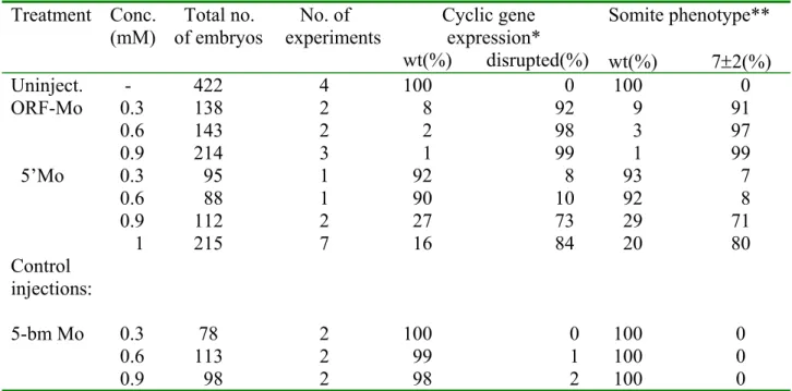
Table 2: Effects of Su(H)-Mo injections on cyclic gene expression and somite morphology
Treatment Conc.
(mM) Total no.
of embryos No. of
experiments Cyclic gene expression*
wt(%) disrupted(%)
Somite phenotype**
wt(%) 7±2(%) Uninject. - 422 4 100 0 100 0 ORF-Mo 0.3 138 2 8 92 9 91 0.6 143 2 2 98 3 97 0.9 214 3 1 99 1 99 5’Mo 0.3 95 1 92 8 93 7
0.6 88 1 90 10 92 8 0.9 112 2 27 73 29 71 1 215 7 16 84 20 80 Control
injections:
5-bm Mo 0.3 78 2 100 0 100 0 0.6 113 2 99 1 100 0 0.9 98 2 98 2 100 0
*Cyclic gene expression was monitored by her1 and her7 in situ hybridisations in 10s stage embryos.
**The somite phenotype, characterised by appearance of only the 7±2 anteriormost somites, was observed in living embryos at the 14s stage under a dissection microscope.
Embryos injected with the ORF-Mo (ORF morphants) resemble the notch1a mutant deadly
seven during the first 20 hours of development (Fig.1 C, H). These embryos develop 7±2
somite borders, which are not properly arranged in comparison to the control embryos
(compare Fig.1 E, J with Fig.1 D, I). Beyond these anterior somites further somitic tissue is
generated but somite borders are missing (Fig.1 C, E, H, J). At the end of somitogenesis
the ORF-Mo injected embryos exhibit a more severe phenotype than the des mutant. The
ORF morphants show a curved trunk and tail phenotype as it has been observed after
overexpression of a DNA-binding mutant of the Xenopus Su(H) homologue in zebrafish
(Fig.1 L, P, R; Lawson et al. 2001). In addition ORF morphants are shorter than the
comparable control embryo and lack posterior trunk pigmentation (compare Fig.1 O, Q
with Fig.1 P, R). They survive up to 200 hpf, show severe defects in blood vessel
formation and develop a large heart oedema (data not shown). Almost the same phenotype
has been observed for the zebrafish mutant mindbomb, which codes for a defective E3
ubiquitin ligase (Itoh et al. 2003; Jiang et al. 1996). Since this ubiquitin ligase is necessary
for the endocytosis of Delta the mutation leads to a breakdown of delta dependent Notch
signalling. This suggests that the phenotype observed here for the ORF morphants is typical for defective Notch signalling in zebrafish.
However, embryos injected with the 5’Mo (5’ morphants) show morphologically a much stronger effect, which is similar to the respective RBP-J κ
null-mutant in mouse (Oka et al.1995). The embryos are always developmentally retarded and drastically reduced in size when compared to their wildtype counterpart and the ORF morphant (Fig.1). As a consequence of the reduction in size, these embryos show an incomplete rotation around the yolk after 16 hpf (Fig. 1 B). Measuring the length of the knockdown embryos after 16 hpf shows that they have only approximately 2/3 of the length of a comparable wildtype embryo. The 5’ morphants form, like the ORF morphants, only 7±2 somites, which show segmental defects and are irregularly arranged when compared to their wildtype complement (Fig.1 B, G). After 16 hpf distinct areas of degenerating cells can be detected in the head of the embryo (Fig.1 B) and during the next hours of development degenerating cells can also be observed in the trunk and tail (data not shown). Since RBP- J κ
-/-mutant mice embryos show also distinct areas of cell degeneration and none of the control injections leads to this effect one might assume that the effect is specific to the knockdown of Su(H). However, it is not possible to exclude that this is a toxic effect caused by Mo injection (for review see Heasman 2002). After the formation of the first 7±2 somites the embryos essentially stop growing and it appears that no further somitic tissue is generated.
The embryos develop for a few more hours and die after approximately 35-60 hpf.
To analyse the identity of the mesodermal tissue generated in Su(H)-knockdown embryos
in situ hybridisations for MyoD were performed. The somites express MyoD in Su(H)
morphants, indicating that muscle cells are still forming (Fig.1 N). However, while MyoD
expression is normally restricted to the posterior parts of newly formed somites (Weinberg
et al. 1996), this distinct expression is disturbed in the Su(H)-knockdown embryos when
compared to wildtype embryos of the same stage (Fig.1 M, N). This suggests that not only
the somite borders are missing beyond the anterior somites but that there is also
disturbance of the A-P polarity of the somitic tissue.
Fig. 1:
Influence on morphology after Su(H) morpholino injections and effects on MyoD expression. (A), (F), (K), (O), (Q) wildtype embryos at 16, 18, 28, 45, 75 hpf, respectively. (B), (G) Su(H)-5’Mo injected embryos at 16, 20 hpf, respectively. (C), (H), (L), (P), (R) Su(H)-ORF-Mo injected embryos at 16, 18, 28, 45, 75 hpf, respectively. (D), (I) higher magnification of the somites in wildtype embryos at 16 and 18 hpf, respectively. (E), (J) higher magnification of the somites in ORF-Mo injected embryos at 16 and 18 hpf, respectively. Arrows in (D), (E) indicate anterior somite borders in wildtype embryos and ORF morphants, respectively. (A)-(L), (O)-(R) lateral view, dorsal to the top. (M) wildtype embryo at 16 hpf stained for MyoD, (N) Su(H)-ORF-Mo injected embryo at 16 hpf stained for MyoD (0.9mM, 2 experiments, n=131, 98% affected). (M), (N) dorsal view, anterior to the top, flat mounted embryos.