her15, a novel gene with oscillating mRNA expression domains and its potential role in zebrafish somitogenesis
I n a u g u r a l - D i s s e r t a t i o n
zur
Erlangung des Doktorgrades
der Mathematisch-Naturwissenschaftlichen Fakultät
der Universität zu Köln
vorgelegt von
Sunita Sathy Shankaran
aus Kerala, Indien
Köln, Dezember 2004
Berichterstatter:
Prof. Dr. Diethard Tautz
Prof. Dr. Jürgen Dohmen
Tag der mündlichen Prüfung: 15 Februar 2005
Table of Contents
Acknowledgements i
Abbreviations iii
Zusammenfassung iv
Summary vi
1. Introduction 1
1.2 Origin of the somitic territory in the vertebrate blastula 2
1.3 Convergence-extension gives rise to presomitic mesoderm 2
1.4 Somitogenesis and the Segmentation clock 3
1.5 The Notch signaling pathway 9
1.6 Delayed negative feed back loops give rise to oscillations 13
1.7 A model for the zebrafish somitogenesis oscillator 14
1.8 Wnt-β-catenin signaling and the segmentation clock-upstream and downstream 15
1.9 Mesenchymal to Epithelial transition-a closer look! 17
1.10 Somite differentiation 20
1.11 Medical relevance of somitogenesis studies 21
1.12 Central Theme of Doctoral thesis 22
2 Materials 24
2.1 Buffers 24
2.1 Primers and RZPD clones 24
2.3 Sequence of morpholinos used in gene knock down experiments 26
2.4 Vector maps 26
2.5 Computer system 27
2.6 Software 27
3.1 Zebrafish methods 28
3.1.1 Rearing of zebrafish and collection of embryos 28
3.1.1.1 Origin of zebrafish 28
3.1.1.2 Growth conditions 28
3.1.1.3 Zebrafish embryos 29
3.1.2 Dechorionisation and storage of zebrafish embryos 29
3.1.2.1 Mechanical dechorionisation of embryos 29
3.1.2.2 Storage of embryos 30
3.1.3 In situ hybridization of whole embryos with digoxygenin labeled RNA probes 30
3.1.3.1 Heat treatment of zebrafish embryos 31
3.1.3.2 Treatment with acetanhydrid 31
3.1.3.3 Prehybridization 32
3.1.3.4 Hybriization 32
3.1.3.5 Washing steps 32
3.1.3.6 Incubation in antibody 32
3.1.3.7 Color substrate reaction 33
3.1.3.8 Double in situ hybridization 33
3.1.4 Analysis of embryos after in situ hybridization 35
3.1.4.1 Analysis by whole-mount preparations 35
3.1.4.2 Analysis by flat-mount preparations 35
3.2 Molecular biology protocols 36
3.2.1 RZPD clones 36
3.2.2 Diatamaceous earth plasmid DNA miniprep 37
3.2.3 Quantification of DNA by spectrophotometric analysis 39
3.2.4 Sequencing of DNA 39
3.2.4.1 Sequencing reaction 40
3.2.4.1 Reaction profile 40
3.2.4.2 Cleaning of the reaction products 40
3.2.4.3 Analysis 40
3.2.5 Linearisation of plasmid DNA by restriction enzyme digestion 41
3.2.5.2 Restriction enzyme digestion of plasmid DNA 41
3.2.5.3 Phenol-Chloroform extraction 41
3.2.7 Invitro transcription for the production of in situ probes 43
3.2.8 Preparation of total RNA and cDNA form zebrafish embryos 43
3.2.9 Polymerase chain reaction (PCR) 44
3.2.10 Ligation 45
3.2.11 Cloning 45
3.2.11.1 Preparation of competent cells 45
3.2.11.2 Transformation 46
3.3 Preparation of capped mRNA for zebrafish injections 46
3.4 Synthesis of morpholinos 46
3.5 Injection of zebrafish embryos 47
3.6 Confocal imaging 47
3.7 Eppon embedding and sectioning 47
4 Results 48
4.1. hairy (h) and enhancer of split (E(spl)) related genes in zebrafish 48
4.1.1 Molecular nature of her15 48
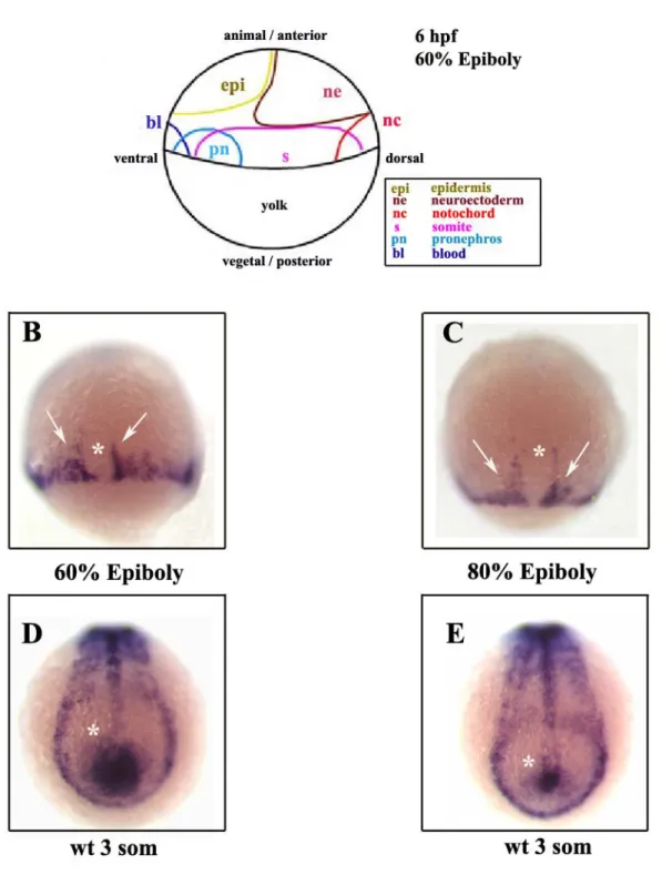
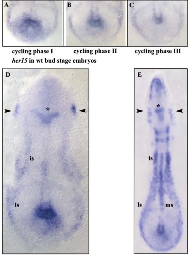
4.1.2 Analysis of her15 mRNA expression domains 49
4.1.3 The stripes of her15 62
4.1.4 her15 oscillation in the posterior PSM is a target of Delta-Notch signaling 70
4.1.5 her15 dynamics in the posterior PSM is independent of fss/tbx24 72
4.1.6 Early and late somitogenesis 82
4.1.7 her15 oscillation in the posterior PSM is independent of her1 and partially dependent on her7 86
4.1.8 Micropulsing her15 domain is uncovered by left-right asymmetric and dynamic mRNA expression 90
4.1.9 Confocal and microtome sectioning of her15 stained embryos for details pertaining o the micropulsing posterior PSM domain 93
4.1.10 Functional analysis of her15 94
4.1.10.1 her15 morpholino knockdown approach 96
4.1.11.2 her15 misexpression approach 96
4.2 Selective screening of the NIH zebrafish cDNA in situ expression database 103
4.2.3 ZfChp mRNA is maternal and ubiquitously expressed 107
4.2.4 Mapping of ZfChp 108
4.2.5 ZfChp is a target of Notch signaling 108
4.2.5 Functional analysis-ZfChp morpholino gene knockdown 116
4.2.7 Functional analysis- ZfChp misexpression 116
5. Discussion 117
5.1 her15 is a novel oscillating her gene in zebrafish 118
5.1.1 Is her15 a component of the zebrafish somitogenesis oscillator or just an just an output of the clock 120
5.1.2 The stripes of her15 are expressed at double segmental distance in the anterior PSM 121
5.1.3 her15 mRNA is expressed in the posterior wall of the PSM 122
5.1.4 her15 shares conserved mRNA expression domains with other mouse Hes5 homologues in chick, mouse and Xenopus 124
5.1.5 Synergistic interactions between Hes1 and Hes5 providing a possible explanation for the lack of penetrant phenotypes in mouse 127
5.1.6 Mouse Hes7, Hes5 and Hes1 homologues in zebrafish 128
5.2 Clone-3259-ZfChp 129
5.2.1 ZfChp and its Rho family GTPase domains 129
5.2.2 ZfChp as a possible link between somite prepatterning and MET in zebrafish embryos. 130
5.2.3 Proposal for functional analysis of ZfChp 130
5.2.4. ZfChp dominant negative and constitutively active constructs 131
6 References 133
7. Appendix 153
Erklarung 164
Lebenslauf 165
Figure 1. Temporal landmarks during zebrafish somitogenesis Figure 2. Overview of zebrafish somitogenesis
Figure 3. The oscillatory expression of deltaC
Figure 4: A model for the Presomitic Mesoderm Oscillator in chick embryos Figure 5: The Notch signaling pathway.
Figure 6: A summary of the genetic analysis of the functions of her1 and the notch pathway during somitogenesis.
Figure 7 Cells Undergo Mesenchymal-to-Epithelial Transition at Somite Boundaries.
Figure 8. The four cell types of zebrafish somite Figure 9. Protein architecture of her15
Figure 10. her15 mRNA expression pattern during late gastrulation and 3 somite stages Figure 11. her15 in bud stage and 3-6 somite stage
Figure 12. her15 is dynamically expressed in the posterior PSM Figure 13. her15 is expressed throughout somitogenesis
Figure 14. her15 mRNA expression during somitogenesis stages Figure 15. The stripes of her15
Figure16: her15 is a target of Notch signaling
Figure 17: her15 in Delta-Notch pathway somitogenesis mutants Figure 18: her15 is activated by Delta-Notch signaling
Figure 19. her15 in fss/tbx24 mutant embryos
Figure 20. her15 in bea and Su(H) knock down, bud stage embryos
Figure 21. her15 mRNA expression following her1 and her7 gene knockdown
Figure 22: her15 mRNA shows asymmetric distribution on left-right halves of the embryo Figure 23. Confocal and sagittal sections of her15 micropulsing domain
Figure 25. her1 and deltaC are disrupted after her15 misexpression Figure 26. Phylogenetic analysis of ZfChp and other Rho GTPases Figure 27. ZfChp mRNA is maternal ubiquitously expressed Figure 28. Mapping of ZfChp
Figure 29. ZfChp is a target of the Notch signaling.
Figure 30.Gradual progression of gene expression in the posterior PSM wall of wild type zebrafish embryos
Figure 31. A model of the chick embryo showing the primitive streak and oscillating expression domains of mouse Hes5
Figure 32. Comparison of mRNA expression domains of mouse Hes5 homologues in zebrafish and Xenopus.
Table1 genes implicated in the vertebrate segmentation oscillator mechanism Table2: Primers Used
Table 3: RZPD clones corresponding to the ESTs from the NIH in situ expression database and their cloning vectors
Table 4. Protein sequence comparison of her15 with other mouse Hes5 homologues Tables 5. Analysis of her15 stripes and her4 stripes
Acknowledgements
To Prof.Dr.Diethard Tautz, for giving me the opportunity to do my PhD in his lab. You have fostered in me a love for evolution and developmental biology which I hope to pursue with added fervor in the coming years. I thankyou for giving me the confidence to move forward at a point where I felt I would certainly fail. Thankyou for believing in me and for always welcoming my queries and doubts, with a smile.
To Dr.Martin Gajewski, “Doktorshen”, for having taught me everything I know about zebrafish somitogenesis, for your patience and understanding while teaching me and answering my queries.
To Dirki, for all the help and suggestions, for your patience in tutoring me on how to poke a needle deep enough into a zebrafish embryo to make it worthwhile, for our friendship and all the endless conversations
To Carmen, for help, suggestions, amity and for a sympathetic ear when things just were not going my way
To Burkhard, for friendly dialogue at times when I needed it
To Klaus Rohr and Dr.David Arnosti for research suggestions. Thankyou for helping me prep up my interview presentation which definitely made the difference between a good presentation and just an average one
To all the members of the Tautz lab, for their help from time to time
A special token of gratitude to Prof.Dr.Diethard Tautz, Dr.Klaus Rohr and Prof.Dr.Maria Leptin for excellent recommendation letters.
To all the Professors and students of the Graduate School for research suggestions and engaging dialogue during Graduate school meetings
To Dr.Brigitte and Dr.Sebastian, for helping me with all the official formalities of being a graduate student
To my fellow collegues from Batch 2001 of the Cologne University Graduate School for their friendship. It still feels like yesterday when we all came here to do our PhD and 3 years have gone by so quickly
To Aswani, Bhupi, Michael, Palani, Sam and Salin for their goodwill and alacrity whenever situations demanded it. Having you guys around has made Cologne home for me and I will never forget the meals and conversations we shared during our time in Efferren and beyond.
To the many scientists from the zebrafish field and farther, whom I have contacted for suggestions and reagents
To my family in India and for their continual support and love.
Abbreviations
AP: Alkaline Phosphatase
bp: Base pairs
BSA: Bovine serum albumine
ddNTP: Dideoxynucleotidetriphosphate DEPC: Diethylpyrocarbonate
dNTP: Deoxynucleotidtriphosphate hpf: hours post fertilisation
Hybmix: Hybridization Mix
L Litre
MO: Morpholino antisense Oligomer
nt: Nucleotide
ORF Open reading frame
PBS: Phosphate buffered saline
PBST: Phosphate buffered saline + Tween-20 PCR: Polymerase chain reaction
PFA: Paraformaldehyde PSM: Pre Somitic Mesoderm
RACE: Rapid amplification of cDNA ends
SDS: Sodium Dodecyl Sulphate
RT: Room temperature
s/som Somites
SSC: Sodium chloride/Sodium citrate
SSCT: SSC + Tween-20
TAE: Tris-Acetate EDTA (Electrophoresis Buffer) wt: Wildtype
Zusammenfassung
Die Somitogenese ist der entscheidene entwicklungsbiologische Prozess, welcher die Grundlagen für eine strukturierte Körperachse in Vertebraten und Cephalochordaten festlegt. Während der Somitogenese wird der Embryo in transiente, segmentale Strukturen (Somiten) unterteilt, welche später zu Muskeln und Wirbeln des Rumpfes und Schwanzes differenzieren. Somiten entstehen aus dem unsegmentierten, präsomitischen, paraxialen Mesoderm (PSM), in welchem ein komplexer Musterbildungsprozess abläuft. Für den koordinierten Ablauf dieses Prozesses ist ein Oszillator Mechanismus („Segmentation clock“) verantwortlich. Dieser beinhaltet eine wellenartige, von posterior nach anterior verlaufende Expression verschiedener Gene. Die konservierten Hauptkomponenten dieses Oszillator Mechanismus sind der Delta-Notch Signaltransduktionsweg sowie zahlreiche hairy/(E(spl)-C)-homologe Gene. Zwei der hairy/(E(spl)-C)-homologen Gene, her1 und her7, spielen eine entscheidene Rolle während der Somitogenese im Zebrafisch. Offen bleibt jedoch die Frage, wieviele her Gene Hauptkomponenten des Oszillator Mechanismus sind und wie diese miteinander interagieren. Um diese Frage zu beatworten wurde eine Suche nach weiteren hairy/(E(spl)-C)-homologen Genen von Sieger et. al, (2004) im Zebrafisch durchgefürt. Es konnten so drei weitere her Gene mit oszillierender Expression im PSM identifiziert werden. Eines dieser Gene ist her15, das in dieser Arbeit charakterisiert wurde.
Das her15 Gen zeigt im posterioren PSM eine oszillierende Expressionsdomäne, welche hauptsächlich in drei Phasen unterteilbar ist: eine breitere, eine intermediäre sowie eine punktartige Expression. Vergleichbar mit anderen her Genen zeigt auch her15 eine streifenartige Expression im anterioren PSM. Diese Streifen sind jedoch im Gegensatz zu allen anderen her Genen in einem doppelt segmentalen Abstand exprimiert. Zusätzlich konnte gezeigt werden, dass einer der her15 Streifen die posteriore Grenze des letzten Somiten markiert. Durch Morpholino „knock down“ Studien wurde gezeigt, dass die oszillierende her15 Expression in gewisser Hinsicht vom Her7 Protein beeinflusst wird, jedoch völlig unabhängig von Her1 ist. Die oszillierende her15 Expression im posterioren PSM zeigt gewisse Unterschiede zwischen der rechten und linken Seite des Embryos, welches ein klarer Hinweis darauf ist, dass das oszillierende Signal auf beiden Körperseiten autonom generiert wird. Missexpressionsstudien deuten auf eine mögliche Beteiligung von her15 an der Bildung der Somitengrenzen, sowie eine mögliche Funktion
in der Regulation der oszillierenden Genexpression im PSM hin. Die Injektion von Morpholinos gegen her15 zeigte jedoch weder Effekte auf die Bildung der Somitengrenzen noch auf die Expression verschiedener Gene des Delta-Notch Signalweges.
ZfChp ist das zweite Gen, welches in dieser Arbeit charakterisiert wurde. Dieses Gen entstammt einer Suche nach Kandidatengenen in der „NIH cDNA in situ expression database“. ZfChp zeigt eine dynamische, streifenartige Expression im intermediären PSM, einer Region in welcher die Zellen vom mesenchymalen in den epithelialen Zustand übergehen („Mesenchymal to Epithelial transition“-MET), und damit zu Somiten zu differenzieren. Das ZfChp Protein weist die molekulare Struktur einer zur Rho Familie gehörenden GTPase auf (basierend auf den konservierten Rho GTPase Domänen). Durch eine Mutantenanalyse konnte gezeigt werden, dass der Delta-Notch Signalweg die streifenartige Expression des ZfChp Gens im intermediären PSM postiv reguliert. Dies ist der erste molekulare Hinweis auf eine Verknüpfung des Musterbildungsprozesses im PSM und dem Übergang vom mesenchymalen in den epithelialen Zustand (MET).
Abstract
Somitogenesis is the key developmental process which lays down the framework for an organised body plan in vertebrates and cephalochordates. Somitogenesis divides the body axis into transient segmental structures called somites, which later give rise to muscles and vertebrae of the trunk and tail. Somites are generated from the unsegmented presomitic mesoderm (PSM) by an intricate process of prepatterning. Prepatterning is driven by a segmentation clock referred to as the presomitic mesoderm oscillator. This oscillator consists of certain gene members with oscillating mRNA expression compartments that sweep like a wave from the posterior to the anterior end of the embryonic PSM. The Delta- Notch pathway and various genes belonging to the hairy-(h) and Enhancer of split – [E(spl)] related family, are the core conserved components of this oscillator. h/E(spl) genes in zebrafish are commonly referred to as her genes. her1 and her7, play very important roles in the regulation of somitogenesis. The open question is how many her genes are core components of the zebrafish presomitic mesoderm oscillator and how do they interact with one another? To answer this, an in situ screen for h/E(spl) genes in zebrafish was conducted by Sieger et al., (2004). Three new her genes with oscillating mRNA expression domains were identified and one of them is her15, which has been further characterized in this PhD thesis.
her15 mRNA is expressed as a distinct oscillatory posterior PSM domain which shows three primary phases, namely broad, intermediate and dot-like. Comparable to other her genes, her15 also showed stripes in the anterior PSM but unlike others, these stripes were found to be expressed at double segmental distance. Additionally, the her15 stripe was found to label the posterior border of the last somite. Morpholino gene knock down studies showed that the oscillating expression of her15 is partly dependent on her7 regulation, but independent of her1. Oscillating her15 mRNA signals in the posterior PSM displays fluctuations with respect to left and right halves of the embryonic PSM, suggestive of the autonomy of both halves of the embryo in generating the signal. Misexpression studies suggest a prospective role for her15 in the regulation of somite border formation and oscillatory gene expression in zebrafish PSM. Morpholinos against her15 did not result in morphological border disruption, or in changes in mRNA expression of genes of the Delta- Notch pathway.
ZfChp, the second candidate gene which has been analyzed in the present thesis, came out of a screen of the NIH cDNA in situ expression database. ZfChp exhibits dynamic stripes of mRNA expression in the intermediate PSM region, a dynamic zone where tail bud mesenchymal cells undergo transition to epithelial state (mesenchymal to epithelial transition-MET), thus giving rise to somites. It the molecular signature of a Rho family GTPase with respect to conserved Rho GTPase domains. The Delta-Notch signaling pathway positively regulates the dynamic stripes of ZfChp in the intermediate PSM region.
This provides the first molecular evidence supporting a link between prepatterning of the PSM and MET in zebrafish embryos.
1. Introduction
Designing a body plan is an architectural challenge. Both invertebrates and vertebrates have addressed this problem by first establishing repeated units of equivalent identity (segments), and later coordinating these motifs into regionally specialized and integrated structures. Formation of the anteroposterior (AP) axis of vertebrates occurs progressively in the wake of the rostral-to-caudal regression of the embryonic organizer-known as the node and Hensen’s node in mammals and birds, and the Spemann organizer and the shield in amphibians and fishes, respectively. This regression movement results in the production of a variety of tissues, which includes the paraxial mesoderm that gives rise to head mesoderm anteriorly and to somites at the body level (Psychoyos and Stern, 1996, Tam et al., 2000). The most distinct feature of vertebrate mesoderm segmentation is the somite. Somites are transient segments of the paraxial mesoderm that are present in developing cephalochordates and vertebrates. The specific paraxial mesoderm region which gives rise to somites is called the presomitic mesoderm (PSM) and the process of somite formation is referred to as somitogenesis. Important landmarks in somitogenesis are periodicity, segmentation, epithelialization and differentiation. Somitogenesis begins soon after gastrulation and the process lasts until the number of somites characteristic of the species is reached. Somitic derivatives become later regionalized into different morphological domains-such as cervical, thoracic, lumbar and sacral regions as a result of the specific combination of Hox genes expressed in these domains. This body plan is highly variable within a given species indicating that the mechanism of segmentation and regionalization of the axis have to be tightly coordinated during development. Indeed cell fate in the immature somite is flexible and dependent on local environmental signals.
Consequently, somitogenesis has generated considerable interest and the somite now serves as a paradigm for investigating how naive cells adopt identity.
Somites were first identified at the beginning of the last century, and much of our understanding of somite development comes from morphological observations and experimental manipulations in the avian embryo (Christ and Ordahl, 1995), and more
recently, from embryo culture and genetic studies in mice (Gossler and Hrabe de Angelis, 1997). The rapidly developing zebrafish model promises to unite these approaches.
1.2 Origin of the somitic territory in the vertebrate blastula
Studies on the amphibian model system have provided most of the details pertaining to the dynamic cellular interactions that lead to the formation of the paraxial mesoderm during gastrulation (Keller 2000). In Xenopus and zebrafish, the paraxial mesoderm arises from deep layers of symmetrical tissue, located on either side of the Spemann organizer, at the marginal zone. The Spemann organizer corresponds to the shield in zebrafish (Kimmel et al., 1990; Warga and Kimmel, 1990) (for review see (Pourquie, 2001) .
1.3 Convergence-extension gives rise to presomitic mesoderm
Morphogenetic movements in the gastrulating embryo lead to the establishment of the paraxial mesoderm, which consists of the PSM region. It is this PSM region which gives rise to somites. These morphogenetic movements are called convergence-extension movements. Both in zebrafish and Xenopus, the prospective paraxial mesoderm region converges towards the blastopore during gastrulation, where they undergo invagination and subsequent elongation along the antero-posterior axis. First, the head mesoderm involutes and actively migrates towards the anterior (Niehrs et al., 1994). This is followed by the involution of the somitic mesoderm regions, which progressively elongates along the medio-lateral axis by a process of medio-lateral intercalation of cells, which results in the formation of PSM. The segmentation of the PSM into somites then takes place at the rostral end (Shih and Keller, 1992; Wilson et al., 1989). Convergence-extension of the paraxial mesoderm is crucial for the antero-posterior elongation of the vertebrate body axis. It is widely accepted that axis elongation is driven by the organizer and its major derivative the notochord, which is also formed by similar convergence-extension movements. But the paraxial mesoderm plays a prominent role in axis elongation and this is supported by the observation that embryos which the lack the notochord are capable of
axis elongation, while those embryos which are deprived of paraxial mesoderm are not (Malacinski and Youn, 1982; Scharf and Gerhart, 1980).
In lower vertebrates like zebrafish, gastrulation proceeds until the closure of the blastopore. During this phase, the somitic mesoderm which involutes, gives rise to the anterior most mesoderm, i.e., the head mesoderm and the anterior somites. Additionally, convergence-extension movements give rise to the anterior region of the paraxial mesoderm. Production of the more posterior somitic mesoderm occurs by a different process which occurs at the end of the classical gastrulation period. In Xenopus and zebrafish, the 12 anterior most somites are formed as a result of involution occurring during gastrulation (Kanki and Ho, 1997; Keller, 2000).
Gastrulation comes to an end with the closure of the blastopore in lower vertebrates such as zebrafish, or complete regression of the primitive streak, as in the case of higher vertebrates, such as chick. Subsequently, caudal somites are produced from the tail bud, which is located at the caudal end of the embryo. The tail bud consists of an apparently homogeneous mass of cells, namely mesenchymal cells. In fish, somites caudal to the twelfth are produced from the tail bud. The trunk-tail boundary lies at the level of somite 17, which consists of 5 trunk somites and all of the tail somites (Kanki and Ho, 1997;
Prince et al., 1998)
1.4 Somitogenesis and the Segmentation Clock
Vertebrates take their name from the segmented column of bones and joints that give rise to the main body axis. These elements, with their associated muscles, ribs and other connective tissue, derive from the somites which are blocks of mesodermal cells that form on either side of the central axis of the early embryo (Hirsinger et al., 2000). In all vertebrates, somites are generated sequentially from the caudal mesenchymal portion of the unsegmented paraxial mesoderm called the presomitic mesoderm (PSM) present at the tail end of the embryo. At the anterior end of the PSM, clefts appear, splitting off the successive blocks of somitic tissue; simultaneously, the embryo continues growing caudally, thereby maintaining the amount of PSM tissue approximately constant.
Accordingly, a new somite is generated in zebrafish every 30 mins; in the Xenopus every 40 mins, in the chick every 90 mins and in the mouse every 120 min.
Somites are laid down sequentially and this gives rise to a final spatially periodic pattern.
This spatial periodicity reflects a temporally periodic process, an oscillation, at the growing end of the developing organism, where anterior and posterior halves of somites are generated alternately. The possibility of there being a biological and molecular clock driving vertebrate segmentation and somitogenesis was put forward in 1976 by Cooke and Zeeman. They presented theoretical evidence for a ‘clock and wavefront’ model controlling the number of repeated structures formed during animal morphogenesis (as described in their own words). This abstract suggestion came to life after 30 years in 1997, with the discovery of certain genes whose mRNA expression oscillates at the tail end of the vertebrate embryo (Palmeirim et al., 1997). This ticking molecular clock is central to the process of segmentation of the vertebrate body. The first cycling or oscillating gene to be discovered in vertebrates was c-hairy1 in chicken which is a homologue of the Drosophila pair rule gene hairy (Palmeirim et al., 1997). hairy in Drosophila regulates segmentation and is expressed as stripes in alternate segments numbering a total of 7 stripes in the embryo (Ish-Horowicz et al., 1985). The fact that the first cycling gene to be discovered is a homologue of the Drosophila pair rule segmentation gene, suggests that certain aspects of invertebrate and vertebrate segmentation have been conserved. An overview of somitogenesis or segmentation in zebrafish is presented in Figures 1 and 2.
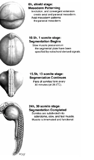
Figure 1. Temporal landmarks during zebrafish somitogenesis. This figure and legend has been taken from Stickney et al., (2000).
Figure 2. Overview of zebrafish somitogenesis. (A) Scanning electron micrograph of a 19 somite embryo.
Shortly after somites form, they change from a cuboidal to a chevron shape. Reproduced from Waterman and McCarty (1977) with permission of Scanning Microscopy International. (B) Live, lateral view of somitogenesis in a 20 somite embryo. The notochord is out of focus, medial to the somites and presomitic mesoderm. (C-F) Time lapse views of an embryo undergoing somitogenesis, as observed from dorsal. The notochord, in the center, is flanked on either side by paraxial mesoderm. Arrowheads indicate the positions of somitic furrows. Somitic furrows are first visible in the lateral part of the paraxial mesoderm. (C) Six somites have formed; arrowheads bracket somite 6. (D) The furrow on the right side between somite 6 and the future somite 7 has begun to form in the lateral presomitic mesoderm. (E) The furrow between somite 6 and the future somite 7 is nearly complete on both sides and a new furrow between somites 7 and 8 has begun to form. (F) Somite 7 has fully separated from the presomitic mesoderm. (G) Horizontal section through a 20 somite embryo at the level of the notochord. Epithelial boundaries and loosely packed central cells are visible in several of the somites. Reproduced with permission, from Waterman (1969). Scale bars
= 100 µm (A); 50 m (B); 25 µm (C-F); 25 µm (G).This figure and legend has been taken from Stickney et al., (2000).
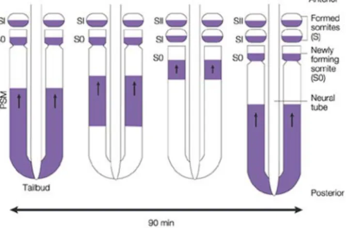
Research in the last couple of years has brought to light a significant number of genes with dynamic and oscillating mRNA expression domains which form the core of the molecular oscillator driving vertebrate somite segmentation. In the PSM, a particular subset of genes display oscillating mRNA expression, which switches on and off at the rate corresponding to the formation of one somite in each clock cycle (for review see Giudicelli and Lewis, 2004). The tempo for the entire process is set in the region where all the cells originate, namely, the posterior part of the PSM. The posterior PSM defines the periodicity of the entire process and hence this region behaves like a pace maker. One by one, each cohort of cells mature and pass from the posterior to the anterior part of the PSM. Eventually, at the anterior end of the PSM, each cohort of cells slow down its oscillation and become arrested in either an ‘on’ or ‘off’ state, according to its time of exit from the PSM. Cells arrested in the one state are stamped with the character assigned to the anterior part of the somite; while those arrested in the other, are stamped as posterior. Accordingly, phases of the oscillation cycle are recorded along the antero- posterior axis of the body, thus generating a pattern of gene expression, which appears to govern the physical process of segmentation, presumably through effects on cell-cell adhesion (Saga and Takeda, 2001).
As the oscillation gradually slows down in the anterior region of the PSM, anterior cells become retarded in their phase relative to the posterior cells. This gives rise to a spatio- temporal pattern of waves of expression that appear to sweep forward through the PSM tissue, prepatterning the somites. This phenomenon has been illustrated in oscillating expression of zebrafish deltaC shown in Figure 3. The maturation wavefront is the moving interface between the PSM region where, gene expression oscillates and the somitic mesoderm where oscillation is halted and overt differentiation begins.
Figure 3. The oscillatory expression of deltaC. The periodic spatial pattern of somites represents the trace of a temporal oscillation of gene expression at the tail end of the embryo. In this figure, the pattern of expression of deltaC, coding for the Notch ligand DeltaC, is shown by in situ hybridization in a zebrafish embryo at the 10 somite stage; other oscillating genes show a similar pattern. Somites are formed sequentially as growth continues at the tail end of the embryo. In the posterior region of the presomitic mesoderm (PSM), the mRNA levels rise and fall with a period of 30 min (at 28°C). As cells emerge into the anterior PSM, the oscillation in each of them slows down, finally halting as somite formation begins.
The prominent stripes in the anterior PSM corresponds to the cells that are still oscillating, but in different phases of their cycle; in the region of formed somites, oscillation has stopped, leaving cells arrested in different phases according to their time of exit from the PSM (Lewis, 2003). This figure and legend has been taken from Giudicelli and Lewis, (2004).
Figure 4: A model for the Presomitic Mesoderm Oscillator in chick embryos. It shows cyclic c-haiy1 mRNA expression (chick hairy homologue) in the PSM. These oscillations are bilaterally synchronous, and appears as antero-posterior waves of expression that sweep across the PSM approximately every 90 min (see figure in which c-hairy1 expression is shown in purple), which equals one cycle of somite formation in the chick embryo (Palmeirim et al., 1997). This figure and legend have been taken from Saga and Takeda, (2001).
1.5 The Notch signaling pathway
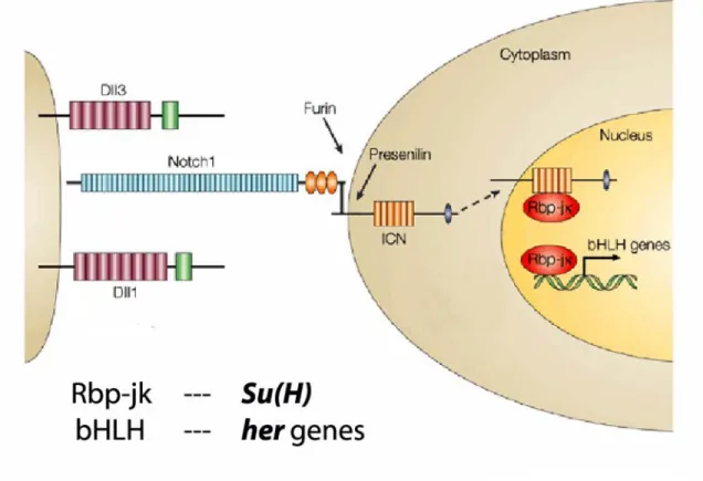
The Notch signaling cascade is crucial for both the generation and regulation of the is shown in Figure 5 (for review see Weinmaster and Kintner, 2003). Genetic studies in mice have shown that inactivation of many components of the Notch pathway result in dramatic segmentation defects and severe impairment in the periodic expression of the cyclically expressed genes (Barrantes et al., 1999). The Notch pathway mutants in mouse and fish lose the dynamic expression of cyclically expressed genes, indicating that Notch signaling is required for their periodic expression, or is required to coordinate the oscillations between the PSM cells. It has therefore been proposed that the Notch pathway is either part of the core mechanism of the segmentation clock (Pourquie, 2000), or acts as a cofactor to synchronize the cyclical gene expression in neighboring cells (Jiang et al., 2000). A further potentially important role for the segmentation clock is to periodically activate Notch signaling in the rostral presomitic mesoderm, thereby generating the periodic formation of somite boundaries.
Figure 5: The Notch signaling pathway. The figure has been modified from Sawada and Takeda, (2001).
Genetic studies have shown that Notch signaling is necessary for somite formation and for periodic expression of the cyclically expressed genes (Pourquie, 1999). Periodic expression of genes of the Notch signaling pathway provide a link between the signaling cascade and the segmentation clock. Notch is a large transmembrane receptor, which is able to recognize two sets of transmembrane ligands, Delta and Serrate. Upon ligand binding, Notch undergoes a proteolytic cleavage at the membrane, leading to the translocation of its cytoplasmic domain into the nucleus, where together with the transcription factor Su(h)/RBPjk, it activates the transcription of downstream genes such as those of the Enhancer of split complex in the fly or Her/Hes in vertebrates (Artavanis- Tsakonas et al., 1999). In the fly, Notch signaling has been implicated in several distinct developmental processes such as lateral inhibition or boundary formation between compartments.
Notch pathway directs somitogenesis in vertebrates and other phyla. The mechanisms utilised during somitogenesis in vertebrates and segmentation in lower animals such as in an annelid, the leech and in an arthropod, the fruit fly Drosophila, exhibit gross differences, at both molecular and morphological levels. These differences have led many to conclude that metameric body plans evolved independently, at least three times over the course of animal evolution. However, leeches and flies both exhibit evolutionarily
‘derived’ modes of segmentation and the molecular mechanisms that underlie these may be unrepresentative of their phyla. In this context, recent work on the spider Cupiennius salei is of great interest, for spiders are chelicerates and thus represent a branch of the arthropods that diverged very early from the insect/crustacean lineage (Cook et al., 2001).
Stollewerk et al., (2003) recently demonstrated that the similarity between vertebrate somitogenesis and opisthosomal segmentation in Cupiennius salei, is not merely morphological in nature. In both cases, the Notch signaling pathway is involved and thus provides the evolutionarily conserved link between somitogenesis in vertebrates and segmentation in lower animals.
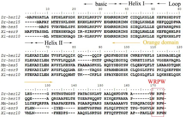
All the cyclically expressed genes characterized until now can be grouped into four categories. The first one corresponds to the glycosyl-transferase, Lunatic fringe, in chick and mouse, which acts as a modulator of Notch affinity for its ligands Delta and Serrate
(Aulehla and Johnson, 1999; Forsberg et al., 1998; McGrew et al., 1998). The second includes the zebrafish deltaC, one of the Notch ligands (Jiang et al., 2000). The third category is related to known or supposed direct downstream targets of Notch signaling, namely the hairy/Enhancer of split (Hes/her) family of transcription factors including chairy1, chairy2, cHey2 in chick, Hes1, Hes7 and mHey2 in mouse as well as her1, her7, her11, her12 and her15 in zebrafish and esr9 and esr10 in Xenopus (Bessho et al., 2001;
Gajewski et al., 2003; Henry et al., 2002; Holley et al., 2000; Jouve et al., 2000;
Leimeister et al., 2000; Li et al., 2003; Oates and Ho, 2002; Palmeirim et al., 1997;
Sawada et al., 2000; Sieger et al., 2004; Winkler et al., 2003). The fourth category consists of genes belonging to the Wnt-β catenin signaling pathway which includes Axin2 and Nkd1 (Aulehla et al., 2003; Ishikawa et al., 2004). All the genes implicated in the vertebrate segmentation oscillator mechanism and the corresponding references have been listed in Table1.
Table 1. Genes implicated in the vertebrate segmentation oscillator mechanism Notch signaling
components
hairy/E(spl) related Transcription factors
Wnt/β-catenin pathway components Cycling
genes
Mouse
Lfng [Aulehla et al; 1999]
Chicken
Lfng [McGrew et al., 1998]
Zebrafish
deltaC [Jiang, et al., 2000]
Xenopus
Xdelta2 [Jen et al., 1997]
Mouse
Hes1 [Jouve et al; 2000]
Hes7 [Bessho et al; 2001]
Chicken
c-hairy1 [Palmerim, et al., 1997]
c-hairy2 [Jouve et al., 2000]
c-hey2 [Leimeister et al.,2000]
Zebrafish
her1 [Holley et al.,2000]
[Sawada et al.,2000]
her7 [Oates &Ho,2002]
[Henry et al.,2002]
Xenopus
esr4 [Jen et al., 1999]
esr5 [Jen et al., 1999]
esr9 [Li et al., 2003]
esr10 [Li et al., 2003]
Mouse
Axin2 [Aulehla et al., 2003]
Nkd1 [Ishikawa et al.,
2004]
_
_
_
Gene functions required for
oscillations
Mouse
Cbf1 [Morales et al., 2002]
[Barrantes et al., 1999]
Delta1 [Jouve et al., 2000]
Lfng [Dale et al., 2003;
Serth et al., 2003]
Zebrafish
deltaD [Jiang et al., 2000]
[Holley et al., 2000]
mindbomb [Jiang et al., 2000]
[Holley et al., 2000]
notch1a [Holley et al., 2002]
deltaC [Scott Holley, personal communication]
Su(H) [Sieger et al., 2003]
Mouse
Hes7 [Bessho et al., 2001]
[Bessho et al., 2003]
Zebrafish
her1/her7 [Holley et al., 2002]
[Oates & Ho,2002]
[Henry et al., 2002]
[Gajewski et al., 2003]
Mouse
Wnt3a [Aulehla et al.,
2003]
Zebrafish
Receptor protein tyrosine
phosphatase (RPTP ψ) [Aerne et al.,2004]
1.6 Delayed negative feed back loops give rise to oscillations
In many biological clocks, such as the circadian rhythm, molecular oscillations rely on a delayed negative feed back loop. This implies that a particular component of the system is able to switch between active and inactive states and is auto inhibitory, so that in switching on it somehow sends itself a delayed signal to switch off, and in switching off it sends itself a delayed signal to switch on. The resulting system can then oscillate between the active and inactive states, with a cycle time determined by the delay in the feedback loop.
It has been suggested that the Hes/her genes, which behave as transcriptional repressors downstream of the Notch pathway, act by repressing Notch activity and thus directly participate in the generation of the transcriptional periodicity seen in the PSM cells.
Hes/her genes have characteristic repressor domains which led to the conclusion that they behave as repressors in all situations. However, the mice mutant of the cycling gene Hes1 does not display somitic defects or show disruption of the periodic expression of other cyclically expressed genes (Jouve et al., 2000; Ohtsuka et al., 1999). Recently a new member of the Hes family, Hes7, has been characterized in mouse. This gene acts as a repressor of its own transcription and is a target of Notch signaling (Bessho et al., 2003;
Bessho et al., 2001). It is expressed in a periodic fashion in the PSM like other cycling genes and homozygous mice for a null allele of Hes7 present strong segmentation defects. In these mice, expression of other cyclically expressed genes like Hes genes, Hes1 or mHey2 is lost, whereas Lunatic fringe remains expressed in the PSM but its expression is no longer dynamic (Bessho et al., 2001). Therefore, Hes7 represents the first Hes gene shown to be involved in regulating the periodic expression of cyclically expressed genes in the mouse PSM.
Further evidence pointing to a pivotal role of hes7 during mouse somitogenesis has been put forward in a recent publication which provides conclusive evidence that the instability of the Hes7 protein is crucial for sustained oscillation and for its function in the somite segmentation clock (Hirata et al., 2004). Mice were generated expressing a mutant version of Hes7 protein with a longer half-life (approximately 30 min as
compared to approximately 22 min for wild-type Hes7), but normal repressor activity. In these mice, oscillatory expression and somite segmentation became severely disorganized after a few normal cycles of segmentation. The observed effect could also be simulated mathematically using a direct auto repression model.
In the zebrafish embryo, the disruption of the expression of the two cyclically expressed her genes namely, her1 and her7 by morpholino antisense oligonucleotide injections as well as the misexpression of her1 lead to a disruption of the dynamic expression of cyclically expressed genes (Gajewski et al., 2003; Holley et al., 2002; Oates and Ho, 2002; Takke and Campos-Ortega, 1999) and disruption of somite border formation.
Remarkably, the sequences of her1 and her7 are much more closely related to that of Hes7 than that of Hes1. Recent studies in zebrafish present a slightly different picture regarding the negative feed back loop of her1 and its activity as an auto repressor. Based on morpholino gene knock down studies and exon probe in situ experiments, Holley et al., (2002) and Oates and Ho, (2002), have proposed that her1 acts as a repressor on its own transcription. But a recent publication by Gajewski et al., (2003), has challenged this by using an intron probe to detect the nascent transcript of her1. The intron probe studies show that her1 does not repress itself but instead acts as an activator for its own anterior- most PSM stripes. All these data, especially the studies in mouse, argue in favor of a central role for a subfamily of Hes/her genes in the mechanism of the oscillator.
1.7 A Model for the zebrafish somitogenesis oscillator
after eight (aei; dld), deadly seven (des, notch1), fused somites (fss, tbx24), beamter (bea, deltaC) and white tail/mindbomb (wit,mib) are the five genes that are necessary for normal somite formation which were isolated in the zebrafish genetic screen done in Tübingen (van Eeden et al., 1996). In zebrafish, oscillations in the expression of a hairy- related transcription factor, her1 and the notch ligand deltaC precede somite formation.
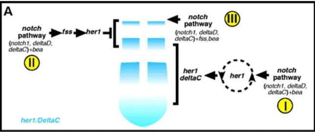
Scott Holley has proposed a model depicted in Figure 6, which suggests that her1 and the notch pathway have cyclical functions at the center of the somitogenesis oscillator.
Figure 6: A summary of the genetic analysis of the functions of her1 and the notch pathway during somitogenesis. (A) Anterior upwards. (I) The notch pathway and bea are required to generate the oscillating expression of deltaC and her1 in the posterior and intermediate PSM.
Her1 probably functions within the oscillator and feeds back on the notch pathway to create the oscillating pattern of both deltaC and her1. (II) fss functions downstream of the notch pathway but upstream of her1 in the anterior PSM. (III) Slightly later, the notch pathway, bea and fss function in the anterior most PSM/somitic mesoderm. The figure and legend has been taken from Holley et al; (2002).
1.8 Wnt/β-catenin signaling and the segmentation clock - upstream and downstream!
Wnt/β-catenin signaling cascade plays a major role in the segmentation clock and in the control of segmentation in mouse (Aulehla et al., 2003). Axin2, a negative regulator of Wnt signaling, shows oscillating mRNA transcription in the presomitic mesoderm (PSM) and the tail bud. Cyclic Axin2 alternates with Lfng expression, a Notch pathway cycling gene, and occurs even when Notch signaling is impaired. In contrast, Lfng is down regulated in the posterior PSM when Wnt3a activity is lacking and does not show cyclic expression anymore. This implies that Wnt3a indirectly controls Notch signaling.
Moreover, misexpression of Axin2 in the PSM resulted in ectopic upregulation of Lfng, disrupting its cyclic expression pattern, and impairing the segmentation process.
Therefore, Notch signaling appears to act downstream of Axin2. In addition, it was shown that Axin2 is a direct target of Wnt/β-catenin signaling in the PSM and acts downstream of Wnt3a, strongly suggesting that Wnt3a controls Notch signaling via Axin2.
Furthermore, misexpression of Axin2 in the PSM resulted in enlarged somites, while expression of Wnt3a from NIH3T3 cells transplanted on beads into the PSM of chick embryos had the opposite effect, the formation of smaller somites. There is also indirect evidence for a graded distribution of Wnt3a activity in the PSM. Thus, it can be concluded that Wnt3a plays a major role during mouse and chick segmentation process and lies upstream of the Notch signaling cascade.
A very recent publication had identified a novel gene Nkd1, which is a Wnt antagonist transcribed in an oscillatory manner (Ishikawa et al., 2004). The transcription of Nkd1 is extremely down regulated in the PSM of vestigial tail (vt/vt), a hypomorphic mutant of Wnt3a. Nkd1 oscillations have a similar phase to lunatic fringe (L-fng) transcription and they are arrested in Hes7 (a negative regulator of notch signaling), deficient embryos.
The results suggest that the transcription of Nkd1 requires Wnt3a, and that its oscillation pattern depends on the function of Hes7. Previously Wnt3a has been postulated to be upstream of Notch signaling but the present study demonstrates that a Wnt-signal-related gene may also be regulated by Notch signaling.
A new model for vertebrate somitogenesis in which the clock and the gradient are joined together has been proposed (Aulehla and Herrmann, 2004). Axin2 is the first gene found to be linked to both central components controlling segmentation, namely the transcriptional oscillator and the gradient. Axin2 displays oscillating transcription with periodicity similar to the segmentation process and is a direct target of Wnt3a gradient.
All other genes known to play a role in somitogenesis are either linked to the clock or the gradient, but not to both. Additionally, Wnt3a controls Fgf8 expression in the tail bud and oscillating Notch activity in the PSM. Therefore, Wnt3a and the Wnt signaling cascade must play a central role in the segmentation clock and in the gradient controlling somitogenesis.
Compelling and direct evidence for the involvement of the Wnt/β-catenin signaling cascade in somitogeneis has only been obtained till date from mouse embryos. But, a recent paper from Aerne and Ish-Horowicz (2004), reveals a potential role for the non- canonical Wnt-signaling pathway, which is independent of β-catenin, playing a role in zebrafish somite formation. The paper unveils a new gene namely receptor protein tyrosine phosphatase ψ (RPTPψ), which is essential for normal functioning of the somitogenesis clock in zebrafish. RPTPψ gene knock down in zebrafish embryos result in severe disruption of somite border formation and a loss of cyclic gene expression.
Impairing RPTPψ activity also interferes with convergent-extension during zebrafish gastrulation. Convergent-extension is a processof cell polarisation and intercalation that leads to lengthening and narrowing of the embryonic body during gastrulation and is regulated by the non-canonical Wnt signaling pathway. The authors state that a plausible explanation for the dual effect of RPTPψ on the somite oscillator and convergent extension is that changes in RPTPψ activity influences both adhesion and migration processes during convergentextension movements, and on Wnt-directed transcriptional regulationof the somite oscillator. This paper thus provides the first hint for a possible role for the non-canonical Wnt-signaling pathway in the regulation of zebrafish somitogenesis.
1.9 Mesenchymal to Epithelial transition during somitogenesis – a closer look!
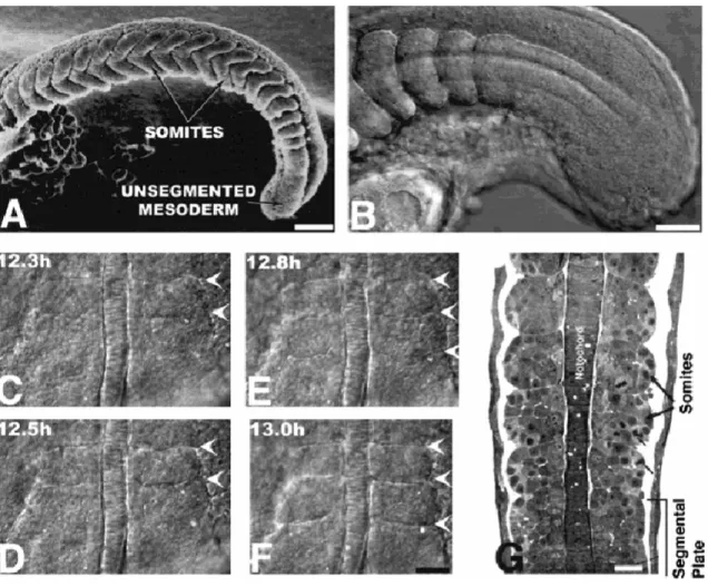
In the growing vertebrate embryos for the first 24 hours, starting at around 10 hpf, mesenchymal cells in the tail bud region undergo epithelial transition and thus give rise to somites. This dynamic process is called MET-Mesenchymal to Epithelial transition. MET involves extensive cytoskeletal changes. In chick embryos these changes are mediated by members of the Rho GTPase super family, namely Rac1, and Cdc42 (Nakaya et al., 2004).
During somite formation, a palisade- like structure called intersomitic furrow is formed by the alignement of the PSM cells positioned on either side of the prospective intersomitic boundary (Fig 4A). In zebrafish, there is virtually no cell movement during this process (Wood and Thorogood, 1994), as opposed to the situation in chick embryos (Kulesa and Fraser, 2002). Nevertheless, transformation from a mesenchymal to epithelial morphology brings about changes in the boundary cells. Aspects of epithelialization characteristic of cells at somite boundaries are
(i) Acquisition of a columnar shape
(ii) Accumulation of molecules associated with adhesion complexes, such as β- catenin, at the apical pole of cells (Fig 4B)
(iii) Basally directed relocalization of cell nuclei towards the somite boundary (Fig 4C)
(iv) Apical relocalization of centrosomes (Figure 4D)
These changes are initiated simultaneously with, and not prior to, intersomitic boundary formation. The cells within the core of the somite remain mesenchymal, while the cells at the boundary become epithelialized.
Figure 7 Cells Undergo Mesenchymal-to-Epithelial Transition at Somite Boundaries. (A–L) Dorsal views of the left-sided paraxial mesoderm of embryos labeled with Bodipy ceramide (which reveals cell morphology; [A], [E], and [I]) or with Bodipy 505-515 (which reveals nuclear position, [C], [G], and [K]) or immunostained for β-catenin ([B], [F], and [J]) or for γ-tubulin (which labels centrosomes) and stained with phalloidin (which labels actin) ([D], [H], and [L]). Anterior is oriented toward the top. (A–D) Cells at somite boundaries in wild-type embryos. The arrowheads point to the intersomitic boundary. (E–H) Cells in the presomitic mesoderm (PSM) of wild-type embryos. The arrows point to epithelial adaxial cells in which centrosomes are apically localized (H), as also seen in epithelial cells at somite boundaries (D).
Centrosomes are randomly positioned in other PSM cells. (I–L) Cells in the somitic mesoderm of fss−/−
embryos. n, notochord; ac, adaxial cells. The scale bars represent 10 µm.This figure and legend has been taken from Barrios et al., (2003).
In zebrafish embryos, Eph receptor/Ephrin signaling plays a significant role in driving the morphogenetic events associated with somite epithelialization. Numerous members of the Eph family of transmembrane receptor tyrosine kinases and other Ephrin ligands are expressed in a segmental pattern in the rostral presomitic mesoderm (Durbin et al., 1998) This pattern leads to the establishment of a receptor-ligand interface at each site of somite furrow formation. In the fused somites (fss/tbx24) mutant embryos, the cells in the paraxial mesoderm fail to undergo MET, as a result of which they fail to form intersomitic boundaries and epithelial somites. As the cells fail to epithelialize, they remain mesenchymal (Fig 4I), β-catenin appears localized homogeneously throughout the cell membrane (Fig 7J), nuclei remain at the centre of the cells (Figure 7K), and
centrosomes are distributed randomly within the cytoplasm (Figure 7L). The PSM cells in fss mutant displays the characteristics of mesenchymal morphology and not of epithelial morphology, hence they resemble the mesenchymal cells present in the core of the PSM in wild type embryos (Figures 7E-H). Additionally, cells with epithelial morphology can be seen in the PSM of wild type embryos, at sites where the paraxial mesoderm borders with the notochord, neural and surface ectoderm, and lateral plate (the arrows in the Figures 4E-H) (Barrios et al., 2003).
The fused somites fss/tbx24 mutants are also characterized by the absence of Eph/Ephrin signaling interfaces. Restoration of the Eph/Ephrin signaling interface is capable of rescuing the formation of morphological distinct boundaries in the paraxial mesoderm of fused somites mutant embryos (Barrios et al., 2003; Durbin et al., 2000).
1.10 Somite Differentiation
Somites give rise to the axial skeleton and the skeletal muscle of the trunk. Zebrafish require large muscles to locomote through their relatively viscous aquatic environment.
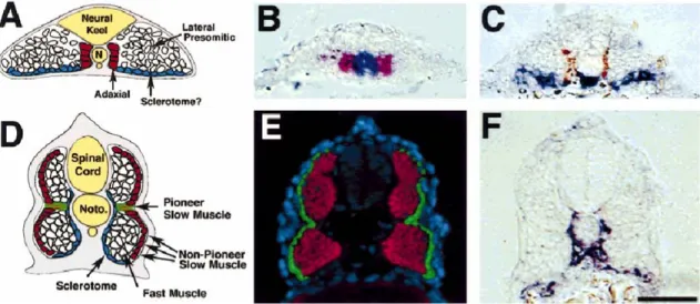
As the fish is supported by the buoyancy of water and their swim bladder, it has no use for the robust skeleton needed to support the terrestrial vertebrates. Hence zebrafish somite is predominantly mytome which gives rise to muscles as opposed to sclerotome from which the skeletal components such as vertebra and ribs are derived. Zebrafish sclerotome can be identified morphologically, shortly after somite formation, as a cluster of cells on the ventromedial surface of the somite which lies ventral to the myotome.
pax9 and twist are expressed in this cluster of mesenchymal cells (Morin-Kensicki and Eisen, 1997; Nornes et al., 1996), many of which will migrate dorsally to encircle the spinal cord and notochord, forming the vertebrae (Fig. 8C, F). In the adult, zebrafish muscle fibers can be subdivided into two broad classes (Bone, 1978). Slow muscle fibers, which are specialized for slow swimming and fast muscle fibers, used during bursts of rapid swimming. Slow muscle fibres are found in a wedge-shaped triangle on the lateral surface of the adult myotome whilst fast muscle fibers, are located in the deep portion of
the myotome. Slow fibers are smaller, darker, and more heavily vascularized than fast fibers (see review by Stickney et al., 2000).
Figure 8. The four cell types of zebrafish somite. Depicted in this figure are the starting and final positions for cells expressing markers of sclerotome (twist), slow muscle (S58, F59) and fast muscle (ZM4). (A-C) Cross-sections of zebrafish embryos at 13 hr. (A) Schematic cross-section through the anterior psm, showing the relative positions of fast muscle precursors (lateral presomitic), twist expressing cells (sclerotome?), slow muscle precursors (adaxial). (B) Adaxial cells express myoD (red) while still adjacent to the shh-expressing notochord (blue) in the segmental plate. (C) twist expressing cells (blue) are initially ventral to the myotome and are separated from the notochord by the medial-most adaxial cells that give rise to slow muscle fibers (brown, F59). Dark blue staining directly beneath the notochord is twist labeling of the hypochord. (D-F) Cross-sections of zebrafish embryos at 24 hr. (D) Schematic cross-section through a 24 hr zebrafish embryo, showing the positions of the four characterized cell types. (E) Slow muscle cells (green) form a superficial monolayer whereas fast muscle cells (red) remain deep. (F) twist expressing cells (blue) at 24 hr are found ventral and medial to the myotome, such that expression is directly adjacent to the notochord and ventral spinal chord. Dorsal is to the top. Scale bar = 100 µm (B, C, E, F).This figure and legend has been taken from Stickney et al, (2000).
1.11 Medical relevance of somitogenesis studies
Spondylocostal dystostosis (SCD) is a term given to a heterogeneous group of disorders in humans, with severe axial skeletal malformation, characterized radiologically by multiple vertebral segmentation defects. Additionally, in these patients the ribs are frequently misaligned, with points of fusion and sometimes reduction in number.
Mutations in Delta-like 3 (DLL3), which codes for one of the Delta ligands for the Notch receptor in humans; as well as MESP2, a basic-helix-loop-helix transcription factor, have been shown to cause SCD in humans. Somitogenesis studies in lower vertebrates such as zebrafish and mouse, may someday provide detailed information facilitating better understanding and treatment of human SCD (Whittock et al., 2004).
1.12 Central Theme of Doctoral thesis
The objective of the present doctoral thesis was to identify new genes, which regulate the zebrafish segmentation clock or somitogenesis oscillator. The search was initiated by looking for genes which interact with or belong to the Notch pathway or Wnt/β-Catenin signaling pathway, as there is compelling evidence that both these pathways play significant roles in regulating somitogenesis in both zebrafish and mouse. To identify new candidates, I participated in two cDNA RNA in situ screens conducted the lab namely
(i) Screen for hairy (h) and enhancer of split (E(spl)) family genes in zebrafish (Sieger, D et al., 2004)
(ii) Selective rescreening of the NIH zebrafish cDNA in situ expression database from the lab of Dr.Igor Dawid (Kudoh et al., 2001).
The experimental strategy was to select potential new genes involved in somitogenesis by analyzing the mRNA expression patterns in wild type zebrafish embryos and later in the somitogenesis mutants namely, after eight (aei /mutant for deltaD), deadly seven (des/
mutant for notch1a), beamter (bea/mutant for deltaC) and fused somites (fss/mutant for tbx24). This would give a preliminary hint as to which among them would possibly function in regulating somitogenesis. All of these mutants encode genes belonging to the Notch signaling cascade, with the exception of fused somite (fss/mutant for tbx24) which codes for a T-box gene.
I focused on two candidate genes which came out of the screens namely her15 and ZfChp. her15 was analysed for its potential role as a novel component of the zebrafish presomitic mesoderm oscillator. ZfChp, on the other hand was examined for its role as a link between somite prepatterning and downstream processes such as somite border formation, as suggested by its mRNA expression pattern in the intermediate PSM region.
2. Materials
2.1 Buffers
Buffers and solutions have been prepared according to Sambrook et al., (1989).
2.2 Primers and RZPD clones
The primers used were synthesized by the company Metabion and are listed in the following table. The lyophilized primer was dissolved in the appropriate volume of water to obtain a concentration of 100µM.
Table2. Primers used
No Name Sequence from 5’ to 3’ Hybridization
Temperature 1 T3 Primer AAT TAA CCC TCA CTA AAG GG 50°C
2 T7 Primer TAA TAC GAC TCA CTA TAG GG 50°C 3 SP6 Primer ATT TAG GTG ACA CTA TAG 50°C SP6 primer
pCS2
CCC AAG CTT GAT TTA GGT GAC 50°C T7 primer
pCS2
AAT ACG ACT CAC TAT AG 50°C 4 Zf Chp For-
Cla1
ACA TCG ATA TGC CAC CTC AAA TGG AT
56°C 5 Zf Chp rev-
Xho1
TAC TCG AGT CAG ATG AAG CAG AAG AA
56°C 6 T3-esr7up AAT TAA CCC TCA CTA AAG GGC TCC
TGC GTA TAT G
55°C 7 T7-
esr7downrc
TAA TAC GAC TCA CTA TAG GGT CTC CAG AGC GGA G
55°C 8 H15overexp
Cla1for
GCA TCG ATA TGG CTC CTG TGT ATA TGAC
58°C
9 H15overexp Xho1rev
TAA CTC GAG CTA CCA GGG TCT CCA GAG
58°C 10 H15traceFor
Amp
GTG GGA AAG CTA ATC CTG AC 52°C 11 H15trace
RevAmp
TGC TTG ATG TGT GTG TGC TG 52°C 12 H15 For
5'UTR check
ATC AGT GCA CGC TGA TGT TC 52°C
13 2H15 For 5'UTR check
GAT GCC TCT TCC ATT GTG TG 52°C
14 H15 Rev 5'UTR check
TGC TTG ATG TGT GTG TGC TG 52°C
15 T3-her1-29 AAT TAA CCC TCA CTA AAG GGT GTA TCG TCT TCT T
45°C 16 T7-her1-
1037
TAA TAC GAC TCA CTA TAG GGT CTC
CAC AAA GGC T 45°C
Table 3. RZPD clones corresponding to the ESTs from NIH cDNA in
situ expression database and their cloning vectorsNo NIH cDNA in situ expression database
RZPD clone ID Vector
1 Clone No 5116 IMAGp998D158968Q3 pSPORT1
2 Clone No 5096 IMAGp998E028969Q3 pSPORT1
3 Clone No 5144 IMAGp998A178977Q3 pSPORT1
4 Clone No 3259 IMAGp998K0510339Q3 pSPORT1
5 Clone No 2247 IMAGp998C028964Q3 pSPORT1
6 Clone No 5038 LLKMp964N1517Q2 pBK-CMV
2.3 Sequence of morpholinos used in gene knock down experiments
her1 morpholino - 5’-AGT ATT GTA TTC CCG CTG ATC TGT C-3’
her7 morpholino – 5’-ATG CAG GTG GAG GTC TTT CAT CGA G-3’
Su(H) morpholino – 5’-CAA ACT TCC CTG TCA CAA CAG GCG C-3’
ZfChp morpholino – 5’-ACC CTC TGC TTA CCC GAG AAG ACG T-3’
her15 morpholino – 5’-CAG GAG CCA TTG CTT CTT CAG GAG A-3’
2.4 Vector maps
pCS2+ is the standard vector used in zebrafish studies for misexpression experiments. As control for these functional studies, a modified pCS2+eGFP is used. pCS2+eGFP vector was constructed by cloning the Eco47III/XhoI-EGFP fragment from the pEGFP-C1 vector into the StuI/XhoI site of the CS2+ vector.
2.5 Computer system
The data acquisition as well as the picture formating and word processing were done using personal computers with operating systems Windows 2000 and Windows XP Professional.
2.6 Software
Acrobat Reader 5.0 (Adobe) AxioVision 2.0.5.3 (Zeiss)
BioEdit 5.0.7 (Tom Hall, Department of Microbiology, North Carolina State University) Entrez (National Centre for Biotechnology Information = NCBI)
Photoshop 6.0 (Adobe)
Vector NTI 6.0 (Info Max, Inc.) Microsoft Office 2000 Premium
Moreover, the services of PubMed (NCBI), Blast (Altschul et al., 1997), Zebrafish EST Database (http://zfish.wustl.edu/) (Washington University, St. Louis) and ZFIN (www.zfin.org) (Sprague et al., 2001; Sparague et al, 2003) have been utilized and are acknowledged.
3.Methods
3.1 Zebrafish methods
3.1.1 Rearing of zebrafish and collection of embryos 3.1.1.1 Origin of the zebrafish
The zebrafish Danio rerio is a fresh water fish from the Ganges that belongs to the family Cyprinidae. The animals in our facility were acquired from pet shops in Cologne and Gottingen. These parent animals were then further bred.
3.1.1.2 Growth conditions
Starting from day2, zebrafish were grown in an aquarium, consisting of several serial 12L tank units, with a water temperature between 26 and 28°C (Mullins et al., 1994). The maximum extent of utilization of a unit amounted to 40 fish per liter. The aquarium was supplied continuously with fresh water, whereby daily 1/10 of the liquid volume was replaced by fresh water. One half of the fresh water was adjusted by means of an ion exchange resin to a total hardness between 6-10 degrees of hardness units; the other half was transmitted from a reverse osmosis plant. Within the aquarium, the water was circulated by a pump system. Suspended particles were sieved by integrated filter units from the water and the filtered water was sterilized afterwards by UV irradiation. The accumulation of toxic substances (e.g. nitrite) was prevented by using a bacterial filter.
Feeding of the fish took place thrice daily. Beside the usual trockenfutter (Tetramin), Artemia and Bosmina were fed, in order to ensure balanced nutrition. The light and darkness rhythm was adjusted to 14 hours light and 10 hours darkness.
3.1.1.3 Zebrafish embryos
The collection of zebrafish embryos for various experiments like total RNA isolation and in situ hybridization took place in the morning, directly after the dark phase came to an end. The previous evening the male and the female zebrafish pair were put in plastic boxes with a divider that separated them. The males and females can be differentiated by the shape of the under belly. The under belly of the female fish will be rounder. These plastic boxes contained normal aquarium water and the bottom was lined with marbles to prevent the eggs from being eaten by either of the parents after they had been laid. The divider was removed in the morning as soon as the light phase had started. The fish were then allowed to mate for 30 min to 1 hr and the eggs were collected in petridishes containing autoclaved aquarium water. These embryos were then allowed to develop at 28.5°C in an incubator and the desired stages were collected.
Solutions
Autoclaved aquarium water
3.1.2 Dechorionisation and storage of zebrafish embryos 3.1.2.1 Mechanical dechorionisation of embryos
Embryos of the desired growth stage were fixed in 4% paraformaldehyde (PFA) in PBS (phosphate buffered saline). This fixation can take place for 2 hr at room temperature (RT) or overnight at 4°C. After fixation, the embryos were dechorionated with a pair of sterile, fine-pointed watch-makers forceps.
Solutions
20X PBS 2,76 M NaCl50 mM KCl 160 mM Na2HPO4 50 mM KH2PO4
3.1.2.2 Storage of Embryos
The dechorionated embryos were then dehydrated by a series of methanol:PBST solutions (33% methanol/PBST, 66% methanol/PBST) and twice in 100% methanol. The incubation was for 10 min in each solution. After this, the embryos were stored at -20°C.
3.1.3 In situ hybrdization of whole embryos with digoxygenin labeled RNA probes
In situ hybridization by means of Digoxygenin labeled probes is a non-radioactive procedure, which makes it possible, to determine the spatial expression of mRNA (Tautz and Pfeifle, 1989). The embryos were incubated with digoxygenin labeled anti-sense RNA probes. The hybridized probes were then detected immunochemically, by means of alkaline phosphatase (AP) conjugated anti-digoxygenin Fab fragments, whereby the enzymatic conversion of specific substrates resulted in the production of colored precipitates.
For in situ hybridization of zebrafish embryos the protocol by Schulte-Merker et al., (1992) was followed with slight modifications. The Proteinase K treatment was replaced by heat treatment and the composition of some of the solutions was modified. All in situ hybridizations were carried out in the automated, InsituPro machine (Abimed) (Plickert et al., 1997)
![Figure 7 Cells Undergo Mesenchymal-to-Epithelial Transition at Somite Boundaries. (A–L) Dorsal views of the left-sided paraxial mesoderm of embryos labeled with Bodipy ceramide (which reveals cell morphology; [A], [E], and [I]) or with Bodipy 505-515 (wh](https://thumb-eu.123doks.com/thumbv2/1library_info/3625861.1502001/35.918.139.785.102.409/undergo-mesenchymal-epithelial-transition-boundaries-paraxial-mesoderm-morphology.webp)