A Genetic Screen for Genes Involved in the FGF Signalling Pathway in
Drosophila melanogaster
Inaugural-Dissertation zur
Erlangung des Doktorgrades
der Mathematisch-Naturwissenschaftlichen Fakultät der Universität zu Köln
vorgelegt von Min-yan Zhu
aus Sichuan, P. R. China
Köln 2003
Berichterstatter: Prof. Dr. Maria Leptin
Priv.-Doz. Dr. Frank Sprenger
Tag der mündlichen Prüfung: 4 Juli 2003
Table of contents
1. Introduction 1
1.1 Receptor tyrosine kinase signals 1 1.1.1 The activation of the receptors 1 1.1.2 Bring the substrates close to the membrane 3 Adapter proteins and docking proteins 3 Function of the lipids during signal transduction 4 1.1.3 Different downstream pathways 5
PLCγ pathway 5
PI3K pathway 5
Ras-MAPK pathway 6 1.1.4 Signals to nucleus 6 1.1.5 Signals to cytoplasm 7 1.1.6 Signal termination 8 1.2 Retain the specificity of different receptor tyrosine kinase signals 8 1.3 Fibroblast growth factor signalling in vertebrate development 9
1.4 Drosophila development and signal transduction 10 1.4.1 Signals required during Drosophila eye development 10 Drosophila eye development 10 The specification of photoreceptor cells 10 Epithelial planar polarity 11 1.4.2 FGF signalling during Drosophila embryonic development 12 Mesoderm migration 12 Tracheal branching 13 The transduction of FGF signalling 14
1.5 Approaches 14
1.6 Aim of the thesis 14
2. Results 15
2.1 The gain-of-function screen 15 2.1.1 The ‘GMR>λ-btl, dof’ eye phenotype 15 2.1.2 Tests of the potential candidates 17 2.1.3 The gain-of-function screen 20
2.2 Candidates 23
2.2.1 The description of the candidates 23 2.2.2 Summary of the candidate genes 40
2.3 Genetic interaction of the candidates with other signalling pathways 41 2.3.1 Testing for genetic interactions of the candidates with other signalling pathways 41 2.3.2 The results from other gain-of-function screens in comparison to my Screen 43
2.3.3 Summary 44
2.4 Studies on three of the candidates-EP0719, EP3575 and EP0863 45
2.4.1 EP0719 45
CG3542 is homologous to a formin binding protein 45 The analysis of functions of CG3542 during Drosophila development 47
2.4.2 EP3542 51
2.4.3 EP0863 55
2.4.4 Summary 58
2.5 Studies on the mutants of X-144 58
2.5.1 Mapping of the mutant 59
2.5.2 The mutant phenotype 60
3. Discussion 70
3.1 The screen is likely to have some specificity for the FGF signal 71
3.2 Many candidates are possibly involved in various receptor tyrosine kinase
signals 72
3.3 Various cellular processes may regulate FGF signalling 73 3.4 The involvement of the Ras85D-MAPK pathway in FGF signal transduction 74 3.5 The effects of epithelial planar polarity signal 75
3.6 Detailed analysis of some of the candidate genes 76 Analysis of a cytoskeleton related protein-CG3542 76 Analysis of a protein involved in secretion pathway-Sar1 76 Analysis of a predicted kinase-CG6386 77 Potential approach to characterize the possible roles of CG3542, Sar1 and CG6386
in FGF signalling 77 Characterisation of the mutant X-144 that is close to EP1340 78
3.7 Conclusions 78
4. Materials and methods 79
4.1 Reagents 79
4.2 Fly genetics 79
Drosophila stocks 79
Genetic crosses 81
4.3 Histochemistry 82
Embryo fixation 82
In situ hybridization 83
Antibody staining 83
Enbedding stained embryos in Araldite 84
Isolation of genomic DNA 85
Genomic PCR 85
PCR sequencing 86
4.5 Construction of transgenic flies 86
Molecular cloning 86
Microinjection 86
Establishing the lines 86
4.6 Analysis of protein-protein interactions 86 Molecular cloning 86
Yeast two-hybrid 86
4.7 Microscopy and image analysis 87
4.8 DNA sequence analysis 87
5. Bibliography 88
Appendix 103
Abstract 109
Zusammenfassung 110 Abbreviations
Acknowledgements Lebenslauf
Erklärung
Introduction
1 Introduction
Cell-cell communications are the fundamental bases of the involvement of multi-cellular organisms. Cells engage various ways to communicate. They send signals by ions, small organic molecules, and secreted proteins and also by membrane proteins. Signals can be detected by various receptors, which can be located at the membrane or within the cytoplasm. Upon binding to their ligands, receptors are capable of triggering a series of events, which can change cell behavior. A well-studied family of receptors are the receptor tyrosine kinases (RTK). Receptor tyrosine kinases respond to different signals, and have distinct consequences, although they share many common downstream components. How these processes are regulated presents a great challenge for biologists.
The embryonic development of Drosophila melanogaster provides a good model system to study the communication between cells. During the embryonic development, a fertilized egg forms a single layer of cells after 13 synchronized divisions. Morphogenesis movements start with ventral furrow invagination, followed by germband extension and posterior mid gut invagination. At mean time, invaginated mesoderm cells migrate along the lateral ectoderm to form a monolayer beneath the overlying ectoderm. Later on, specified tracheal placodes invaginate and branch out during germband retraction and dorsal closure, to form an interconnected tubule network of the larvae respiration system.
RTK signals are required extensively during morphogenesis of the embryo development.
Both mesoderm migration and trachea formation require fibroblast growth factor (FGF) signals. Compare to vertebrates, many downstream targets for RTK signals are conserved in Drosophila. The work presented in this thesis is an approach to identify other components involved in the FGF signalling pathway.
1.1 Receptor tyrosine kinase signals
RTKs are transmembrane glycoproteins that are activated by binding to their ligands. The ligand binding changes the conformation of the receptors, which leads to the receptor oligomerization and autophosphorylation. The phosphorylated tyrosine residues on the receptor provide binding sites for various signal molecules. By binding to and phosphorylating their substrates, receptor tyrosine kinases are able to trigger a series of signal events (Fig.1-1), such as Phospholipase Cγ (PLCγ) signal, Phospholinositide 3- Kinase (PI3K) pathway, Ras-mitogen-activated protein kinase (Ras-MAPK) pathway, and JAK-STATs pathway (Hubbard et al., 2000; Schlessinger, 2000)
1.1.1 The activation of the receptors
The protein family of RTKs includes receptors for insulin and growth factors, such as
Epidermal Growth Factor (EGF), Fibroblast Growth Factor (FGF), Platelet-Derived
Introduction Growth Factor (PDGF), Vascular Endothelial Growth Factor (VEGF) and Nerve Growth Factor (NGF). All the RTKs consist of an extracellular portion that binds polypeptide ligands, a transmembrane helix, a cytoplasmic portion that possesses tyrosine kinase catalytic activity. The extracellular portion normally contains one or several of immunoglobulin-like domains, fibronectin type IV-like domain, Cystein-rich domains and EGF-like domains. The cytoplasmic region of the receptor can be subdivided to a juxtamembrane region, a tyrosine kinase catalytic region and a carboxy-terminal region.
Fig.1-1. A simplified schematic representation of receptor tyrosine kinase signal (receptor activation after example of Epidermal Growth Factor Receptor). Signals are sent via ligands.
Receptors exist mainly as monomers in their silent state on the membrane. The binding of the ligands to receptors leads to the activation of the receptors. Activated receptors recruit various adaptor and docking proteins, which triggers different pathways and therefore activates transcriptional factors or alters the cytoskeleton.
Except Insulin Receptor (IR), all known RTKs exist as monomers on the membrane in
their silent state. They oligomerize upon ligand binding. Different ligands employ
different strategies to activate the receptor. Recent solved structure of complex of human
EGF (TGF-α) and the extracellular domains of its receptor (EGFR) revealed that EGF
binds to its receptor in a 1:1 fashion. The binding causes the conformational change of the
receptors, which leads to the direct interaction of the extracellular domains of the
Introduction receptors. This interaction is likely to be the key events of the dimerization and activation of the EGFRs (Garrett et al., 2002; Ogiso et al., 2002). The activation of FGF receptor (FGFR) requires heparin sulfate proteoglycan in addition of FGF. FGF binds to its receptor in a 1:1 manner similar to the binding of EGF to EGFR. In a crystallized complex, heparins make numerous contacts with both FGF and FGFR. These contacts are thought to stabilize the binding of FGF and its receptor. In addition, heparin also interacts with FGFRs in the region that two FGF-FGFR complexes adjoin, which is probably important for the dimerization of the receptors (Plotnikov et al., 1999). The oligomerization leads to the trans-phosphorylation of the receptors in their kinase domains and therefore activates the receptor. The activated receptors are thought to cis- phosphorylate their tyrosine residues in the juxtamembrane region and C-terminal region.
These phosphorylated tyrosines provide binding sites for numerous signal molecules (Hubbard et al., 2000).
1.1.2 Bring the substrates close to the membrane
One important feature of the RTK signal transduction is that upon the activation of the receptor, numerous signal molecules are translocated to the membrane, either by direct interaction with receptor or the receptor binding proteins, or by interaction with the newly synthesized lipids such as phosphatidylinositol-3,4,5-trisphosphate or the proteins that bind to the lipids. This membrane translocation is important not only for the interaction of the molecules in the signal cascade, but also the increasing of local concentration of the downstream components, which is thought to ensure the specificity and efficiency of the signal transduction. For example, Ras is targeted to the inner leaflet of the plasma membrane through ER and Golgi by posttranslational modification of a C-terminal CAAX motif (Choy et al., 1999; Clarke et al., 1988), therefore to activated Ras would require the upstream molecules also localize to the membrane.
Proteins with different modules are able to recognize the phosphorylated tyrosine residues in different sequence motifs. The multiple tyrosine sites on the receptors are generally specific for binding of certain protein and therefore are responsible for specific signalling branches when phosphorylated. A well-studied case is PDGFRβ (Heldin et al., 1998).
Activated PDGFRβs bind to signalling molecules at specific tyrosine residues. For instance, it binds PLCγ1 at tyrosine 1021, PI3K at tyrosine 740 and 751, the src class kinases at tyrosines 579 and 581. It also binds to proteins such as Grb2 (growth factor receptor-bound) and Shc (Src and collagen homolog). Proteins that are able to bind to receptors in RTK signal cascades are termed adaptor proteins and docking proteins.
Adapter proteins and docking proteins
Adapter proteins in RTK pathways generally contain modules that can recognize and bind to phosphorylated receptors. They are the direct targets of the receptor tyrosine kinases.
They can be classified to two groups. One group includes proteins also having enzymatic
function, such as Src kinases, PLCγ and PI3K. The other group includes proteins mainly
function to recruit other proteins, such as Grb and Shc.
Introduction
SH2 domains and PTB domains are the domains that help adapter proteins recognize and bind to specific phosphorylated tyrosine residues. SH2 domain is the abbreviation for src homology 2 domain which consists of about 100 residues that binds phosphotyrosine residues in a specific sequence motif. It is first identified in Src kinase. PTB domain is the abbreviation of phosphotyrosine binding domain which consists of about 60 residues that recognize phosphorylated tyrosine in a distinct sequence context (Forman-Kay et al., 1999; Shoelson, 1997).
In addition to the domains that are required for the binding of receptor, adaptor proteins normally also contain other protein-protein interaction modules such as SH3 domain (src homologue 3) domain, WW domain, PH (pleckstrin homology) domain, PDZ (post synaptic density/disc-large/zo-1) domain and PYVE domain. SH3 domain and WW domain recognize similar but distinct proline rich motifs (Bedford et al., 2000). PH domain has affinity for phosphoinositides or their soluble head groups (Lemmon et al., 1996). PDZ domain binds specifically to hydrophobic residues at the C termini of their target proteins (Sudol, 1998). PYVE domains specifically recognize PtdIns-3-P (Misra et al., 1999).
There are also a group of proteins called docking protein that are associated with the cell membrane by either a myristyl anchor or a transmembrane domain. Examples are FRS2, which contains a myristyl anchor (Kouhara et al., 1997), and LAT, which has a transmembrane domain (Zhang et al., 1998). Most of the docking proteins have a PH domain at their N-termini, which also confers them abilities to bind to phosphoinositides on the membrane. Docking proteins contain specific domains such as PTB domains that enable them bind different receptors. Docking proteins are thought to function as platforms for the recruitment of other signalling proteins in response to receptor stimulation. One example for docking protein is FRS2. It contains a consensus myristylation sequence and a PTB domain. In addition it has several tyrosine sites that are the potential target for phosphorylation. It is myristylated and targeted to the cell membrane. It binds to activated FGFR and being phosphorylated. The phosphorylated FRS2 are able to recruit Grb2 to the cell membrane and therefore transduce the signal (Kouhara et al., 1997; Rabin et al., 1993).
Function of the lipids during signal transduction
One of the major consequences of the RTK signal is the change of the lipid environment
on the membrane. The major targets for the RTK signals are the phosphatidylinositol-4,5-
phosphotate (PtdIns(4,5)P
2). Of particular interest is the phosphorylation of the D-3
position of the inositol ring in the PtdIns(4,5)P
2molecule. This phosphorylation generates
a structure that can be recognized and bound by particular protein modules, such as PH
domain and FYVE domain. Many signal molecules are translocated to the membrane via
their PH domains binding to the PrdIns(3,4,5)P
3. Examples include serine-threonine
kinases Atk (or protein kinase B, PKB), Phosphoinositide-Dependent Kinase 1 (PDK1),
the Arf exchange factor Grp1, the docking protein Gab1, and PLCγ1 (Czech, 2000; Rameh
Introduction et al., 1999). The association of Atk and PDK1 to the membrane leads to the phosphorylation of Atk by PDK1 (Lawlor et al., 2001).
The model that some lipids are capable of aggregating on the membrane has emerged during the last decade. This model proposes the existence in biological membranes of lipid microdomains or rafts that have a high sphinogolipid and cholesterol content, which makes the rafts more ordered and less fluid than else where in the plasma membrane. As a consequence, these membranes are resistant to solubilization with nonionic detergents at low temperatures (Brown et al., 1998; Brown et al., 1992; Moldovan et al., 1995). Rafts are abundant at the plasma membrane but are also found in exocytic and endocytic compartments, such as Golgi apparatus and caveolae (Alonso et al., 2001; Caroni, 2001;
Puertollano et al., 2001). GPI-anchored proteins have been first reported to associate with those detergent resistant membrane complexes in 1983 (Hoessli et al., 1983). Later, src family tyrosine kinases are also reported to be associated with GPI-anchored proteins in the rafts (Cinek et al., 1992; Stefanova et al., 1991). It was proposed that the rafts may act as platforms for conducting a variety of cellular functions such as vesicular trafficking and signal transduction (Simons et al., 1997). The reports that heterotrimeric G proteins, Ras and PKC can bind to caveolae and be inactivated further supported the proposal by Simon and Ikonen (Okamoto et al., 1998). The fact that PtdIns(4,5)P
2also accumulates in the membrane rafts (Pike et al., 1998) suggests that rafts may play an important role in regulating RTK signal.
1.1.3 Different downstream pathways PLCγ pathway
PLCγ (phospholipase Cγ) contains one PH domain, one EF hand, two catalytic domains, two SH2 and one SH3 domains, and one C2 domain (Schlessinger, 2000). PLCγ is phosphorylated and recruited to membrane by binding to the activated receptor via its SH2 domains. It hydrolyzes PtdIns(4,5)P
2to form two second messengers, diacylglycerol and Ins(1,4,5)P
3. By binding to specific intracellular receptors, Ins(1,4,5)P
3stimulates the release of Ca
2+from intracellular stores. Ca
2+then binds to calmodulin, which in turn activates a family of Ca
2+/calmodulin-dependent protein kinases. In addition, both diacylglycerol and Ca
2+activate members of PKC family of protein kinases. These and other events generated by the activation of PLCγ finally lead to the changes in both transcription and cytoskeleton.
PI3K pathway
To date, all the PTKs can activate the phospholipid kinase PI3 kinase (Cantley, 2002;
Katso et al., 2001; Schlessinger, 2000). Although multiple forms of PI3 kinases exist in
higher eukaryotes, the class Ia enzymes are the primary targets of growth factors (Fruman
et al., 1998). Group Ia PI3 kinases are heterodimers composed of a regulatory subunit (p85
in mammalian and p60 in Drosophila) and a catalytic subunit p110. The regulatory subunit
which contains two SH2 and one SH3 domains, is responsible for binding to
phosphorylated tyrosine sites on activated receptors or with tyrosine phosphorylated
Introduction docking proteins such as IRS (insulin receptor substrates) and Gab1 via its SH2 domains.
Activated PI3 kinase phosphorylates PtdIns(4)P and PtdIns(4,5)P
2to generate the second messengers PtdIns(3,4)P
2and PtdIns(3,4,5)P
3. The latter induce the translocation of various signal molecules to the membrane, through which signal transduces to Akt and other components such as Arf6, Cdc42 and Rac, finally results in transcription and cytoskeleton changes.
Ras-MAPK pathway
All known RTKs and many other cell surface receptors stimulate the exchange of GTP for GDP on the small G protein Ras (Schlessinger, 2000). The guanine nucleotide exchange factor for this process is Sos (son of sevenless) protein. The adaptor protein Grb2 forms a complex with Sos via its SH3 domains. In order to activate Ras, Sos has to be recruited to the membrane where Ras is concentrated. There are several ways to achieve this. One way is that the complex binds to an activated RTK by the SH2 domain of Grb2, and thus brings the Sos to the membrane. Alternatively, the complex can bind to another adaptor protein such as Shc or a membrane linked docking protein such as IRS1 or FRS2α, which then can recruit the complex to the membrane. Vertebrate Grb cannot bind to FGFR directly. In the case of FGF signal, Grb/Sos complex is brought to the membrane through FRS2α/SNT1 (Kouhara et al., 1997; Wang et al., 1996).
Activated Ras can activate Raf and PI3 kinase. The activated Raf phosphorylates MAP- kinase kinase (MAPKK or MEK), which consequently activates MAPK. Activated MAPK phosphorylates a variety of cytoplasmic and membrane linked substrates. In addition MAPK is rapidly translocated into the nucleus where it phosphorylates transcription factors. This signal cascades from Ras to MAPK is highly conserved in yeast, invertebrates and vertebrates.
1.1.4 Signals to nucleus
Upon activation MAPK is translocated into nucleus. The major targets of MAPK are Ets domain transcription factors. In flies, Pointed and Yan are both directly phosphorylated by activated MAPK upon EGF signalling in the embryonic ventral ectoderm. This phosphorylation stimulates the activity of Pointed, a transcriptional activator, but inhibits Yan, a transcriptional repressor. Both proteins have similar binding preferences. Therefore the activation of MAPK is thought to promote transcription by causing a switch of activated Pointed for Yan at Ets sites (Gabay et al., 1996). C-Jun is another important target of MAPK. Its Drosophila homologue is Jra (Jun-related antigen) (Perkins et al., 1990; Zhang et al., 1990).
RTKs can also induce gene transcription by activating the JAK/STAT signalling pathway.
Activated RTKs can activate JAK which subsequently tyrosine phosphorylates STATs.
STATs then are able to form homodimers or heterodimers. The dimeric STATs translocate to the nucleus to activate transcription of targeted DNA sequence (Darnell et al., 1994;
Ihle, 1995).
Introduction
1.1.5 Signals to cytoplasm
The cytosolic events include cytoskeleton rearrangement, changes in vesicle transport and metabolism. How exactly these events are achieved are largely unknown. Many components involved in the signal network have potential to regulate cytoskeleton.
Raucher et al. have shown that PtdIns(4,5)P
2regulates the adhesion energy between the cytoskeleton and the plasma membrane. They overexpressed PtdIns(4,5)P
2–specific PH domain in cells, and measure the interaction force between plasma membrane and the cytoskeleton by pulling membrane tethers using optical tweezers (Raucher et al., 2000).
They found that when they sequenster PtdIns(4,5)P
2by overexpression of PH domain, the adhesion force between plasma membrane and cytoskeleton is decreased. They obtained same affect with overexpression of membrane targeted 5’-specific PtdIns(4,5)P
2phosphotase. Further more, they showed that by simulate the activation of PLCγ they could reduce the adhesion force between the cytoskeleton and cell membrane. Their results imply a direct way that RTK signals affect cytoskeleton. Although PtdIns(4,5)P
2can bind to many actin regulatory proteins in vitro, there is no direct evidence on how membrane associated PtdIns(4,5)P
2can regulate the cytoskeleton. One possible answer comes from the recent studies on GAP43-like proteins. PtdIns(4,5)P
2is enriched together with GAP43-like proteins in the membrane rafts (Laux et al., 2000). GAP43-like proteins are capable of binding to PtdIns(4,5)P
2, calcium/calmodulin, PKC and actin filament.
They can induce filopodia and microspikes at the periphery of cells in the overexpression experiment. Studies have pointed to raft association as critical determinant of the protein function (Caroni, 2001).
Small GTPase such as Rho1, Rac and Cdc42 are important for cytoskeleton rearrangement. In Swiss 3T3 cells, the activation of Rho1 results in the formation of stress fibers (Ridley et al., 1992a), while the activation of Rac leads to polymerization of actin at the plasma membrane, producing lamellipodia and membrane ruffles (Ridley et al., 1992b). Cdc42 can bind to WASP family proteins, which are adaptor molecules that bind multiple signalling and cytoskeletal proteins such as Arp2/3 complex (actin related protein 2/3 complex). These three molecules form a complex to regulate the actin skeleton (Carlier et al., 1999; Mullins, 2000). Recent studies by Fukata et al. have shown that activated Rac1 and Cdc42 bind to IQGAP and CLIP-170 to form a tripartite complex. The latter two proteins are microtubule binding proteins. The forming of tripartite complex attaches the microtubule to actin network, which is essential for cell polarization (Fukata et al., 2002). This paper presents a direct evidence of how RhoGTPase regulates the microtubule network.
PI3K upon the activation by IR, can promote the glucose transport and metabolism, which
is partially regulated through the cytoskeleton rearrangement caused by activating Rac
(Katso et al., 2001). Activated Cdc42 interact with Sec3p, one of the eight components of
exocyst, and is required for targeted secretion in yeast S. cerevisiae (Zhang et al., 2001).
Introduction 1.1.6 Signal termination
The RTK signals have to be tightly controlled. The attenuation and termination of the signal events is as important as the initiation of the events. All the signal pathways engaged negative feedback to ensure the signals are terminated efficiently.
Some feedbacks function at receptor level. For instance, the activation of EGFR leads to the expression of a secreted EGF-like protein, such as Argos in Drosophila. Argos competes the binding domain for Spitz on the receptors but cannot activate the receptor, and therefore attenuate the EGF signalling (Jin et al., 2000a). Receptor endocytosis and degradation provide a more direct and efficient way of terminating the signal. Growth factor stimulation results in rapid endocytosis and degradation of both the receptor and the ligand. Ligand binding induces receptor clustering in coated pits on the cell surface, followed by endocytosis, migration to multivesicular bodies and eventual degradation by lysosomal enzymes. The kinase domain of the receptor is important for the process (Ullrich et al., 1990). It is not clear how this process is regulated. In case of EGFR and PDGFR, they are ubiquitinated by Cbl and degraded (Joazeiro et al., 1999). Recently identified cell adhesion molecule-echinoid is a negative regulator of EGFR signal during the formation of Drosophila eyes. Echinoid is localized to the cell membrane of every cell through out the eye disc. Its expression does not dependant on EGFR signalling (Bai et al., 2001).
Sprouty has been found to be a negative regulator of both EGF and FGF signals (Hacohen et al., 1998; Taguchi et al., 2000). It functions probably downstream of the receptors and upstream of Ras (Casci et al., 1999). The SH2-containing phosphatases (SHIP1 and SHIP2) dephosphorylate the 5 position of the inositol ring to produce PtdIns(3,4)P
2and therefore reduce the amount of PtdIns(3,4,5)P
3on the cell membrane. This dephosphorylation is important for insulin signals (Clement et al., 2001). The phosphatase PTEN dephosphorylates the 3 position of PtdIns(3,4,5)P
3to produce PtdIns(4,5)P
2and therefore terminate the signal (Goberdhan et al., 1999; Maehama et al., 1999).
1.2 Retain the specificity of different receptor tyrosine kinase signals
Fambrough et al. in 1999 tried to address the question of if the activated RTKs lead to
similar transcription response by analyzing the RNA expression induced by PDGFRβ
using DNA microarray technique. They mutated the specific tyrosine binding sites for
different signal molecules in PDGFRβ, and tested the ability of the mutated receptor to
induce transcription of a set of ‘immediate early genes’ (IEG). They found that although
signals from the same receptor divert, these divert signal pathways virtually induce a set of
largely overlapping IEGs in NIH3T3 cells if they provide saturating amount of the ligand,
M-CSF. Even more, they found when they stimulated NIH3T3 cells with saturating
amount of PDGF or FGF for 1 hour, both PDGFRβ and FGFR induce almost the same set
of IEGs. In contrast, EGF stimulation results in a similar, but clearly distinct,
Introduction transcriptional response. These results lead to their conclusion that different downstream pathways of RTKs induce a set of overlapping, rather than independent genes in vitro (Fambrough et al., 1999). However, their experimental settings have intrinsic differences in comparison to the in vivo situation. Firstly, they provided saturating amount of the ligand stimuli, which is unlikely to be always the case in vivo. Secondly, they have only studied the response in one cell type.
The studies in C. elegans provide good examples of how the regulation of RTK signals happens in vivo. The Let-23 signal (EGFR signal) in C. elegans is required in multiple tissues for specific responses. Lesa and Sternberg found that different tyrosine containing domains in the cytoplasmic part of the receptor are required differently for Let-23 function in viability and vulval induction in comparison to the development of hermaphrodite gonad, which suggests that different downstream pathway is activated in different processes (Lesa et al., 1997). The significance of this finding is that, activation of different downstream pathways of EGFR signal is important for different outcome in vivo.
In an in vivo system cells with different developmental history are intrinsically different.
Therefore even if they receive same signals, they interpret the signals differently. A good example comes from studies of C.elegant vulva development. A Hox gene lin-39 is expressed low level in the vulval precursor cells (VPCs) before the vulval induction occurs. The induction of vulva requires EGF signal stimulated Ras activity as well as a later notch signal. The activation of Ras increases lin-39 expression in the VPCs. The increased expression of lin-39 is crucial for vulval induction. However, without the presence of low level fully functional Lin-39 at first place, activated Ras alone can not increase the lin-39 expression (Maloof et al., 1998).
These examples and others (Pawson et al., 1999; Simon, 2000), suggest that RTKs are likely to activate different combination of downstream molecules in vivo and the signals that RTKs send also interpreted by the receiving cells according to their developmental states. To fully understand the biological function of RTK signals, we have to study signal events in an in vivo system. The studies of FGF signal in vertebrate development provide a good example.
1.3 Fibroblast growth factor signalling in vertebrate development
FGF signals are involved in many aspects of animal development. During the development of Xenopus embryos, FGF signals are required in controlling mesoderm production and maintenance as well as morphogenetic movements during gastrulation.
This mesoderm production has been shown to dependent on Ras1-MAPK signal
(Umbhauer et al., 1995; Whitman et al., 1992), and PLCγ does not appear to affect
mesoderm induction in Xenopus ectodermal explants (Muslin et al., 1994). The
association of Grb/Sos complex to receptor requires the docking protein FRS2, which
activates Ras1 (Ong et al., 2000). In addition, a member of the src-related gene family-
Introduction Laloo appears to act specifically downstream of FGF signal during mesoderm formation of Xenopus embryos, although it was not tested if Laloo could also proceed signals from other RTKs (Weinstein et al., 1998). The Low-Molecular-Weight Protein Tyrosine Phosphatase1 (LMW-PTP1) found in Xenopus also involves in FGF signal during mesoderm formation. Injection of morpholino antisense specific for LMW-PTP1 inhibits Ras1-MAPK signal and therefore blocks FGF signal during mesoderm formation.
Additional phenotypes were also observed which suggests the gene does not function merely in FGF signalling pathway (Park et al., 2002). However, it is still very little that we know about how the signal is transduced.
1.4 Drosophila development and signal transduction
Receptor tyrosine signals have been studied in Drosophila extensively. They are involved in many aspects of Drosophila development. Here I will mainly focus on signals required during Drosophila eye development and FGF signals during embryo development.
1.4.1 Signals required during Drosophila eye development Drosophila eye development
The Drosophila eye is composed of approximately 800 ommatidia, each of which comprises eight photoreceptor cells (R1-R8), four lens secreting cone cells and eight accessory cells including three classes of pigment cells and a bristle complex. The eye morphogenesis initiates during the third larval instar of development as a morphogenetic furrow moves across the disc from posterior to anterior (Ready et al., 1976). Cell differentiation starts at the posterior edge of the morphogenetic furrow. R8 is first differentiated, which in turn recruits R2/R5 and R3/R4 in pairs. R1/R6 are differentiated afterwards, and the R7 is the last to differentiate. The differentiation of the cone cells and accessory cells occurs almost simultaneously with R1/R6 and R7 (Wolff et al., 1993). The photoreceptor cells in each ommatidium are organized into a polarized pattern. In a cross section of a mature ommatidium, R8 lies beneath the R7, the R1 to R6 surround the R7 (or R8) to form a trapezoid shape (Fig.1-2). The relative position of R3 and R4 determines the polarity of the ommatidium. Ommatidia are arranged in mirror-symmetry across the dorsoventral midline, the equator. This chirality of the ommatidia arrangement is termed epithelial planar polarity (EPP) in Drosophila eye (Mlodzik, 1999).
The specification of photoreceptor cells
EGF signals are required for the differentiation of all cell types in the eye. In addition, it is also essential for proper furrow initiation, proliferation, spacing, recruitment and survival of cells in the eye discs. These functions are achieved mainly through MAPK pathway to inhibit the ETS domain repressor Yan and activate the activator Pointed.
The Sevenless (Sev) receptor tyrosine signal was first identified to be involved in the
determination of a single cell type R7. Later it has also been found to be important for the
Introduction determination of R2/R5 fate (Zipursky et al., 1994). The ligand for Sev is Boss (Bride of sevenless). The activated Sev binds to Drk (Downstream of receptor kinase), the Drosophila homologue of Grb2, which recruits Sos and in turn activates Ras1-MAPK pathway. Corkscrew, a protein-tyrosine phosphotase and Dos, a docking protein, have also been shown to be able to transduce the signal to Ras85D. The Drosophila MAPK pathway consists of Raf, Dsor1 (MEK), and Rolled (Rl, MAPK). Kinase Suppressor of Ras (KSR) is genetically function downstream of Ras85D and upstream of Raf (Raabe, 1998).
Recently, Roy et al. have reported that KSR can bind independently to Raf and MEK in Schneider cells and function as a scaffold protein to promote the signal transduction from Raf to MEK (Roy et al., 2002).
It has been shown that the function of Sev for the fate determination of R7 can be replaced by an activated version of EGFR (Freeman, 1996). It is not surprising since all the RTKs share common downstream components. However, it brings a question that how the specific cell fate in the eye is determined. A study on the specification of cone cells provides a clue for the cell fate specification. The expression of D-Pax2 (the Drosophila homologue of Pax2) is required for cone cell specification. The transcription regulatory region of D-Pax2 is capable of binding to Suppressor of Hairless (Su(H)), Yan, Pointed (Pnt) and Lozenge (Lz). Su(H) is the target of Notch signal, while Yan and Pnt are the target of RTK signalling. Lz encodes a Runt domain containing transcription factor. By examine the expression of D-Pax2 in cone cells in mutant background of Lz, EGFR and Notch, Flores et al. have shown that the specification of cone cell dependants on the presence of Lz in addition to Notch and EGFR signals (Flores et al., 2000). This work gives a good example of how multiple signals work together to specify the cell fate. In this case, EGFR signal is part of the signal network, and the specific cell fate is the results of the integration of various inputs.
Epithelial planar polarity
Signals from the seven transmembrane receptor, Frizzled (Fz) is required to establish the correct epithelial planar polarity (Vinson et al., 1989). In the Fz mutant, the mirror-image symmetry is lost. Genes involved in establishing the EPP in Drosophila eye includes dishevelled (dsh), flamingo (fmi), misshapen (msn), rhoA, and rac (Fanto et al., 2000;
Paricio et al., 1999; Strutt et al., 1997). Genetic evidence has established Dsh as an adaptor
protein, which transduces the signal from Fz to the downstream targets. Msn, RhoA and
Rac function downstream of Dsh (Mlodzik, 1999; Van Aelst et al., 2002). Fmi is a seven
transmembrane cadherin that function genetically downstream of Fz (Usui et al., 1999).
Introduction
Fig.1-2. The schematic representation of the arrangement of epithelial planar polarity in Drosophila compound eye. In each ommatidium, seven photoreceptor cells (R1 to 7) are arranged in a trapezoid shape, R8 lies beneath the R7. The relative position of R3 and R4 determines the polarity of each ommatidium. Ommatidia are arranged in mirror-symmetry across the dorsoventral midline, the equator (left panel). The polarity of each ommatidium is indicated by arrows (right panel).
1.4.2 FGF signalling during Drosophila embryonic development
Two processes of embryo development require FGF signals exclusively. One is the mesoderm migration, the other is the trachea branching.
Mesoderm migration
Soon after ventral furrow invagination, the cells of the mesoderm primordium that are close to the ectoderm make contact with ectoderm. After the initiation of the contact, mesoderm cells can migrate along the ectoderm dorsally to form single layer of cells (Fig.1-2A). A gene encodes for an FGFR-heartless (htl) is indispensable for this process.
In the htl mutant, mesoderm cells invaginate normally but fail to migrate dorsally (Beiman et al., 1996; Gisselbrecht et al., 1996). The FGF ligand for Htl is currently unknown. In the homozygous dof (downstream of FGF) mutant embryos, mesoderm cell migration is also defective (Fig.1-2B) (Imam et al., 1999; Michelson et al., 1998; Vincent et al., 1998).
Later on, the Htl signal together with other inductive signals such as Decapentaplegic
(Dpp) signal are required for the differentiation of certain cardiac and somatic muscle cells
(Michelson et al., 1998).
Introduction
Fig.1-3. The phenotype of homozygous dof mutant embryos. Comparing to wild type embryo in similar stage (A&C), in dof mutant, mesoderm cells cannot spread normally (B), and tracheal pits do not branch (D). Mesoderm cells are visualized by anti-twi antibody in brown (A&B). Trachea branches are visualized by 2A12 in brown as well (C&D). The mesoderm phenotype of dof is similar to htl mutant, while the tracheal phenotype is similar to btl or bnl mutant. Courtesy of R.
Wilson (Vincent et al., 1998).
Tracheal branching
The larval tracheal system is a net of interconnected hollow tubules with openings to the environment that provides oxygen to the different tissues by passive diffusion. The development of the trachea starts at the end of the germband extension during embryo development. The placodes of approximately 40 cells per hemisegment differentiate within the surface ectoderm. These cells invaginate while undergo their final round of cell division, forming tracheal pits. Tracheal pits then initiate first, second and third branches to form an interconnected network of hollow tubules (Fig.1-2C). Several signal pathways have been shown to be important for the development of trachea, such as FGF signal, Dpp signal, Notch signal and EGF signal. FGF signal is important for the primary and secondary branching as well as some fine third branching. Branchless (Bnl, a FGF homologue), secreted by mesoderm cells, gives instructive information to the tracheal cells where to extend (Sutherland et al., 1996). The receptor for Bnl is Breathless (Btl) (Klambt et al., 1992). In the btl or bnl or dof mutants, no branching occurrs during the trachea development (Fig.1-2D) (Imam et al., 1999; Klambt et al., 1992; Sutherland et al., 1996), (Michelson et al., 1998; Vincent et al., 1998).
The transduction of FGF signalling
Introduction How the two FGF signals in mesoderm and trachea are transduced is largely unknown.
The activated forms of either FGFRs, which are the FGFR chimera composed of the dimerization domain of λ repressor and the transmembrane and cytoplasmic domains of FGFRs (Lee et al., 1996; Michelson et al., 1998), can not rescue the dof mutant phenotype.
Together with its cytoplasmic location, these results imply that Dof functions downstream of FGF. Overexpression of constitutive active form of Ras85D in mesoderm or trachea can rescue the dof mutant phenotype partially which place Ras85D genetically downstream or parallel to Dof. In addition, activated Rl (MAPK) have been found at the tip of migrating mesoderm and trachea cells (Gabay et al., 1997). The staining specific for activated Rl disappears in htl or dof homozygous mutant embryos (Imam et al., 1999; Michelson et al., 1998; Vincent et al., 1998). Sprouty, which is a common negative regulator of RTK signals, is required for proper FGF signal during tracheal branching (Hacohen et al., 1998). However, how exactly the signals are transduced in mesoderm and trachea is not clear. As an attempt to understand FGF signals in morphogenesis, I first tried to address how the FGF signal is transduced in general, that is what the other components in the FGF signals are. A general approach to address this question genetically is to perform a screen searching for mutants of genes that affect FGF signals.
1.5 Approaches
A number of screens have been done to search for new components involved in Ras85D- MAPK pathways using Drosophila compound eye as a model system (Huang et al., 2000;
Karim et al., 1996; Rebay et al., 2000; Therrien et al., 2000). The major advantages of this system are that Drosophila eye is not required for survival, and is highly sensitive to signal changes and subjective to pattern disruption. Either a gain-of-function (GOF) or a loss-of- function (LOF) screen have been employed in the screens that have been published. It has been estimated that over 2/3 of all Drosophila genes show no obvious LOF phenotypes when mutated, perhaps due to functional redundancy (Miklos et al., 1996). However, over- or mis-expression of these genes could provide information of their functions.
Therefore Rorth et al. have generated a collection of flies carrying enhancer P-element (EP-element) that can be used for a GOF screen (Rorth, 1996). Flies carrying the EP- element are able to overexpress the gene downstream of the EP insertions when Gal4 is present.
1.6 Aim of the thesis
The aim of this thesis is to identify molecules involved in the FGF signalling pathway.
Results
2 Results
2.1 The gain-of-function screen
2.1.1 The ‘GMR>λ-btl, dof’ eye phenotype
Flies expressing the constitutively active form of Btl or Htl (λ-Btl or λ-Htl) respectively together with Dof by GMR-Gal4 had a rough eye phenotype (Robert Wilson, Fig.2-1D).
However, expression of λ -btl or λ -htl or dof with GMR-Gal4 had no effect on the morphology of the fly eyes in comparison to that of gmr-Gal4 flies (Fig.2-1, compare B&C to A). This observation implies that the major downstream targets of the FGF signal are probably present during the Drosophila eye development and therefore offers us a possibility to utilize the Drosophila compound eye to screen for components that are involved in the FGF signal. A forward gain-of-function (GOF) screen was hence designed to identify critical components involved in the FGF signalling cascade. This screen is based on the hypothesis that when the dose of a critical downstream component of the FGF signalling cascade is changed, the strength of the ectopic FGF signal will be affected and thus the ‘GMR>λ-btl, dof’ eye phenotype can be modified.
Fig.2-1. Misexpression of Dof and λ-btl by GMR-Gal4 produces a rough eye phenotype.
Expression of GMR-Gal4 alone (A), λ-Btl by GMR-Gal4 (B), dof by GMR-Gal4 (C) do not show any rough eye phenotype, while expression of both dof and λ-Btl by GMR-Gal4 (D) produces a rough eye phenotype. The other FGFR homologue in Drosophila, htl has similar effect to btl. Flies were raised at 22°C. Courtesy of R. Wilson.
For the convenience of the screen, transgenes of gmr-Gal4, UAS- λ -btl, and UAS-dof were
recombined onto one chromosome. The flies carrying all three transgenes on one
Results chromosome were denominated as ‘GMR>λ-btl, dof’. Flies with one copy of the chromosome show a rough eye phenotype, two copies cause lethality.
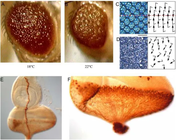
Fig. 2-2. The ‘GMR>λ-btl, dof’ eye phenotype and the expression pattern of the gmr-Gal4. The eyes of
‘GMR>λ-btl, dof’ flies are rougher and smaller at 22°C (B) than at 18°C (A). Sections through the eye of a
‘GMR>λ-btl, dof’ fly raised at 18°C show that the orientation of the photoreceptors are irregular (D) in comparison to wild type (C), which suggests that ‘GMR>λ-btl, dof’ eye has an epithelial planar polarity (EPP) defect at 18°C. In addition, there are loss of photoreceptor cells, shortening of rhabdomeres as well as EPP defects in the eyes of ‘GMR>λ-btl, dof’ flies raised at higher temperature (Curtiss J., personal communication). Rhabdomeres were stained with toluidine blue. The red dash in C marks the equator. Scale Bar for C & D is 25µm. The gmr-Gal4 is expressed in Bolwig’s nerve (E) and all the developing photoreceptor cells in the eye discs of 3rd instar larvae (F). GMR-Gal4 was visualized by overexpression of lacZ and anti-beta-Gal staining. The schematic drawing for the orientation of photoreceptors followed the example of Fanto et al. 2000.
Before starting the screen, I examined the ‘GMR>λ-btl, dof’ eye phenotype in more detail.
The eye phenotype is mild at 18°C. The eyes become smaller and rougher as the
Results temperature increases (Fig.2-2, A&B), which is probably due to the increased activity of Gal4 transcription factor. The change of eye size at higher temperature is likely to be due to the loss of photoreceptor cells. The epithelial planar polarity (EPP) in Drosophila compound eye is established in the mirror-symmetric arrangement of ommatidia relative to the dorsoventral midline, the equator (Fig.2-2C). Sections through the eye of the
‘GMR>λ-btl, dof’ fly raised at 18°C show that the photoreceptor cells did not orient properly compare to wild type (Fig.2-2D). This suggests that the ectopic FGF signal has an EPP defect. In addition, the eyes of the ‘GMR>λ-btl, dof’ flies raised at higher temperature show defects in loss of photoreceptor cells, and shortening of rhabdomeres (Curtiss J., personal communication). In order to find out in which cells the ectopic FGF signal is induced, the flies carrying the gmr-Gal4 that were used to express dof and λ -btl, were crossed to flies with a UAS-lacZ transgene. GMR-Gal4 can drive expression of lacZ in all the differentiated photoreceptor cells and in Bolwig’s nerve in eye disks of the third instar larvae (Fig.2-2, E&F). Therefore the ectopic FGF signals in the ‘GMR>λ-btl, dof’
fly are induced in all the differentiated photoreceptor cells and Bolwig’s nerve.
2.1.2 Tests of the potential candidates
To test some potential candidates and also to determine if the eye phenotype of ‘GMR>λ- btl, dof’ could be modified in a dose sensitive manner, I carried out a loss-of-function test for those candidates. Several classes of known signalling molecules were tested for modification of the ‘GMR>λ-btl, dof’ eye phenotype when half of their gene products were mutated. Firstly genes that are involved in Ras85D-MAPK pathway were examined.
I have chosen to test ras85D, raf (the Drosophila homologue of MEKK), dsor1 (the Drosophila homologue of MEK), rl (rolled, the Drosophila homologue of MAPK), csw (corkscrew), dos (daughter of sevenless), drk (downstream of receptor tyrosine kinase), ksr (kinase suppressor of Ras), gap1 (GTPase activating protein), sos (son of sevenless), and 14-3-3 ζ . The adaptor protein, Dock (Dreadlock), has been found to interact genetically with Ras85D (Schnorr et al., 2001), and therefore the dock mutant was tested.
Since ‘GMR>λ-btl, dof’ flies show defects in planar polarity, the mutants of genes that mediate planar polarity signals were included in the experiment. These are dsh (disheveled), fmi (flamingo), and fz (frizzled). RhoA has also been shown to have a function in EPP signalling. In addition, overexpression of RhoA by Btl-Gal4 shows defects in the formation of dorsal trunk of the trachea (Lee et al., 2002). Given that RhoA is an important effector for cytoskeletal rearrangement, mutants for rhoA and one of its activators rhoGEF2 (Barrett et al., 1997) were also tested. Genes, like Notch and cyclinA, which have distinct functions and are unlikely to be directly regulated by the FGF signalling pathway, were tested as controls.
Flies carrying mutations in the candidate genes were crossed to the ‘GMR>λ-btl, dof’
stock. The progeny were raised at 18°C. Eyes of those progeny containing one copy of the
mutant gene and one copy of the ‘GMR>λ-btl, dof’ chromosome were compared to their
Results siblings with only one copy of the ‘GMR>λ-btl, dof’ chromosome. The results of this experiment are summarized in table 2-1.
Mutants of raf, rhoA, rhoGEF and fmi enhanced the ‘GMR>λ-btl, dof’ eye phenotype.
Other mutants such as rl, ras85D, csw, dos, drk, Dsor1, ksr, gap1, sos, 14-3-3 ζ , dock, dsh, and fz did not show any visible modification of the ‘GMR>λ-btl, dof’ eye phenotype.
Mutants of Notch and cyclinA did not affect the roughness of the eyes of ‘GMR>λ-btl, dof’ flies.
The fact that among the MAPK signalling cassette only the raf mutant enhanced
‘GMR>λ-btl, dof’ eye phenotype is surprising. It may be explained by the fact that Raf is on the top of the hierarchy of the enzymatic pathway and therefore more sensitive to the dose change in the test. The significance of the other modifiers is difficult to explain based on a single test. The results suggest that it is possible to identify components involved in the FGF signalling cascade when the amount of the molecule in the Drosophila eye is changed in the presence of ‘GMR>λ-btl, dof’ chromosome. Neither Notch nor cyclinA mutants show modification of ‘GMR>λ-btl, dof’ eye phenotype suggesting that the test has some specificity.
Both Cdc42 and Rac1 are known to be downstream targets of RTK signals and to regulate signals leading to cytoskeleton rearrangement, vesicle transport and cell differentiation (Katso et al., 2001; Schlessinger, 2000). During eye development, signalling by Rac1 is also required in the establishment of epithelial planar polarity (Fanto et al. 2000). To test if they also interact with the ectopic FGF signal in fly eyes, I crossed ‘GMR>λ-btl, dof’ flies to transgenic flies carrying UAS-cdc42
L89.4or UAS-rac
N17respectively. Cdc42
L89.4and rac
N17are dominant negative alleles (Benlali et al., 2000; Lou, 2001). Neither of the co- overexpression modified the ‘GMR>λ-btl, dof’ eye phenotype at 18°C, which may imply that Cdc42 and Rac1 are not critical components for this ectopic FGF signal.
The results of this test indicated that it was possible to find components that are important
for the ectopic FGF signalling in a dose sensitive screen. Given that other RTK signal
pathways are used extensively during the Drosophila eye development and most of the
downstream components are shared by different RTK signals, only candidates that are
common for RTK signals during eye development could be found in a loss-of-function
screen. Many genes are expressed ectopically in a gain-of-function (GOF) screen, which
offers a possibility to find components that are more specific for FGF signals in a GOF
screen. Therefore, a GOF screen was carried out to search for genes that are important for
the FGF signalling pathway.
Results Table 2-1. The results of the tests of the potential candidates
~: no obvious difference; −: not determined; LOF: loss of function Genes
tested: Genotype of the flies examined: Allele class Eye phenotype of the progeny compared to that of ‘GMR>λ-btl, dof’
ras85D sev d2 ; Ras1 e2f / TM3
hypomorph ~
RasC40B FRT82B/TM3ftz
amorph ~
raf raf EA75 / FM7
LOF Rougher
raf C110 / FM7
hypomorph Rougher
dsor1 y w dsor1 LH110 FRT / FM7a
amorph ~
rl Df(2R)rl 10a / CyO −
~
rl EMS698 / SM1 −
~
csw y cswEsev1A–eOP sevd2 / FM7
antimorph ~
dos w-; dosP115FRT 2A / TM3LOF ~
drk w; drk 24/1 / CyO −
~
drk e0A
antimorph ~
ksr ksr s721 / TM3,Sb e ry sev-RasV12
LOF ~
gap1 Gap11-16 / TM3 ry −
~
sos sev d2;sos e4G / CyO −
~
14-3-3ζ 14-3-3ζP07103 cn / CyO; ryhypomorph ~
rhoA FRT rhoA R2 / CyO −Rougher
rhoGEF2 FRT DrhoGEF / CyO −Rougher
dock dock P04723 cn 1 / CyO; ry506amorph ~
y1 w67 c23; dock P13421 / CyO
amorph ~
dsh dsh1
hypomorph ~
fmi y w; fmi E59 / CyO (y+)
LOF Rougher
fz In(3LR)fz / TM1, Me ri sbd1amorph ~
cyclinA If / CyO; cycAC8 / TM3 UbxLacZ
null ~
Notch Df(1) N 81K / FM6 ; UAS FLP / TM2null ~
Results
2.1.3 The gain-of-function screen
A gain-of-function screen was performed at 18°C to identify new components of the FGF signal (Fig.2-3A). ‘GMR>λ-btl, dof’ flies were crossed to the EP lines individually. The eyes of the progeny containing an EP insertion and the ‘GMR>λ-btl, dof’ chromosome were compared to their siblings carrying only the ‘GMR>λ-btl, dof’ chromosome. Those EP lines showing a modified ‘GMR>λ-btl, dof’ eye phenotype in the F1 generation were selected as potential candidates. When the screen was completed, in total, 153 candidates emerged. Among these candidates, there were 81 enhancers and 72 suppressors.
Fig.2-3 Schematic representation of the crosses for the screen. A, illustrates the crosses of the screen.
‘GMR>λ-btl, dof’ flies were crossed to the individual EP lines. The offspring were scored for the modification of ‘GMR>λ -btl, dof’ eye phenotype. B, illustrates the retest of the candidates. The retest is similar to the screen with one modification. The offspring of the suppressors were raised at 22°C while the enhancers were raised at 18°C. C, illustrates the test for EP candidates whether they give rough eye phenotype with GMR-Gal4 alone. The offspring were raised in a similar way to ‘b’.
‘GMR>λ-btl, dof’: gmr-Gal4 UAS-lambda-btl UAS-dof / CyO.
The ‘GMR>λ-btl, dof’ eye phenotype is temperature sensitive (Fig.2-2, A&B). It is mild at 18°C but can be enhanced dramatically by raising the temperature by 0.5 to 1 degree.
However the eye phenotype does not visibly change when flies are raised between 19-
Results 22°C. This observation implies that the conditions giving rise to ‘GMR>λ-btl, dof’ eye phenotype at 18°C are finely balanced, and a slight change in the conditions can dramatically affect the eye phenotype. Therefore it is possible that some of the enhancers were picked up by chance due to the temperature fluctuation during the period of eye development. It is difficult to determine a potential suppression of the ‘GMR>λ-btl, dof’
at 18°C due to its mild phenotype. To confirm the effect of a suppressor, I retested their effect on the ‘GMR>λ-btl, dof’ phenotype at 22°C. To confirm the potential enhancement, they were retested at 18°C (Fig.2-3B). This retest and the test described below were carried out simultaneously and the results of the two tests were combined. Only those candidates that passed both tests were considered further.
It is possible that some EP lines produce a rough eye phenotype with gmr-Gal4 alone that act independent of the ectopic FGF signal, in which case the enhancement phenotype observed in the screen may be the additive effect and have nothing to do with the FGF signal. To exclude this class of modifiers, the candidates were crossed to gmr-Gal4 to test if the progeny with gmr-Gal4 and EP insertions cause a rough eye phenotype (Fig.2-3C).
These crosses were carried out in such a manner that the candidates for enhancers were crossed and raised at 18°C, and those for suppressors were at 22°C in accordance to the retest. Those enhancer candidates giving a rough eye phenotype with gmr-Gal4 alone were excluded. However, a suppressor giving a rough eye phenotype with gmr-Gal4 alone could still be considered to be a specific modifier. All the confirmed suppressors had passed the test despite four of them giving mild rough eye phenotype with gmr-Gal4 alone. These four EP lines are EP1413, EP0355, EP1455 and EP0622.
In the end, there were 26 enhancers and 24 suppressors that passed both the retest and the
test for its overexpression phenotype by GMR-Gal4 alone. These 50 candidates are
summarized in table 2-2 according to their cytological localization. The candidates are
described in the next chapter.
Results Table 2-2 The list of the candidates found in the screen
EP lines
Cytological region
Phenotype in the screen
Phenotype with GMR- Gal4
Genes possibly affected Additional remarks Viability
1200 S N viable
1408 S N viable
1413 3F2
S Sli. rough
CG2829 (kinase) GOF
viable 1340 7A1 S N fz4, RE54930, LD39940 viable
1342 E N viable
1453 7B6
E N
ches-1-like (checkpoint suppressor
homologue-1-like) viable
1207 9F5 S N CG1679 No flanking seq. info. viable 1503 10E2 E N CG2446 (containing DNA-
glycosylase domain)
can be overexpressed viable
1335 S N viable
1390 S N viable
1508 S N viable
1353
12A6-10
S N
CG11172 (NFAT, nuclear factor of activated T-cells homology)
GOF
viable 0355 14B16-17 S Sli. rough dsp1 (dorsal switch protein 1) can be overexpressed viable 1455 18D3 S Sli. rough CG14217 (kinase) can be overexpressed viable 1323 19A2 S N amnesiac No flanking seq. info. viable
1B14 skpA
1216
13C7-8
E N
no information available No flanking seq. info.
viable 2582 22A2 slight S N robo2 (roundabout 2, or leak) can be overexpressed viable 1211 23B1-2 E N NTPase No flanking seq. info. viable 0719 23C2 E N CG3542 (formin binding protein) LOF no 2204 25C1 S N msp300 (muscle specific protein
300)
viable
2510 28D2 E N CG7231 viable
2171 34D4 S N CG8954 can be overexpressed viable
2571 42E5 S N tetraspanin 42Ef viable
0622 48A2 S Sli. rough tou (toutatis) GOF viable 0988 54C3-7 slight S N mesr4 (misexpression suppressor of
ras 4)
GOF viable 2258 57C6-7 S N CG4266 (containing RNA binding
domain)
no
2516 E N no
2034 57F3
E N CG10433
viable 1222 57F5 strong E N CG10321 (transcription factor) viable
2319 E N viable
0436 E N viable
2310 E N no
0712 strong E N viable
2440 57F6
E N
CG10082 (kinase)
no
0541 E N viable
2494 E N viable
2421 57F8
E N
tim10, or CG30290
no
2444 E N viable
0467 57F9
E N
hmgD (high mobility group protein
D) viable
1135 64A12 strong S N ago (archipelago) GOF viable 0595 66C S Darker* CG6765 (transcription factor) viable
3659 66F4 E N boule GOF no
3348 69E2-4 E N CG10967 or CG11006 viable 3443 78A2-4 S Darker* pap (poils aux pattes) GOF viable 3468 78C1-2 E N eip78C (Ecdysome-induced protein
78C)
No flanking seq. info. viable 3028 82A5 E N CG1090 (Na+/Ca+ exchanger) No flanking seq. info. no
3634 90F7 E N dlc90F (dynein light chain 90F) no
3575 94A4 E N sar1 GOF no
0863 97D2 S N CG6386 (kinase) GOF no
3280 100B2 S N dco (discs overgrown) GOF no Legend: S, suppressor; E, enhancer; N, no modification; Sli. Rough, slightly rough; can be overexpressed, the EP can drive expression
the gene with a proper gal4 driver; GOF, gain-of-function phenotype; LOF, loss-of-function phenotype; No flanking seq. info., no flanking sequence information is available from internet; * , the eye colour becomes darker; no, not viable.