• Keine Ergebnisse gefunden

Role of the

N/A
N/A
Protected

Academic year: 2021

Aktie "Role of the "

Copied!
143
0
0

Wird geladen.... (Jetzt Volltext ansehen)

Volltext

(1)

Role of the

mitochondrial ATP-dependent Clp protease in mammalian metabolism

Inaugural-Dissertation

zur

Erlangung des Doktorgrades

der Mathematisch-Naturwissenschaftlichen Fakultät der Universität zu Köln

vorgelegt von

Christina Becker

aus Werl

Köln, 2017

(2)

B erichterstatter: Prof. Dr. Aleksandra Trifunovic Prof. Dr. Elena Rugarli

Tag der mündlichen Prüfung: 25.04.2017

(3)

To my family and Ludwig

(4)

Table of Contents

List of Figures ... VII List of Tables ... IX Abbreviations ... X Abstract ... XIII Zusammenfassung ... XIV

1 Introduction ... 1

1.1 Mitochondria ... 1

1.1.1 Mitochondrial structure and function ... 1

1.1.2 Mitochondrial genetics ... 2

1.2 Mitochondrial quality control... 3

1.3 ClpXP protease... 4

1.3.1 ClpXP- structure and function ... 4

1.3.2 Physiological functions of ClpXP ... 6

1.4 Metabolism ... 8

1.4.1 Cellular metabolism ... 10

1.4.1.1 Cytosolic metabolism ... 10

1.4.1.2 Mitochondrial metabolism... 13

1.4.2 Whole body metabolism... 16

1.4.2.1 Endocrine regulation of metabolism ... 16

1.4.2.2 Organ interrelation in metabolism with regard to mitochondrial function ... 17

1.5 Mitochondrial dysfunction and metabolic diseases ... 19

1.6 Objectives ... 20

2 Materials and Methods ... 22

2.1 Animal care ... 22

2.2 Experimental mouse models ... 22

2.3 Phenotyping ... 23

2.3.1 Body weight ... 23

2.3.2 Body composition ... 23

2.3.3 Indirect calorimetry ... 23

2.3.4 Blood glucose measurement ... 23

2.3.5 Serum analyses ... 24

2.3.6 Glucose tolerance test and insulin tolerance test ... 24

2.3.7 Insulin signaling ... 24

2.3.8 Acute cold exposure and measurement of rectal body temperature ... 24

2.3.9 Femur length ... 25

(5)

2.3.10 Micro computed tomography ... 25

2.4 Molecular biology ... 25

2.4.1 Isolation of genomic DNA from mouse ear biopsies ... 25

2.4.2 Isolation of genomic DNA from mouse tissues ... 25

2.4.3 Isolation of total RNA ... 25

2.4.4 Quantification of nucleic acids ... 26

2.4.5 Polymerase chain reaction ... 26

2.4.6 Northern blot analysis for mitochondrial mRNA levels ... 27

2.4.7 Reverse transcription and quantitative real-time PCR... 27

2.5 Biochemistry ... 29

2.5.1 Protein extraction from mouse tissues... 29

2.5.2 Isolation of mitochondria from liver and brown adipose tissue ... 29

2.5.3 Isolation of mitochondria from skeletal muscle ... 29

2.5.4 Blue native polyacrylamide gel electrophoresis ... 30

2.5.5 SDS-PAGE and Western blot ... 30

2.5.6 Mitochondrial respiration ... 32

2.5.7 Palmitate oxidation rate ex vivo... 32

2.5.8 Sucrose gradient fractionation of mitochondrial ribosomes ... 33

2.5.9 In organello translation ... 33

2.6 Histological analysis ... 33

2.6.1 Embedding of tissues in paraffin ... 33

2.6.2 Cryostat sections ... 34

2.6.3 Hematoxylin and Eosin staining ... 34

2.6.4 Periodic acid Schiff’s reaction ... 34

2.6.5 Oil red O staining ... 34

2.6.6 COX-SDH staining ... 34

2.6.7 Transmission electron microscopy ... 35

2.7 Cell Culture ... 35

2.8 Label-free quantification of the liver proteome ... 35

2.8.1 In-solution digest ... 35

2.8.2 Liquid chromatography and mass spectrometry ... 36

2.9 Computer analyses ... 36

2.9.1 Software ... 36

2.9.2 Statistical analyses ... 36

2.10 Chemicals ... 36

3 Results ... 39

3.1 Phenotypic analysis of whole body CLPP deficient mice ... 39

3.1.1 Loss of CLPP does not affect lifespan ... 39

3.1.2 Clpp -/- mice have increased energy expenditure and improved glucose homeostasis despite lower locomotor activity ... 42

3.1.3 CLPP deficiency causes a fasting-like phenotype... 45

(6)

3.1.4 Lack of CLPP decreases glycogen content in liver and skeletal muscle ... 48

3.1.5 Effect of CLPP deficiency on fatty acid β-oxidation ... 49

3.1.6 Loss of CLPP severely impairs cold tolerance ... 55

3.1.7 Loss of CLPP leads to enhanced browning of WAT ... 59

3.2 Phenotype of Clpp -/- mice is independent of FGF21 ... 63

3.3 Tissue-specific disruption of Clpp... 66

3.3.1 Liver restricted loss of CLPP does not affect body weight and glucose homeostasis ... 66

3.3.2 Liver restricted loss of CLPP alters FAO enzyme levels and leads to moderate mitochondrial dysfunction ... 67

3.3.3 CLPP ablation in skeletal muscle and heart does not alter body weight and glucose homeostasis ... 68

3.3.4 Muscle restricted loss of CLPP induces FAO oxidation profile and causes mitochondrial dysfunction ... 70

3.3.5 Clpp MKO mice exhibit functional shivering activity ... 71

3.4 High fat feeding impairs hepatic mitochondrial translation in the absence of CLPP ... 72

3.4.1 Clpp -/- mice maintain lean phenotype and enhanced glucose metabolism during HFD-feeding ... 72

3.4.2 Label-free quantitative proteomic profiling of liver reveals deregulated OXPHOS subunit levels due to HFD-feeding upon loss of CLPP ... 77

3.4.3 Loss of CLPP during HFD-feeding leads to strong decrease only in hepatic mitochondrial respiratory chain complexes ... 79

3.4.4 Decreased hepatic mitochondrial complex subunits in HFD fed Clpp -/- mice are not caused by increased turnover ... 84

3.4.5 Loss of CLPP causes increased mitochondrial mRNA levels and reduced de novo protein synthesis with an aggravating effect of HFD ... 88

3.4.6 Loss of CLPP does not impair hepatic mitoribosome assembly ... 92

3.4.7 Hepatic translation defect of CLPP deficient liver mitochondria is not caused by an increased association of ERAL1 with the 28S ribosomal subunit ... 93

4 Discussion ... 98

4.1 Loss of CLPP alters whole body metabolism ... 99

4.2 FGF21 is not a metabolic regulator of the CLPP phenotype ... 103

4.3 Tissue-specific CLPP deletion does not have systemic implications ... 105

4.4 CLPP is required for hepatic mitochondrial translation in metabolic stress conditions ... 106

4.5 Summary ... 109

References ... 110

Acknowledgments ... 125

Erklärung ... 126

Curriculum vitae ... 127

(7)

List of Figures

Figure 1.1 Mitochondrial DNA. ... 3

Figure 1.2 Schematic presentation of the ClpXP degradation cycle. ... 6

Figure 1.3 OXPHOS system. ... 15

Figure 3.1 Validation of ubiquitous CLPP deletion. ... 40

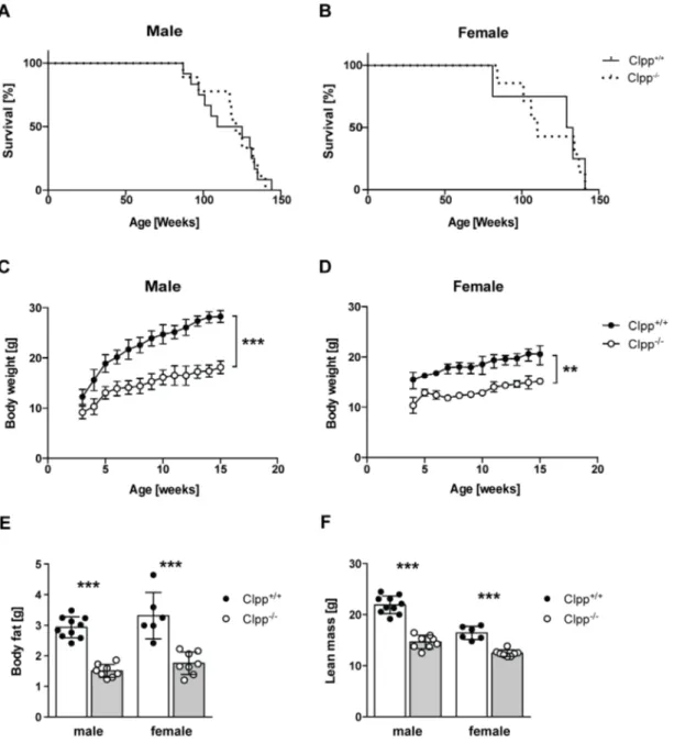
Figure 3.2 Phenotypic characterization of Clpp -/- mice. ... 41

Figure 3.3 Clpp -/- mice exhibit growth defect. ... 42

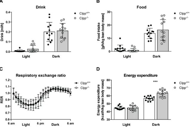
Figure 3.4 Loss of CLPP increases energy expenditure. ... 43

Figure 3.5 Reduced locomotor and rearing activities in Clpp -/- mice. ... 44

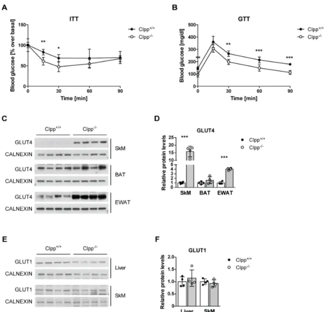
Figure 3.6 Improved glucose homeostasis in Clpp -/- mice due to increased peripheral glucose uptake. ... 45

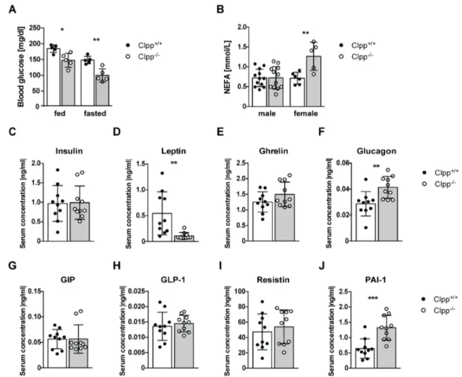
Figure 3.7 Serum parameters of control and CLPP deficient mice. ... 47

Figure 3.8 AMPK activation in SkM of CLPP deficient mice. ... 48

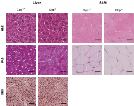
Figure 3.9 Loss of CLPP decreases glycogen content in liver and SkM... 49

Figure 3.10 CLPP deficient mice exhibit decreased FAO in liver and SkM. ... 50

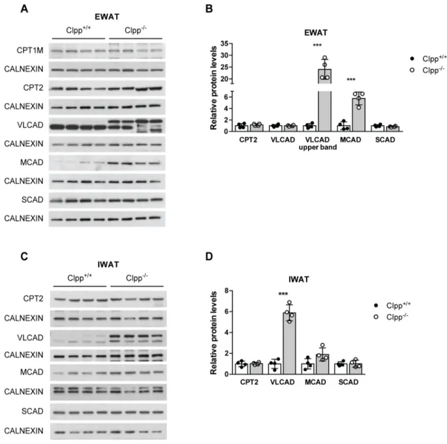
Figure 3.11 Profile of FAO enzymes in Clpp -/- mice (1). ... 52

Figure 3.12 Profile of FAO enzymes in Clpp -/- mice (2). ... 53

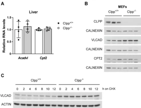
Figure 3.13 VLCAD is a putative substrate of CLPP. ... 55

Figure 3.14 Lipid accumulation in BAT of Clpp -/- mice. ... 57

Figure 3.15 CLPP is essential for cold induced thermogenesis. ... 59

Figure 3.16 Browning of WAT in Clpp -/- mice. ... 61

Figure 3.17 Systemic induction of Fgf21 and increased circulating levels in Clpp -/- mice ... 63

Figure 3.18 Loss of FGF21 in CLPP deficient mice does not normalize whole body physiology. ... 65

Figure 3.19 FGF21 deletion in CLPP deficient liver alters LONP1 and TFAM abundance. ... 66

Figure 3.20 Liver restricted loss of CLPP does not alter body weight and glucose homeostasis. ... 67

Figure 3.21 Hepatic deficiency of CLPP causes mitochondrial dysfunction and similar FAO profile as observed for Clpp -/- mice. ... 68

Figure 3.22 Skeletal muscle and heart restricted loss of CLPP does not alter body weight and glucose homeostasis. ... 70

Figure 3.23 Muscle deficiency of CLPP causes mitochondrial dysfunction and similar FAO profile as observed for Clpp -/- mice. ... 71

Figure 3.24 Cold induced adaptive thermogenesis is not impaired in Clpp MKO mice. ... 72

Figure 3.25 Loss of CLPP protects against HFD induced obesity. ... 74

Figure 3.26 Serum parameters of control and CLPP deficient mice fed HFD. ... 75

(8)

Figure 3.27 Clpp -/- mice are protected against HFD induced hypertrophy of EWAT. ... 76 Figure 3.28 CPT2 is upregulated upon HFD-feeding in Clpp -/- mice. ... 77 Figure 3.29 Quantitative assessment of liver proteomes of CLPP deficient mice

fed NCD or HFD. ... 79 Figure 3.30 High dietary fat content causes mitochondrial dysfunction in liver. ... 80 Figure 3.31 Decreased steady state levels of mitochondrial and nuclear-encoded

complex I subunits in 5-week-old and HFD fed Clpp -/- mice. ... 82 Figure 3.32 Complex composition is not affected by HFD-feeding in CLPP deficient

SkM mitochondria. ... 84 Figure 3.33 Decreased mitochondrial complex subunits in liver do not result from

increased turnover or altered fusion events rather from increased

biogenesis. ... 87 Figure 3.34 Mitochondrial transcript levels are increased in liver of CLPP deficient

mice. ... 89 Figure 3.35 High fat feeding exacerbates the mitochondrial translation defect in

liver in the absence of CLPP. ... 90 Figure 3.36 Mitochondrial translation defect in 5-week-old CLPP deficient liver is

comparable to HFD fed Clpp -/- mice. ... 91 Figure 3.37 Loss of CLPP does not affect mitoribosomal protein levels of the small

and large subunit and mitoribosome assembly in liver. ... 93 Figure 3.38 Mild comigration of the CLPP substrate ERAL1 with the small ribosomal

subunit in liver irrespective of diet. ... 94 Figure 3.39 Difference in hepatic mitochondrial translation of NCD and HFD fed

CLPP deficient mice might be affected by altered ratios of mitochondrial

translation factors. ... 97

(9)

List of Tables

Table 2.1 Oligonucleotides used for genotyping ... 26

Table 2.2 Primers used for quantitative real-time PCR ... 28

Table 2.3 Taqman Probes used for quantitative real-time PCR ... 28

Table 2.4 Primary antibodies used for Western blot analysis ... 31

Table 2.5 Chemicals ... 36

(10)

Abbreviations

°C degree Celsius

3’ three prime end of DNA sequences 5’ five prime end of DNA sequences

A adenosine

ADP adenosine diphosphate ATP adenosine triphosphate BAT brown adipose tissue

bp base pairs

BN blue native

BSA bovine serum albumin

C cytosine

cDNA complementary DNA

cAMP cyclic AMP

Ci Curie

Cre bacteriophage P1 derived site-specific recombinase

Cyt cytochrome

CHX cycloheximide

Da Dalton

ddH 2 O double distilled water

DKO double knockout

DNA deoxyribonucleic acid

dNTP deoxyribonucleotide-triphosphate ECL enhanced chemiluminescence EDTA ethylenediaminetetraacetic acid EGTA ethylene glycol tetraacetic acid ETC electron transport chain

EtOH ethanol

ETS electron transfer system

EWAT epididymal white adipose tissue FADH 2 flavin adenine dinucleotide, reduced FAO fatty acid β-oxidation

FCCP carbonylcyanide p-trifluoromethoxyphenylhydrazone Fe-S iron-sulfur

g gram

G guanine

(11)

H 2 O 2 hydrogen peroxide HCl hydrochloric acid

HEPES N-2-hydroxyethylpiperazine-N-2-ethansulfonic acid HFD high fat diet

HSP heavy strand promoter

i.e. id est

IMM inner mitochondrial membrane IMS inter membrane space

IWAT inguinal white adipose tissue

k kilo

KCl potassium chloride

KO knockout

KOH potassium hydroxide

l liter

fl loxP flanked

LSP light strand promoter

m milli

M molar

MgCl 2 magnesium chloride mtDNA mitochondrial DNA

mRNA messenger RNA

NCD normal chow diet

nDNA nuclear DNA

NaCl sodium chloride

NADH nicotinamide adenine dinucleotide, reduced

NADP nicotinamide adenine dinucleotide phosphate, reduced NaF sodium fluoride

NAH 2 PO 4 monosodium phosphate NaHCO 3 sodium bicarbonate NaOH sodium hydroxide

OMM outer mitochondrial membrane OXPHOS oxidative phosphorylation

PAGE polyacrylamide gel electrophoresis PBS phosphate buffered saline

PCR polymerase chain reaction

Pi Phosphates

RNA ribonucleic acid

(12)

rRNA ribosomal RNA RNase ribonuclease

ROS reactive oxygen species rpm revolutions per minute

RT room temperature

rtPCR reverse transcription polymerase chain reaction SD standard deviation

SDS sodium dodecyl sulfate SkM skeletal muscle

TBE tris-borate-EDTA buffer

TE tris-EDTA buffer

Tris 2-amino-2-(hydroxymethyl)-1,3-propandiole

tRNA transfer RNA

TWEEN polyoxethylene-sorbitan-monolaureate

U units

UPR mt mitochondrial unfolded protein response

V volt

v/v volume per volume

w/v weight per volume

WT wildtype

β-me β-mercaptoethanol

μ micro

(13)

Abstract

Mitochondria are fundamental for cellular metabolism and take center stage in the regulation of systemic energy metabolism. They are origin and target of nutrient intermediates of converging metabolic pathways. Thus, it is essential to maintain mitochondrial homoeostasis. Cells harbor a large set of mitochondrial proteases involved in quality control, including the ATP-dependent Clp protease (CLPP).

However, CLPP not only degrades misfolded or damaged proteins, CLPP is also involved in highly regulated proteolytic activities. Many bacterial and mammalian ClpXP substrates have been identified in various metabolic pathways. This study analyzes the role of CLPP in mammalian metabolism using whole body and tissue-specific Clpp knockout mouse models.

The ubiquitous loss of CLPP under normal dietary conditions leads to a lean phenotype with enhanced glucose metabolism. The absence of CLPP further facilitates increased energy expenditure in part by WAT browning, despite decreased physical activity.

Moreover, CLPP was shown to be involved in fatty acid oxidation by the regulation of its putative substrate VLCAD. In addition, CLPP has a critical role in BAT homeostasis and cold induced thermogenesis. The mild to moderate mitochondrial dysfunction caused by the loss of CLPP in various tissues, induced the expression of the mitokine FGF21. However, using Clpp/Fgf21 double knockout mice, it could be shown that FGF21 is not mediating the metabolic changes observed in CLPP deficient mice.

The tissue-specific depletion of CLPP in liver or skeletal muscle and heart revealed a dispensable role for CLPP with regard to whole body metabolism, although tissue restricted mitochondrial dysfunction was present. Finally, ablation of CLPP was demonstrated to protect against HFD induced obesity and insulin resistance.

Remarkably, HFD-feeding impaired hepatic mitochondrial translation in the absence of

CLPP resulting in decreased complex subunits and decreased supercomplexes levels

in an alternate mechanism as previously described for the heart. Thus, mitochondrial

CLPP has a critical role in metabolic stress conditions in particular during cold

exposure and high fat diet feeding. Therefore, by analyzing CLPP protease function

and its link to metabolic stress this study may help to understand pathologies with

deregulated Clpp expression.

(14)

Zusammenfassung

Mitochondrien sind für den Zellstoffwechsel von grundlegender Bedeutung und nehmen eine zentrale Rolle in der Regulierung des systemischen Energiestoffwechsels ein. Sie sind Ursprung und Zielort von Nährstoffzwischenprodukten konvergierender Stoffwechselwege. Daher ist es von besonderer Bedeutung, die mitochondriale Homöostase aufrechtzuerhalten. Zu diesem Zweck beherbergen Zellen eine Vielzahl von mitochondrialen Proteasen, die an der Qualitätskontrolle beteiligt sind, einschließlich der ATP-abhängigen Clp-Protease (CLPP). Allerdings ist CLPP nicht nur an der Degradierung fehlgefalteter oder beschädigter Proteine, sondern auch an regulierten proteolytischen Aktivitäten beteiligt. CLPP Substrate vieler Bakterien und Säugetiere spielen eine Rolle in verschiedenen Stoffwechselwegen. In dieser Studie sollte daher die Rolle von CLPP im Stoffwechsel mit Hilfe von Ganzkörper und Gewebespezifischen Clpp Knockout Mausmodellen analysiert werden.

Der ubiquitäre Verlust von CLPP unter Bedingungen einer Normaldiät führt zu einem schlankeren Phänotyp mit erhöhtem Glukosestoffwechsel. Weiterhin weisen CLPP- defiziente Mäuse, trotz einer verminderten körperlichen Aktivität, einen erhöhten Energieverbrauch auf, was teilweise bedingt ist durch die Bräunung des weißen Fettgewebes. Darüber hinaus wurde gezeigt, dass CLPP eine entscheidende Rolle in der Homöostase des braunen Fettgewebes und der Kälte induzierten Thermogenese spielt und durch die Regulierung seines potentiellen Substrats VLCAD an der Fettsäureoxidation beteiligt ist. Die leichte bis mäßige mitochondriale Dysfunktion, die durch den Verlust von CLPP in verschiedenen Geweben verursacht wird, induzierte dabei die Expression des Mitokins FGF21. Unter Verwendung eines Clpp/Fgf21 Doppel Knockout Mausmodells konnte jedoch gezeigt werden, dass FGF21 nicht ursächlich für die metabolischen Veränderungen ist, die bei CLPP-defizienten Mäusen beobachtet wurden. Der spezifische Verlust von CLPP in der Leber oder dem Herz- und Skelettmuskel zeigte eine unbedeutende Rolle für CLPP im Hinblick auf den Ganzkörper-Stoffwechsel, obwohl eine mitochondriale Dysfunktion in den jeweiligen Geweben vorhanden war. Schließlich wurde gezeigt, dass die Ablation von CLPP gegen eine hochkalorische Diät induzierte Fettleibigkeit und Insulinresistenz schützt.

Bemerkenswerterweise führte die Fütterung der fettreichen Diät in Abwesenheit von CLPP zu einer Beeinträchtigung der mitochondrialen Translation in der Leber.

Allerdings liegt dem ein anderer Mechanismus zugrunde, als welcher zuvor für das

Herz beschrieben wurde. Dabei konnte eine verminderte Anzahl von Untereinheiten

der Atmungskette und Superkomplexen beobachtet werden. Die mitochondriale CLPP

Protease hat daher eine entscheidende Rolle in metabolischen Stresskonditionen,

(15)

insbesondere bei Kälteexposition und der Fütterung mit hochkalorischer Diät. Die

Analyse der Funktion von CLPP und deren Verknüpfung mit metabolischem Stress

kann somit dazu beitragen, Pathologien mit deregulierter Clpp Expression besser zu

verstehen.

(16)

Introduction

1 Introduction

1.1 Mitochondria

Two billion years ago, when the oxygen levels in the earth’s atmosphere were changing, an α-proteobacterium entered an ancestral host cell to start a symbiotic relationship (Kurland and Andersson, 2000). This is known as the endosymbiotic theory, which is suggesting the origin of eukaryotic cells from prokaryotes (Margulis, 1975). Mitochondria are thought to have evolved from these anaerobic bacterial endosymbionts, introducing the oxidative respiratory system into the host cell.

1.1.1 Mitochondrial structure and function

Mitochondria (Greek mitos "thread” and chondrion "granule”) are found in almost all eukaryotic cells with the exception of erythrocytes. They form a highly dynamic network within the cell undergoing constant fusion and fission and occupying about 20% of its volume (McBride et al., 2006). Mitochondria are compartmentalized by two membranes composed of phospholipid bilayers, namely the outer (OMM) and the inner mitochondrial membrane (IMM) that are separated by the inter membrane space (IMS).

The inner membrane has several invaginations referred to as cristae and encloses the

matrix. The matrix harbors the mitochondrial DNA (mtDNA) a unique feature of

mitochondria denoting its prokaryotic origin. The mtDNA has great similarity to the

genome of the bacterium Rickettsia prowazekii (Kurland et al., 1998). During the

course of evolution, the majority of the ancestral bacterial genes have been lost and

transferred to the nuclear genome, through a process known as endosymbiotic gene

transfer (Timmis et al., 2004). Mammalian mitochondria therefore maintain a genome

encoding only for 13 core proteins of the respiratory chain complexes, 2 rRNA and 22

tRNAs (Chan, 2006; Wallace, 2005). Mitochondria harbor their own replication

(17)

Introduction machinery for the propagation of the mtDNA, as well as separate systems for the transcription and translation of the mitochondrial proteins. The vast majority of the mitochondrial proteome, roughly 1500 proteins, is encoded in the nucleus and needs to be imported into the mitochondria.

The hallmark ability of mitochondria is the generation of cellular energy, adenosine triphosphate (ATP), through the process of oxidative phosphorylation (OXPHOS) (see section 1.3.2.3). Moreover, mitochondria are involved in diverse other cellular processes, such as iron-sulfur (Fe-S) cluster biogenesis, lipid biosynthesis, Calcium homeostasis, apoptosis and generation of reactive oxygen species (ROS) (Chan, 2006; Lill and Mühlenhoff, 2005; Nunnari and Suomalainen, 2012; Wallace, 2005).

1.1.2 Mitochondrial genetics

Mitochondria contain their own genome (mtDNA), which is a closed circular, double- stranded DNA of approx. 16.6 kb in size. Despite the rather small genome size compared to the nuclear genome, point mutations or depletion of mtDNA can lead to severe pathologies. Mitochondrial DNA comprises a total of 37 genes of which 13 are encoding core subunits of the mitochondrial respiratory complexes I, III, IV and V.

Additionally, mtDNA encompasses genes for 2 ribosomal RNAs (rRNA) and 22 transfer RNA (tRNA) that are essential for mitochondrial translation. Only a small portion of the mtDNA (about 1100 bp) comprises a non-coding control region encompassing the heavy and light strand promoter (HSP, LSP) for transcription and the origin of replication of the heavy strand (O H ), whereas the origin of replication for the light strand (O L ) is located in a distant position (Figure 1.1) (Wallace, 2007).

Transcription of polycistronic transcripts is initiated from the two promoters and they are further processed into mature mRNAs, tRNAs and rRNAs. Those transcripts also function as primer for mtDNA replication, which is accomplished by “strand displacement” (Holt and Reyes, 2012).

Unlike the nuclear genome, mitochondria contain between 2-10 copies of mtDNA as result of relaxed replication. Although mammalian mitochondria harbor a DNA repair system, mutations of mtDNA still occur and can lead to a heterogeneous population of mtDNA sequences (Boesch et al., 2009). This condition is termed heteroplasmy, whereas the presence of identical mtDNA copies in a cell is described as homoplasmy (Larsson, 2010).

The mtDNA underlies a non-Mendelian inheritance pattern. It is maternally transmitted,

while the paternal mtDNA is degraded after fertilization and is not passed to the

offspring (Al Rawi et al., 2011). This implies that also mtDNA molecules carrying

mutations are transmitted maternally. When the load of mutated mtDNA molecules

(18)

Introduction exceeds a certain threshold in the cell, this can lead to mitochondrial dysfunction (Wong, 2007). The threshold effect is different for each mutation and tissue, and determines the manifestation and the outcome of a mitochondrial disease. This is also important during cell division, since mitochondria are randomly distributed to the daughter cells (Stewart and Chinnery, 2015).

Mitochondrial DNA mutations commonly affect the OXPHOS system, leading to diverse and multifactorial mitochondrial pathologies (Wallace, 2013).

1.2 Mitochondrial quality control

The maintenance and surveillance of the mitochondrial proteome is crucial for mitochondrial function and cellular homeostasis. The dual origin of the mitochondrial proteins represents a particular challenge. Most mitochondrial proteins are encoded in the nucleus and have to be translocated across one or two membranes, sorted into one of the four compartments and properly folded (Neupert and Herrmann, 2007). In addition, the mitochondrial-encoded proteins are synthesized in the matrix and are subsequently assembled into respiratory chain complex. The timed coordination of the two genomes and the arrangement of all proteins and complexes into the respective

Figure 1.1 Mitochondrial DNA.

The mitochondrial DNA (mtDNA) encodes 37 genes, including 7 subunits of Complex I (red), 1 subunit of

Complex III (orange), 3 subunits of Complex IV (purple), 2 subunits of F O F 1 -ATP synthase (yellow),

2 ribosomal RNAs (rRNA, green) and 22 transfer RNAs (tRNA, blue) (Schon et al., 2012).

(19)

Introduction compartments requires a stringent quality control system. Additionally, mitochondrial protein homeostasis can be perturbed during different stress conditions like nutrient starvation or excess, oxidative stress or age dependent pathologies. A hierarchical system of quality control mechanisms allows the surveillance of mitochondrial proteostasis and the selective removal of proteins or entire organelles (Rugarli and Langer, 2012).

During the import of nuclear-encoded proteins, the mitochondrial targeting sequence is cleaved by the mitochondrial processing peptidase (MPP). The translocation and folding of the newly imported proteins is facilitated by the molecular chaperone mtHSP70. In addition, mtHSP70 prevents the aggregation of misfolded proteins to allow proteolysis (Liu et al., 2001; Wagner et al., 1994). Another matrix chaperone, HSP60, is also involved in folding activity of unfolded proteins after import and is further needed during mitochondrial stress conditions (Bender et al., 2011; Ostermann et al., 1989). The proteolytic removal of proteins is required for terminally damaged, misfolded and non-assembled proteins to prevent proteotoxic stress. There are four different ATP-dependent proteases performing quality control surveillance within the mitochondria. The metalloproteases i-AAA and m-AAA are localized in the IMM with their catalytic site oriented towards the IMS or the matrix, respectively and they execute quality control for the mitochondrial respiratory system (Gerdes et al., 2012).

The mitochondrial matrix harbors two proteases LONP1 and ClpXP. Whereas LONP1 is important for the degradation of oxidized proteins, besides other cellular functions (Ngo and Davies, 2007), the function of ClpXP is less clear and implicated in the degradation of misfolded proteins (Zhao et al., 2002). At the organelle level, sustained stress may lead to mitochondrial hyperfusion or fragmentation, depending on the degree of mitochondrial dysfunction (Rugarli and Langer, 2012). Hyperfusion allows mitochondria to exchange genetic material and increase ATP production to alleviate stress. Mitochondrial fragmentation facilitates the segregation of damaged mitochondria and their subsequent degradation through mitophagy (Kusminski et al., 2014). The failure of this quality control system results in dysfunctional mitochondria and ultimately in cell death, which might trigger pathological processes.

1.3 ClpXP protease

1.3.1 ClpXP- structure and function

ClpXP is a AAA+ protease (ATPases associated with various cellular activities) that

utilizes energy of ATP binding and hydrolysis to catalyze the unfolding and degradation

of proteins and thereby regulate many mitochondrial processes. The ClpXP complex

consists of two distinct proteins, the AAA+ ATPase CLPX and the peptidase CLPP and

(20)

Introduction is localized within the mitochondrial matrix. Both proteins are highly conserved and are found in almost all bacteria, chloroplast and mitochondria of eukaryotic cells, yet S.cerevisiae and S.pombe are lacking a ClpXP homolog (Yu and Houry, 2007).

Most of what is known today about ClpXP structure, substrates and functions was revealed from prokaryotic studies, especially E.coli and much less is known about the mammalian protease. The crystal structure of ClpXP has been solved in different organisms including humans (Glynn et al., 2009; Kang et al., 2004; Wang et al., 1997).

Bacterial CLPP consist of two homoheptameric rings that are stacked to form a

spherical degradation chamber with an axial channel. Human CLPP was shown to

exists as a single heptameric ring and only upon binding CLPX the double ring is

formed (Kang et al., 2005). Each CLPP subunit harbors a serine-histidine-aspartate

catalytic triad, a hallmark of serine proteases. The 14 proteolytic active sites are facing

the inside of the chamber. This allows to control the proteolytic activity of CLPP, since

the opening of the channel is restricting the access of native proteins resulting in a low

peptidase activity of CLPP, being able to degrade peptides less than 15 amino acids

long (Thompson et al., 1994). To obtain proteolytic activity CLPP assembles on one or

both ends with a hexameric ring of CLPX to form a holoenzyme complex (Kang et al.,

2005). In bacteria, in addition to CLPX, the ATPases CLPA and CLPC associate with

CLPP (Sauer and Baker, 2011). The CLPX chaperone functions in substrate

recognition, ATP-dependent protein unfolding and translocation of the substrate into

the proteolytic chamber of CLPP (Baker and Sauer, 2012). The targeting of substrates

for degradation and the recognition by CLPX is mediated by recognition motifs called

degrons at the N- or C-terminus of the target protein (Flynn et al., 2003). Upon binding

of such a peptide sequence to the axial opening, CLPX is denaturing and translocating

the native protein into CLPP in a process requiring numerous cycles of ATP hydrolysis

to catalyze the conformational change of CLPX to excerpt the mechanical pulling

(Ortega et al., 2000). The actual function of CLPP is to cleave the translocated protein,

typically resulting in peptides of 3-7 amino acids in length (Choi and Licht, 2005) that

exit the chamber and are further degraded by exopeptidases (Figure 1.2).

(21)

Introduction

1.3.2 Physiological functions of ClpXP

Protein degradation by ClpXP is a vital process contributing to protein quality control by limiting misfolded or damaged proteins, however, ClpXP is also involved in highly regulated proteolytic activities thereby sustaining cellular homeostasis. The selective degradation of substrates based on substrate recognition represents an important mechanism of proteolytic regulation.

In E.coli several recognition motifs have been identified. In general these peptide sequences contain 3-10 amino acids and are located at the N- or C-terminal end of the target protein (Flynn et al., 2003; Gottesman et al., 1998). The best-studied recognition motif in E.coli is the ssrA tag. When ribosomes stall during protein synthesis, the 11 amino acid residue is cotranslationally added to the nascent polypeptides by the tmRNA system (Keiler et al., 1996). This allows termination of translation and the recycling of the ribosome machinery. The ssrA tagged peptide is then degraded by ClpXP. Mutagenesis experiments have shown that the last three residues (Leu-Ala- Ala) are sufficient for CLPX recognition (Flynn et al., 2001), while the other residues are bound by the SspB adaptor protein to enhance ClpXP mediated degradation (Flynn et al., 2004). So far the recognition motifs of the mammalian CLPX are unknown.

ClpXP is not only involved in the quality of de novo protein synthesis but also in the regulation of specific stress responses of in E.coli. The DNA repair protein RecN genetically encodes a ClpXP recognition motif (Leu-Ala-Ala), which constantly targets it for degradation by ClpXP. Upon DNA damage RecN is highly induced as part of the SOS regulon and accumulates within the cell irrespective of the ClpXP degradation (Neher et al., 2006). As soon as the stress condition is solved the transcription slows

Figure 1.2 Schematic presentation of the ClpXP degradation cycle.

The CLPX hexamer recognizes the substrate upon binding of the recognition tag to axial pore. The protein

substrate is then unfolded and translocated into the CLPP chamber for proteolysis (Baker and Sauer,

2012).

(22)

Introduction down and RecN is efficiently degraded by ClpXP. Similarly, the general stress response regulatory protein σ S is regulated by ClpXP degradation together with the adapter protein RssB (Hengge, 2009; Zhou et al., 2001). During exponential growth RssB binds to σ S and targets it for degradation, however, in the stationary phase or other stress conditions the σ S is stabilized due to decreased binding of RssB (Becker et al., 2000). Furthermore, CLPP was demonstrated to be important for the stress tolerance of many prokaryotes, including Staphylococcus aureus, Streptococcus mutants and Haemophilus parasuis (Frees et al., 2003; Hou et al., 2014; Huang et al., 2016). CLPP also plays an important role in the virulence of the mammalian pathogens Salmonella typhimurium and Listeria monocytogenes (Gaillot et al., 2000; Yamamoto et al., 2001). Different than in bacteria, in the fungus Podospora anserine ablation of CLPP leads to an increase in lifespan and this it is not accompanied by decreased health, which is commonly observed for life expanding interventions (Fischer et al., 2013).

Moreover, ClpXP dependent degradation of CtrA is required for the cell cycle progression in Caulobacter crescentus (Brown et al., 2009). In HEK293 cells, ClpXP was suggested to be involved in the regulation of mitophagy through the degradation of PINK1 together with MPP, m-AAA protease and LONP1 (Greene et al., 2012).

In C.elegans, CLPP-1 was implicated in the retrograde signaling of the mitochondrial unfolded protein response (UPR mt ) (Haynes et al., 2007). In response to an increased accumulation of unfolded or unassembled proteins or a mitonuclear imbalance that is exceeding the folding capacity of chaperones, the nuclear expression of mitochondrial chaperones and proteases is induced (Houtkooper et al., 2013). CLPP-1 has been proposed to degrade unfolded proteins to small peptides, which are exported by the ABC transporter HAF (Haynes et al., 2010). The efflux of those peptides might serve as an initial signal for the activation and nuclear translocation of the bZIP transcription factor ATFS-1, which activates expression of hsp-60 and hsp-6 (HSP60 and mtHSP70) (Nargund et al., 2012). In mammals far less is known about the signaling of the UPR mt . In response to unfolded mitochondrial protein stress, the transcription factor CHOP (C/EBP homology protein), the chaperones HSP60 and mtDNAJ, as well as CLPP as a protease were shown to be specifically induced in mammalian cells (Zhao et al., 2002).

However, unlike in C.elegans, in mammals the induction of those UPR responsive genes is not dependent on CLPP (Seiferling et al., 2016), suggesting a different signaling mechanism or the involvement of another protease for the UPR mt in mammals.

Recently, a novel role for ClpXP in the regulation of mitochondrial translation through

the degradation of ERAL1 has been proposed (Szczepanowska et al., 2016). ERAL1

(23)

Introduction binds to the small ribosomal subunit and acts as a chaperone for the mitochondrial 12S rRNA during ribosome assembly (Dennerlein et al., 2010). In the absence of CLPP, ERAL1 cannot be degraded and is not removed from the small ribosomal subunit, which leads to an impaired mitoribosomal assembly and in turn impairs mitochondrial translation (Szczepanowska et al., 2016).

In humans, recessive mutations in CLPP were found to cause Perrault Syndrome, which is characterized by premature ovarian failure, sensorineural hearing loss and growth retardation besides some other symptoms described for the patients with Clpp mutations (Jenkinson et al., 2013; Mytinger et al., 2016). The mouse model lacking Clpp expression represents a faithful model of the disease with similar pathologies (Gispert et al., 2013).

Although ClpXP has been extensively characterized in prokaryotes, including a comprehensive description of ClpXP substrates, the role of mammalian ClpXP is far less understood. A recent study identified 48 potential CLPP interaction proteins using a proximity based labeling technique in human cells (Cole et al., 2015). Most of these proteins were subunits of the mitochondrial respiratory chain and enzymes involved in mitochondrial metabolism. Intriguingly, another study reported overlapping candidates of ClpXP interaction partners and substrates in mouse heart, using a more sophisticated method to distinguish interactors from near neighbors and to additionally identify putative substrates (Szczepanowska et al., 2016). Hence, ClpXP seems to be involved in the regulation of mitochondrial metabolism and thereby also cellular and whole body metabolism.

1.4 Metabolism

Metabolism is the entity of all essential biochemical reactions taking place in a cell of living organisms. These reactions are organized into metabolic pathways, which begin with a particular molecule that is altered and converted into another molecule or molecules in a series of defined steps. Metabolic pathways can be divided into catabolic and anabolic processes. Catabolic pathways produce chemical energy through the degradation of organic molecules, whereas anabolic pathways require energy for biosynthetic reactions. Depending on the energy condition within the cell, some pathways can be either anabolic or catabolic (Berg et al., 2002). The generation of energy is essential for all types of cells. However, the metabolic requirement of each cell type is determined by the tissue function (see section 1.4.2.1) and environmental factors, like the type and availability of nutrients.

Due to the interconnection and interdependence of all metabolic pathways, their

activity requires a fine coordination and tight regulation to ensure metabolic

(24)

Introduction homeostasis in a dynamically changing environment. The metabolic regulation is taking place at different levels (Metallo and Vander Heiden, 2013). The communication between tissues is mediated through hormone signaling and other extracellular factors (see section 1.4.2.2). At the cellular level, the abundance of enzymes or regulatory factors is regulated tissue- or context-specifically by mRNA transcription, splicing, stability or translation (Cairns et al., 2011). Transcriptional regulation for example involves transcription factors such as PPARα or PPARγ or transcription coregulators like PGC1α. The nuclear receptor peroxisome proliferator-activated receptors (PPARs) are activated by ligand binding and control the expression of gene networks involved in lipid metabolism, adipogenesis and inflammation (Ahmadian et al., 2013). Additionally, these transcription factors are modulated by the peroxisome-proliferator-activated receptor coactivator 1α (PGC1α), the master regulator of mitochondrial biogenesis.

Another means to regulate enzyme activities and functions are posttranslational modifications through the covalent addition of a chemical moiety, such as acetate or phosphate to amino acid residues, proteolytic cleavage or degradation of the whole protein (Zhao et al., 2010). Enzyme activities can be further modulated by allosteric effects in the presence of small molecules, which alter the conformation of an enzyme either increasing or decreasing the affinity for its substrate (Phillips and Ainsworth, 1977). Finally, feedback regulation is a mechanism known to modulate the flux through the pathway. When a metabolite reaches a certain concentration, the upstream enzyme activity is regulated.

These mechanisms controlling enzyme activities and therefore metabolic flux can act separately or in conjunction. An important example for the interplay of two regulatory processes is the control of AMP-activated kinase (AMPK) activity. AMPK functions as an energy sensor and master regulator of metabolism (Yuan et al., 2013). Cellular energy levels are sensed by AMPK through the direct binding of AMP or ADP (allosteric regulation), which leads to a conformational change of AMPK. This conformational change allows AMPK to be phosphorylated by upstream kinases leading to its activation. In turn AMPK coordinates diverse metabolic responses through the phosphorylation of its downstream targets to increase catabolic pathways for the rapid modulation of ATP production and consumption.

All these processes aim to regulate metabolic flux according to the requirements of the

individual cells.

(25)

Introduction

1.4.1 Cellular metabolism

Metabolic pathways in the cell are compartmentalized to the cytosol, nucleus, mitochondria, peroxisomes, endoplasmatic reticulum and lysosomes. Here, we focus on the main metabolic reactions allocated to the cytosol and the mitochondria.

1.4.1.1 Cytosolic metabolism

Cytosol is the liquid component of the cytoplasm surrounding the nucleus and the cytoplasmic organelles. The biochemical reactions taking place in the cytosol are glycolysis, pentose phosphate pathway, fatty acid synthesis, amino acid metabolism and glycogen synthesis. The nutrient derived carbohydrates, lipids and proteins are taken up by the cell in the form of glucose, fatty acids and amino acids, which is facilitated by specialized membrane proteins. Glucose enters the cell through glucose transporters (GLUT1-4) and fatty acid uptake is mediated by fatty acid transporters, like the fatty acid translocase (FAT/CD36), tissue-specific fatty acid transport proteins (FATP) and plasma membrane fatty acid binding protein (FABPpm) (Fillmore et al., 2014).

Glycolysis

Glycolysis (Greek glykos „sweet“ and lysis “degradation”) is an ancient catabolic

pathway derived from times when there was less oxygen in the atmosphere. It is

conserved in almost all aerobe and anaerobe living organisms (Fothergill-Gilmore,

1986). In a series of 9 enzymatic steps one molecule of glucose is converted to two

molecules of pyruvate. The glycolytic pathway can be divided into three stages. In the

initial three reactions, glucose is converted to fructose-1-6-biphosphate, requiring two

molecules of ATP. This first stage facilitates the trapping of glucose within the cell and

its destabilization for the following reactions. The second stage is the cleavage of the

six-carbon molecule fructose-1-6-biphosphate into two three-carbon molecules

glyceraldehyde 3-phosphate and dihydroxyacetone phosphate. In the last stage ATP is

harvested when the two three-carbon molecules are oxidized to pyruvate. This reaction

sequence yields two molecules of ATP and two molecules of NADH (reduced

nicotinamide adenine dinucleotide) (Hue and Hers, 1983). NADH is transported into

mitochondria through the malate-aspartate and glycerol-3-phosphate shuttles for

subsequent oxidation reactions (Stein and Imai, 2012). The fate of pyruvate depends

on the cellular conditions. In the absence of oxygen, pyruvate can be reduced to

lactate by the lactate dehydrogenase, while regenerating NAD + (oxidized NAD)

required for upstream reactions of glycolysis, which is important in skeletal muscle

when the energy demands exceed the oxygen supply or in erythrocytes due to the lack

(26)

Introduction of mitochondria. Although this reaction is energetically unfavorable as compared to the entire oxidation of glucose via glycolysis, the tricarboxylic acid cycle (TCA) and oxidative phosphorylation, which is strictly dependent on the availability of oxygen.

Increased blood lactate concentrations can be also evident for mitochondrial dysfunction due to an increased reliance on glycolysis and reduced oxidation of pyruvate within the mitochondria.

Glycolysis is regulated at three distinct steps, through feedback inhibition of the hexokinase catalyzing the initial step of glucose to glucose-6-phosphate conversion and the allosteric regulation of phosphofructokinase and pyruvate kinase by AMP, ADP, ATP or citrate depending on the energy state of the cell.

Gluconeogenesis

Gluconeogenesis is an anabolic pathway generating glucose from non-carbohydrate precursors. During prolonged fasting and starvation conditions gluconeogenesis is required to prevent hypoglycemia. The major precursors are pyruvate, lactate, gluconeogenic amino acids and glycerol. They are either first converted to pyruvate or enter the pathway through later intermediates. Gluconeogenesis is often considered the reverse reaction of glycolysis, however, the irreversible reactions of glycolysis have to be bypassed during gluconeogenesis by four different enzymes. The first reaction of gluconeogenesis is located in the mitochondria and requires a distinct enzyme pyruvate carboxylase (PC), the second reaction can be carried out within mitochondria or the cytosol by phosphoenolpyruvate carboxykinase (PEPCK) for the conversion of pyruvate in the cytosol and in the ER. The other distinctive enzymes required for gluconeogenesis are fructose 1,6-biphosphatase and glucose 6-phosphatase, which catalyze the hydrolysis reactions (Jitrapakdee, 2012). Gluconeogenesis is reciprocally regulated with glycolysis, therefore the same key steps are regulated (Berg et al., 2002).

Glycogen metabolism

Glycogen is a multi-branched polymer of glucose and functions as storage form of

glucose within tissues, especially in liver and skeletal muscle. However, glycogen

serves different functions in both tissues. Hepatic glycogen is required to maintain

blood glucose levels during fasting conditions, whereas muscle glycogen is expended

only for muscular activity as during intense exercise. The synthesis of glycogen

(glycogenesis) requires a primer consisting of a glycogenin protein attached to α-1,4

linked glucose oligosaccharide. This primer is used by the glycogen synthase for the

elongation with UDP-glucose molecules, an activated form of glucose generated from

(27)

Introduction glucose-1-phosphate. Additionally, a branching enzyme is required for the formation of α-1,6 linked glucose branches to obtain the polysaccharide glycogen.

The degradation of glycogen (glycogenolysis) is controlled by the glycogen phosphorylase. Together with a bifunctional debranching enzyme, the phosphorylase mediates the shortening of the oligosaccharide chains to release glucose-1-phosphate.

The synthesis and degradation of glycogen are reciprocally regulated through the activity of glycogen synthase and phosphorylase. Hormone stimulated cyclic AMP (cAMP) cascade acting through protein kinase A is facilitating the switch between the active and inactive forms of glycogen synthase and phosphorylase (Bollen et al., 1998).

Fatty acid synthesis

In a high-energy state, when glycogen stores are already filled, a surplus of glucose results in the synthesis of fatty acids and the formation of triglycerides. Fatty acids are synthesized from acetyl-CoA, which is exported from the mitochondria as citrate and reconverted to acetyl-CoA in the cytosol. Acetyl-CoA is carboxylated by the acetyl-CoA carboxylase (ACC) to malonyl CoA. This reaction is the rate-limiting step during fatty acid synthesis and is highly regulated at the level of ACC (Hillgartner et al., 1995; Kim, 1997). The subsequent reaction steps of the de novo lipogenesis are catalyzed by the multifunctional enzyme fatty acid synthase (FAS) (Wakil, 1989). Malonyl CoA functions as a two-carbon donor for the elongation of acetyl-CoA. This step is repeated six times in a cyclic manner resulting in C16.0 palmitic acid. During the reaction nicotinamide adenine dinucleotide phosphate (NADPH), derived from the pentose phosphate pathway, functions as a reducing agent. The palmitic acid produced by the FAS can be further modulated by elongation or desaturation. In order to be stored in the liver or adipose tissue, three fatty acids are esterified with glycerol-3-phosphate resulting in the formation of triacylglycerol.

As mentioned earlier fatty acid synthesis and in parallel also fatty acid β-oxidation is

regulated by ACC activity. Malonyl CoA generated by ACC, acts as an inhibitor of

CPT1 and is thus controlling fatty acid uptake into mitochondria for β-oxidation. The

activity of ACC is modulated by phosphorylation, dephosphorylation and also

allosterically activated by citrate (Brownsey et al., 2006). The upstream kinase AMPK

is mediating the inhibiting phosphorylation of ACC in low energy conditions. Hence,

AMPK and ACC are stringently regulating fatty acid metabolism according to

physiological needs.

(28)

Introduction

1.4.1.2 Mitochondrial metabolism Fatty acid β-oxidation

During fatty acid β-oxidation (FAO) fatty acids originating from the diet or the hydrolysis of triglycerides during fasting conditions are degraded to generate energy in the form of ATP. Most of the dietary fats are composed of long chain fatty acids, in particular palmitic acid (C16:0) (Rustan, 2009). Long chain fatty acids in the cytosol are converted and thereby activated in a two-step process with an acyl adenylate intermediate to acyl-CoA by acyl-CoA synthases (ACS) (Grevengoed et al., 2014).

Long chain acyl-CoAs are unable to diffuse through the IMM and are esterified to acylcarnitines by the carnitine palmitoyltransferase 1 (CPT1). This permits the transport of the acylcarnitines across the IMM by the carnitine acylcarnitine translocase (CACT) in exchange for carnitine. Once inside the mitochondria, CPT2 reconverts the acylcarnitines to acyl-CoAs, which can finally undergo FAO (Eaton et al., 1996).

Medium and short chain fatty acids diffuse through the mitochondrial membrane and are activated in an ATP-dependent reaction in situ before undergoing FAO.

The β-oxidation cycle comprises a series of four enzymatic reactions, in which a two- carbon acetyl-CoA is released from the acyl-CoA ester per cycle. In the first reaction acyl-CoAs are oxidized to enoyl-CoA by chain length specific acyl-CoA dehydrogenases: short-chain (C4-C6) acyl-CoA dehydrogenase (SCAD), medium- chain (C6–C12) acyl-CoA dehydrogenase (MCAD), long-chain (C12–C16) acyl-CoA dehydrogenase (LCAD) and very long-chain (C14–C20) acyl-CoA dehydrogenase (VLCAD) (Ojuka et al., 2016). The three subsequent reactions are a hydration, oxidation and thiolation that are catalyzed either by the mitochondrial trifunctional protein (MTP) for very long chain fatty acyl-CoAs or three separate enzymes (cronotase, medium/short-chain hydroxyacyl-CoA dehydrogenase and medium-chain ketoacyl-CoA thiolase) for short-, medium- and long-chain fatty acids. Each cycle results in the formation of one FADH 2 , one NADH, one acetyl-CoA and a 2-carbon shortened acyl-CoA, which re-enters the cycle until two acetyl-CoA molecules are produced. The reducing equivalents NADH and FADH 2 transfer their electrons either directly to complex I of the respiratory chain or via the electron transfer flavoprotein (ETF) to ubiquinone, respectively. Acetyl-CoA can be further oxidized in the TCA cycle or exported from the mitochondria for de novo lipogenesis. Furthermore, it can also be converted into ketone bodies during starvation conditions.

TCA cycle

The TCA cycle, also referred to as citric acid cycle or Krebs cycle, constitutes a central

metabolic hub. It represents the final oxidation pathway for metabolic fuels derived

(29)

Introduction from carbohydrates, fats and proteins. Under aerobic conditions pyruvate derived from glycolysis in the cytosol is converted to acetyl-CoA by the pyruvate dehydrogenase, a reaction yielding one molecule of NADH. The acetyl-CoA is then utilized in the TCA cycle for its complete oxidation to CO 2 thereby producing three NADH, one FADH 2 and GTP. The electrons of NADH and FADH 2 are again allocated to the respiratory chain.

The enzyme generating the FADH 2 , succinate dehydrogenase (complex II) takes not only part in the TCA cycle, it is also part of the respiratory chain therefore electrons from the oxidation of succinate directly reduce FAD + , a prosthetic group of complex II (Akram, 2014).

The TCA cycle additionally provides many precursors and metabolites for biosynthetic pathways including gluconeogenesis, amino acid metabolism or porphyrin synthesis (Owen et al., 2002). Many of the TCA cycle intermediates are taken from the cycle by cataplerotic reactions, like α-ketoglutarate or oxaloacetate to form amino acids.

However, they can also be placed back by anaplerotic reactions. Oxaloacetate can be redirected for gluconeogenesis or replenished by the pyruvate carboxylase converting pyruvate to oxaloacetate. Citrate represents an important source of acetyl CoA when exported from the mitochondria for fatty acid synthesis and ketone body formation. The TCA cycle is also important in providing succinyl-CoA for the initial reaction of the heme biosynthesis (Atamna, 2004).

Oxidative phosphorylation

The oxidative phosphorylation (OXPHOS) is the final biochemical pathway for the generation of the largest proportion of the cellular energy. The mitochondrial OXPHOS system consists of four electron transferring protein complexes namely complex I (NADH: ubiquinone oxidoreductase), complex II (succinate dehydrogenase), complex III (ubiquinol:cytochrome c oxidoreductase), complex IV (cytochrome c oxidase, also referred to as COX) and the F O F 1 -ATP synthase, which are embedded in the inner mitochondrial membrane (Figure 1.3) (Barrientos et al., 2002; Fontanesi et al., 2006;

Lazarou et al., 2007; Rutter et al., 2010). The mobile redox carriers ubiquinone (coenzyme Q) and cytochrome c transfer electrons between the individual complexes (Figure 1.3). Additionally, ubiquinone is an entry point for electrons derived from FAO, which are transferred by the ETF.

In the beginning, electrons are transferred via redox reactions from the electron carrier

NADH to complex I and from succinate (via FADH 2 ) to complex II and are passed down

the respiratory chain to the terminal electron acceptor oxygen. These redox reactions

drive the proton translocation from the matrix to the IMS by complex I, complex III and

complex IV and are thereby generating an electrochemical proton gradient. The energy

(30)

Introduction stored within this gradient drives the F O F 1 -ATP synthase to synthesize ATP from ADP and inorganic phosphate (Jonckheere et al., 2012). The protons that flow back to the matrix through F O F 1 -ATP synthase are combined with the reduced oxygen to form H 2 O.

The mitochondrial OXPHOS complexes are not simply arranged in sequence, however, they have a supramolecular organization (Chaban et al., 2014; Schägger and Pfeiffer, 2000). Complexes I, III and IV assemble into supercomplexes with different stoichiometry CoI 0-1 +CoIII 2 +CoIV 0−4 (Dudkina et al., 2010; Genova et al., 2008).

Supercomplexes comprising all three complexes (CoI+CoIII 2 +CoIV 2 ) are referred to as respirasomes (Marques et al., 2007; Wittig and Schägger, 2009). These supramolecular structures are proposed to facilitate channeling of the substrates ubiquinone and cytochrome c or to catalytically enhance the reactions due to shorter diffusion times. These assumptions are strengthened by the finding that OXPHOS complexes are predominantly localized to the protein rich mitochondrial cristae, which are structurally separated from the inner boundary membrane by cristae junctions (Frey et al., 2002; Gilkerson et al., 2003; Lippe et al., 1988; Schägger and Pfeiffer, 2000; Vogel et al., 2006). Another crucial function of the supercomplex formation is the stabilization of complex I by complex III and complex IV (Diaz et al., 2006; Schägger et al., 2004). The ATP synthase dimerizes and forms oligomeric chains, which is suggested to impose a curvature of the IMM and thereby increasing the local proton concentration (Strauss et al., 2008). Complex II is the only complex that has not been detected in higher order structures, which might be due to its dual function in the TCA cycle and OXPHOS. Noteworthy, it was shown that supercomplex formation is

Figure 1.3 OXPHOS system.

The mitochondrial respiratory chain complexes generate an electrochemical gradient across the inner

mitochondrial membrane. NADH and succinate are oxidized to NAD+ and fumarate by Complex I and

Complex II, respectively. The electrons (e - ) are transferred via coenzyme Q (CoQ) to Complex III and

further to Complex IV via cytochrome c (Cyt c). At Complex IV electrons are transferred to the final

electron acceptor oxygen that forms H 2 O. The proton gradient produced by Complex I, Complex III and

Complex IV is used by the F O F 1 -ATP synthase for ATP synthesis. The nuclear-encoded proteins are

colored in blue and the mitochondrial-encoded subunits in other colors. Reprinted with modifications from

Schon et al., 2012.

(31)

Introduction important for mitochondrial function and cell physiology (Ikeda et al., 2013; Wittig and Schägger, 2009).

1.4.2 Whole body metabolism

1.4.2.1 Endocrine regulation of metabolism

Metabolism is tightly regulated through the concerted actions of endocrine hormones (Randle, 1963). It involves an extensive interrelation of metabolites and hormones to control and direct metabolic pathways. Here the endocrine actions of insulin, glucagon, leptin, ghrelin and incretins in the regulation of glucose and lipid metabolism and whole body energy expenditure are considered.

Blood glucose levels are maintained in a narrow range despite alternate fasting and feeding cycles. The glucose concentration in the blood is balanced by the absorption from the intestine, glucose production and secretion from the liver and to a lesser extent from the kidneys and the clearance by peripheral organs (Saltiel and Kahn, 2001).

Cellular responses to endocrine hormones are mediated through receptors on the cell surface. Hormone binding induces signaling via G protein coupled receptors and second messengers like cAMP (glucagon, ghrelin), tyrosine kinase receptors and a direct phosphorylation of downstream substrates (insulin) or cytokine receptors with bound Janus kinase (JAK) (leptin) (Schwartz et al., 2000).

Insulin is one of the most important anabolic peptide hormones. In response to increased blood glucose levels, insulin is secreted from pancreatic β-cells (Saltiel and Kahn, 2001). Insulin is mainly responsible for reducing blood glucose levels by increasing peripheral glucose uptake and reducing glucose production in the liver, by reducing gluconeogenesis and glycogenolysis (Rizza et al., 1981). Specifically it modulates enzyme levels by decreasing transcription of phosphoenolpyruvate carboxykinase (PEPCK), glucose-6-phosphatase, and fructose-1,6-biphosphatase and increasing transcription of glucokinase and pyruvate kinase (S J Pilkis and Granner, 1992). The peripheral insulin dependent glucose disposal is stimulated by membrane translocation of GLUT4 in skeletal muscle and adipose tissue, whereas skeletal muscle is clearing about 75% (Klip and Pâquet, 1990). Moreover, insulin facilitates storage of metabolic fuels in liver, skeletal muscle and adipose tissue by stimulating glycogenesis and lipogenesis.

Glucagon is secreted from pancreatic α-cells in order to increase blood glucose

concentration. It dominates hepatic metabolism during fasting, by increasing

glycogenolysis and gluconeogenesis (Campbell and Drucker, 2015). Additionally,

(32)

Introduction glucagon induces lipolysis in adipose tissue to provide more substrates for gluconeogenesis (glycerol) and FAO (fatty acids) (Slavin et al., 1994).

Two important hormones implicated in the regulation of whole body energy expenditure are leptin and ghrelin. Leptin is commonly known as the satiety hormone. It is secreted from adipocytes and acts on the hypothalamus in the central nervous system thereby regulating food intake, energy expenditure and body weight (Schwartz et al., 2000).

Furthermore, leptin stimulates FAO through AMPK activation and is thus regulating lipid metabolism (Minokoshi et al., 2002). The loss of leptin leads to obesity and insulin resistance in mice (ob/ob) (Pelleymounter et al., 1995). However, circulating leptin levels are increased in obesity, since leptin secretion is proportional to adipose tissue weight, a condition often referred to as leptin resistance.

Ghrelin functions as the counter regulatory hormone to leptin. Its circulating levels are alternating and peaking pre-prandial, which was suggesting the function as hunger signal (Yin et al., 2009). It is secreted from the stomach and acts like leptin on the central nervous system (Abizaid et al., 2006). Ghrelin stimulates appetite and thus regulates body weight (Tschöp et al., 2000). Additional physiological functions are the stimulation of GH secretion and promotion of gastric motility (Yang et al., 2014).

The incretin hormones GLP-1 and GIP are secreted from the intestinal cells rapidly after food ingestion. They facilitate quick disposal of nutrients by facilitating insulin secretion from pancreatic β-cells (Baggio and Drucker, 2007). GLP-1 was also shown to promote satiety.

1.4.2.2 Organ interrelation in metabolism with regard to mitochondrial function Each organ has specific functions in the body and varies widely in energy needs. The functional requirements of the mitochondria are reflected in the tissue-specific variations of the mitochondrial proteome, mitochondrial morphology, as well as differences in mitochondrial number, ranging from a few hundred to a few thousand per cell (Pagliarini et al., 2008; Thor Johnson et al., 2007). These differences are even apparent within one tissue, as for example in the different skeletal muscle fiber types.

The liver is the key metabolic organ governing whole body energy metabolism. It

represents the metabolic hub providing fuels to other tissues and is the central organ in

the regulation of glucose metabolism. During fasting conditions the liver increases

glycogenolysis and gluconeogenesis in response to glucagon signaling in order to

increase blood glucose levels. Additionally, the liver is the main organ generating

ketone bodies from fatty acids during starvation, which serve as fuel for the brain. In

the postprandial state, insulin stimulates glycogenesis and de novo lipogenesis in the

liver (Rui, 2014). Hepatocytes harbor some distinctive enzymes in order to perform

Abbildung

Figure 1.2 Schematic presentation of the ClpXP degradation cycle.
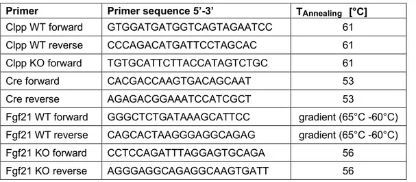
Table 2.1 Oligonucleotides used for genotyping  Primer sequences are displayed in 5’-3’ order
Figure 3.2 Phenotypic characterization of Clpp -/-  mice.
Figure 3.4 Loss of CLPP increases energy expenditure.
+7

Referenzen

ÄHNLICHE DOKUMENTE

The main system starts from an existing emotional word dictionary (Liu et al., 2008), and use those words with strong polarities (3, the strongest) as cue for selecting sentences

Since 1981, the JEW has been organising an annual poll of projections of crude oil prices, economic growth, primary en- ergy consumption and production, the energy

The Workshop offers a unique forum for researchers working on energy perspectives to compare their sce- narios and projections on future crude oil prices, economic

the first was the recent series of political and economic upheavals which had re- sulted in sweeping changes to systems which had dominated the region since

Hence, for the pre-culture models, the number of days in ICU before blood culture extraction, the presence of catheters, fever and the presence of symptoms related to the source

In February 2011, the Greek Minis- ter for Economic Affairs, Michael Chryso- choidis, suggested that the main culprit for the economic underdevelopment of Greece is the

The main objective of the consultation was to identify the preferences of PARADIGM stakeholders with regards to core components addressing the long-term sustainability of PE.

29 Of the 617 weights attributed by Pernice to Athens the following are omitted: 1-4 early bronze pieces, 13-17 astragaloi which are not staters and have no legends, 44