Genomic approaches to dorsoventral patterning and functional studies of epithelial morphogenesis in
the short-germ beetle Tribolium castaneum
Inaugural-Dissertation
zur
Erlangung des Doktorgrades
der Mathematisch-Naturwissenschaftlichen Fakultät der Universität zu Köln
vorgelegt von
Nadine Frey aus Lindau (Bodensee)
Köln, 2017
2 Berichterstatter: Herr Professor Dr. Siegfried Roth
Herr PD Dr. Michael Kroiher
Vorsitzender der
Prüfungskommission: Frau Professor Dr. Ute Höcker
Tag der mündlichen Prüfung: 19.12.2017
3
Table of contents
Zusammenfassung ... 7
Abstract ... 9
CHAPTER I Reconstructing the dorsoventral gene regulatory network of Tribolium castaneum ... 10
1 Introduction ... 10
1.1 Gene regulatory networks (GRNs)... 10
1.2 Tribolium as an emerging model organism ... 11
1.3 The evolution of dorsoventral patterning ... 13
1.3.1 Formation of the dorsoventral axis in Drosophila melanogaster ... 14
1.3.2 The dorsoventral gene regulatory network of Tribolium castaneum ... 15
1.4 Identification of new DV-patterning genes by differential expression analysis ... 17
1.4.1 Generation of knockdown embryos lacking key DV patterning genes ... 17
1.4.2 Validation of the knockdown efficiency ... 20
1.4.3 Identification of differentially expressed genes ... 20
1.5 Aims of the study ... 22
2 Results ... 23
2.1 Verification of the RNA-seq data by an in-situ hybridization screen ... 23
2.1.1 Detailed analysis of two subgroups of differentially expressed genes ... 24
2.1.2 Identification of genes regulated by Dpp ... 24
2.1.3 Expression patterns of genes regulated by Toll and/or Twist ... 26
2.2 Functional analysis of genes potentially involved in the DV-GRN of Tribolium ... 28
2.2.1 Functional analysis of Sog/Dpp targets ... 29
2.2.2 Functional analysis of Toll and Twist targets ... 31
3 Discussion ... 36
3.1 Verification of the RNA-sequencing data ... 36
3.2 Expression patterns of some candidates suggest an involvement in DV patterning ... 37
3.2.1 Tc-uninflatable knockdown affects the formation of abdominal segments ... 38
3.2.2 Tc-tartan is an early regulator in neurogenesis ... 38
3.2.3 Involvement of the FGF pathway in formation of mesodermal derived tissue ... 39
4
3.2.4 Extracellular matrix components (ECMs) and their role in mesoderm formation ... 40
3.2.5 Zfh1 - A new essential regulator maintaining late Tc-twist expression ... 41
3.3 Possibilities and limitations of comparative analysis by RNA-sequencing ... 42
3.3.1 Stronger influence of Tribolium homologs on early embryonic development ... 42
3.3.2 Comparative analysis for de novo identification of DV-GRN components ... 43
3.3.3 Reasons for the detection of false positives ... 44
3.4 Conclusion ... 45
3.5 Outlook ... 46
CHAPTER II Fog signaling and its role in epithelial morphogenesis in Tribolium castaneum ... 47
1 Introduction ... 47
1.1 The role of cell shape changes in embryonic development ... 47
1.2 The Fog signaling pathway ... 49
1.2.1 The Pathway components and their interaction ... 49
1.2.2 Regulation of Fog signaling ... 52
1.2.3 Fog and T48 - Two distinct mechanisms of myosin localization ... 52
1.3 Conservation of Fog signaling in insect morphogenesis ... 54
1.4 Different modes of mesoderm internalization... 55
1.5 Morphogenetic movements during Tribolium gastrulation ... 56
1.6 Aims of the study ... 59
2 Results ... 60
2.1 Expression patterns of the Fog signaling key components in Tribolium castaneum ... 60
2.2 Knockdown of Fog signaling components in Tribolium results in a variety of defects ... 65
2.3 Fog signaling is required for primitive pit formation ... 66
2.4 Mesoderm invagination is delayed upon Tc-fog knockdown ... 70
2.5 Positioning of the primordial germ cells is controlled by Fog signaling ... 75
2.6 Regulation of Fog signaling in Tribolium ... 77
2.7 Serosa spreading - A novel function of Fog signaling ... 81
2.8 Knockdown of smog does not result in morphogenetic defects ... 83
2.9 T48 does not contribute to mesoderm internalization in Tribolium castaneum ... 85
5
3 Discussion ... 86
3.1 Fog and its conserved role in tissue internalization during gastrulation ... 86
3.2 Genes with less influence on apical constrictions in Tribolium compared to Drosophila ... 88
3.3 The importance of posterior amniotic fold formation by Fog signaling ... 89
3.4 Positioning of the primordial germ cells is influenced by Fog signaling ... 90
3.5 The regulatory network underlying Fog signaling ... 92
3.6 Serosa spreading and cellularization - A novel function of Fog signaling ... 93
3.6.1 Fog signaling regulates flattening of the serosa cells ... 93
3.6.2 A role of Fog signaling in the blastoderm ... 95
3.7 Conclusion ... 97
Material &Methods ... 98
1 Strains ... 98
2 Animal keeping and embryo collection ... 98
3 Total RNA isolation ... 99
4 cDNA synthesis ... 99
5 Primer design ... 100
6 Standard Polymerase Chain Reaction (PCR) ... 100
7 Gel electrophoresis... 101
8 In-situ hybridization (ISH) ... 101
8.1 Generation of mRNA probes ... 101
8.1.1 Two-step Polymerase Chain Reaction ... 101
8.1.2 Probe synthesis... 102
8.2 Embryo fixation ... 103
8.3 Detection of mRNA expression ... 103
9 Double in-situ hybridization (dISH) ... 104
10 Fluorescence staining ... 106
10.1 Phalloidin staining ... 106
10.2 Nuclear staining ... 107
10.2.1 DAPI staining... 107
10.2.2 Sytox staining ... 107
11 Generation of knockdown embryos by RNA interference (RNAi) ... 108
11.1 Production of double-stranded RNA (dsRNA) ... 108
6
11.1.1 2. PCR for amplification of a dsRNA template ... 108
11.1.2 dsRNA synthesis ... 108
12 Adult or pupal injections (pRNAi) ... 109
13 Embryonic injections (eRNAi) ... 110
14 Cuticle preparation ... 111
15 Cryosections ... 111
16 Microscopy ... 112
17 Live-imaging ... 112
References ... 113
Appendix A: Primerlist RNA-Seq ... 123
Appendix B: Genes differentially expressed upon Tc-dpp kd and Tc-sog kd ... 124
Appendix C: Genes differentially expressed upon Tc-twist vs Tc-Toll knockdown ... 125
Appendix D: Marker gene expression in Tc-sog and Tc-dpp knockdown embryos ... 127
Appendix E: Marker gene expression in Tc-Toll and Tc-twist knockdown embryos ... 128
Appendix F: Varity in knockdown strength of Tc-LanB1 ... 129
Appendix G: Primerlist Fog signaling ... 130
Appendix H: Movies ... 130
Acknowledgements ... 131
Erklärung zur Dissertation ... 132
Lebenslauf ... 133
7
Zusammenfassung
Ein entscheidender Schritt in der frühen Embryonalentwicklung ist die Festlegung verschiedener Zellschicksale entlang der dorsoventralen (DV) Achse. Das Zellschicksal wird durch die räumliche und zeitliche Expression bestimmter Gene festgelegt, die durch genregulatorische Netzwerke (GRN) reguliert werden. Diese Arbeit befasst sich mit der Evolution des DV-GRN von Insekten. In der Gruppe der stark spezialisierten Drosophiliden ist die dorsoventrale Musterbildung durch den Toll Signalweg reguliert, welcher den BMP Signalweg kontrolliert. Während das dorsoventrale Netzwerk in Drosophila melanogaster gut untersucht ist, ist nur wenig über das genregulatorische Netzwerk bekannt, welches die Bildung der dorsoventralen Achse des Roten Reismehlkäfers Tribolium castaneum reguliert. Das Ziel dieser Arbeit war es neue potentielle Dorsoventral-Gene in Tribolium zu identifizieren. Um dieses Ziel zu erreichen, wurde eine vergleichende Transkriptom-Analyse nach der Herunterregulierung verschiedener Bestandteile des Toll und BMP Signalwegs durch RNAi durchgeführt. Ich habe die Expressionsmuster ausgewählter Gruppen der 796 differentiell exprimierten Genen untersucht. Des Weiteren, wurden funktionelle Untersuchungen für Gene durchgeführt, die ein typisches dorsoventrales Expressionsmuster aufweisen. Die Ergebnisse weisen darauf hin, dass einige der konservierten Dorsoventral-Gene (z.B. Tartan) einen stärkeren Einfluss auf die dorsoventrale Musterbildung in Tribolium haben als ihre Drosophila Homologe. Zusätzlich unterscheiden sich die Expressionsdomänen von einigen Notch Signalweg Bestandteilen erheblich von denen ihrer Homologe in Drosophila. Interessanterweise wird Zfh-1 anders als in Drosophila, für die Aufrechterhaltung der Tc- twist Expression während der Keimbandstreckung benötigt und ist daher essenziell ist für die Entwicklung des Mesoderms in Tribolium.
Neben der Bildung der Köperachsen, tragen auch morphogenetische Bewegungen von Epithelien erheblich zur Entwicklung komplexer Embryonalstrukturen bei. In Drosophila ist der Fog Signalweg an der Kontrolle morphogenetischer Bewegungen beteiligt. Fog induziert apikale Zellkonstriktionen, welche die Invagination von Epithelien ermöglichen. Die Inaktivierung des Fog Signalwegs führt zu Defekten in der Internalisierung des Mesoderms und des hinteren Mitteldarms. Bisher wurde davon ausgegangen, dass der Fog Signalweg ausschließlich während Gastrulation einiger stark abgeleiteter Fliegen eine Rolle spielt. Im zweiten Teil dieser Arbeit wird die Funktion des Fog Signalwegs im Tribolium Embryo untersucht, welcher eine im Vergleich zu Drosophila ursprünglichere Form der Embryonalentwicklung aufweist. Interessanterweise führt die Herunterregulierung wichtiger Fog Signalwegbestandteile in Tribolium zu einer verzögerten Mesoderminternalisierung und einem vollständigen Verlust der Internalisierung des Hinterdarms und posterioren Endoderms. Dies weist auf eine Konservierung des Fog Signalwegs für die Gewebeinternalisierung während der Gastrulation außerhalb von höheren Dipteren hin. Zusätzlich werden in dieser Arbeit bisher unbekannte Funktionen
8 des Fog Signalwegs bei der Bildung der posterioren Amnionfalte, bei der Positionierung der primordialen Keimzellen und in der Ausweitung der extraembryonalen Serosa erläutert. Auch deuten die Ergebnisse auf eine weitere konservierte, allgemeinere Rolle von Fog für die Koordinierung von Zellformveränderungen im Blastoderm hin.
9
Abstract
A crucial step in early embryonic development is the determination of different cell fates along the dorsoventral (DV) axis. These cell fates are defined by the spatially and temporally restricted expression of gene sets, controlled by complex gene regulatory networks (GRN). Here, we address the evolution of the DV-GRN within insects. In the highly derived group of the drosophilids, dorsoventral patterning is dominated by Toll-signaling, which directly controls BMP-signaling. Whereas the DV-GRN of Drosophila melanogaster is well understood, little is known about the GRN that acts during establishment of the DV axis in more basally branching insects like the red flour beetle Tribolium castaneum. This work aimed to identify new potential DV patterning genes in Tribolium. To achieve this goal, a comparative transcriptome analysis after knockdown of Toll signaling and BMP signaling components by RNAi was performed. I analyzed the expression patterns of selected subgroups of the 796 differentially expressed genes. Furthermore, functional studies for genes exhibiting typical dorsoventral expression patterns were performed. The results suggest that some of the conserved DV- GRN components (e.g. Tartan) have a stronger and earlier influence on DV patterning in Tribolium. In addition, the expression of Notch signaling components clearly differs from the expression pattern of their Drosophila homologs. But the most striking finding was the identification of Zfh-1, which is required for maintaining Tc-twist expression during germ band extension and is thus essential for mesoderm development in Tribolium.
Besides the establishment of the body axis, morphogenetic movements of epithelial sheets significantly contribute to the development of complex embryonic structures. The Fog signaling pathway is one of the best studied processes in initiating early morphogenetic movements by cell shape changes. Modifications of the acto-myosin cytoskeleton by Fog signaling result in apical cell constrictions which enable bending of epithelial sheets. In Drosophila, loss of Fog signaling causes defects in mesoderm and posterior midgut internalization. So far, it was assumed that Fog signaling is exclusively involved in gastrulation in some highly derived flies. In the second part of this work the role of Fog signaling was analyzed in Tribolium, which possesses a more ancestral mode of embryogenesis.
Interestingly, knockdown of important Fog signaling components in Tribolium cause similar defects in ventral furrow formation and internalization of the posterior endoderm. This indicates the conservation of Fog signaling in tissue internalization during gastrulation outside of higher dipterans.
In addition, this work presents so far unknown functions of the Fog signaling pathway in formation of the posterior amniotic fold, positioning of the primordial germ cells and spreading of the serosa.
Furthermore, the results suggest another conserved more general role of Fog in coordinating cell shape changes in the blastoderm.
10
CHAPTER I
Reconstructing the dorsoventral gene regulatory network of Tribolium castaneum
1 Introduction
1.1 Gene regulatory networks (GRNs)
Pathways are a simple way to visualize gene interactions. In a pathway each component acts upon a substrate or another gene product. Nevertheless, a pathway is often limited in describing the various complex gene interactions. In fact, genes or gene products are not only affecting single targets, but are also influenced by numerous gene products, deriving from multiple pathways. This complex system of interaction between different pathways can be best described as networks (Stern, 2010). Gene regulatory networks (GRNs) consist of various regulatory inputs like binding of gene products to enhancers, silencers and insulators (cis-regulatory elements) (Levine and Davidson, 2005). The diversity of cis-regulatory elements establishes compartmentation by enabling the independent control of gene transcription in different body parts (Carroll, 2005).
Transcription factors (TFs) are often the most essential parts of GRNs and act as key regulators.
Different transcription factors can concurrently bind to one enhancer. Depending on the set of bound TFs at a specific time, the enhancer activates the expression of a specific gene set, thus resulting in functional output (Levine and Davidson, 2005). Furthermore, gene expression can be influenced by genetic circuits. By feed-forward loops components of a network can modify their own expression by either enhancing or repressing it. These circuits act directly by negative or positive self-regulation or indirectly via regulation of target genes (Peter and Davidson, 2011).
Embryogenesis is controlled by large gene-regulatory networks, which generate spatially and temporally refined patterns of gene expression, enabling progressive restriction of cell fates from pluripotent fields of cells to complex organs and tissues. (Sandmann et al., 2007). Evolutionary alterations of the functional organization of the gene regulatory networks that control development of the body plan cause changes in animal anatomy (Peter and Davidson, 2011). In fact, instead of changes in protein structure, evolutionary changes in morphology occur primarily through changes in regulatory sequences (Carroll, 2005). The dorsoventral gene regulatory network (DV-GRN) of Drosophila was already intensively studied (Bonn and Furlong, 2008; Stathopoulos and Levine, 2002, 2005). However, elucidating the genes and their interlinkage in the DV-GRN of more ancestral insects like Tribolium or Oncopeltus, might provide insights in the integration of Toll signaling in the regulation of early developmental processes like cell-fate determination.
11
1.2 Tribolium as an emerging model organism
Due to their diversity and the easy husbandry conditions, insects are suitable for evolutionary comparative analysis. Drosophila is a member of the evolutionary relatively young order of Diptera, and many aspects of its embryogenesis are highly derived. Tribolium castaneum (Tc) is a more basally branching member of the insect lineage, with ~300 million years of independent evolution from Drosophila (Rousso et al., 2010). Thus, it shares many features with other insects, particularly a more ancestral mode of embryonic development, compared to Drosophila. In long germ-type embryos, like those of Drosophila melanogaster, the segments are formed synchronously in the blastoderm. In short germ-type insects, like Tribolium castaneum, only head and thorax segments are generated in the blastoderm stage, while the abdominal segments derive from a posterior segment addition zone (Fig.
1.1) after gastrulation. In addition, Tribolium embryos develop two distinct extraembryonic membranes: amnion and serosa. The fruit fly possesses only a reduced amnioserosa (Tautz and Sommer, 1995). However, the localization of the three germ layers (mesoderm, neurogenic ectoderm and dorsal ectoderm) along the DV axis does not differ between both insects.
Figure 1.1: Fate determination and segmentation in D. melanogaster and T. castaneum
Drosophila as well as Tribolium mesoderm, neurogenic ectoderm and dorsal ectoderm are specified in the blastoderm. In long germ-type embryos like Drosophila, all segments are formed synchronously in the blastoderm. In short germ-type embryos, like Tribolium castaneum the segments are successively added at the posterior end after gastrulation. Only the most anterior segments are formed in the blastoderm. Drosophila possesses a single extraembryonic tissue, the amnioserosa.
Tribolium has two extraembryonic membranes: amnion and serosa.
modified from (Sommer, 2009)
12 Many tools for functional analysis are available in Tribolium castaneum. Diverse methods for generation of transgenic lines (Berghammer et al., 2009; Gilles and Averof, 2014; Gilles et al., 2015;
Lorenzen et al., 2003; Pavlopoulos et al., 2004) and for transient gene expression (Benton et al., 2013) could be established, enabling advanced techniques like live-imaging. In addition, Tribolium shows a strong RNAi response (parental as well as embryonic) (Bucher et al., 2002; Posnien et al., 2009), providing many possibilities for functional studies. Furthermore, the availability of a well annotated genome (Tcas 5.2/OGS 3) as well as various transcriptomic data (different embryonic stages) and a database, containing functional information on genes based on a genome-wide RNAi screen (iBeetleBase, Schmitt-Engel et al., 2015), qualify Tribolium as a perfect candidate for comparative analysis. Although the DV-GRN of Tribolium shows some regulatory differences compared to the Drosophila DV-GRN, the flour beetle has still an evolutionary intermediate position compared to other insects. For example, the jewel wasp Nasonia vitripennis (Hymenoptera), or the milk weed bug Oncopeltus (Hemiptera) show both a stronger reliance on BMP signaling (Lynch and Roth, 2011; Ozuak et al., 2014; Sachs et al., 2015). The evolution of the insect DV-GRN will be described in detail in the next section.
13
1.3 The evolution of dorsoventral patterning
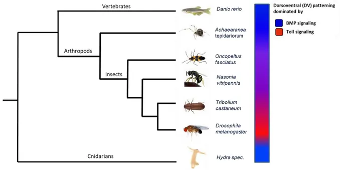
The formation of the two body axes, the anterior-posterior (AP) axis and the dorsoventral (DV) axis is a crucial early step in establishing the body of any animal (Wilson et al., 2014). Though, early embryogenesis and corresponding formation of the axes varies in different species. BMP (bone morphogenic protein) signaling and its role in dorsoventral patterning is highly conserved (De Robertis and Kuroda, 2004). However, in the model organism Drosophila melanogaster the Toll pathway plays the major role in dorsoventral patterning, whereas BMP acts downstream and only on the dorsal side of the embryo (Lynch and Roth, 2011) (Fig. 1.2). Although, Toll signaling has a conserved function in innate immunity of almost all bilaterians (Caamano and Hunter, 2002; Lemaitre et al., 1996; Rosetto et al., 1995), its role in axis formation appears to be an evolutionary novelty in insects (Leulier &
Lemaitre 2008). This leads to the conclusion that Toll was integrated into an existing patterning pathway during insect evolution. Regarding its evolutionary changing role in DV-patterning, the Toll- NFκB/Dorsal pathway is an interesting subject that promises insights into evolution of gene regulation and into functional constraints, underlying gene regulatory network (GRN) evolution. Comparative studies using more basally branching insects might help to understand the mechanisms of such modifications. The emerging model organism Tribolium castaneum is an insect with more ancestral features of embryogenesis and is thus, a perfect candidate for comparative analysis. The following section concentrates on Drosophila in order to highlight the best understood DV patterning system, which is the foundation for comparative studies in this thesis.
Figure 1.2: Evolution of dorsoventral patterning
The BMP pathway (blue) plays a conserved role in the establishment of dorsoventral polarity in vertebrates as well as in arthropods. However, during insect evolution the Toll pathway (red) which has a conserved role in innate immunity, became dominant in establishing the DV axis.
14
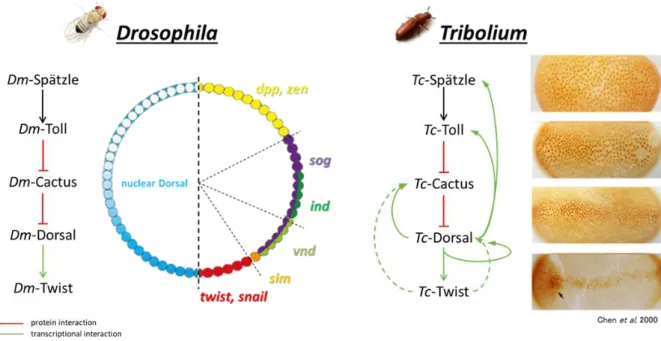
1.3.1 Formation of the dorsoventral axis in Drosophila melanogaster
One of the best studied developmental processes is the dorsoventral axis formation of Drosophila melanogaster. Multiple former studies uncovered the fine-tuned, spatio-temporal gene expression which leads to the determination of different cell fates.
The DV-axis is established by migration of the oocyte nucleus from a central-posterior towards an anterior-cortical location (Peri and Roth, 2000; Roth, 2003; Roth and Lynch, 2009). The maternal mRNA of the TGFα-like ligand Gurken is located around the nucleus and activates upon translation, the EGF receptor (Roth, 2004) in the overlying follicle cells (Schupbach and Roth, 1994). EGFR signaling restricts the expression of the sulfotransferase Pipe to the ventral side where it modifies several components of the vitelline membrane (Nilson and Schupbach, 1998; Roth, 2003). This dorsoventral polarity of the egg chamber is transmitted to the early embryo via an extracellular proteolytic cascade in the perivitelline space (Fig. 1.3).
The final step of the protease cascade is the cleavage of Spätzle. After cleavage, Spätzle binds and activates the Toll receptor. Probably, the cleaved Spätzle protein is present in a concentration gradient.
Cleavage of the Toll-ligand Spätzle generates a gradient of Toll receptor activation on the ventral side of the embryo (Morisato, 2001; Moussian and Roth, 2005). The activation of the Toll receptor leads to the phosphorylation and degradation of Cactus. Usually Cactus binds the NF-κB transcription factor Dorsal, which is consequently retained in the cytoplasm. Upon degradation of Cactus, Dorsal can translocate into the nuclei which leads to the establishment of a nuclear Dorsal gradient (Fig. 1.3) with peak levels at the ventral midline (Moussian and Roth, 2005; Roth et al., 1991). The nuclear Dorsal gradient in Drosophila is stable and leads to the transcriptional regulation of 60-70 different target genes (Type I, Type II, Type III) which are activated or repressed by Dorsal in a concentration- dependent manner (Reeves and Stathopoulos, 2009) (Fig. 1.3). Genes like twist and snail are expressed, due to high nuclear Dorsal levels (Type I), on the ventral-most side of the embryo and specify the presumptive mesoderm (Jiang et al., 1991; Leptin, 1991). In more lateral regions of the egg circumference, intermediate nuclear Dorsal concentrations activate the transcription of genes like ventral nervous defective (vnd) (Type II), which trigger formation of the neuroectoderm.
15 Figure 1.3: Establishment of the dorsoventral axis in Drosophila melanogaster
Activation of EGF signaling (blue) in the dorsal ovary follicle cells by Gurken (green) restricts Pipe (red) to the ventral side.
Pipe activates a protease cascade in the perivitelline space. The cleaved and thus active ligand Spätzle binds to Toll receptors which finally results in the establishment of a nuclear Dorsal gradient with peak levels at the ventral midline (brown).
Dependent on the nuclear Dorsal concentration, different target genes are expressed along the dorsoventral axis.
Two subgroups of Type III genes are regulated by low nuclear dorsal concentrations. Type III+ genes like short gastrulation (sog) are activated, while Type III- genes like the BMP homolog decapentaplegic are repressed (Hong et al., 2008; Reeves and Stathopoulos, 2009). dpp is expressed on the dorsal most side, where no nuclear Dorsal is present. Sog acts as an inhibitory modulator for BMP signaling and antagonizes the BMP2/4-like ligand Decapentaplegic (Dpp) in lateral regions (Reeves et al., 2012). Dpp gets bound and transported to the dorsal side, where it is cleaved by the metallo-protease Tolloid.
Released Dpp can than bind to its receptors (Nunes da Fonseca et al., 2008). This mechanism restricts the activation of BMP signaling to the dorsal side, which is necessary for the specification of the dorsal non-neurogenic ectoderm and the amnioserosa (Nunes da Fonseca et al., 2008). The differences of the DV-GRN of Tribolium will be discussed in the following section.
1.3.2 The dorsoventral gene regulatory network of Tribolium castaneum
DV patterning in Tribolium shows many similarities to DV-patterning in Drosophila melanogaster.
However, the mRNA of the Tribolium TGFα homolog is not localized in the ovaries. Thus, the process of DV polarity transmission from the ovary to the embryo remains unclear (Lynch and Roth, 2011).
Former studies showed that Toll is also a key component in DV patterning of Tribolium castaneum. Like in Drosophila, the activation of Tc-Toll by binding of Tc-Spätzle leads to nuclear uptake of Tc-Dorsal.
But in contrast to Drosophila, the resulting Tc-Dorsal gradient is spatio-temporally dynamic (Chen et al., 2000; Nunes da Fonseca et al., 2008). The Tc-Dorsal gradient refines from a broad domain in the early blastoderm to a narrow stripe along the ventral midline (Fig. 1.4). Finally, it disappears from the germ rudiment before the onset of gastrulation (Chen et al., 2000). In addition, Toll and cactus are zygotically expressed genes in Tribolium, whereas they are maternally expressed in the fruit fly (Lynch and Roth, 2011). Thus, the dynamism of the Dorsal gradient is caused by several negative and positive feedback mechanisms that act on zygotic level (Nunes da Fonseca et al., 2008). Previous studies (Lynch
16 and Roth, 2011; Nunes da Fonseca et al., 2008) showed that Tc-Cactus is activated by Tc-Dorsal itself and in addition by the Tc-Dorsal target Tc-Twist. This Dorsal-/Twist-dependent activation of Tc-Cactus simultaneously leads to an enhanced repression of Tc-Dorsal. In addition, Tc-Dorsal regulates its own activator Tc-Toll in a positive feedback loop (Fig. 1.4). Furthermore, ChIP sequencing experiments revealed a potential self-regulative input of Tc-Dorsal and a positive regulation of Tc-Dorsal on the Toll ligand Tc-Spätzle (Stappert, PhD Thesis 2014).
Another difference is that the BMP pathway plays a more important role in DV-patterning of Tribolium castaneum, and that the Toll pathway influences less target genes compared to the derived patterning system of Drosophila. It was shown, that loss of sog in Drosophila results in weaker defects than the Tc-sog kd, which leads to a strong dorsalization of the embryo, indicating that Tc-Dorsal is not a direct repressor of Tc-dpp (Nunes da Fonseca et al., 2010). Moreover, dpp knockout in Drosophila does not affect the mesoderm, while knockdown of dpp in Tribolium results in a slight expansion of the mesoderm (Nunes da Fonseca et al., 2008). These results strengthen the presumption that Toll's role in DV patterning is derived, and that the DV-GRN changed since the last common ancestor of flies and beetles. The approach to elucidate these changes is described in the following sections.
Figure 1.4: Regulation of dorsoventral patterning genes
The Drosophila nuclear Dorsal-gradient is stable and regulates the transcription of ~ 50 target genes in a concentration- dependent manner. The same signaling cascade initiates the expression of Tc-twist and other DV patterning genes in Tribolium castaneum. However, in contrast to Drosophila the nuclear Tc-Dorsal gradient is dynamic due to feedback mechanisms. Tc-cactus is regulated by Dorsal and by the Dorsal target gene twist. This activation establishes a negative feedback loop, as Cactus has a negative influence on the nuclear uptake of Dorsal. In addition, Tc-Dorsal regulates its own activator Tc-Toll in a positive feedback loop.
modified from (Chen et al., 2000; Nunes da Fonseca et al., 2008; Stappert, PhD Thesis 2014)
17
1.4 Identification of new DV-patterning genes by differential expression analysis
Comparative studies are often performed by candidate gene approaches, requiring sequence identification of already known pathway components in the organism of interest by DNA or protein BLAST. Many important evolutionary studies are based on this approach. However, analyzing evolutionary changes in gene regulatory networks by a candidate gene approach depends on the presence of the respective pathway components, rendering discovery of new genes involved in a GRN impossible. Modern techniques like whole genome sequencing, identification of binding regions by ChIP-sequencing or generation of transcriptomes by RNA-sequencing (RNA-seq), facilitate new opportunities in the wide field of evolution and development. To get more insights into the DV-GRN of Tribolium castaneum and the differences in composition and wiring of the genes to the DV-GRN of Drosophila melanogaster, I performed a genome wide comparative differential expression analysis after RNAi. The establishment of this modern technique for comparative studies in Tribolium was performed together with Dominik Stappert (Frey, Master Thesis 2013; Stappert, PhD Thesis 2014).
1.4.1 Generation of knockdown embryos lacking key DV patterning genes
To identify new potential target genes, transcriptomes of Tc-Toll1, Tc-twist, Tc-sog and Tc-dpp knockdown (kd) embryos were generated by parental RNA interference (pRNAi). Wildtype embryos derived from uninjected mothers served as control. Considering possible false positive results by genes activated via the injection procedure itself (e.g. inflammatory genes, RNAi machinery genes), an additional control was used. Therefore, female pupae were injected with dsRNA for dsRed which is derived from Discosoma spec. and is not present in the Tribolium genome. Thus, dsRed knockdown embryos should not show differential gene expression.
The RNA for transcriptome analyses was isolated from embryos derived from dsRNA injected mothers and staged to 7.5h to 11.5h AEL. At that stage, the embryo proceeds from the early differentiated blastoderm stage to the horseshoe stage. The carefully selected time window captures a critical time in DV patterning. During the differentiated blastoderm numerous direct target genes of the key regulators Dorsal and Twist are expressed. These genes are especially important for tissue specification and early tissue differentiation during the gastrulation of the embryo. The embryos were stained with DAPI and surveyed for phenotypic penetrance (Fig. 1.5).
1 Knockdown of Tc-dorsal results in a higher death rate of the injected females and to a reduced number of eggs. Tc-Toll RNAi is upstream of Tc-dorsal and thus phenocopies Tc-dorsal with regard to its dorsoventral phenotype.
18 dsRed kd embryos do not show any morphological defects and look similar to embryos from untreated wildtype control females (Fig. 1.5 A-A`). Although knockdown of Tc-twist results in complete loss of the mesoderm (Stappert et al., 2016), the visible defects in DAPI-stained Tc-twist kd embryos are rather mild. Older embryos show characteristic finger-like structures at the posterior end (Fig. 1.5 B-B’’).
These structures are the malphigian tubules which are usually wrapping the connection of the hindgut and the midgut. Compared to wildtype (WT) blastoderm stage embryos (Fig. 1.5 A`), which show an oblique borderline between the big anterior serosa cells and the more compact posterior germ band cells, the border becomes straight and is shifted towards the posterior pole after knockdown of Tc-Toll (Fig. 1.5 C’). Elongating germ bands look like a thin tube which is interrupted at different places (Fig.
1.5 C’’). Due to their respective defects, Tc-sog kd and Tc-dpp kd embryos show antagonistic phenotypes. Whereas the knockdown of Tc-sog results in loss of the neurogenic ectoderm and the head structures (Fig. 1.5 D), Tc-dpp kd embryos lack the dorsal ectoderm (Fig. 1.5 E). Similar to Tc-Toll kd, Tc-sog-RNAi and Tc-dpp-RNAi embryos reveal already strong DV-defects in the differentiated blastoderm stage. Tc-sog kd embryos have a straight serosa-germ band border. Furthermore, the border shifts towards the posterior pole (Fig. 1.5 D’). Also, the serosa-embryo border of Tc-dpp- deficient embryos loses its obliqueness, but shifts to a more anterior position (Fig. 1.5 E’), compared to wildtype embryos. Furthermore, extending germ bands show the typically reduced head upon loss of Tc-sog and tube-shaped remaining segments (Fig. 1.5 D’’). The tube-like shape of the germ band was also seen for Tc-dpp-deficient embryos. In addition, some tissue at the posterior end does not get invaginated and is thus not covered by the serosa (Fig. 1.5 E’’).
19 Figure 1.5: Fate maps of dorsoventral phenotypes
(A-E) Fate maps of Tribolium embryos at the beginning of gastrulation2. The different colors indicate different cell fates as displayed in the legend. (A’-E’) DAPI stained embryos corresponding to A-E. The dashed lines represent the embryo-serosa border. (A’’-E’’) DAPI stained elongated germ bands of the respective knockdowns shown in (A-E). Anterior to the left; kd = knockdown. (A) Fate map of a wildtype embryo or of embryos from mothers injected with dsRNA of dsRed. The embryo- serosa border at beginning of gastrulation is oblique (A’). (A’’) Fully extended germ band stage wildtype embryo. (B) Fate map of Tc-twist knockdown embryos in which the mesoderm is lost. In Tc-twist kd embryos, the embryo-serosa border is not affected (B’), while germ bands show a misplacement of the malpighian tubules (B’’). (C) Fate map of dorsalized Tc-Toll knockdown embryos. The dorsal serosa is expanded and its border becomes straight (C’). The mesoderm is lost and the neurogenic ectoderm is also affected. (C’’) Germ band embryos after Tc-Toll kd have a tube-like morphology. (D) Fate map of dorsalized Tc-sog knockdown embryos. The mesoderm is present, but the neurogenic ectoderm is lost and the dorsal serosa is expanded. Similar to Toll kd embryos, the serosa is shifted towards the posterior pole (D’) and the elongated germ band forms a tube with reduced head structures (D’’). (E) Fate map of ventralized Tc-dpp knockdown embryos. The dorsal serosa is lost and the neurogenic ectoderm is expanded. (E’) The embryo-serosa border is shifted towards the anterior pole and germ band embryos show a typical tissue ball at the posterior end, which is not surrounded by serosa (E’’).
modified from (Fonseca et al., 2009)
2 In former schemes the amnion comprised the whole dorsal-most side reaching to the primitive pit. Due to unpublished results by Matthew A. Benton, the borders of the amnion were modified in A.
20
1.4.2 Validation of the knockdown efficiency
Before the isolated RNA was sequenced, the knockdown efficiency of the samples was analyzed by qRT-PCR (Stappert et al., 2016). Five replicates of wildtype, Tc-Toll kd and Tc-twist kd and 3 replicates of each dsRed kd, Tc-sog kd and Tc-dpp kd were chosen due to the best results for phenotypic penetrance; analysis of the knockdown efficiency by qRT-PCR and quality of the isolated RNA and send to the Cologne Center for Genomics (CCG)3. After an additional quality control, the samples were sequenced by Ilumina sequencing (HiSeq 2000, paired end mode, read size of 100 bp) (Stappert, PhD Thesis 2014).
1.4.3 Identification of differentially expressed genes
The bioinformatic analysis of the RNA-sequencing data was performed by Dominik Stappert. In this study, only genes which showed up as differentially expressed upon analysis with two different programs, DESeq2 and edgeR were considered. Details about the used pipeline and algorithms are described in the PhD Thesis of Dominik Stappert, 2014 and in our corresponding paper (Stappert et al., 2016). A comparison of the transcriptomes of uninjected wildtype embryos compared to embryos derived from mothers injected with dsRed dsRNA revealed that the injection procedure itself has no influence on gene regulation in the embryos. For future generation of Tribolium transcriptomes it is thus sufficient to use control embryos from uninjected mothers.
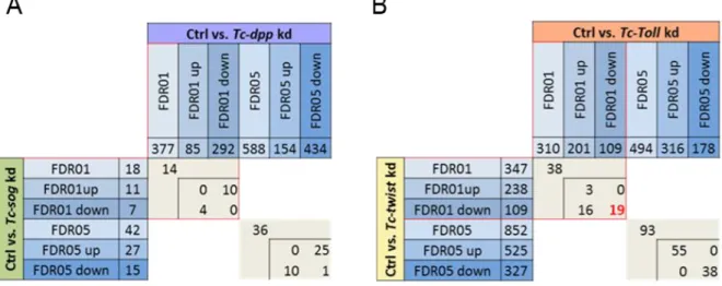
To identify differentially expressed genes, the sequenced transcriptomes of the respective knockdown embryos were first compared to the transcriptomes of control embryos (WT and dsRed-kd). The identified genes should be regulated either by Tc-Toll and/or Tc-twist or by BMP signaling. The comprehensive transcriptome analysis revealed in total 796 genes (Fig. 1.6 A, table), which were differentially expressed in the knockdown embryos compared to the control with a false discovery rate (FDR) of 1%. The data set identified 310 differentially expressed genes in Tc-Toll kd embryos compared to the control, 347 upon Tc-twist kd compared to the control. 377 genes showed up as potentially regulated by Dpp. However, only 18 differentially expressed genes could be identified by comparing the transcriptomes of Tc-sog knockdown embryos and the control (detailed list in the supplement of (Stappert et al., 2016)). Although the fate shift seen in Tc-sog kd embryos is severe (massive expansion of the neuroectoderm), the effect on more downstream components is restricted. Sog is an inhibitory modulator of BMP signaling (van der Zee et al., 2006) and might not have as many target genes as a transcription factor. A heat map (Fig. 1.6 B) shows the 222 genes up- or downregulated in more than one knockdown condition.
3 6 samples were then send to GATC (Konstanz) and 18 samples to the Cologne Center for Genomics (CCG) for sequencing (in total 24 RNA samples). Due to a first test of the procedure, the first six samples were produced and sent separately from the other samples.
21 Figure 1.6: 222 genes are differentially expressed in more than one knockdown condition
(A) Venn diagram depicting the number of genes which are differentially expressed upon kd of Tc-twist, Tc-Toll, Tc-dpp, Tc- sog as compared to wildtype embryos. Each field in the Venn diagram shows the number of genes that are exclusively found in the overlap of conditions indicated by the diagram. (B) Heat map showing if genes that are found in the overlapping fields of the Venn diagram are up regulated (yellow) or down regulated (blue). The numbers in the figure correspond to the numbers in the fields of the Venn diagram.
(Stappert et al., 2016)
22
1.5 Aims of the study
Although they interact differently, the transcription factors Dorsal and Twist are key regulators of dorsoventral patterning in Tribolium and Drosophila, and likely control major parts of the DV-GRN in both organisms. However, BMP signaling has a stronger influence on DV patterning in Tribolium, while Toll signaling seems to control less genes compared to Drosophila. Since little is known of the components involved in the Tribolium DV-GRN, a simple candidate gene approach might not be sufficient to reveal all evolutionary changes compared to the DV-GRN of Drosophila.
The goal of this study was to identify new potential DV pattering genes via unbiased comparative differential transcriptome analysis after knockdown via pRNAi of (Tc-Toll, Tc-twist, Tc-short gastrulation and Tc-decapentaplegic) followed by high-throughput RNA sequencing. Providing direct information on differential gene expression, this approach facilitates analysis of GRNs. Based on the RNA-sequencing data, two subgroups of potential DV patterning genes will be chosen and further investigated by in-situ hybridization, concerning their mRNA expression domains. A selection of these genes will be functionally analyzed by the generation of knockdown embryos via pRNAi. The identified differences in the DV-GRNs of Tribolium and Drosophila will be discussed in an evolutionary context.
23
2 Results
2.1 Verification of the RNA-seq data by an in-situ hybridization screen
For the identification of genes involved in the dorsoventral gene regulatory network (DV-GRN), the transcriptomes of Tc-Toll, Tc-twist, Tc-sog and Tc-dpp knockdown embryos were compared to a control (Stappert et al., 2016).
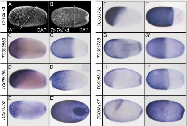
201 genes showed up as upregulated upon Tc-Toll kd compared to the control. To verify the bioinformatics analysis, the expression patterns of a random selection of them was investigated. I analyzed the mRNA expression for some of the genes in wildtype embryos and compared it to Tc-Toll deficient embryos. The expression patterns in Tc-Toll knockdown embryos reflect the data of the differential transcriptome analysis. As the serosa expands after knockdown of Tc-Toll 8 of 10 the genes are strongly expressed in the serosa (Fig. 2.1). TC008197 is only expressed in the dorsal serosa (Fig. 2.1 I). Especially this part is strongly expanded in Toll knockdown embryos (Fig. 2.1 I’). The verification of the RNA-sequencing data correlates well with the RNA-seq data set. Thus, subgroups of potential dorsoventral patterning genes had to be chosen for further analysis.
Figure 2.1: Expression patterns of genes differentially expressed upon knockdown of Tc-Toll
The expression pattern of selected genes which were upregulated in Tc-Toll kd embryos compared to the control. (A) DAPI staining of a wildtype embryo in the differentiated blastoderm stage. The dashed line indicates the oblique border between the serosa (anterior) and the embryo proper (posterior). (B) In Tc-Toll knockdown embryos, the embryo-serosa border is straight and shifted towards the posterior. (B-I) Expression patterns as identified by ISH; (B’-H’) Expression of the same gene as depicted in pictures labeled with corresponding capital letters in Tc-Toll kd embryos. All embryos shown in lateral view – with exception to G, which is shown in a ventral view. Anterior to the left. All genes are expressed in the serosa. Note that TC008197 (I) is just expressed in the posterior-dorsal part of the serosa and that TC006785 (G) is also expressed in two ventral stripes. The expression pattern in Tc-Toll knockdown embryos reflects the data from the differential transcriptome analysis.
As the serosa expands upon knockdown of Tc-Tollmost of the genes show a wider expression domain.
24
2.1.1 Detailed analysis of two subgroups of differentially expressed genes
Facing a large numbers of potential target genes (796, FDR of 1%), it was necessary to reduce the data set for screening. Thus, I concentrated on those 222 genes which were differentially expressed in more than one knockdown condition (Fig. 1.6 B). The mesoderm is missing in Tc-Toll and Tc-Twist knockdowns, hence this comparison should predominantly reveal genes specifying the mesodermal fates of the embryo (Fig. 1.5 B and C). The opposing influence of Tc-sog and Tc-dpp kd is especially affecting the ectoderm. Thus, the comparisons of their transcriptomes should result in a list of genes mainly expressed in the ectoderm (Fig. 1.5 D and E). According to the resulting fate shift of the respective knockdown conditions, differentially expressed genes after knockdown of both Tc-Toll and Tc-twist (Fig. 2.2 A), as well as after knockdown of Tc-dpp and Tc-sog (Fig. 2.2 B) were most interesting for first analysis.
Figure 2.2. Genes differentially expressed upon Tc-Toll kd vs. Tc-twist kd and Tc-sog vs. Tc-dpp kd
ctrl - control embryos; kd - knockdown; FDR - false discovery rate; up - upregulated; down - downregulated. Transcriptomes of different knockdown conditions were first compared to the transcriptome of control embryos. The number of significantly differentially expressed genes was detected with a FDR of 1%. To increase the dataset, also genes with a FDR of 5% were included. (A) Number of genes differentially expressed between Tc-sog kd and Tc-dpp kd. (B) Number of genes differentially expressed between Tc-twist kd and Tc-Toll kd. For the identity of corresponding genes see Appendix B and C.
2.1.2 Identification of genes regulated by Dpp
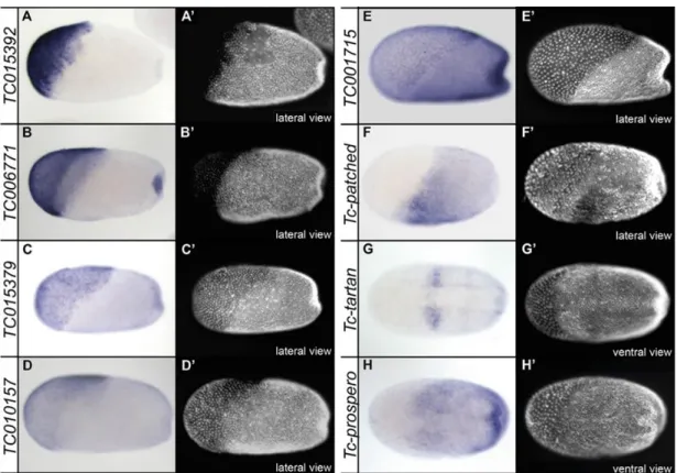
The first group consists of all genes which were differentially expressed in Tc-dpp and Tc-sog knockdown embryos with a FDR of 1% (Fig. 2.2 A). As expected, the identified 14 genes which were up- or downregulated in Tc-dpp knockdown embryos are regulated in the opposite direction after Tc- sog knockdown. Out of this group of 14 genes, 10 are downregulated in Tc-dpp kd embryos compared to knockdown of Tc-sog. Thus, these genes should be expressed either in the serosa or in ectodermal regions of the embryo.
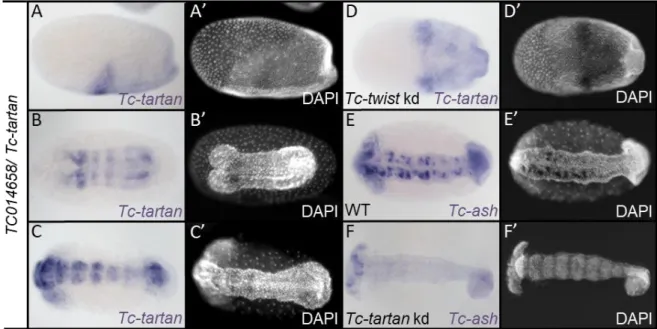
25 An in-situ hybridization screen revealed that 5 of these 10 genes are strongly expressed in the extraembryonic serosa (Fig. 1.5 A-E). The other 5 genes did not show a localized mRNA expression pattern. This finding can be explained by the prominent shift of the embryo-serosa border in different directions (Fig. 1.5 D’ and E’). The remaining 4 genes showed up as being upregulated upon Tc-dpp kd, whereas they are downregulated in Tc-sog deficient embryos. These 4 genes are likely acting in the neurogenic ectoderm of the embryo and are therefore interesting candidates. Among these genes, Tc- patched (TC004745) is expressed mainly in the head anlagen of differentiated blastoderm stage embryos (Fig. 2.3 F). This expression domain is also visible for Tc-tartan (TC014658), but in addition Tc- tartan, as well as Tc-prospero (TC010596), show an additional broad neuroectodermal expression (Fig.
2.3 G and H). To enlarge the data set and to increase the chances to identify promising candidates, also the 14 genes (exclusively on this list) which showed up as differentially regulated with a false discovery rate of 5% were analyzed. 7 of them showed a localized expression pattern: 4 are expressed in the serosa, 2 are expressed in segmental stripes and one shows mRNA expression in the neuroectoderm (see Appendix B).
Figure 2.3: Expression patterns of genes differentially expressed upon knockdown of Tc-sog and Tc-dpp (A-H) Expression patterns as identified by ISH; (A’-H’) Same embryo as depicted in pictures labeled with corresponding capital letters, nuclei stained with DAPI for staging; Embryos in A-D in primitive pit stage; Embryos in E-H are gastrulating; Embryos in A-E: lateral view; embryos in F-H ventral view; Anterior to left. A - D) TC015392, TC006771, TC015379, and TC010157 are expressed in the serosa. Note that TC015392 is just expressed in part of the serosa and that TC006771 is also expressed in the primitive pit. E) TC001715 is expressed in the serosa, but also in the germ rudiment. F) TC004745/Tc-patched is not expressed in the serosa, but in the embryo proper. A DV stripe of strong expression posterior to the presumptive head lobes is visible. G) TC014658/Tc-tartan is expressed in the same domain in which strong expression for TC004745/Tc-patched is detected. The expression is absent from the mesoderm. H) TC010596/Tc-prospero is not expressed in the serosa, but inside the germ rudiment, in which the expression is absent from the future mesoderm, although the expression borders are not well defined.
26
2.1.3 Expression patterns of genes regulated by Toll and/or Twist
The second subgroup of 38 genes showed up as differentially expressed after knockdown of both, Tc- Toll and Tc-twist with a false discovery rate of 1% (Fig. 2.1 A).
As the mesoderm is missing in both knockdown conditions, the 19 genes which are downregulated in both Tc-Toll and Tc-twist kd, are of special interest. The 14 genes which showed a specific mRNA expression domain, include already known dorsoventral patterning genes like Tc-twist itself, Tc-cactus and Tc-snail (Fig. 2.4 A, B and C). Also, the known Drosophila Twist targets heartless (Fig. 2.4 G) and downstream-of-fgf (dof) (Fig. 2.4 H) showed up as downregulated in Tc-Toll and Tc-twist kd embryos.
Like the remaining genes, both show a clear ventral expression domain in the presumptive mesoderm of blastoderm Tribolium embryos (Fig. 2.4 A-D and G-L). Furthermore, I could identify Tc-Delta (Fig. 2.4 D). In Drosophila, Delta is expressed in the neuro-ectoderm, whereas it is repressed in the presumptive mesoderm (Vassin et al., 1987). However, in Tribolium Tc-Delta seems to be positively regulated by Toll and Twist in the mesoderm. Its expression is furthermore co-localized with two other components of the Delta/Notch pathway: Tc-E(spl)1 and Tc-E(spl)3. Both enhancer of split homologs of Tribolium are expressed in a mesodermal domain with enhanced levels at its lateral borders (Fig. 2.4 E and F).
Similar to Delta, E(spl)1 and E(spl)3 are expressed in the neurogenic ectoderm in Drosophila (Knust et al., 1992; Wech et al., 1999). The only gene showing a typical anterio-posterior (AP) expression pattern was TC008064 (sloppy paired/slp). Tc-slp is a pair-rule gene (Choe and Brown, 2007) which is expressed in segmental stripes along the AP axis (Fig. 2.4 M). Some of the candidates from this group (TC003461, TC003606, TC007056, TC009862, TC010195 and TC013142), did not show a specific mRNA expression pattern (data not shown). Of the remaining 19 genes which are upregulated in Tc-Toll kd embryos and downregulated in Tc-twist embryos, only two showed a distinct expression pattern. Interestingly, they are expressed in the serosa, which is expanded after loss of Tc-Toll. Similar to the analysis of potential Tc-Sog vs Tc-Dpp targets, the expression patterns of 20 genes (exclusively on this list) which showed up as differentially expressed with a FDR of 5% were also analyzed. 5 of them are expressed in the mesoderm, 3 in the serosa and 2 are expressed in segmental stripes (see Appendix C).
27 Figure 2.4: Expression patterns of genes downregulated upon knockdown of Tc-Toll and Tc-twist
(A-N) Whole-mount ISH of embryos at blastoderm stage. (A-C) Primitive pit stage (E-H, J, L, M, N) or early gastrulation stage (D, I, K). (A′-N′) DAPI staining of the respective embryos. (A-H, M) Ventral surface views. (I-L, N) Lateral views with dorsal side pointing upwards. The anterior pole points to the left. All genes (except in G, H and M) are expressed in the presumptive mesoderm. Note that Tc-cactus (B) is also expressed in ventral parts of the serosa. Tc-E(spl)1 (G) and Tc-E(spl)3 (H) are expressed in narrow lateral stripes at the border of the mesoderm.
28
2.2 Functional analysis of genes potentially involved in the DV-GRN of Tribolium
To further investigate the role of selected candidate genes in the DV-GRN of Tribolium castaneum, functional studies using pRNAi were performed. The survival of the mothers injected with the respective dsRNA, the phenotypic penetrance in the embryos derived from these mothers and analyzed the expression of different marker genes in the knockdown (see Appendix D and E) were determined for each kd condition. Tc-twist was used as mesodermal marker, Tc-achaete-scute homolog (Tc-ash) as neuronal marker (Wheeler et al., 2003), Tc-pannier as marker for the amnion and the dorsal ectoderm (van der Zee et al., 2005) and Tc-engrailed (Brown et al., 1994) (not shown in Fig.
2.5) or Tc-gooseberry (Davis et al., 2001) as segmental markers (Fig. 2.5).
Figure 2.5: Expression of marker genes
(A-D) Expression of Tc-twist, Tc-achaete-scute homolog (Tc-ash), Tc-pannier and Tc-gooseberry (Tc-gsb) in wildtype embryos.
(A) Tc-twist is expressed in a strong mesodermal stripe in differentiated blastoderm embryos. (B) Amniotic Tc-pannier expression along the embryo-serosa border and in the dorsal ectoderm around early gastrulation. (C) Expression of neuronal precursor marker Tc-achaete-scute homolog during germ band extension. (D) Expression of segmental marker Tc-gooseberry during germ band extension.
29
2.2.1 Functional analysis of Sog/Dpp targets
2.2.1.1 Neuroectodermal expression of Tc-uniflatable
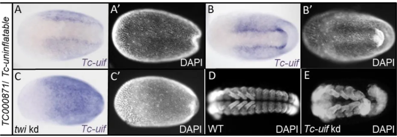
A candidate showing an interesting neuroectodermal expression pattern could be identified among the differentially expressed genes with a false discovery rate of 5% (see Appendix B). Tc-uninflatable (TC000871) is a potential candidate, as it is known to be involved in Notch signaling in Drosophila (Xie et al., 2012; Zhang and Ward, 2009).
Tc-uif is upregulated in Tc-dpp kd and downregulated in Tc-sog kd embryos. In blastoderm embryos, it is expressed in a broad neuroectodermal domain, while it is absent from the mesoderm on the ventral side (Fig. 2.6 A). Shortly before gastrulation the expression becomes enhanced in two stripes flanking the mesoderm (Fig. 2.6 B). In elongating germ bands, Tc-uif is expressed uniformly with exception of the mesoderm (data not shown). Interestingly, uif also shows an early expression in ectodermal cells in Drosophila embryos (Zhang and Ward, 2009). No changes in the early expression of the marker genes in Tc-uif kd embryos could be detected (see Appendix D). However, the neuroectodermal expression domain is expanded to the ventral side after Tc-twist knockdown (Fig. 2.6 C). This result suggests a negative regulation of Tc-uif by either Twist itself, or by a Twist target gene.
Knockdown of Tc-uninflatable results in a consistent phenotype with a high phenotypic penetrance (61.45%; N=182). The knockdown embryos show thinner abdominal segments compared to wildtype embryos (Fig. 2.6 D and E). In addition, the posterior segments were often bended. Although, the mRNA shows an early localization before gastrulation, the defects are restricted to the segment addition zone of extending germ band embryos. However, affecting the segment addition zone indicates an important role of Tc-uif in embryonic development.
Figure 2.6: Functional analysis of Tc-uninflatable
(A-C) Expression patterns detected by ISH. (A’-C’) Corresponding embryos, nuclei stained with DAPI for staging; All embryos in ventral view; Anterior to left. (A-C) Expression of TC000871/Tc-uniflatable (Tc-uif) in wildtype embryos. (A) In primitive pit stage embryos, Tc-uif is expressed in broad lateral domains. (B) When gastrulation proceeds, expression of TC000871 enhances in stripes flanking the mesoderm. (C) In Tc-twist kd embryos the neuroectodermal expression domain expands towards the ventral side. E shows DAPI staining of older embryos (~24h-48h AEL) after Tc-uif knockdown via pRNAi. The abdominal segments of these embryos are thinner and often bent compared to WT (D).