The Role of TGF-β Signaling in Development and Maintenance of the Ocular Vasculature
Dissertation
Zur Erlangung des Doktorgrades der Naturwissenschaften (Dr. rer. nat.) der Fakultät der Biologie und Vorklinischen Medizin der Universität Regensburg
Durchgeführt am Lehrstuhl für Embryologie und Humananatomie der Universität Regensburg
vorgelegt von Anja Schlecht
aus Roding
2016
Das Promotionsgesuch wurde eingereicht am: 21.10.2016.
Die Arbeit wurde angeleitet von: Prof. Dr. Ernst R. Tamm
Unterschrift:
Manuscripts included in this thesis:
Schlecht A, Leimbeck SV, Tamm ER, Braunger BM. Tamoxifen-containing eyedrops successfully trigger Cre-mediated recombination in the entire eye. Adv Exp Med Biol 2016, 854:495-500.
Braunger BM, Leimbeck SV, Schlecht A, Volz C, Jägle H, Tamm ER. Deletion of ocular transforming growth factor β signaling mimics essential characteristics of diabetic retinopathy. Am J Pathol. 2015, 185(6):1749-68.
Schlecht A, Leimbeck SV, Jägle H, Feuchtinger A, Tamm ER, Braunger BM. Ablation of endothelial TGF-β signaling causes choroidal neovascularization. Submitted to Journal of Clinical Investigation.
Authors‘ contribution is noted in each figure legend.
_________________________
Anja Schlecht
Table of content
Chapter 1
1 General Introduction ... 2
1.1 TGF-β signaling ... 2
1.2 Cre loxP system ... 3
1.3 The ocular vasculature ... 5
1.3.1 Development of the retinal vasculature ... 6
1.3.2 Blood retinal barrier ... 7
1.4 Pathologies of the ocular vasculature ... 9
1.4.1 Diabetic retinopathy ... 9
1.4.2 Age-related macular degeneration ... 11
1.5 Aim of the study ... 13
Chapter 2 2 Tamoxifen-Containing Eye Drops Successfully Trigger Cre-Mediated Recombination in the Entire Eye ... 16
2.1 Abstract ... 16
2.2 Introduction ... 17
2.3 Material and Methods ... 18
2.3.1 Mice ... 18
2.3.2 Tamoxifen Treatment ... 18
2.3.3 PCR Analysis ... 18
2.3.4 β-galactosidase Staining ... 19
2.4 Results - Localization of Cre-mediated Recombination in Ocular Tissues ... 20
2.5 Discussion ... 22
Chapter 3 3 Deletion of ocular TGF-β signaling mimics essential characteristics of diabetic retinopathy ... 24
3.1 Abstract ... 24
3.2 Introduction ... 25
3.3 Materials and Methods ... 27
3.3.1 Mice ... 27
3.3.2 Western blot analysis ... 27
3.3.3 Microscopy and morphometry ... 28
3.3.4 LacZ staining ... 29
3.3.5 Immunohistochemistry ... 29
3.3.6 Dextran perfusion and retinal whole mounts ... 31
3.3.7 Apoptosis ... 31
3.3.8 Trypsin digest ... 31
3.3.9 Electroretinography ... 32
3.3.10 Fundus imaging and angiography ... 32
3.3.11 RNA analysis ... 32
3.3.12 Statistical analysis ... 34
3.4 Results ... 35
3.4.1 Conditional deletion of TβRII in the eye following local tamoxifen treatment ... 35
3.4.2 Conditional deletion of TGF-β signaling results in leaky retinal capillaries that form microaneurysms ... 37
3.4.3 Lack of pericyte differentiation upon absence of retinal TGF-β signaling ... 39
3.4.4 Hemorrhages and vessel loss in TGF-β-deficient retinae ... 45
3.4.5 Lack of ocular TGF-β signaling leads to hyaloid vasculature persistence ... 47
3.4.6 Absence of TGF-β signaling induces microglia reactivity in the retina ... 49
3.4.7 Continuous lack of retinal TGF-β signaling leads to proliferative retinopathy ... 51
3.5 Discussion ... 56
3.5.1 TGF-β signaling determines pericyte differentiation during development of the retinal vasculature ... 57
3.5.2 Lack of retinal TGF-β signaling leads to vascular leakage ... 58
3.5.3 Lack of retinal TGF-β signaling induces the formation of capillary microaneurysms
... 59
3.5.4 Lack of retinal TGF-β signaling induces capillary closure and reactive microglia .... 60
3.5.5 Phenotypic changes in retinae progress to proliferative retinopathy ... 61
3.5.6 Conclusion ... 62
Chapter 4 4 Ablation of endothelial TGF-β signaling causes choroidal neovascularization ... 64
4.1 Abstract ... 64
4.2 Introduction ... 65
4.3 Material and Methods ... 67
4.3.1 Mice ... 67
4.3.2 Induction of Cre recombinase ... 67
4.3.3 mT/mG reporter mice ... 68
4.3.4 Tgfbr2 deletion PCR ... 69
4.3.5 Morphology and microscopy ... 69
4.3.6 Immunohistochemistry ... 69
4.3.7 Dextran Perfusion ... 71
4.3.8 Electroretinography (ERG) ... 71
4.3.9 Fundus Imaging and Angiography ... 72
4.3.10 RNA Analysis ... 72
4.3.11 Deep tissue imaging by 3D light-sheet fluorescence microscopy ... 74
4.3.12 Statistical Analysis ... 74
4.4 Results ... 75
4.4.1 Deletion of ocular TGF-β signaling in mouse pups leads to choroidal neovascularization ... 75
4.4.2 TGF-β signaling is required to prevent choroidal neovascularization in late-induced Tgfbr2Δeyemice ... 80
4.4.3 Expression of angiogenic factors and immune modulating cytokines in Tgfbr2Δeye mice ... 85
4.4.4 Formation of basal lamina deposits in Tgfbr2Δeye mice ... 88
4.4.5 Cell-specific conditional deletion of Tgfbr2 in RPE and vascular endothelium ... 91
4.5 Discussion ... 96
4.5.1 TGF-β functions at the retinal/choroidal interface to prevent CNV ... 96
4.5.2 TGF-β and VEGF as part of a homeostatic system to maintain integrity of the choriocapillaris ... 97
4.5.3 Potential neuroprotective effects of TGF-β ... 98
4.5.4 The formation of BlamD-like material is not required for CNV ... 99
4.5.5 Neovascularization is associated with macrophage accumulation ... 100
4.5.6 TGF-β signaling and endothelial proliferation ... 100
4.5.7 TGF-β signaling in human patients with AMD ... 102
4.5.8 Conclusion ... 102
4.6 Supplementary Data ... 103
5 General discussion ... 105
5.1 Tamoxifen eye drops: A new delivery method without side effects? ... 106
5.2 TGF-β signaling is required for pericyte differentiation ... 108
5.3 Early-induced deletion of ocular TGF-β signaling leads to formation of multiple microaneurysms... 109
5.4 Early-induced deletion of ocular TGF-β signaling results in non-perfused capillaries and activation of the ocular immune system ... 110
5.5 Early-induced deletion of ocular TGF-β signaling causes vitreal neovascularization, impaired retinal function and retinal detachment ... 111
5.6 Early-induced deletion of ocular TGF-β signaling leads to an AMD-like phenotype ... 111
5.7 Late-induced deletion of ocular TGF-β signaling results in CNV and a alterations of the Bruch’s membrane ... 113
5.8 Deletion of endothelial TGF-β signaling leads to development of CNV and changes in the Bruch’s membrane ... 114
6 Summary ... 116
7 Table of Figures ... 118
8 List of Tables ... 120
9 References ... 121
10 Abbreviations ... 154
11 Acknowledgments - Danksagung ... 157
12 Curriculum vitae ... 159
1
Chapter 1
General Introduction
2
1 General Introduction
Visual impairment and blindness can be caused by various diseases and may affect people of every age and social class. The leading causes of blindness worldwide are, for example, cataract, glaucoma, age-related macular degeneration and diabetic retinopathy (Thylefors et al.
1995). Every disease has its own possible risk factors. Among them are environmental factors, aging, smoking and genetic predispositions. Dysregulation of signaling pathways which are critical for development and homeostasis of the ocular environment may also cause malfunctions which can then lead to blindness.
1.1 TGF-β signaling
During development and adulthood, TGF-β signaling modulates essential processes e.g.cell growth and differentiation, tissue maintenance and apoptosis (Massagué 2012, Heldin et al.
1997). However, TGF-β has divers functions which are discussed controversially, as the effects of TGF-β depend on the affected cell type and the cellular environment (Massagué 2012). To date, three different isoforms of the cytokine TGF-β are known in mammals (TGF-β1-3) (Massagué 1992, Kingsley 1994). These ligands have the ability to bind TGF-β receptor type II (Tgfbr2, TβRII) which is essential for TGF-β signaling. TGF-β receptor type I recognizes this process, joins the complex and is phosphorylated by receptor type II (Wrana et al. 1992, Wrana et al. 1994). TGF-β receptor type II as well as type I are transmembrane serine/threonine kinases. Phosphorylation correlates with their kinase activity (Massagué et al. 1994, Wrana et al.
1994). Upon complex formation, the activated type I receptor phosphorylates the receptor- regulated (R-) Smads, Smad2 and 3, which form a complex with the co-mediator Smad, Smad4.
This complex translocates into the nucleus to regulate the expression of target genes (Figure 1) (Visser and Themmen 1998, Massagué 1998).
3 Figure 1: TGF-β signaling pathway. Binding of TGF-β to Tgfbr2 leads to recruitment and phosphorylation of Tgfbr1 which posphorylates and thereby activates the R-Smads. The R-Smads form a complex with the Co-Smad Smad4. This complex translocates into the nucleus and regulates the expression of target genes.
1.2 Cre loxP system
To understand the function of a specific gene in vivo, a common experimental approach is to analyze its function by generating animal models that harbor an inactivation or mutation of the gene (Rajewsky et al. 1996). However, when working with genes that have important functions during cell development or maintenance, researchers are frequently confronted with the problem of embryonic lethality (Branda and Dymecki 2004, Maddison and Clarke 2005). The establishment of the Cre loxP system in mouse models greatly enhanced the opportunities in this context. When using a Cre that is expressed in mice carrying a loxP targeted gene, the gene modification can be restricted to certain cell types or developmental stages depending on the use of specific promotors.
In general, two specific components, both derived from the bacteriophage P1, are necessary to achieve recombination using this system, namely a Cre recombinase and loxP sites: Cre stands for causes recombination and denotes a 38 kDa protein that recognizes and recombines loxP flanked DNA sequences. LoxP is an abbreviation for locus of crossover (x) in P1 bacteriophage
4 and describes a 34 bp asymmetric DNA sequence (Kuhn and Torres 2002, Sternberg and Hamilton 1981).
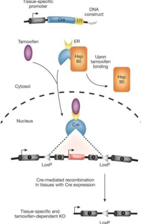
Tamoxifen inducible Cre recombinases are known as a special variant of the Cre loxP system (Figure 2). These allow recombination of the target gene at any desired time point. A tamoxifen- inducible Cre recombinase is characterized by its fusion with a mutated form of an estrogen receptor binding domain (ERTM). This mutation prevents the binding of endogenous 17β- estradiol but makes the ERTM domain susceptible to tamoxifen. Furthermore, this fusion leads to a sequestration of the Cre in the cytoplasm by interaction with Hsp90, thus preventing the Cre’s access to the nucleus and therefore recombination. The binding of tamoxifen causes a disruption of this complex. Thus, the Cre is able to translocate into the nucleus to initiate recombination (Hayashi and McMahon 2002).
Figure 2: Tamoxifen-inducible Cre–loxP system. The Cre recombinase is fused to a modified estrogen receptor (ER) and controlled by a specific promoter. In the inactivated state, heat shock protein 90 (Hsp90) binds to the ER, retaining it in the cytosol. Upon tamoxifen treatment, tamoxifen binds the ER, the Hsp90 protein is released, and the Cre–ER fusion protein can translocate in the nucleus to initiate recombination. Slightly modified from (Gunschmann et al. 2014).
5 1.3 The ocular vasculature
The oxygenation of the retina during initial development is ensured by two different and independent circulatory systems: the choroidal vessels and the hyaloid system. The mature and fully developed retina has the highest oxygen consumption per unit weight of any human tissue and thus needs a perfect system for oxygen supply (Saint-Geniez and D'amore 2004).
Therefore, during ocular development and maturation, the embryonic, hyaloid vascular system is replaced by the mature retinal vasculature which consists of three vascular plexus, namely the superior, intermediate and deep plexus (Stahl et al. 2010).
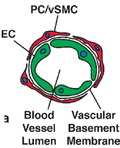
The inner wall of capillaries consists of a single layer of endothelial cells (ECs). These cells are densely coated with either several layers of vascular smooth muscle cells (in larger arteries) or a single layer of non-overlapping pericytes (in smaller arteries and arterioles) (Saint-Geniez and D'amore 2004). Together, the endothelial cells and the pericytes form the vascular basement membrane (Figure 3) (Mandarino et al. 1993). In general, pericytes have long cytoplasmatic processes which they use for contacting neighbouring endothelial cells and therefore can intgrate signals along the vessel length (Bergers and Song 2005). Furthermore, they show various characteristics correlating with muscle-cell activity and have the ability to produce vasoconstriction and vasodilation to regulate capillary blood flow (Rucker et al. 2000).
Figure 3: Composition of capillaries. Endothelial cells (EC, green) form the inner wall of capillaries, surrounded by the basement membrane in which pericytes (PC, red) or,vascular smooth muscle cells are embedded. Drawing taken from (Bergers and Song 2005)
6 1.3.1 Development of the retinal vasculature
There are two mechanisms responsible for the formation of new blood vessels, namely vasculogenesis and angiogenesis. The term vasculogenesis stands for the de novo formation of blood vessels which begins with the formation of clusters consisting of primitive vascular cells or hemangioblasts (Doetschman et al. 1987). These clusters form tube-like structures that fuse to form a vascular network (Vokes 2004). Angiogenesis, however, refers to the formation of vessels by the sprouting of capillaries from already existing vessels (Risau et al. 1988). In the human retina, both processes occur during vascularization, whereas in mice, retinal vascularization appears to be mediated only via angiogenesis (Hughes et al. 2000, Jiang et al.
1995, Fruttiger 2007, Ashton 1970).
During early stages of retinal development, oxygen supply is provided by the hyaloid vasculature. The hyaloid artery enters the optic cup through the optic nerve and forms an arterial network in the vitreous (Fruttiger 2007, Anand-Apte and Hollyfield 2010). In the more developed retina, the hyaloid vascular system regresses and is replaced by the retinal vasculature. This phenomenon occurs in humans around mid-gestation, in mice regression starts around birth and is finished at approx. P7 (Fruttiger 2007, Lang and Bishop 1993). The regression of the hyaloid arteries starts with an atrophy of the capillaries a process that is mediated via macrophage activity and WNT signaling (Fruttiger 2007, Diez-Roux and Lang 1997, Lobov et al. 2005).
Herein, the occlusion of the capillaries by macrophages seems to be a critical step to induce their atrophy. (Anand-Apte and Hollyfield 2010).
Simultaneously with the regression of the hyaloid system, the development of the retinal vasculature starts. Endothelial cells migrate from the optic nerve head over the inner surface of the retina toward the periphery of the retina and form the superficial plexus (Figure 4A). This migration is guided by a gradient of angiogenic factors, e.g. VEGF (vascular endothelial growth factor), expressed from a preexisting astrocytic template (Figure 4E) (Dorrell et al. 2002, Hughes et al. 2000, Ling et al. 1989). The endothelial cells follow the VEGF gradient of the astrocytic network (Stone et al. 1995). Endothelial cells at the vascular front possess filopodia and are called “tip cells” (Figure 4D). Astrocytes guide these tip cells to the remaining avascular areas (Siemerink et al. 2013).
7 Figure 4: Development of the retinal vasculature in the mouse. A. Growth of the retinal vasculature starts at the optic nerve head and extends to the periphery to build the superficial plexus. B. Secondary branches grow into the retina and reach the outer plexiform layer to develop the deep vascular plexus. C.
From the deep plexus, vessels extend to the inner plexiform layer, where the intermediate plexus is located. D. Endothelial cell at the vascular front shows long filopodia (arrows). E. Astrocytes (red) guide endothelial cells (green) to form the superficial plexus. INL, inner nuclear layer; ONL, outer nuclear layer;
GCL, ganglion cell layer; NBL, neuroblast layer. Pictures taken from (Ye et al. 2010), slightly modified.
In mice, the vessels of the superficial plexus reach the periphery of the retina on postnatal day 9 (P9). Subsequently, the vessels from the already existing plexus are growing toward the outer plexiform layer to form the deep vascular plexus (Figure 4B). Just a few days later, the intermediate plexus begins to develop from vertical sprouting of the deep plexus upwards to the inner plexiform layer (Figure 4C) (Stahl et al. 2010). Studies assume that the intermediate and deep vascular plexus are built independently of astrocyte guidance (Fruttiger 2007). There are some differences in the development of the retinal vasculature between humans and mice: In mice the development of the retinal vasculature is completed at P21 and in humans already at birth. Also, the chronology of plexus formation in humans is different compared to mice: After the development of the superficial plexus, the intermediate plexus is formed, and finally the deep vascular plexus (Gariano 2003).
1.3.2 Blood retinal barrier
The blood retinal barrier (BRB) plays an essential role in the microenvironment of the retina and retinal neurons, as it regulates ion, protein and water in- and outflow of the retina (Cunha-Vaz et al. 2011). This barrier allows the neural retina to develop and maintain specific substrate and ion
8 concentrations which are substantial for proper neuronal function (Cunha-Vaz et al. 2011).
Furthermore, it regulates infiltration of immune competent cells, toxins and various other factors that could have negative effects on the neurons e.g. impede with their function (Cunha-Vaz 2009, Phillips et al. 2007). The blood supply for the retina is secured by two vascular systems, one at the level of the retinal vasculature, and the other at the interface of the choroid and RPE retinal pigment epithelial. Both systems have a BRB which is consequently, subdivided into two major components: the inner blood retinal barrier (iBRB) which comprises the endothelium of the retinal vessels and an interaction with glial cells and perivascular cells like pericytes, and the outer blood retinal barrier (aBRB) which is formed by the retinal pigment epithelium (RPE) (Phillips et al. 2007, Cunha-Vaz 1976, Chen et al. 2008, Klaassen et al. 2013).
The iBRB is formed by tight junctions between the neighboring endothelial cells of the retinal vessels. These junctions make the endothelial cells impermeable to any molecules that are larger than approximately 30 kDA. Small molecules, like glucose and ascorbate, are transported via facilitated diffusion (Chen et al. 2008). The continuous endothelial layer at the inside of the vessel’s basal lamina, comprises the main structure of the inner blood retinal barrier. However, at the outside, the.basal lamina is covered by pericytes, which together with endothelial cells form the basal lamina, processes of astrocytes and Müller glia cells (Bergers and Song 2005). It is of interest to note that the pericytes do not form a continuous layer (Jordan and Ruiz-Moreno 2013). However, morphological studies could show that pericytes may act as supressors of endothelial cell growth and contact between these two cell types is necessary for the activation of latent TGFβ-1 (Antonelli-Orlidge et al. 1989) Activation of TGFβ-1 inhibits proliferation and migration of endothelial cells (Orlidge 1987). Furthermore, TGFβ-1 is an important player during vessel formation, as it induces differentiation of mesenchymal stem cells and neuro crest cells into pericytes (Chen and Lechleider 2004, Ding et al. 2004, Bergers and Song 2005)
Astrocytes, pericytes and Müller glia cells influence the activity of retinal endothelial cells and the iBRB by transmitting regulatory signals to ECs (Chen et al. 2008, Bergers and Song 2005). In humans, a loss of pericytes, a phenomenon called “pericyte-dropout” in patients suffering from diabetic retinopathy, results in a weakening of the vascular walls and can result in the development of microaneurysms (Wilkinson-Berka et al. 2004, Hammes et al. 2002). However, in mice, lacking >50% of pericytes, retinal microaneurysms are not a prominent finding (Enge 2002, Hammes et al. 2002, Hammes 2005). In general, a breakdown of the inner blood retinal barrier is associated with diseases like diabetic retinopathy (Cunha-Vaz 2009, Cunha-Vaz 1976, Chen et al. 2008)
9 The outer blood retinal barrier is formed by tight junctions (zonulae occludentes) between the neighboring cells of the retinal pigment epithelium (Peyman and Bok 1972, Strauss 2005). The RPE consists of a single layer of pigmented cells and disconnects the neural retina from the underlying fenestrated endothelium of the choriocapillaris and therefore plays an essential role in extracting nutrients from the blood and making them accessible to the photoreceptors.
Furthermore, the RPE eliminates debris and maintains retinal adhesion. The relationship between the retinal pigment epithelium and the photoreceptor layer is essential for maintaining visual function (Cunha-Vaz 2009, Strauss 2005). Alterations in the tightness of the outer blood retinal barrier may result in diseases like age-related macular degeneration (AMD) (Cunha-Vaz 1976, Cunha-Vaz 2009).
1.4 Pathologies of the ocular vasculature 1.4.1 Diabetic retinopathy
Diabetic retinopathy (DR) is the leading cause of blindness in the developed world (Cai and McGinnis 2016). This disease can be divided into two stages: nonproliferative DR and proliferative DR (Engerman 1989). An early sign of nonproliferative diabetic retinopathy is the appearance of multiple microaneurysms (Speiser et al. 1968). These microaneurysms are presumed to be the result of loss of pericytes, a phenomenon that is called “pericyte dropout”
(Wilkinson-Berka et al. 2004, Hammes et al. 2002). The loss of pericytes initiates the death of ECs and therefore leads to a weakening of the vascular walls. When microaneurysms rupture, microhemorrhages (Figure 5) can be observed throughout the retina (Wilkinson-Berka et al.
2004, Bandello et al. 2014). Further symptoms for nonproliferative diabetic retinopathy are a thickening of the basement membrane, development of hard exudates, cotton wool spots (Figure 5) and occlusion of retinal vessels (Cai and McGinnis 2016, 2016, Bandello et al., Cai and Boulton 2002, Engerman 1989). When diabetic retinopathy changes toward a proliferative phenotype, neovascularizations can be observed. Neovascularizations towards the vitreous can in the following lead to vitreous contraction, hemorrhage and tractional retinal detachment (Cai and McGinnis 2016, Cheung et al. 2010, Antonetti et al. 2012).
10 Figure 5: Diabetic retinopathy. A. Drawing of a healthy eye and an eye with diabetic retinopathy.
Pathological changes like hemorrhages, abnormal growth of blood vessels, aneurysms, cotton wool spots and hard exudates can be seen in the eye with diabetic retinopathy. B. Funduscopy of an healthy eye (left panel, macula is marked by black circle) and an eye with severe diabetic retinopathy chracterized by several widespread hemorrhages inside and outside the major vascular arcades, cotton wool spots ( black arrows ) A. Slightly modified from www.shutterstock.com. B. Slightly modified from (Jager et al. 2008, Bandello et al. 2014).
The pathological changes which occur during diabetic retinopathy are well described, albeit the underlying molecular mechanisms remain still unclear. Undoubtedly, diabetes represents the greatest risk for developing DR. Furthermore, type I diabetes is more likely to result in vision loss than type II. Along with diabetes, further important risk factors for developing DR are race, smoking, hyperglycemia and hyperlipidemia (Das et al. 2015). A key element in the development of diabetic retinopathy is VEGF (vascular endothelial growth factor) which is clearly elevated in diabetic retinal tissue without overt retinopathy. It is presumed to be the initiator of increased
©Tefi/Shutterstock.com
A
11 permeability of the retinal vasculature in diabetes (Amin et al. 1997, Sone et al. 1997, Clermont et al. 1997). Increased VEGF levels lead to a decreased tight junction protein expression which is strongly associated with a breakdown of the inner blood retinal barrier (Antonetti et al. 1998).
Another growth factor that may contribute to the pathogenesis of diabetic retinopathy is TGF-β.
Several studies have shown that levels of TGFβ-2 were elevated in the vitreous of patients with diabetic retinopathy, especially in those who suffer from a proliferative form of the disease (Kita et al. 2006, Hirase et al. 1998). These findings suggest that TGF-β signaling may be involved in the molecular pathogenesis of diabetic retinopathy.
1.4.2 Age-related macular degeneration
In people of 50 years and older, age-related macular degeneration (AMD) supersedes diabetic retinopathy as the leading cause of severe vision loss in the developed world (Pascolini et al.
2004, Congdon et al. 2004). In general, two forms (wet and dry) of AMD can be distinguished (Bowes Rickman et al. 2013). In the early stage, the disease develops slowly and remains mostly asymptotic for years (Coleman et al. 2008, Hogg and Chakravarthy 2006). The late stage, however, is characterized by severe vision loss (de Jong, Paulus T V M 2006, Sarks et al.
1999). Typically, focal deposition of acellular debris between the retinal pigment epithelium and the Bruch`s membrane occur during aging (Bird et al. 1995). These deposits are called drusen and are usually the first clinical finding of dry age-related macular degeneration (Figure 6A) (Bird et al. 1995). They can be observed in the macula as well as in the peripheral retina. Drusen can be classified in three different stages by their diameter. There are small (< 63µm), medium (between 63µm and 124µm) and large drusen ( 124µm) (Bird et al. 1995). Furthermore, drusen can be characterized as soft or hard, based on the appearance of their margins. While hard drusen show discrete margins, soft drusen are characterized by indistinct edges (Bird et al.
1995). Excessive formation of drusen can subsequently lead to a damage of the retinal pigment epithelium. This effect together with a chronic inflammatory response can result in areas of retinal atrophy, also known as “geographic atrophy” (Figure 6B) (de Jong, Paulus T V M 2006, Maguire and Vine 1986, Sunness et al. 1999). In the following, an induction of angiogenic cytokines like VEGF can occur, leading to the formation of choroidal neovascularization (CNV) which is accompanied by increased vascular permeability (Figure 6C). These newly formed vessels grow from the choriocapillaris through the Bruch`s membrane toward and into the subretinal space (Pauleikhoff 2005, van Lookeren Campagne et al. 2014). The development of
12 these neovascularization is directly related to serious hemorrhages and detachment of the RPE (de Jong, Paulus T V M 2006, Jager et al. 2008).
Figure 6: Funduscopy – age-related macular degeneration. A. Funduscopy of an eye with intermediate age-related macular degeneration showing large drusen. B. Geographic atrophy involving the centre of the fovea, with sharply demarcated loss of normal retinal pigment epithelial cells and evidence of deeper larger choroidal vessels. C. Neovascular age-related macular degeneration, with retinal haemorrhage, lipids, or retinal hard exudate and subretinal fluid. Slightly modified from (Coleman et al.
2008).
The strongest risk factor for developing age-related macular degeneration is age (Mitchell et al.
1995, Klein et al. 1999, Klein et al. 1992, Friedman et al. 1999). Other than that, smoking as well as genetic factors may be involved in causing AMD (Evans et al. 2005, Thornton et al. 2005, Scholl et al. 2007, Haddad et al. 2006). A recent study could demonstrate that high temperature requirement factor A1 (HTRA1) influences the risk of AMD (Yang et al. 2010).
Recently it was shown, that HTRA1 can interact with members of the TGF-β signaling pathway and plays a critical role in the regulation of angiogenesis via TGF-β signaling (Friedrich et al.
2015). Growth differentiation factor 6 (GDF6), which is a member of the TGF-β family is involved in ectoderm patterning and eye development. Furthermore, GDF6 could be identified as a novel disease gene for AMD (Zhang et al. 2012). In summary, these findings implicate a potential role of TGF-beta signaling in the molecular pathogenesis of age related macular degeneration.
13 1.5 Aim of the study
The systemic knockout of TGF-β is embryonically lethal, because of haematopoietic and vascular defects in the yolk sack (Dickson et al. 1995). Therefore, scientists are confronted with serious problems when investigating the role of TGF-β signaling in vivo during development and in adulthood. The Cre loxP system allows to circumvent embryonic lethality, using Cre recombinases that are time-dependent or tissue specific.
To date, there is no Cre recombinase available with an ubiquitous expression of Cre that is restricted to all cells of the eye. Therefore our group established a method to obtain efficient Cre mediated recombination in the entire eye. In previous studies we could show that tamoxifen eye drops successfully achieve the activation of Cre when applied to the eyes of 4 days old mouse pups carrying an tamoxifen dependent cre recombinase which is under control of an ubiquitously expressed chicken β actin promotor (CAGG-Cre ERTM) (Hayashi and McMahon 2002). When crossing heterozygous CAGG-Cre ERTM mice with mice carrying loxP sites flanking exon 2 of Tgfbr2 (Chytil et al. 2002), the administration of tamoxifen led to a deletion of Tgfbr2 in all ocular tissue. The downregulation of TGF-β signaling resulted in marked morphological changes of the retinal vasculature. Most strikingly, Tgfbr2 deficient mice (Tgfbr2Δeye) developed multiple microaneurysms, similar to those that can be seen in patients suffering from diabetic retinopathy.
Furthermore, the expression levels of Vegf-a and Hif1-α were significantly increased in the retinae of 4 week-old mice. At the age of 6 weeks the mice additionally developed choroidal neovascularization (CNV) which are a hallmark for the wet form of age related macular degeneration.
The overal goal of my thesis was to characterize the newly established system of tamoxifen administration and to investigate the phenotype of Tgfbr2 deficient mice with an early and late induced deletion of the TGF-β signaling pathway. In particular following aims were pursued:
What is the effect of an early-induced deletion of the TGF-β signaling pathway on the pericytes? Is the formation of microaneurysms in the Tgfbr2Δeye animal model a result of pericyte loss, a scenario that is called “pericyte-dropout” in diabetic retinopathy? Are there further similarities to diabetic retinopathy in humans?
Are the choroidal neovascularization in the early-induced Tgfbr2Δeye animal model, a primary effect caused by the deletion of ocular TGF-β signaling or are they a result of the irregularly developed retinal vasculature and the herein resulting hypoxia?
14
To discriminate these two possibilities I inhibited the TGFβ-signaling pathway after the retinal vasculature had been developed. The late-induced Tgfbr2Δeye animal model was analyzed using a broad range of histological and molecular techniques.
Which cell type is responsible for the formation of the choroidal neovascularization? To answer this question, I will delete Tgfbr2 specifically in the retinal pigment epithelium or in the endothelial cells using appropriate inducible Cre mouse lines.
15
Chapter 2
Tamoxifen-Containing Eye Drops Successfully Trigger Cre- Mediated Recombination in the Entire Eye
(adapted from: Anja Schlecht*, Sarah V. Leimbeck*, Ernst R. Tamm, Barbara M. Braunger Tamoxifen-Containing Eye Drops Successfully Trigger Cre-Mediated Recombination in
the Entire Eye. Adv Exp Med Biol. 2016;854:495-500)
*contributed equally to the study
16
2 Tamoxifen-Containing Eye Drops Successfully Trigger Cre-Mediated Recombination in the Entire Eye
2.1 Abstract
Embryonic lethality in mice with targeted gene deletion is a major issue that can be circumvented by using Cre-loxP-based animal models. Various inducible Cre systems are available, e.g. such that are activated following tamoxifen treatment, and allow deletion of a specific target gene at any desired time point during the life span of the animal. In this study, we describe the efficiency of topical tamoxifen administration by eye drops using a Cre- reporter mouse strain (R26R). We report that tamoxifen-responsive CAGG Cre-ERTM mice show a robust Cre-mediated recombination throughout the entire eye.
17 2.2 Introduction
When working with genes associated with germline null alleles that are required for major developmental or cell maintenance pathways, scientists frequently face the problem of embryonic lethality after constitutional targeted deletion of their gene of interest (Branda and Dymecki 2004, Maddison and Clarke 2005). The use of Cre loxP- based animal models has greatly expanded the possibilities for scientists to delete essential genes in the mouse and thus circumvent the embryonic lethality, as this approach allows the generation of tissue- or cell- specific conditional deletions (Kühn and Torres 2002). Moreover, different inducible Cre systems are available, like such that are tamoxifen-responsive, and allow gene deletion at any desired time point. In this study, we used CAGG Cre-ERTM mice (Hayashi and McMahon 2002) that carry the Cre-ERTM fusion protein, which is comprised of the Cre-recombinase fused to a mutant form of the mouse estrogen receptor (Hayashi and McMahon 2002). The fusion protein is restricted to the cytoplasm and Cre- ERTM will only access the nucleus after exposure to tamoxifen. Thus, exposure to tamoxifen in a spatially-defined manner allows tissue-specific targeted gene deletion. In this article, we describe a protocol that efficiently causes Cre- mediated recombination following topical tamoxifen treatment by applying tamoxifen-containing eye drops. Using a Cre- reporter mouse strain (R26R), we show a robust Cre-mediated recombination throughout the entire eye.
18 2.3 Material and Methods
2.3.1 Mice
All procedures conformed to the tenets of the National Institutes of Health Guidelines on the Care and Use of Animals in Research, the EU Directive 2010/63/E, and institutional guidelines.
Mice that were heterozygous for CAGG Cre-ERTM were crossed with homozygous Cre-reporter (R26R) (Soriano 1999) mice. R26R mice carry a loxP-flanked DNA segment that prevents the expression of the downstream lacZ gene. However, when R26R mice are crossed with a Cre transgenic strain, the Cre expression results in the removal of the loxP-flanked DNA segment and lacZ is expressed in all cells or tissues where Cre is expressed. In this study, CAGG Cre- ERTM/R26R mice were used as experimental mice, and R26R littermates as control mice.
Genetic backgrounds were 129SV (R26R) or C57Bl6 (CAGG Cre-ERTM).
2.3.2 Tamoxifen Treatment
To induce the nuclear trans-localization of the Cre recombinase and its activation, CAGG Cre- ERTM/R26R mice and R26R littermates were treated with tamoxifen-containing eye drops. To this end, tamoxifen (Sigma) was diluted in corn oil (Sigma) to a final concentration of 5 mg/ml and the solution was pipetted as eye drops (10 μl/ drop) onto the closed eyelids of mouse pups three times per day in 4 h intervals. Our treatment started at p8 and lasted to p12, which obviously can be adjusted for other time points depending on the gene and molecular processes of interest.
2.3.3 PCR Analysis
Genotypes were screened by isolating genomic DNA from tail biopsies and testing for transgenic sequences by PCR as described previously (Braunger et al. 2013b). The following PCR primers were used: Cre genotyping (5′-CAC CCT GTT ACG TAT AGC-3′ and 5′-CTA ATC GCC ATC TTC CAG-3′) and LacZ genotyping (5′- ATC CTC TGC ATG GTC AGG TC-3′ and 5′-CGT GGC CTG ATT CAT TCC-3′). The thermal cycle profile was denaturation at 96 °C for 30 s, annealing at 57 °C (Cre), or 60 °C (LacZ) for 30 s, and extension at 72 °C for 1 min for 35 cycles.
19 2.3.4 β-galactosidase Staining
Lac-Z-staining was performed in mixed CAGG Cre-ERTM/R26R and R26R mice following a previously published protocol (Baulmann et al. 2002). Briefly, after enucleation, eyes were fixed in LacZ fixative solution (2 mM MgCl2, 5 mM EGTA (pH 7.3), 0.2 % glutaraldehyde in 0.1 M phosphate buffer (pH 7.3) at 4 °C for 30 min. After three 10 min rinses in LacZ wash buffer (0.01
% sodium deoxycholate, 0.02 % NP-40, 2 mM MgCl2 in 0.1 M phosphate buffer (pH 7.3)), β- galactosidase activity was visualized in X-Gal staining solution (500 mM K4 Fe(CN)6 × 3 H20, 500 mM K3Fe(CN)6, 1 mg/ml X-gal in LacZ wash buffer). The eyes were stained in X-Gal solution at 37 °C for 24 h, rinsed in LacZ wash buffer (3 × 10 min) followed by one 10 min rinse in phosphate buffer and then processed to paraffin embedding. Paraffin sections (6 μm thick) were analyzed as mentioned previously (Braunger et al. 2013a).
20 2.4 Results - Localization of Cre-mediated Recombination in Ocular Tissues
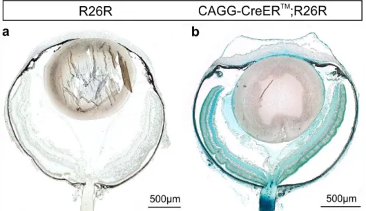
After topical tamoxifen treatment with eye drops, we used β-galactosidase staining to localize Cre-mediated recombination in the eye. Eyes of CAGG Cre-ERTM/R26R mice (Figure 7b) showed an intense β-galactosidase reaction throughout the entire organ while control eyes (R26R) were essentially negative (Figure 7a and Figure 8a, c, e, and g).
Figure 7. Localization and activation of Cre recombinase in the eye following tamoxifen containing eye drops. An intense β-galactosidase staining throughout the entire eye in 14 days old CAGG Cre ERTM/R26R mouse (b) indicates a successful activation of the Cre recombinase in ocular tissue following treatment with tamoxifen eye drops. Control littermates (R26R) (a) did not show a positive reaction.
Experiment performed by Anja Schlecht and Sarah V. Leimbeck.
The detailed analysis of CAGG Cre-ERTM/R26R eyes showed an intense β-galactosidase reaction in the anterior eye segment. We observed in particular a strong β-galactosidase staining in the structures of the chamber angle outflow pathway, in the ciliary body (Figure 8b) and in the cornea, as well as in the epithelium of the lens (Figure 8d). In the posterior eye segment of CAGG Cre-ERTM/R26R eyes, the sensory retina, the retinal pigment epithelium (RPE) and the choroid (Figure 8f) stained positive for β-galactosidase indicating a successful Cre-mediated recombination in basically every ocular cell type. In addition, in sections where the optic nerve was cut, we observed positive staining along the sheaths surrounding the nerve indicating that tamoxifen had been distributed outside the eye (Figure 8h).
21 Figure 8. Detailed localization and activation of Cre recombinase in the eye. Detailed magnification of the β-galactosidase staining in the structures of the chamber angle outflow pathway (b), the cornea and the lens epithelium (d), the retina and choroid (f) and the optic nerve (h) of a 14 days old CAGG Cre- ERTM/R26R mouse. The control littermate did not show a positive reaction for β-galactosidase (a, c, e and g). RGC retinal ganglion cells, INL inner nuclear layer, ONL outer nuclear layer, RPE retinal pigment epithelium, C choroid, ON optic nerve, CB ciliary body, CO cornea, TM Trabecular meshwork, SC Schlemm’s canal, LE lens. Experiment performed by Anja Schlecht and Sarah V. Leimbeck.
22 2.5 Discussion
Our results show that induction of Cre recombinase by using tamoxifen-containing eye drops is a suitable method to induce a tamoxifen-dependent Cre-mediated recombination in ocular tissues.
The topical application of tamoxifen-containing eye drops provides several advantages. As a non-invasive method it greatly reduces or even avoids the potential risk of infections, which might eventually result from intra-peritoneal injections (Leenaars et al. 1998, Leenaars and Hendriksen 2005), which is a common method to administer tamoxifen. Intravitreal tamoxifen injections harbor the same risks of infection. Furthermore this route might influence the expression level of potential genes of interests because intravitreal injection of the vehicle alone already results in the activation of microglia and/or an elevated expression of neuroprotective molecules (Braunger 2014, Seitz and Tamm 2014). In our study, we noticed staining along the optic nerve outside the eye, a finding that appears to indicate that tamoxifen is distributed to tissues outside the eye. One could avoid this and achieve even greater spatial control of Cre expression by reducing the duration of tamoxifen treatment, e.g. from 5 days, to 3 days or maybe even less. Of course, this approach could in turn result in a Cre-mediated recombination gradient in the eye itself. This scenario might be of great interest for scientist focusing on the anterior segment of the eye like the cornea or the chamber angle outflow pathway. Here, a reduced exposure time might reduce the tamoxifen- induced Cre- mediated recombination in other parts of the eye or the body to an even greater extend. As a side note, our system also allows the usage of strong promoters like CMV or β- actin that would drive Cre- expression in every cell. The expression of Cre, however, can be spatially controlled, as the tamoxifen is applied topical. Finally, considering tamoxifen induced toxicity, which may influence cell viability or even promote cell death (Kim et al. 2014), the topical administration of tamoxifen using eye drops could obviously reduce this risk, too. In summary, our approach may be of great interest for scientists in the field of experimental eye research.
23
Chapter 3
Deletion of ocular TGF-β signaling mimics essential characteristics of diabetic retinopathy
(adapted from: Barbara M. Braunger, Sarah V. Leimbeck, Anja Schlecht, Cornelia Volz, Herbert Jägle and Ernst R. Tamm. Deletion of ocular TGF-β signaling mimics essential
characteristics of diabetic retinopathy. Am J Pathol. 2015 Jun;185(6):1749-68)
24
3 Deletion of ocular TGF-β signaling mimics essential characteristics of diabetic retinopathy
3.1 Abstract
Diabetic retinopathy, a major cause of blindness, is characterized by a distinct phenotype. The molecular causes of the phenotype are not sufficiently clear. Here we report that deletion of TGF-β signaling in the retinal microenvironment of newborn mice induces changes that largely mimic the phenotype of non-proliferative and proliferative diabetic retinopathy in humans. Lack of TGF-β signaling leads to the formation of abundant microaneurysms, leaky capillaries, and retinal hemorrhages. Retinal capillaries are not covered by differentiated pericytes, but by a coat of vascular smooth muscle-like cells and a thickened basal lamina. Reactive microglia is found in close association with retinal capillaries. In older animals, loss of endothelial cells and the formation of ghost vessels are observed, findings that correlate with the induction of angiogenic molecules and the accumulation of retinal HIF-1α indicating hypoxia. Consequently, retinal and vitreal neovascularization occurs, a scenario that leads to retinal detachment, vitreal hemorrhages, neuronal apoptosis and impairment of sensory function. We conclude that TGF-β signaling is required for the differentiation of retinal pericytes during vascular development of the retina. Lack of differentiated pericytes initiates a scenario of structural and functional changes in the retina that mimics those of diabetic retinopathy strongly indicating a common mechanism.
25 3.2 Introduction
Diabetic retinopathy is a leading cause of visual impairment and blindness in the developed world (Sivaprasad et al. 2012, Congdon et al. 2003). Clinically, diabetic retinopathy begins with typical microvascular signs in the retina of an individual with diabetes mellitus. The earliest clinical signs of diabetic retinopathy include the formation of microaneurysms, small outpouchings from retinal capillaries, and of dot intraretinal hemorrhages (Frank 2004). Number and sizes of hemorrhages increase as the disease progresses. In addition, the barrier of retinal capillaries becomes impaired which may lead to retinal edema, to the deposition of plasma lipid and lipoprotein contents, and the formation of hard exudates. In parallel to the increase in vascular permeability, occlusion and loss of retinal vessels is observed, a scenario that is thought to cause the formation of ischemic infarcts of the nerve fiber layer (cotton-wool spots).
Because of the high metabolic requirements of retinal neurons, capillary damage in the retina causes hypoxia and the release of potent angiogenic factors such as vascular endothelium growth factor (VEGF). As a result, diabetic retinopathy progresses from a non-proliferative stage to a fibrotic, proliferative stage that is characterized by the growth of new capillaries in retina and vitreous. Diabetic neovascularization leads to preretinal or vitreous hemorrhages, and to tractional retinal detachment caused by proliferative vitreoretinopathy (Cheung et al. 2010, Antonetti et al. 2012).
The characteristic histopathological lesion that occurs early in diabetic retinopathy, is the relative selective loss of pericytes from retinal capillaries (Kuwabara 1963). The loss of pericytes is followed by the loss of capillary endothelial cells and apoptosis is thought to be the mechanism that is largely responsible for the loss of both cell types (Mizutani et al. 1996, Barber et al. 2011).
Similarly, pericyte dropout and acellular occluded capillaries were observed in numerous diabetic animal models (Hammes 2005). Pericyte loss appears to be causatively involved in capillary occlusion as genetically modified mice with specific deficiency of platelet-derived growth factor-β (PDGF- β) in endothelial cells show a varying degree of pericyte loss in parallel with occlusions of retinal capillaries (Hammes 2005). Comparable findings were observed in heterozygous PDGF-β-deficient diabetic mice (Hammes et al. 2002). Still, other characteristics of diabetic retinopathy such as sprouting of retinal capillaries and the formation of microaneurysms are not commonly observed in mice with pericyte dropout (Hammes et al.
2002). Accordingly, besides pericyte dropout, other mechanisms appear to contribute to the structural changes in diabetic retinopathy and immunological processes are likely candidates (Adamis and Berman 2008, Kern 2007).
26 Evidence suggests that transforming growth factor (TGF)-β signaling plays an important role in the proliferation and the differentiation of both pericytes and endothelial cells during development (Armulik et al. 2011). The specific action of TGF-β signaling in this context appears to depend on the respective concentrations of the TGF-β isoforms and the availability of appropriate receptors, and may inhibit or promote proliferation and differentiation of the vascular cell types (Gaengel et al. 2009). In the retina, constitutive TGF-β signaling appears to be important for maintaining the structure and function of retinal capillaries. Accordingly, the inhibition of TGF-β signaling by systemic expression of soluble endoglin results in the apoptosis of vascular cells, the formation of leaky capillaries and the impairment of retinal perfusion (Walshe et al. 2009). Moreover, TGF-β signaling is thought to serve an important immunosuppressive role in the retina (Chytil et al. 2002). Still, the role of TGF-β signaling for the formation and maintenance of the retinal vasculature has only been incompletely studied, since the deletion of most of the different TGF-β signaling pathway genes results in embryonic lethality during midgestation (Armulik et al. 2011, Gaengel et al. 2009). Here we report on the phenotype of mice with a conditional deletion of the essential TGF-β type II receptor (TβRII) in ocular tissues. We provide evidence that impairment of TGF-β signaling in the retinal microenvironment promotes the development of structural changes in the retina that largely mimic the phenotype of non-proliferative and proliferative diabetic retinopathy. Our results point towards an important role of TGF-β signaling in the pathogenesis of diabetic retinopathy.
27 3.3 Materials and Methods
3.3.1 Mice
Mice with two floxed alleles of Tgfbr2 (Chytil et al. 2002) were crossed with Tgfbr2flox/flox;CAGG Cre-ERTM animals that were heterozygous for transgenic CAGG Cre-ERTM. Resulting Tgfbr2flox/flox;CAGG Cre-ERTM animals were used as experimental mice, and littermates with two unrecombined Tgfbr2fl/fl alleles are referred to as control mice. Genetic backgrounds were 129SV (Tgfbr2) or C57Bl6 (CAGG Cre-ERTM). All mice were reared in 12 h light–12 h dark cycles (lights on at 7 am). Genotypes were screened by isolating genomic DNA from tail biopsies and testing for transgenic sequences by PCR. For Cre-PCR analysis primers were: 5'- ATGCTTCTGTCCGTT TGCCG-3' (sense) and 5'-CCTGTTTTGCACGTTCACCG-3' (antisense).
The thermal cycle profile was denaturation at 96°C for 30 s, annealing at 57°C for 30 s, and 1 min extension at 72°C for 35 cycles. For genotyping of TGF-β-R2flox/flox animals, primers were 5’- GCAGGCATCAGGACCCAGTTTGATCC-3’ (sense) and 5’-AGAGTGAAGCCGTGGTAGGT GAGCTTG-3’ (antisense). The thermal cycle profile was denaturation at 96°C for 30 s, annealing at 61°C for 30 s, and 1 min extension at 72°C for 35 cycles. To induce Cre recombinase, mice were treated with tamoxifen (Sigma) eye drops. Tamoxifen was diluted in corn oil to a final concentration of 5 mg/ml. From P4 to P8, eye drops at a volume of 10 µl were pipetted onto the closed eyelids of mouse pups three times a day. All procedures conformed to the tenets of the National Institutes of Health Guidelines on the Care and Use of Laboratory Animals (National Research Council (U.S.) and Institute for Laboratory Animal Research (U.S.) 2011), the EU Directive 2010/63/E, and institutional guidelines, and were approved by the local authority (Regierung der Oberpfalz, Bavaria, Germany, 54-2532.1-44/12).
3.3.2 Western blot analysis
Retinal proteins were isolated following the manufacturer’s instructions (Invitrogen) for TRIzol protein isolation. Proteins were separated by SDS-PAGE (6%, 8% or 10% gels, depending on the molecular weight of the protein of interest) and transferred by semidry blotting onto a polyvinyl difluoride membrane (PVDF, Milipore). PVDF membranes were incubated with TBS containing 0.1% Tween 20 (TBST; pH 7.2) and blocking reagent (Table 1) overnight. Antibodies were used as described in Table 1. After washing with TBS-T, secondary antibodies (1:2000) were added. Chemiluminescence was detected on a LAS 3000 imaging workstation. For normalization, blots were labeled with antibodies against glyceraldehyde 3-phosphate dehydrogenase (GAPDH, 1:10,000, HRP-conjugated, Cell Signaling) or α-tubulin (1:5000,
28 Rockland). Western blots were evaluated by relative densitometry using the Aida Image Analyzer v.4.06 software (Raytest).
Table 1. Antibodies used for Western Blot analysis
Primary antibody Blocking Secondary antibody
TβRII c16 (Santa Cruz) 1:200
5% non-fat dry milk chicken anti-rabbit coupled to horseradish peroxidase (Santa Cruz) pSmad3 (Cell Signaling)
1:200
5% bovine serum albumin chicken anti-rabbit coupled to horseradish peroxidase (Santa Cruz) FGF-2 sc79 (Santa Cruz)
1:200
5% bovine serum albumin chicken anti-rabbit coupled to horseradish peroxidase (Santa Cruz) HIF1- α (Cayman Chemical)
1:200
5% bovine serum albumin chicken anti-rabbit coupled to horseradish peroxidase (Santa Cruz) Collagen IV (Rockland)
1:500
5% bovine serum albumin goat anti-rabbit coupled to alkaline phosphatase (Santa Cruz)
Smooth muscle α-actin (Genetex) 1:500
5% bovine serum albumin goat anti-rabbit coupled to alkaline phosphatase (Santa Cruz)
NG2 (Merck Millipore) 1:1000
5% bovine serum albumin chicken anti-rabbit coupled to horseradish peroxidase (Santa Cruz) GAPDH (Cell Signaling)
1:10000
5% non-fat dry milk directly horseradish peroxidase coupled α-tubulin (Rockland) 1:5000 5% bovine serum albumin goat anti-rabbit coupled to alkaline
phosphatase (Santa Cruz)
3.3.3 Microscopy and morphometry
Mice were deeply anesthetized with ketamine (120 mg/kg body weight, i.m.) and xylazine (8 mg/kg body weight), and perfused with 1 ml of modified Karnowsky’s fixative (Karnovsky 1965) through the left ventricle of the heart. Eyes and optic nerves were isolated, postfixed for 24 h in the same fixative and embedded in Epon (Serva) as described elsewhere (Kritzenberger et al.
2011). Semi-thin sagittal sections (1.0 µm thick) were cut through the eyes and stained after Richardson (Richardson et al. 2009). Transmission electron microscopy was performed according to protocols published previously (Braunger et al. 2013a). Semi-thin sections of retinae and cross semi-thin sections through the optic nerve were stained with paraphenylenediamine (PPD) as described previously (Kroeber et al. 2010) to visualize lipid deposits or myelinated axons, respectively. For quantification of optic nerve axons, the number
29 of PPD-labeled axons in optic nerve cross sections was counted using ImageJ Cell counter as described previously (Braunger et al. 2013b). Thickness of the outer nuclear layer (ONL) was measured on semi-thin sections along the mid-horizontal (nasal-temporal) plane. The distance between ora serrata (OS) and optic nerve head (ONH) was divided into tenths and measured between each tenth (Braunger et al. 2013b). Fluorescence-labeled retinal whole mounts or sagittal sections were investigated on an Axiovision fluorescent microscope (Carl Zeiss) using appropriate Axiovision software 4.8 (Carl Zeiss, Jena, Germany). Analysis of retinal whole mounts by confocal microscopy was performed on a Zeiss Axiovert 200M inverted microscope combined with a LSM 510 laser-scanning device. FITC-dextran was excited using an Ar-laser at 488 nm, CyTM-3 was excited using a HeNe laser at 543nm. The software used for image acquisition and processing was AIM 4.2 (Zeiss).
3.3.4 LacZ staining
Lac-Z-staining was performed in mixed CAGG CreERTM/Rosa-LacZ (Cre-reporter) or Tgfbr2Δeye/TOP-Gal (WNT-reporter) mice (The Jackson Laboratory) as described previously (Braunger et al. 2013a, Baulmann et al. 2002). Paraffin sections (6.0 µm) were cut, placed on glass slides (SuperFrost/Plus; Menzel) and analyzed by light microscopy (Carl Zeiss).
3.3.5 Immunohistochemistry
Prior to TβRII, pSmad3, glial fibrillary acid protein (GFAP), collagen IV and CD31 staining, eyes were fixed for 4 h in 4% paraformaldehyde (PFA), washed extensively in phosphate buffer (PP, 0.1M) and embedded in paraffin according to standard protocols. Paraffin sections (6 µm) were deparaffinized and washed in H2O. For detection of TβRII and pSmad3, sections were treated with boiling citrate buffer (1 x 10 min, ph 6), washed again in H2O and incubated in 0.1M PP. For detection of collagen IV and CD31, sections were pretreated with 0.05 M Tris-HCL (5 min) and covered with Proteinase K (100 µl of Proteinase K in 57 ml Tris-HCl (0.05 M), 5 min), washed in H2O, incubated in 2 N HCl (20 min), and washed again in H2O. Sections were incubated in PP for 5 min. IBA-1, F4/80 and α-smooth muscle-actin (α-SMA) immunohistochemistry was performed on frozen sections. For Iba-1 and α-SMA staining, eyes were fixed for 4 h in 10%
glacial acetic acid, 60% methanol, and 30% chloroform, transferred to 50% and 25% methanol 30 min each, washed in phosphate buffered saline (PBS), incubated in 10%, 20%, 30%
sucrose/PBS overnight at 4°C and shock frozen in tissue mounting medium (DiaTec). For F4/80 staining, eyes were fixed for 4 h in 4% PFA and washed extensively in phosphate buffer (PP,
30 0.1M). For immunohistochemistry, sections were washed three times in TBS (pSmad3) or PP (others) for 5 min each and blocked with 2% BSA in PP/TBS 45 min (F4/80: 1% dry milk, 0.01%
Tween in PBS) at room temperature. Primary antibodies (Table 2) were diluted in a 1:10 dilution of blocking solution in PP/TBS (F4/80: 2% BSA, 0.02% NaN3, 0.01% Triton in PBS) and incubated at 4°C overnight. After three washes in PP/TBS (5 min each), biotinylated antibodies were applied for 1 h diluted in a 1:10 dilution of the blocking solution as an additional step, then anti-biotin or appropriate secondary antibodies (Table 2), diluted in a 1:10 dilution of the blocking solution, were applied for 1 h. Sections were washed again three times and cell nuclei were counterstained with DAPI (Vectashield, Vector Laboratories) 1:10 diluted in fluorescent mounting medium (Serva).
Table 2. Antibodies used for immunohistochemistry
Primary antibody Fixation Secondary antibody
TβRII- L21 (Santa Cruz) 1:20
4% paraformaldehyde (PFA)
anti-rabbit, biotinylated (Vector) 1:500, Streptavidin Alexa 488 (Invitrogen) 1:1000 pSmad3 (Cell Signaling)
1:20
4% PFA anti-rabbit, biotinylated (Vector) 1:500, Streptavidin Alexa 488 (Invitrogen) 1:1000 Iba-1 (Wako) 1:1000 methyl-Carnoy anti-rabbit CyTM-3 conjugated (Jackson
Immuno Research Lab) 1:2000 Collagen IV (Chemicon)
1:100
4% PFA anti-rabbit, biotinylated (Vector) 1:500, Streptavidin Alexa 546 (Invitrogen) 1:1000 α-smooth muscle-actin
(Genetex) 1:50
methyl-Carnoy (section) methanol (whole mount)
anti-rabbit CyTM-3 conjugated (Jackson Immuno Research Lab) 1:2000
NG2 (Merck Milipore) 1:50 methanol anti-rabbit CyTM-3 conjugated (Jackson Immuno Research Lab) 1:2000
CD31 (RD Systems) 1:100 4% PFA anti-goat CyTM-3 conjugated (Jackson Immuno Research Lab) 1:2000
Collagen IV (Abcam) 1:100 4% PFA anti-rabbit Alexa 488 (Invitrogen) 1:1000 F4/80 (Acris Antibodies)
1:600
4% PFA anti-rat CyTM-3 conjugated (Jackson Immuno Research Lab) 1:2000 in PBS
31 3.3.6 Dextran perfusion and retinal whole mounts
Mice were deeply anesthetized with ketamine (120 mg/kg body weight, i.m.) and xylazine (8 mg/kg body weight) and perfused through the left ventricle with 1 ml of phosphate buffered saline (PBS) containing 50 mg high molecular weight (MW = 2,000,000) FITC-dextran (TdB Consultancy). The eyes were enucleated and placed in 4% paraformaldehyde for 2 h. Retinae were dissected and flat mounted using fluorescent mounting medium (Serva). If sagittal sections of FITC-dextran-perfused retinae were used, the perfused eyes were fixed for 4 h in 10% glacial acetic acid, 60% methanol, and 30% chloroform, transferred to 50% and 25% methanol 30 min each, washed in phosphate-buffered saline (PBS), incubated in 10%, 20%, 30% sucrose/PBS overnight at 4°C and shock frozen in tissue mounting medium (DiaTec). Following sectioning, nuclei were counterstained with DAPI (Vectashield, Vector Laboratories) 1:10 diluted in fluorescent mounting medium (Serva). If immunhistochemical labelling was performed using retinal whole mounts, fixation was modified (Tab. 2), and antibodies were applied according to Tab. 2 and as described in the immunhistochemistry section.
3.3.7 Apoptosis
Apoptotic cell death was analyzed by terminal deoxynucleotidyl transferase-mediated dUTP nick end labeling using the Apoptosis Detection System (DeadEnd Fluorometric TUNEL, Promega).
Paraffin sections were treated following manufacturers’ instructions and protocols reported previously (Braunger et al. 2013b). For quantitative analysis, the number of TUNEL-positive nuclei in sagittal sections throughout the entire retina was counted.
3.3.8 Trypsin digest
Eyes were fixed for 48 h in 4% PFA. Retinae were washed in H2O for 75 min, transferred to glass slides and incubated for 90 min with 3% trypsin 0.2 M Tris-HCl at 37°C. Retinae were transferred on a new glass slide, again incubated for 120 min in 3% trypsin 0.2 M Tris-HCl at 37°C and stained with periodic acid–Schiff (PAS).
GFAP (DAKO) 1:1000 4% PFA anti-rabbit Alexa 488 (Invitrogen) 1:1000


