Eur J Clin Chem Clin Biochem 1997; 35(5):365-367 © 1997 by Walter de Gruyter · Berlin · New York
Anti-Insulin Antibodies in Insulin Immunometric Assays:
A Still Possible Pitfall
Remy Sapin
Laboratoire Universitaire de Biophysique, CNRS URA 1173, Faculte de Medecine, Strasbourg, France
Summary: Insulin was assayed directly using radioimmunoassay and immune-metric assay in 31 sera containing anti-insulin antibodies. Anti-insulin antibodies were determined by radio-binding-assay. Insulin measurements were compared with those of free (unbound to antibodies, polyethylene glycol precipitated) insulin measurements. Com- pared with free insulin concentrations, radioimmunoassay and immunometric assay yielded falsely increased insulin results. The degree of overestimation by radioimmunoassay and by immunometric assay correlated with the anti- insulin antibody value. Anti-insulin antibodies still remain a possible pitfall in the insulin-specific immunometric assays which are now being widely used.
Introduction
The availability of monoclonal antibodies to well-de- fined sequences has made it possible to develop non- competitive two-site immunometric assays with a high specificity for the intact insulin molecule (1). Compared with previous competitive one-site radioimmunoassays (2) these new assays, now commercially available, have greatly improved the specificity and sensitivity of plasma insulin measurements (3). However, as recently reviewed by Crowthers et al. (4), many problems are still encountered in measuring insulin: sample collection and storage, standards and quality control samples and matrix effects. Insulin radioimmunoassay measurements were also well-known to be falsified by endogenous anti-insulin antibodies (5, 6). Anti-insulin antibodies are produced during treatment with porcine or human insu- lin, and anti-insulin autoantibodies are present in predia- betes before any treatment with insulin (7). The aim of this study was to establish whether anti-insulin antibod- ies interfere with the now widely used insulin immuno- metric assays and thus influence the interpretation of insulin concentration.
Material and Methods Methods
The presence of free anti-insulin antibodies (not complexed with circulating insulin) was determined using the radioimmunopreci- pitation technique in liquid phase: radio-binding-assay (Sanofi Pasteur, Mames la Coquette, France). As recommended by in- ternational workshops (8) this assay uses a monocomponent A 14 human insulin tracer. The positivity threshold was fixed by the manufacturer at 5.5%. A binding percentage greater than this positivity threshold indicates the presence of anti-insulin antibod- ies.
Insulin concentrations were determined with a radioimmunoassay kit from Sanofi Pasteur involving a guinea-pig polyclonal antibody.
The percentage of cross reaction with proinsulin is 40%. Insulin concentrations were further determined with a microparticle en- zyme immunometric assay kit (Abbott, Abbott Park, USA) which allows insulin determination in the absence of cross-reactivity with proinsulin and proinsulin-like molecules (9). This method is fully automated on the IMx system (Abbott). Both methods are cal- ibrated against the WHO 66/304 standard.
Free (unbound to anti-insulin antibodies) insulin was determined with the radioimmunoassay kit after precipitation of endogenous immune complexes by polyethylene glycol (10). Free insulin repre- sents the biologically active form of insulin.
Specimens
We selected 31 sera from insulin-treated insulin-dependent diabetic patients. These samples contained anti-insulin antibodies in propor- tions determined by radio-binding-assay results ranging from 6.1 to 67%.
Results
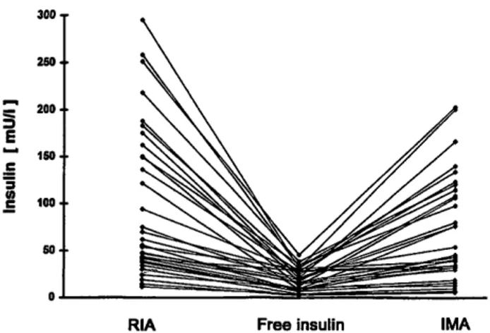
Radioimmunoassay insulin values ranged from 18.3 to 295 mU/1, immunometric assay values from 5.7 to 220.5 mU/1, and free insulin concentrations from 2 to 45.5 mU/1 (fig. 1). Comparison of the paired radioimmuno- assay or immunometric assay results with free insulin concentrations showed a significant fall (t = 6.25, p = 1 · 10~7 and t = 5.82,/? = 2 · 10~6, respectively). Com- pared to radioimmunoassay, immunometric results were significantly lower (t = 3.95, p = 4 · 10~4).
A strong correlation was observed between the overesti- mation of insulin concentration, measured by either radioimmunoassay or immunometric assay, determined as 100 X ([insulin] — [free insulin])/[free insulin] and the percentage of anti-insulin antibodies measured by radio-binding-assay (fig. 2). The correlation was similar for radioimmunoassay (r = 0.79) and immunometric as- say (r = 0.77). The degree of interference, as appreci- ated by the slope of the regression line, was greater with
366 Sapin: Anti-insulin antibodies and insulin immunometric assays 300 τ
150·
RIA Free insulin IMA
Fig. 1 Comparison of insulin measured by radioimmunoassay (RIA) and by immunometric assay (IMA) with free (polythylene glycol precipitated) insulin concentration in 31 anti-insulin anti- body positive sera.
1400
1200
1000
30 60
Anti-Insulin antibodies [ % ]
Fig. 2 Association of insulin overestimation determined as 100 X ([insulin] — [free insulin])/[free insulin] with anti-insulin antibody percent determined by radio-binding-assay. Insulin measured by radioimmunoassay (dark circles) and immunometric assay (open circles).
radioimmunoassay (y = 14.2x + 120) than with immu- nometric assay (y = 9.4x + 47).
Discussion
It was demonstrated, long ago, that anti-insulin antibod- ies interfere with the radioimmunoassay of insulin. The effect of endogenous anti-insulin antibodies is to increase the total amount of labelled insulin bound to endogenous human and exogenous guinea-pig antibod- ies and to reduce the amount of labelled insulin bound to exogenous guinea-pig antibodies. The results of com- petitive one-site radioimmunoassay depend on the sepa- ration method used. Non-specific precipitation of immu- noglobulin complexes, with, for example, polyethylene glycol, leads to false underestimated values due to over- precipitation of the bound tracer. The double-antibody, involved in the Sanofi radioimmunoassay used in this study, or coated tube technology, used in today's radio- immunoassays, results in overestimation, as too little of the tracer is specifically bound.
Our results show clearly that anti-insulin antibodies can also interfere with insulin immunometric determin- ations. Immunometric assay recognised, at least par- tially, insulin bound to endogenous antibodies, yielding increased insulin values as compared with free insulin concentrations. The degree of overestimation in an im- munometric assay depends, at least theoretically and es- pecially for small analyte molecules with a limited number of antibody binding sites, such as insulin, on the comparative avidity of autoantibodies and of the catcher and tracer antibodies used in the assay. If the avidity of the assay antibodies exceeds that of autoantibodies, the measured concentration will tend towards the total (free and antibody-bound) insulin concentration, i. e. yield an overestimation of the biologically active (free) insulin in the sample. But, if the assay antibodies are less avid than autoantibodies, the measured concentration will tend towards the free insulin concentration, as little or no displacement of the autoantibody-insulin complex takes place during the incubation time.
This may explain why the Abbott immunometric assay was more sensitive to interference from anti-insulin anti- bodies than the enzyme immunoassay (ELISA) studied by Andersen et al. (11) who observed insulin concentra- tions close to free insulin in sera containing insulin anti- bodies with a bound fraction below 25%. Under the same conditions, with the Abbott kit, the mean overesti- mation was 165%.
In sera containing anti-insulin antibodies, when com- pared with free insulin concentrations, insulin results by immunometric assay are overestimated, but, when com- pared with total insulin levels, they may be underesti- mated (12). It is generally stated that anti-analyte anti- bodies may yield a positive interference (overestima- tion) when the result is compared with the free analyte concentration (insulin assay for example) or a negative interference (underestimation) when the result is com- pared with the total analyte concentration (thyroglobulin assay for example) (13). The relevant analyte concentra- tion is, in the first case, that which corresponds to its biologically active form (free form), and in the second case, that which shows a residual secretion by thyroid after thyroidectomy (total form).
Conclusion
Interference from anti-insulin antibodies, yielding increased values, still remains a topical pitfall in insulin immunometric assays. Care must be taken in the inter- pretation of immunometric insulin results when anti-in- sulin antibodies are present.
The solid phase antigen immunoassay described by Wood et al. (14) could be an interesting approach to set- tle the issue of an antibody interference-free assay, be-
Sapin: Anti-insulin antibodies and insulin immunometric assays 367
cause free thyroxin assays based on this methodology have been shown to be much less sensitive to anti-thy- roxin autoantibody interference than previous labelled antigen immunoassays (15).
Acknowledgements
The author is very grateful to Mrs. Nathalie Heider for reviewing the English of this manuscript.
References
1. Sobey WJ, Beer SF, Canington CA, Clark PMS, Frank BH, Gray IP, et al. Sensitive and specific two-site immunoradiome- tric assays for human insulin, proinsulin, 65—66 split and 32—
33 split proinsulins. Biochem J 1989; 260:535-41.
2. Yalow RS, Berson SA. Assay of plasma insulin in human sub- jects by immunological methods. Nature 1959; 184:1648—9.
3. Sapin R. Recent insulin immunoassays: improvements and limits. J Clin Ligand Assay 1996; 19 (1 Suppl):100S-110S.
4. Crowthers NJ, Gray IP. Immunometric assays of insulin and its precursors. J Clin Ligand Assay 1996; 19:112-20.
5. Armitage M, Wilkin T, Wood P, Casey C, Loveless R. Insulin autoantibodies and insulin assay. Diabetes 1988; 37:1392-6.
6. Clark PMS, Hales CN. How to measure plasma insulin. Diabe- tes/Metabolism Reviews 1994; 10:79-90.
7. Sodoyez JC, Koch M, Sodoyez-Goffaux F. Anticorps anti-insu- line: methodologie et implications cliniques. Diabetes Metab 1991; 17:255-69.
8. Kuglin B, Kolb H, Greenbaum C, Maclaren NK, Lernmark Ä, Palmer JP. The fourth international workshop on the standardi- sation of insulin autoantibody measurement. Diabetologia
1990; 33:638-9.
9. Monti LD, Sandoli EP, Phan VC, Piatti PM, Costa S, Secchi A, et al. A sensitive and reliable method for assaying true hu- man insulin without interaction with human proinsulin-like molecules. Acta Diabetol 1995; 32:57-63.
10. Arnqvist H, Olsson PO, von Schenck H. Free and total insulin as determined after precipitation with polyethylene glycol: an-
alytical characteristics and effects of sample handling and stor- age. Clin Chem 1987; 33:93-6.
11. Andersen L, Dinesen B, Jorgensen PN, Poulsen F, Roder M.
Enzyme immunoassay for intact human insulin in serum or plasma. Clin Chem 1993; 39:578-82.
12. Gennaro WD, Van Norman JD. Quantitation of free, total, and antibody-bound insulin in insulin-treated diabetics. Clin Chem 1975; 21:873-9.
13. Spencer CA, Takeuchi M, Kazarosyan M. Current status and performance goals for serum thyroglobulin assays. Clin Chem
1996; 42:164-73.
14. Wood WG, Fricke H, von Klitzing L, Strasburger CJ, Scriba PC. Solid phase antigen luminescent immunoassays (SPALT) for the determination of insulin, insulin antibodies and genta- micin levels in human serum. J Clin Chem Clin Biochem
1982; 20:825-31.
15. Sheehan CP, Cristofides ND. One-step, labeled antibody assay for measuring free thyroxin. II. Performance in a multicenter trial. Clin Chem 1992; 38:19-25.
Received January 3/February 17, 1997
Corresponding author: Dr. Remy Sapin, Institut de Physique.
Biologique, Faculte de Medecine, 4 rue Kirschleger, F-67085 Strasbourg Cedex, France