zur
Erlangung der Doktorwürde der
Naturwissenschaftlich-Mathematischen Gesamtfakultät
der
Ruprecht-Karls-Universität Heidelberg
vorgelegt von
Dipl.-Phys. Johann Heinrich Bollmann aus Bremen
Tag der mündlichen Prüfung: 4. Juli 2001
Ausschüttung in einer glutamatergen Synapse des Zentralnervensystems
Gutachter: Prof. Dr. Bert Sakmann Prof. Dr. Christoph Cremer
submitted to the
Combined Faculties for the Natural Sciences and for Mathematics of the
Rupertus Carola University of Heidelberg, Germany
for the degree of Doctor of Natural Sciences
Calcium sensitivity of neurotransmitter release in a glutamatergic synapse of the central nervous system
presented by
Diplom-Physicist Johann Heinrich Bollmann born in Bremen/Germany
Heidelberg, July 4th, 2001 Referees: Prof. Dr. Bert Sakmann
Prof. Dr. Christoph Cremer
Calcium sensitivity of neurotransmitter release in a glutamatergic synapse of the central nervous system
During chemical synaptic transmission, the presynaptic action potential couples to the biochemical release process by the opening of Ca2+ channels and Ca2+-dependent activation of a release sensor that triggers the release of transmitter. Here, the dependence of transmitter release on the intracellular Ca2+
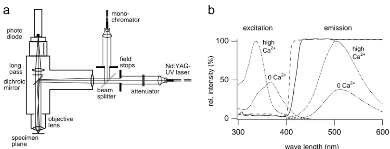
concentration ([Ca2+]) was determined in a glutamatergic calyx-type synapse in slices of the rat brainstem by UV-induced Ca2+ uncaging. Because of the fast speed of glutamatergic synapses, an electrophysiological setup was combined with a rapid fluorescence detection system and a short-pulsed UV laser in order to both evoke and measure uniform [Ca2+] elevations on a fast time scale.
A homogeneous rise in the presynaptic [Ca2+] to 1 µM resulted in a clearly measurable increase in release. The peak release rates depended on presynaptic [Ca2+] with more than the fourth power. A [Ca2+] jump to 30 µM or more depleted the releasable vesicle pool in less than 0.5 ms. A kinetic model was devised to quantify the release rate-[Ca2+] relation measured in this synapse type. A comparison with action potential evoked release in the same synapses suggested that a brief elevation of [Ca2+] to less than 10 µM would be sufficient to reproduce the physiological release pattern. In summary, the Ca2+ sensitivity of synaptic transmitter release is, at least in some synapses, higher than previously thought.
Die Kalziumempfindlichkeit der Überträgerstoff-Ausschüttung in einer glutamatergen Synapse des Zentralnervensystems
Der Signalübertragung an chemischen Synapsen liegt ein Kopplungsmechanismus zwischen dem präsynaptischen Aktionspotential und der biochemischen Überträgerstoff-Ausschüttung zu Grunde.
Dabei werden Ca2+-Kanäle geöffnet und ein Freisetzungssensor Ca2+-abhängig aktiviert, der schließlich die Überträgerstoff-Ausschüttung auslöst. In der vorliegenden Arbeit wurde die Abhängigkeit der Überträgerstoff-Ausschüttung von der intrazellulären Ca2+-Konzentration ([Ca2 +]) in einer glutamatergen kelchförmigen Synapse in Stammhirnschnitten der Ratte unter Verwendung photolytischer Ca2+-Freisetzungen gemessen. Um [Ca2+]-Sprünge auf einer Zeitskala sowohl hervorrufen als auch messen zu können, die der schnellen Übertragungsgeschwindigkeit von glutamatergen Synapsen vergleichbar ist, wurde ein elektrophysiologischer Messstand mit einem schnellen Fluoreszenzdetektor und einem UV-Kurzpulslaser ausgestattet.
Ein deutlich messbarer Anstieg der Überträgerstoff-Ausschüttung wurde bereits bei einer homogenen Erhöhung der präsynaptischen [Ca2+] von ca. 1 µM beobachtet. Der Spitzenwert der Freisetzungsrate wuchs mit mehr als der vierten Potenz der präsynaptischen [Ca2+]-Amplitude. Ein [Ca2+]-Sprung von mehr als 30 µM löste die Aktivierung aller zur Fusion unmittelbar bereitstehenden Vesikel innerhalb von 0,5 ms aus. Die in dieser Synapse beobachtete Beziehung zwischen der Freisetzungsrate und der präsynaptischen [Ca2+] wurde mit Hilfe eines kinetischen Modells quantitativ beschrieben. Ein Vergleich der Modellvorhersagen mit Freisetzungsraten, die in denselben Synapsen während eines Aktionspotentials gemessen worden waren, ergab, dass ein kurzer Anstieg der [Ca2+] auf weniger als 10 µM ausreicht, um den physiologischen Freisetzungsverlauf zu erklären. Die synaptische Überträgerstoff-Ausschüttung reagiert somit zumindest in manchen synaptischen Systemen empfindlicher auf Ca2+ als bisher angenommen.
Contents
1. Introduction
...11.1 Fundamental principles of neural signal processing...1
1.1.1 Electrical signaling in the neuron ... 1
1.1.1.1 Neurons as structural and functional units of the nervous system ... 1
1.1.1.2 Neuronal excitability: resting and action potentials... 2
1.1.2 Synapses ... 4
1.1.3 Synaptic plasticity ... 6
1.2 Exo- and endocytosis...8
1.2.1 Vesicle cycling ... 8
1.2.2 Exocytosis... 10
1.2.2.1 Some molecules involved in exocytosis ... 10
1.2.2.2 The fusion mechanism ... 11
1.2.3 Endocytosis... 12
1.2.4 The role of Ca2+ in exocytosis and endocytosis ... 13
1.3 Motivation...14
2. Theory
...172.1 Intracellular Ca2+ dynamics...17
2.1.1 Time-dependent Ca2+ diffusion-reaction in an aqueous medium ... 19
2.1.2 Ca2+ microdomains ... 21
2.1.3 Simulation of laser-induced [Ca2+] jumps ... 23
2.2 Fluorescence...27
2.3 Kinetic model of vesicle fusion...30
3. Methods
...333.1 Experimental setup...33
3.1.1 Optical components... 34
3.1.1.1 Upright microscope, infrared video microscopy of brain slices ... 34
3.1.1.2 Laser and monochromator... 35
3.1.1.3 Homogeneity of illumination, energy attenuation... 37
3.1.1.4 Fast photodetection with photodiode, photodiode holder ... 38
3.1.2 Electrophysiological components ... 41
3.1.2.1 Pre- and postsynaptic whole-cell voltage clamp... 41
3.1.2.2 Bandwidth and fidelity ... 42
3.2 Extraction of transmitter release rates from compound EPSCs...45
3.3 Preparation and electrophysiological recordings...47
3.3.1 Brain slice preparation, stimulation and extracellular solutions... 47
3.3.2 Whole-cell recordings, intracellular solutions... 48
3.3.3 Adjustment of the DM-nitrophen – CaCl2 equilibrium ... 50
3.4 Optically controlled, intracellular [Ca2+] jumps...50
3.4.1 Ratiometric [Ca2+] measurements... 51
3.4.2 Rapid [Ca2+] elevations, evoked by laser photolysis... 54
3.4.2.1 Choice of the UV-sensitive Ca2+ chelator... 54
3.4.2.2 [Ca2+] uncaging dynamics ... 56
3.4.3 Implementation of a kinetic model for Ca2+ uncaging ... 57
4. Results
...614.1 Temporal analysis of [Ca2+] uncaging in microcuvettes...62
4.1.1 Ca2+ uncaging in the presence of different Ca2+ buffers and indicators ... 62
4.1.2 A refined model of Ca2+ uncaging with DM-nitrophen ... 62
4.2 Electrophysiological characterization of glutamate release...64
4.2.1 Multi-quantal excitatory postsynaptic currents in the giant synapse... 65
4.2.2 Miniature excitatory postsynaptic currents ... 67
4.2.3 Pool size estimate with EPSC trains... 68
4.3 [Ca2+] dependence of glutamate release...72
4.3.1 Glutamate release evoked by UV-induced [Ca2+] jumps ... 72
4.3.2 [Ca2+] dependence of rates and delays of glutamate release... 73
4.3.3 Kinetic model of glutamate release ... 75
4.3.3.1 Release promoter model with five Ca2+ binding steps ... 75
4.3.3.2 Estimate of [Ca2+] during presynaptic action potentials... 77
4.3.3.3 Dependence of EPSCs on extracellular [Ca2+]... 79
4.3.4 Dependence of glutamate release rates on resting [Ca2+] level... 81
5. Discussion
...835.1 Summary...83
5.2 Methodological aspects...84
5.2.1 Optically controlled [Ca2+] elevations in small volumes ... 84
5.2.1.1 Homogeneity of the evoked [Ca2+] jump ... 84
5.2.1.2 Estimate of error for ratiometric [Ca2+] measurements... 84
5.2.1.3 Comparison of predicted release rates with [Ca2+] steps and [Ca2+] spikes ... 86
5.2.2 Methods for measuring exocytosis ... 90
5.2.2.1 Postsynaptic currents, other methods... 90
5.2.2.2 Saturation of postsynaptic receptors ... 91
5.3 Physiology...95
5.3.1 The model of glutamate release ... 95
5.3.1.1 General remarks... 95
5.3.1.2 Asynchronous release and facilitation ... 96
5.3.2 Molecular candidates for the neuronal Ca2+ sensor...100
5.3.3 Exocytosis in other synaptic and endocrine preparations ...102
5.3.3.1 Other synaptic preparations ...102
5.3.3.2 Slow exocytosis in endocrine cells and neurons ...106
5.4 Outlook...107
References...109
Danksagung...123
1. Introduction
Emerging from the two-sided nature of electro- and biochemical neural signaling, tools from originally distinct scientific disciplines are often combined to successfully elucidate the underlying mechanisms. In the present study, a biophysical question is approached with a combination of techniques from electrophysiology, fluorescence microscopy and photochemistry. A detailed description of the methods applied is provided in chapters 2 and 3. The present study draws its motivation from a physiological framework, which shall briefly be introduced in the present chapter.
1.1 Fundamental principles of neural signal processing
The nervous system is composed of cells, which are classified into the group of excitable neurons and non-neuronal glia cells. The human brain consists of about 1010 - 1012 neurons, while the number of glia cells may be more than ten-fold larger. The network properties of nervous systems arise from the neurons’ capability to receive information, to process input and to send resulting information to other neurons. This section will briefly describe the electrical and chemical processes that enable neurons to communicate within a network.
1.1.1 Electrical signaling in the neuron
1.1.1.1 Neurons as structural and functional units of the nervous system
Neuronal cells represent the basic structural elements of biological networks. The cell boundary is defined by a phospholipid bilayer, the plasma membrane, which hosts numerous proteins such as ion channels and transporters. Thus, a neuron is a closed system that can interact with its environment by exchange of substances, energy and information. Although neurons in the central nervous system exhibit a great morphological diversity, they generally possess similar structural features, which were initially proposed to define the preferred direction of information flow within a neuronal network. A century ago, Ramón y Cajal introduced the concept of ‘dynamic polarization’, in which a neuron receives electrical excitation via its finely branched dendrites and sends it to other, receiving neurons via its axon (Fig. 1.1). The sending
neuron contacts the receiving cell at specialized connections, the synapses, at which electrical excitation is transmitted either chemically or electrically (see section 1.1.2).
The soma of the receiving cell integrates the pattern of electrical signals presented by its dendritic tree and, if a threshold is reached, generates an action potential (see next section), which travels along the axon and activates synapses to communicate the information to succeeding neurons.
This unidirectional model successfully describes a principal pathway of information processing in biological networks. However, later observations demanded several extensions to the early concept of neural information flow. Firstly, synaptic contacts not only exist in the classical axon-to-dendrite arrangement (axo-dendritic), but were found also between the axon and the soma (axo-somatic), the two axons (axo-axonic) or between the dendrites (dendro-dendritic) of connected cells. Moreover, neurons often form reciprocal connections, i.e. the output of the receiving cell is communicated back to the sending neuron (Fig. 1.1 b). Furthermore, the somatic action potential was shown to propagate into the dendritic tree, presenting a feedback signal to the ‘receiving’ elements of the cell (Stuart and Sakmann, 1994). Finally, dendrites probably function as independent integrating units of synaptic activity and may generate local regenerative signals (Larkum et al., 1999).
1.1.1.2 Neuronal excitability: resting and action potentials
In biological neural networks, information is encoded and communicated as changes in the membrane potential. The functional basis of potential changes are the properties of the plasma membrane, which is impermeable to ion movement in its purely lipid phase, but possesses numerous proteinaceous ion channels and ion transporters that mediate ion fluxes across the membrane. Ion channels form an aqueous pore, through which ions can diffuse passively. The pore opening may be gated by the surrounding ionic environment, by the membrane potential or by specific ligands. Transporters carry ions actively, i.e. requiring consumption of chemical energy, across the membrane and generate concentration gradients between the intra- and extracellular space. Typical concentration gradients are ([ion]int / [ion]ext): 12 mM / 145 mM for Na+, 155 mM / 4 mM for K+, 10-4 mM / 1.5 mM for Ca2+ and 4 mM / 123 mM for Cl- (Dudel et al., 1996). The concentration gradients give rise to passive ion transport through open ion channels. Since most of the time the permeability for K+ is higher than for Na+, a dynamic equilibrium state evolves that is close to the Nernst potential for the concentration gradient of K+. This equilibrium potential is termed the resting potential and usually ranges between -60 and -90 mV in neurons.
Fig. 1.1: Neural signal processing. (a) Schematic representation of two pyramidal neurons, connected by an axo-dendritic synapse. Preferred direction of signal propagation indicated by solid arrows. Input from dendrites is integrated at the soma of neuron A. Suprathreshold excitation generates an action potential, which travels along the axon and activates the synapse at the basal dendrite of neuron B. If the integrated input at the soma of neuron B exceeds a threshold, an action potential is initiated, which propagates into the axonal tree of B. Concurrently, somatic action potential initiation leads to a feedback signal in the dendritic tree, the back- propagating action potential (open arrows). (b) Light-microscopic reconstruction of two reciprocally connected pyramidal neurons (blue and red, respectively). Discs indicate synaptic contacts. Image kindly provided by Dr. O. Ohana.
Electrical activity in a neuron is characterized by the initiation and propagation of
‘action potentials’, which are stereotypic all-or-none events, being the elementary units of neuronal information processing. The action potential is generated by the self- amplifying opening of Na+ channels if the membrane is depolarized above a threshold potential of approximately –50 mV. The membrane potential rapidly rises towards the Nernst potential for Na+ of about +70 mV due to the increased Na+ permeability. The rise to positive potentials peaks at ca. 40 mV and is invariably terminated by the rapid inactivation of the Na+ channels and by the delayed opening of K+ channels, which initiates the repolarization towards the resting potential. Neuronal action potentials typically last one millisecond or less, and the triggering of a second action potential requires the transition of inactivated Na+ channels into the resting closed state, which may last a few milliseconds. Because of this refractory period, the rate at which neurons can ‘fire’ action potentials is limited to 100-1000 Hz. Generally, action potentials are triggered at the soma or axon hillock owing to its high Na+ channel density, which admits a brief and strong depolarizing Na+ current once the summed
b a
apical dendritic tree
basal dendrites
axonal tree
soma
synapse B
A
synaptic potentials exceed the activation threshold. The action potential travels from the initiation site along the axon towards the nerve terminals by local membrane depolarization and Na+ channel activation. Due to the Na+ channel inactivation, action potentials are not reflected.
1.1.2 Synapses
Synapses are contact sites between two neurons at which electrical activity is transmitted from one neuron to the other. At a synapse, the plasma membranes of the connected neurons are separated by a narrow cleft of ca. 20 nm thickness. During synaptic transmission, the excited, ‘presynaptic’ neuron activates the transmission process and sends a signal to the ‘postsynaptic’ neuron, either in terms of a chemical transmitter substance (chemical synapse) or in terms of an ionic current through membrane spanning, conducting elements (electrical synapse).
A chemical synapse is characterized by pre- and postsynaptic membrane specializations; the presynaptic ‘active zone’ is a region of electron dense material close to the presynaptic membrane and contains clusters of small vesicles (30 –50 nm in diameter) filled with the transmitter substance (Fig. 1.2). Voltage-dependent Ca2+
channels located in or near the active zone mediate local increases in presynaptic [Ca2+] near the vesicles. The ‘postsynaptic density’ is an electron dense thickening of the postsynaptic membrane, which contains ion-permeable receptor channels that are gated by the transmitter substance.
During synaptic transmission, the presynaptic active zone is depolarized and voltage- dependent Ca2+ channels are opened by an incoming action potential. The resultant local [Ca2+] elevation triggers the fusion of vesicles with the presynaptic membrane.
Transmitter released from the vesicles rapidly diffuses across the synaptic cleft and activates the opening of postsynaptic ‘ionotropic’ receptors, thus changing the postsynaptic permeability for selected ion species. Fast chemical transmission occurs within a time window of less than a millisecond from the arrival of the presynaptic action potential to the start of the postsynaptic electrical response.
Chemical synapses can be excitatory or inhibitory, depending on the transmitter and postsynaptic receptor channel types. Excitatory synapses use transmitters such as acetylcholine or glutamate, which activate cation-selective receptor channels. The influx of cations (Na+, Ca2+) results in a depolarization of the postsynaptic compartment and increases the probability that the postsynaptic neuron fires an action potential. The evoked, transient depolarization is called an ‘excitatory postsynaptic
Fig. 1.2: Schematic diagram of synaptic structures. (a) Axo-dendritic synapse, using glutamate as excitatory transmitter. During a presynaptic action potential, voltage dependent Ca2+ channels (VDCC) open and admit Ca2+ influx in the vicinity of small synaptic vesicles, leading to vesicle fusion and release of their content. Postsynaptic glutamate receptor channels (AMPA, NMDA) open and generate an excitatory postsynaptic potential. (b) A calyx-type, axo-somatic synapse, approximately 10-fold larger than small synapses. The key features of glutamatergic transmission, regarding excitation-secretion coupling and postsynaptic receptor activation are conserved.
Channels and vesicles not drawn to scale.
potential’ (EPSP) and the underlying current an ‘excitatory postsynaptic current’
(EPSC). Inhibitory synapses use transmitters such as γ-amino-butyric acid (GABA) or glycine, which activate anion-selective channels. The increased permeability for Cl- leads to a hyperpolarization of the postsynaptic compartment and/or decreases the excitability of the membrane by shunting simultaneously occurring, depolarizing currents. Evoked potential changes and underlying currents are called ‘inhibitory postsynaptic potentials’ (IPSPs) and ‘currents’ (IPSCs), respectively.
Another type of chemical transmission is mediated by ‘metabotropic’ receptors, which do not permit ion flux across the membrane when bound to transmitter, but trigger intracellular signaling cascades by the activation of G-proteins. Their action occurs on a slower time scale and often exerts a modulatory effect on the excitability of a neuron or the transmission efficacy of a synapse (e.g. Nakanishi, 1994; Byrne and Kandel, 1996).
AMPA NMDA
VDCC
post- synaptic pre-
VDCC AMPA
NMDA
VDCC
nucleus
a b
1 µm 10 µm
dendrite spine
bouton
calyx terminal
soma
Electrical transmission is mediated by ‘gap junctions’, which are built of channel forming proteins (connexins) embedded in the apposing membrane regions of contacting neurons. Hexameric connexin complexes connect the cytoplasm of the two neurons by an ion-permeable pore, which is pH- and Ca2+-sensitive. Electrical signals can propagate directly and without delay across the cell-cell border by ionic current flow through gap junctions. In particular heart muscle and smooth muscle cells are electrically coupled by gap junctions; they are also abundantly present in the central nervous system, where they participate in the synchronization of electrical activity within a cell ensemble (Draguhn et al., 1998).
The synapse investigated in this study is a large axo-somatic synapse in the medial nucleus of the trapezoid body (MNTB) in the brainstem, whose presynaptic terminal forms a large calyx-shaped structure around the postsynaptic cell body (Fig. 1.2 b).
The terminal contains several hundred active zones (Sätzler, 2000) and releases glutamate from more than a hundred vesicles upon arrival of a single presynaptic action potential (Borst and Sakmann, 1996). The postsynaptic neuron contains glutamate channels of both the α-amino-3-hydroxy-5-methyl-isoxazole-4-propionic acid (AMPA) and the N-methyl-D-aspartate (NMDA) type. A single EPSC is sufficient for postsynaptic action potential initiation, and therefore ‘suprathreshold’.
1.1.3 Synaptic plasticity
The coupling strength of a chemical synapse can change depending on its previous activation pattern or the presence of neuromodulatory substances. Frequently, chemical synaptic transmission is described using Poisson or binomial statistics, in its simplest form leading to the definition of four quantities (del Castillo and Katz, 1954): Ideally, the amount of transmitter released from one vesicle evokes a postsynaptic signal, whose size is distributed normally, the mean signal corresponding to the ‘quantal size’ q. During an action potential, exactly one vesicle can fuse at a single site, the ‘release site’, with probability p. A synapse contains N such release sites with uniform release probability p. Then the ‘quantal content’ m, i.e. the number of transmitter packets (vesicles) released during one action potential, is:
m = N p (1.1)
Short term changes in synaptic strength, which decay within several hundred milliseconds to seconds, are activity dependent and are known as facilitation, augmentation, post-tetanic potentiation (PTP) and depression. Facilitation refers to the increase in a second or later postsynaptic response compared to that evoked by a conditioning first pulse. It decays on the order of several tens to hundreds of milliseconds. Augmentation and PTP are induced by a conditioning train of stimuli
and decay with a time constant of several seconds or minutes, respectively. All forms of short term enhancement are in part dependent on presynaptic [Ca2+] elevations following stimulation, called ‘residual Ca2+’, which may act by either increasing N or p, or both. Both differences in the kinetics of the Ca2+ removal mechanisms and different molecular targets mediating synaptic enhancement are thought to account for the various components of increased synaptic strength. Other mechanisms can also contribute to short term enhancement, such as facilitation of presynaptic Ca2 + channels or an activity-dependent relief of postsynaptic AMPA receptors from a polyamine block (reviewed by Zucker, 1999). Another form of short term plasticity is synaptic depression, i.e. a decrease in quantal content after repetitive stimulation that usually recovers with time constants of hundreds of milliseconds to several seconds.
A likely mechanism is the depletion of fusion-competent vesicles available at the active zone, corresponding to a reduction in N in Eq. 1.1, due to previous release.
Alternatively, activation of presynaptic metabotropic receptors or a mechanism that changes the Ca2+ sensitivity of the release machinery during prolonged exposure to elevated [Ca2+] (‘adaptation’) may lead to a reduction in p or N (Nakanishi, 1994; Hsu et al., 1996). Other mechanisms include desensitization of postsynaptic transmitter receptors and depletion of Ca2+ in the synaptic cleft (Trussell et al., 1993; Borst and Sakmann, 1999a). Taken together, short term plasticity is often shaped by multiple mechanisms, which may dominate different temporal phases of the observed changes in synaptic strength.
Aside from short term changes, many synapses in the central nervous system exhibit an activity-dependent increase or decrease in synaptic efficacy lasting hours or days (reviewed by Bliss and Collingridge, 1993). Depending on the direction of change, they are called long term potentiation (LTP) and long term depression (LTD). While LTP can be induced by high frequency stimulation (for example, a few pulses at 100 Hz, repeated several times), LTD is induced by sustained low frequency stimulation (1 – 20 Hz). Alternatively, pairing protocols have been used in hippocampal and neocortical connections, where paired pre- and postsynaptic action potentials evoke LTP or LTD, when the postsynaptic action potential succeeds or precedes the presynaptic action potential, respectively (Magee and Johnston, 1997; Markram et al., 1997). Currently, no simple model is able to predict the various forms of LTP/LTD induction found in different synapses suggesting that multiple mechanisms are involved. However, since both long term effects are often found to depend on the degree of postsynaptic [Ca2+] elevation, it is thought that LTP induction requires [Ca2+] to rise above a higher threshold than that imposed by the mechanisms of LTD induction (Bear, 1995). The postsynaptic rise in [Ca2+] can be mediated by NMDA receptor channels, voltage dependent Ca2+ channels or intracellular Ca2+ stores and is
modulated by postsynaptic membrane depolarization. The NMDA receptor channel represents an ideal substrate for coincidence detection of pre- and postsynaptic activity, because it permits appreciable Ca2+ influx only when it is activated by glutamate released from the presynaptic terminal and a simultaneous relief from Mg2+
block by postsynaptic depolarization (Mayer et al., 1984; Nowak et al., 1984).
The capability of pairing protocols to induce LTP/LTD implies that these forms of plasticity may encode persistent information on coincident activity in a neural network. In turn, a shift of synaptic weights driven by coincident activity is the key mechanism suggested by Hebb (1949) to form the basis of learning. Meanwhile, several genetic studies were performed, in which long term plasticity was manipulated and its possible role in learning was analyzed in behavioral tasks (recent studies are, e.g., Tang et al., 1999; Zamanillo et al., 1999). Because interfering with an isolated mechanism may be masked or compensated by other processes in the living animal, it is difficult at present to assign well-defined functions to LTP/LTD in learning and the formation of memory. Thus, intense research currently focuses on evaluating the significance of LTP and LTD in behavior.
1.2 Exo- and endocytosis
1.2.1 Vesicle cycling
A presynaptic terminal is specialized to release transmitter substances with precise timing and at high rates. This is achieved by storage of the transmitter in small packages, the synaptic vesicles, which can fuse with the membrane shortly after a presynaptic action potential. Presynaptic terminals have developed membrane trafficking mechanisms to maintain the supply of vesicles in a release-ready state.
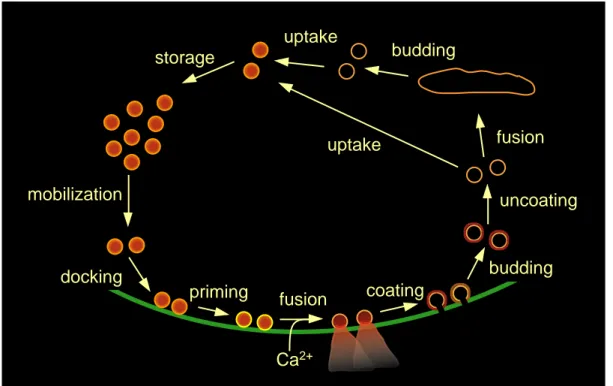
A general model of vesicle cycling in the presynaptic terminal is depicted in Fig. 1.3 (Südhof, 1995; Augustine et al., 1999). In the resting terminal, at least two functional pools of synaptic vesicles can be distinguished (Greengard et al., 1993): (1) a set of vesicles is found in contact with the presynaptic membrane at the active zone, which is regarded as the pool of vesicles readily releasable upon arrival of an action potential. The morphologically docked vesicles found in electron micrographs can be sub-divided into ‘docked only’ and ‘docked and primed’ vesicles, depending on whether they have passed through all molecular priming steps necessary for Ca2+- triggered fusion (Südhof, 1995, see below). (2) Vesicles are observed to form clusters near the active zone, which are stabilized probably because of the binding of the
Fig. 1.3: The synaptic vesicle cycle. Before fusing with the presynaptic membrane, synaptic vesicles have to be translocated to and brought into close apposition with the presynaptic membrane (mobilization and docking). Further molecular reactions (priming) are required until a vesicle can undergo Ca2 +-triggered fusion. Vesicular membrane is recycled after coating with clathrin (coating, budding, uncoating).
Endocytosed vesicles may transit by fusion and budding through intracellular endosomes and are added to the reserve pool (storage). Recycled vesicles are refilled with neurotransmitter by active transport (uptake) (modified from Augustine et al., 1999).
vesicle protein synapsin I with cyto-skeletal elements (Pieribone et al., 1995).
Vesicles from this ‘reserve pool’ are translocated to the active zone and repopulate empty release sites during repetitive stimulation (e.g. Zenisek et al., 2000). After fusion and transmitter release, clathrin-coated membrane segments bud from the presynaptic membrane and form vesicles that are thought to be translocated into the reserve pool, possibly after fusion with and budding from intracellular endosomes (Heuser and Reese, 1973, see section 1.2.3 for an alternative model). During this cycling, vesicles accumulate neurotransmitter by active transport, which is driven by an electrochemical gradient due to a proton pump. Many steps of synaptic vesicle cycling can be associated with specific molecular reactions by disrupting the cycle at different stages. Useful tools are microinjection of peptides or antibodies or genetic manipulations that interfere with one of the putative molecular reactions (Pieribone et
storage
mobilization
docking
priming fusion
Ca2+
budding
budding uncoating
coating
fusion uptake
uptake
al., 1995; Südhof, 1995; Augustine et al., 1999). To clarify the role of the numerous types of presynaptic proteins is still difficult and is the focus of current research. The following sections will briefly introduce more detailed concepts that have been proposed for the key events during synaptic transmission.
1.2.2 Exocytosis
1.2.2.1 Some molecules involved in exocytosis
Chemical transmission requires the precisely timed release of transmitter molecules into the synaptic cleft by the specific fusion of synaptic vesicles with the presynaptic membrane. The abundance of intracellular membrane in the terminal necessitates that this signaling process be highly regulated by specific biochemical binding partners on the vesicle and the target membrane. The vesicular and target membrane contain numerous proteins, of which well characterized super-families are the GTP-binding Rab proteins and the soluble NSF attachment protein receptor (SNARE) proteins (Südhof, 1995). While both protein families may contribute to vesicle transport specificity and target membrane recognition, the SNARE proteins are thought to be important also in mediating the last steps of membrane fusion (Jahn and Südhof, 1999; Chen and Scheller, 2001) The vesicular (v-) SNARE protein synaptobrevin and the target membrane (t-) SNARE proteins SNAP-25 and syntaxin can form a tight complex, the SNARE core complex, which is likely to play a functional role in vesicle targeting to the release site and/or fusion of the two lipid bilayers. In this complex, synaptobrevin and syntaxin both contribute one and SNAP-25 contributes two α-helical domains, which bind by forming a four-stranded coiled-coil structure.
Because syntaxin and synaptobrevin bind in parallel fashion, i.e. with the amino termini at one end and the membrane-anchored carboxy termini at the other end of the core complex, it is thought that the binding reaction between SNARE proteins may occur in a ‘zipper-like’ fashion, thus exerting mechanical force on the lipid membrane anchors. The core complex is heat stable up to 90° C and resistant to protease digestion, biochemical denaturation and cleavage by clostridial neurotoxins, which are able to cut SNARE proteins in the isolated state (Chen and Scheller, 2001). In biological systems, SNARE complexes are disassembled by N-ethyl-maleimide- sensitive fusion protein (NSF) and soluble NSF attachment protein (α-SNAP) under hydrolysis of ATP. This reaction is thought to reactivate previously used SNARE proteins for a new fusion cycle.
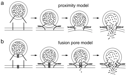
Fig. 1.4: Models of lipid membrane fusion in exocytosis. (a) In the ‘proximity’
model, fusion is induced, when the vesicle and presynaptic membrane are forced into close apposition by the tight binding of v- and t-SNAREs in a zipper-like manner. A precursor state of the fully fused membranes may be ‘hemifusion’ (3rd step in a), where the inner membrane leaflets have mixed, but the outer leaflets are intact. (b) In the ‘fusion pore’ model, the mixing of vesicle and presynaptic membrane lipids is mediated by a proteinaceous channel connecting both membranes. Binding of v- and t-SNAREs may anchor vesicles to the target membrane and promote the formation of fusion pore precursors.
1.2.2.2 The fusion mechanism
The fusion mechanism itself requires that two closely apposed phospholipid bilayers merge in an aqueous environment. Because of the repulsive forces between the polar phospholipid head groups, a high energy barrier must be overcome, which is thought to be mediated by specialized fusion proteins. Two principal hypotheses have been put forward to describe the fusion reaction of the transmitter vesicle with the target membrane mechanistically (Fig. 1.4). In ‘proximity’ models it is proposed that the action of membrane proteins such as SNARE complexes is restricted to reducing the activation energy by forcing the two lipid bilayers into close apposition, from where lipids of the two proximal leaflets can mix and form a hemifusion or stalk state (Fig.
1.4 a). It is unclear whether the hemifused state is immediately followed by the break down and mixing of the distal leaflets, or whether this state is metastable, requiring another catalyzing step for full fusion to occur (Jahn and Südhof, 1999; Chen and Scheller, 2001). In the ‘fusion pore model’, the fusion of apposing membranes is mediated by a proteinaceous channel structure that spans both membranes and promotes the mixing of the two lipid bilayers (Almers and Tse, 1990). This could possibly involve radial expansion of the fusion protein’s subunits within the lipid bilayers, allowing the phospholipids to flow and mix in the expanding space between
a
b
proximity model
fusion pore model
the subunits. As an example, a membrane spanning, proteolipidic sector of the vacuolar H+-ATPase in yeast, a proton pump molecule, was recently shown to form trans-complexes between apposing intracellular membranes and to mediate fusion (Peters et al., 2001), the latter in the absence of SNARE core complexes. Thus, the role of various molecular interactions in fusion is still controversial and it remains to be elucidated to what extent the mechanisms driving membrane fusion are molecularly conserved in different cells and species.
1.2.3 Endocytosis
Synaptic terminal membrane is recycled during and after transmitter release by local infolding of the membrane and subsequent budding of small vesicles and/or larger compartments called cisternae (reviewed by Wilkinson and Cole, 2001). This way, the increase in terminal surface area due to exocytosis is quickly compensated, which is necessary to maintain the supply of membrane material for the formation of new vesicles and to prevent terminal deformation. Based on electron microscopic studies, two alternative recycling pathways were proposed, which differed in several aspects.
In one model, fused vesicles are thought to collapse completely into the presynaptic membrane. Membrane retrieval occurs away from the release site and involves specific membrane recognition mediated by the protein adaptor complex AP2, which triggers the coating and infolding of the membrane segment mediated by clathrin (Pearse et al., 2000). Clathrin-coated vesicles pinch off and subsequently discard the coating; then they are transferred to the reserve pool of vesicles (Heuser and Reese, 1973; Südhof, 1995). In a second model, the fusion of a vesicle is thought to be reversible, leaving the vesicle membrane largely intact and allowing it to reseal and detach from the release site quickly after pore opening and transmitter discharge (‘kiss and run’, Ceccarelli et al., 1973; Fesce et al., 1994). The proposed mechanism does not require clathrin-mediated endocytosis and makes recycled vesicles available close to the release site, which could be advantageous to counteract rapid depletion of the releasable vesicle pool during repetitive stimulation.
Recent experiments using fluorescent membrane markers with different departitioning properties suggest that at least two endocytotic pathways, which resemble those proposed originally, may be at work in synaptic terminals. Thus, endocytosis via the formation of cisternae may serve to supply newly assembled vesicles to the reserve pool on a slow time scale, whereas a rapid endocytotic pathway near the active zone may be capable of locally recycling or reusing vesicles previously located in the readily releasable pool (Pyle et al., 2000; Richards et al., 2000; Stevens and Williams, 2000).
1.2.4 The role of Ca2+ in exocytosis and endocytosis
In the exocytotic event sequence, vesicles are arrested in a primed state at the release site. Only after opening of Ca2+ channels in response to a nerve pulse does a rise of the intracellular Ca2+ concentration trigger the final steps of transmitter release (Katz, 1969). In early experiments it was shown by varying the extracellular [Ca2+] that transmitter release is controlled by the cooperative action of several Ca2+ ions (Dodge Jr. and Rahamimoff, 1967). Later, measurements of the dependence of transmitter release on intracellular [Ca2+] confirmed that at least three Ca2+ ions are required to trigger the fusion of a vesicle, thus providing the release mechanism with a supra- linear sensitivity to variations in [Ca2+] (Heinemann et al., 1994; Heidelberger et al., 1994). Despite considerable effort, the mechanism by which Ca2+ deploys the fusion machinery is still under debate. Many Ca2+-binding proteins present in presynaptic terminals have a modulating function in transmitter release (Burgoyne and Morgan, 1998). The most prominent candidate for a neuronal Ca2+ sensor for transmitter release is synaptotagmin I, which binds to phospholipids and syntaxin and oligomerizes in a Ca2+-dependent manner. Neurons in which synaptotagmin was absent or point mutated showed reduced synchronous transmitter release (Nonet et al., 1993; Broadie et al., 1994; Geppert et al., 1994; Fernández-Chacón et al., 2001).
However, a widely accepted functional model of synaptotagmin-triggered fusion has not yet emerged.
Aside from being the crucial signal for rapid membrane fusion, Ca2+ is likely to regulate also other steps in the synaptic vesicle cycle. Increased [Ca2+] following stimulation may enhance the rate at which the readily releasable pool is replenished (reviewed by Zucker, 1999; but see Wu and Borst, 1999). It has been observed in non- neural exocytosis that even the expansion rate of a granular fusion pore may be a function of intracellular [Ca2+] (Hartmann and Lindau, 1995). Also endocytosis is regulated by Ca2+ in many systems, although the exact dependence is not generally established (Henkel and Almers, 1996). Thus it was observed that rapid endocytosis in endocrine cells is initiated by Ca2+ binding to calmodulin (Artalejo et al., 1996). In hippocampal synapses, intracellular [Ca2+] was observed to up-regulate endocytosis (Sankaranarayanan and Ryan, 2001). In contrast, in synaptic terminals of retinal bipolar cells, increased [Ca2+] levels appeared to inhibit endocytosis (von Gersdorff and Matthews, 1994).
It can be summarized that many processes in the synaptic vesicle cycle are subject to Ca2+-sensitive modulation, implying that the balanced operation of this cycle requires tight regulation of intracellular [Ca2+] levels.
1.3 Motivation
The speed of chemical transmission is limited by the time the final steps of exocytosis require for completion after arrival of a presynaptic action potential. These steps include the opening of presynaptic Ca2+ channels, Ca2+ diffusion to and activation of the Ca2+ sensor for release and ultimately the fusion of the vesicle and target membrane. The activation of the release sensor by Ca2 + is of particular interest because it represents the very relay mechanism by which electrical activity – the flow of ions across the membrane – is coupled to chemical signal transmission – the discharge of chemical transmitter molecules from a fused vesicle. Therefore, the temporal coupling precision will be a main determinant of the synchronicity of pre- and postsynaptic excitation. As mentioned above, synchronicity and coincidence detection are important for the activity-dependent adjustment of synaptic weights, which is considered a possible mechanism for the way a biological network processes and stores information. In addition, the Ca2+-release sensor reaction not only partially determines the fidelity of chemical transmission, but may also be a target where the adjustment of synaptic weights can be implemented. When restricting the view on this element of the signaling cascade, two basic regulatory mechanisms may be considered. First, the Ca2+ signal observed by the Ca2 + sensor during an action potential may be activity-dependent, for instance due to action potential broadening or modulation of Ca2+ channel opening. Second, the efficacy of the Ca2+ sensor to bind Ca2+ and trigger fusion – its sensitivity - may be regulated in an activity-dependent manner.
To better understand the significance of excitation-secretion coupling for transmission fidelity and synaptic plasticity, the general dependence of the process of transmitter release on intracellular [Ca2+] should be determined first. Because of its importance, several investigations have focussed on this issue in other exocytotic systems, for example in hormone releasing cells or in specialized nerve terminals, however with divergent findings that apparently cannot be extrapolated to other synapses (see section 5.3.3 for a more detailed discussion).
In this study, the Ca2+ dependence of rapid synaptic transmission was characterized in a glutamatergic giant synapse in rat brainstem slices, using pre- and postsynaptic current recordings (Forsythe, 1994; Borst et al., 1995). Because of the large size of the pre- and postsynaptic compartment, electrical signals can be detected from both compartments using the whole-cell patch clamp technique. In this method, a recording pipette is tightly sealed to a patch of cell membrane, permitting low resistance access to the intracellular compartment for electrical measurements and diffusional delivery of substances (Neher and Sakmann, 1976; Hamill et al., 1981; Edwards et al., 1989).
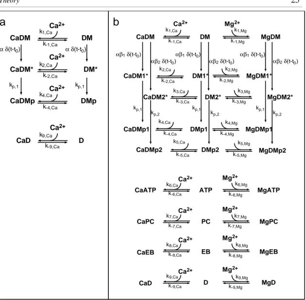
A UV-sensitive Ca2+ chelator loaded with Ca2+ was introduced to the presynaptic terminal together with a fluorescent Ca2+ indicator (Kaplan and Somlyo, 1989). The presynaptic [Ca2+] was artificially raised by brief UV laser pulses and measured using quantitative fluorescence microscopy. By simultaneously measuring the postsynaptic currents, the relation between presynaptic glutamate release and intracellular [Ca2+], in other words the ‘Ca2+ sensitivity of transmitter release’, could be determined for this mammalian synapse. Furthermore, this relation was described in a quantitative model, which was subsequently used to estimate the Ca2+ signal triggering synaptic transmission during physiological activity.
2. Theory
2.1 Intracellular Ca2+ dynamics
Calcium is probably the most versatile signal transduction element in living cells. It controls diverse cell functions, of which the fastest are excitation-secretion coupling in synapses and excitation-contraction coupling in muscle cells, both occurring within milliseconds. On a time scale of minutes or longer, Ca2+ is a main regulator of cell cycling, gene expression and programmed cell death (Clapham, 1995). In general, cells maintain intracellular [Ca2+] at a level ~104-fold lower than the extracellular level. Furthermore, cells must precisely control local and global [Ca2+] signals in order to maintain a high specificity when using a single ion type as a quasi-universal messenger.
In a more abstract view, cells can be regarded as a finite, three-dimensional space delimited by a closed surface, the plasma membrane, and compartmentalized into different spatial and functional units. In this volume, the Ca2+ concentration is a function of space and time, [Ca2+](r,t). The dynamic evolution of [Ca2+](r,t) depends on three regulatory mechanisms: passive diffusion, chemical reaction and active transport. In living cells, all three processes exist and contribute to shaping [Ca2+](r,t).
The first one, diffusion in an aqueous medium, naturally occurs wherever electrochemical gradients are encountered. Thus, Ca2+ diffusion occurs along the electrochemical gradient over the plasma membrane through Ca2+-permeable channel proteins, giving rise to localized Ca2+ influx. Intracellular diffusion of Ca2+ within the cytosol is influenced by diffusional barriers, which contribute to shaping local Ca2+
signals. One example for this compartmentalization of Ca2+ signaling is the restriction of [Ca2+] elevations during synaptic activation to a postsynaptic compartment, the
‘synaptic spine’: the parent dendrite is largely spared from the Ca2+ signal because of the narrower spine neck, forming a bottle-neck for Ca2+ diffusion (Yuste and Denk, 1995).
In the second mechanism, Ca2+ can bind to mobile or immobile proteins in a chemical reaction, which is generally termed ‘Ca2+ buffering’. This mechanism largely contributes to maintaining low global [Ca2+] levels and to confining [Ca2+] elevations to the vicinity of their sources. To date, a large number of Ca2+-binding proteins have been found, of which prominent neuronal members are calbindin, calretinin and
parvalbumin (Blaustein, 1988). Ca2+ buffering reactions are usually described using second order reaction rate equations (see next section).
A third mechanism is the active transport of Ca2+ ‘uphill’, against the electrochemical gradient under consumption of chemical energy. This mechanism is responsible for the sequestration of Ca2+ across the plasma membrane into the extracellular space and for the uptake of Ca2+ into intracellular stores, such as mitochondria or the endoplasmic reticulum. The active transport is taken over by membrane-integral proteins, e.g. the Ca2+-ATPase or the Na+-Ca2+ exchanger.
Under the combined influence of numerous interactions of the above type, a complex spatio-temporal pattern of intracellular [Ca2+] is expected to evolve, especially whenever cellular activity perturbs the electrochemical equilibrium. A special case important for neuronal exocytosis is the rapid opening of Ca2+ channels during action potentials which generates highly localized and short-lived [Ca2+] elevations close to synaptic vesicles at the presynaptic membrane (reviewed by Neher, 1998). These so- called ‘Ca2+ microdomains’ have been difficult to measure directly, mostly because light microscopy lacks the spatial resolution. However, spatially and temporally confined [Ca2+] elevations which probably arise from the opening of a cluster of several channels have been observed (Llinás et al., 1992; DiGregorio et al., 1999;
Yazejian et al., 2000).
On the other hand, mathematical simulations have given much insight into the complex distribution of [Ca2+](r,t), especially during channel opening at synaptic sites (Chad and Eckert, 1984; Simon and Llinás, 1985; Pape et al., 1995; Naraghi and Neher, 1997; reviewed by Neher, 1998). The modeled [Ca2+](r,t), however, is quite sensitive to the kinetic properties of the buffer species present in the vicinity of the Ca2+ channels. For synapses, only little information is currently available regarding the buffers’ identity, nor is much known about the spatial arrangement of Ca2+
channels, buffers and vesicles (but see Harlow et al., 2001).
Therefore, in the present study a different approach was used to analyze the dependence of a cellular process on [Ca2 +](r,t). By uniform uncaging of Ca2+
throughout the presynaptic terminal, the spatial dependence of [Ca2 +](r,t) was removed (Fig. 2.1). By choosing a short-pulsed UV source and a rapidly converting Ca2+ cage and fast Ca2+ indicators, also the temporal dependence of [Ca2+](r,t) was largely reduced to a brief transition period of 100-200 µs.
Fig. 2.1: Spatial pattern of [Ca2+] during Ca2+ influx and UV-induced Ca2+ uncaging.
(a) The opening of a Ca2+ channel in the presynaptic plasma membrane mediates the influx of Ca2+ ions. The concentration profile and the location of vesicles relative to the Ca2+ channels is generally not known, making it difficult to predict the [Ca2+] present at the Ca2+ sensor during action potential-evoked release. (b) UV-induced photolysis of a high-affinity Ca2+ chelator permits spatially homogeneous [Ca2+] elevations. Volume-averaged [Ca2+] measurements then reflect the [Ca2+] at the Ca2+
sensor for release.
In the following, the general approach to model Ca2+ dynamics will be stated based on the three regulatory mechanisms mentioned above. A simple approximation is considered for a single channel Ca2+ domain in order to illustrate the effect of Ca2+
buffers on the spatial extent of the microdomain. Finally, the time course of [Ca2+](t) evoked by Ca2+ uncaging will be calculated using a detailed model, which takes into account those Ca2+ buffers also present in the experiments.
2.1.1 Time-dependent Ca2+ diffusion-reaction in an aqueous medium
Firstly, diffusion of Ca2+ and Ca2+ buffering molecules is described by Fick’s law, if we assume the intracellular volume to be an isopotential space.
∂
∂ [ ]( , )
[ ]( , )
Ca Ca
2+
Ca
r 2+
t r
t = D ∆ t , (2.1)
where ∆ is the Laplace operator and DCa the diffusion coefficient of Ca2+, which also may be a function of space and time. The same applies for the Ca2+ buffers present.
Secondly, the buffering reactions are commonly described with 2nd order reaction rate equations. The rate of concentration changes during a buffer reaction between Ca2+
and a buffer B1:
Ca + B 1 k CaB1
k
1 1
← →
−
(2.2)
a b
vesicle
presynaptic membrane
Ca2+
channel
is proportional to the reactants’ concentrations, with association and dissociation rate constants k1, k-1:
d
dt k k
d
dt k k
[ ]
[ ][ ] [ ]
[ ]
[ ][ ] [ ]
Ca Ca B CaB
CaB Ca B CaB
2
1 2
1 1 1
1 1
2
1 1 1
+ +
−
+ −
= − +
= −
(2.3)
Thirdly, the action of active transport mechanisms cannot be represented in a general form, but it is dependent on chemical energy supply and local [Ca2+]. The transport rate p can often be approximated using a Michaelis-Menten relation, which accounts for the limited transport capacity at saturating [Ca2+]:
p t p t t
t K
([ ], , ) ( , ) [ ]( , )
([ ]( , ) )
Ca max Ca
Ca
2+
2+
2+
m
r r r
= r
+ , (2.4)
where pmax is the maximal pump rate in a given volume element, and Km is the [Ca2+] where p becomes half-maximal.
By combining these mechanisms in the most general form, the spatio-temporal evolution of [Ca2+](r,t) can be described by a system of partial differential equations:
∂
∂
[ ]
[ ][ ] [ ] [ ]
CaBi i Ca2+ Bi i CaBi CaB CaBi
t =k −k− +D i∆ , (2.5)
∂
∂ [ ]
[ ][ ] [ ] [ ]
Bi i Ca2+ Bi i CaBi B Bi
t = −k +k− + D i∆ , (2.6)
∂
∂
[ ]
( [ ][ ] [ ]) [ ]
([ ], , ) ([ ], , )
Ca Ca B CaB Ca
Ca Ca
2+
i 2+
i i i Ca
2+
2+ 2+
t k k D
j t p t
i n
= − − +
+ −
∑
− ∆r r
(2.7)
Here, Bi denotes n different Ca2+ buffer species with association and dissociation rate constants ki and k-i, respectively. Ca2+ influx is summarized as j, which depends on both passive transport through ion channels and active transport, e.g. the Na+-Ca2+
exchanger operating in reverse mode (Regehr, 1997). Furthermore, p comprises the various Ca2+ sequestration mechanisms.
In general, this set of partial differential equations cannot be solved analytically. For different spatial arrangements and buffer conditions, [Ca2+](r,t) has been calculated numerically (e.g. Simon and Llinás, 1985; Yamada and Zucker, 1992; Roberts, 1994).
In parallel, the evolution of [Ca2+](r,t) can be approximated analytically for some limiting cases, for example, when the concentration of the buffer in its free and Ca2+- bound form is assumed to be constant (Neher, 1986), when Ca2+ binding is assumed to
be instantaneous (‘rapid buffer approximation’, Smith, 1996) or when the change in free buffer concentration is small during a perturbation (‘linearized buffer approximation’, Pape et al., 1995; Naraghi and Neher, 1997).
2.1.2 Ca2+ microdomains
Numerical and analytical solutions of [Ca2+](r,t) show that increases in [Ca2+] are defined locally around the site of Ca2+ influx and drop sharply with distance from the channel. To illustrate this point, Ca2+ influx through a single channel is treated as a point source in an infinite plane, corresponding to the molar flux Φ (in units mol.s-1) into a semi-infinite medium (Fig. 2.2 a). It is convenient to use spherical coordinates and first consider the development of the steady-state solution in the absence of Ca2+
buffering and extrusion. Eq. 2.5 - 2.7 reduce to a radial diffusion equation:
∂
∂
∂
∂
∂
∂ [Ca2+] [Ca ] [Ca ]
Ca
2+ 2+
t D
r r r
= +
2 2
2 . (2.8)
Assuming that the [Ca2+] is zero prior to channel opening, which occurs at t = 0, and Φ is constant thereafter, the solution to Eq. 2.8 is given by (Crank, 1975):
[Ca2+]( , ) erfc
Ca Ca
r t D r
r
= D t
Φ
4π 2 . (2.9)
Two characteristics of Ca2 + microdomains can readily be understood from this equation. Firstly, if a channel is continuously open, a microdomain will reach a steady-state profile, since the error function erfc will approach unity for large t. With a value for DCa = 220 µm2 s- 1 (Naraghi and Neher, 1997), steady-state will be established in a range of 10-100 nm from the channel within ~10 µs to ~1 ms.
Secondly, assuming a single channel current of iCa = 0.2 pA, (corresponding to a flux Φ = iCa/F ≈ 2×10-18 mol.s-1; F Faraday’s constant), the steady-state [Ca2+] in the immediate vicinity of a channel (≤ 10 nm) can reach 70 µM or more and drops with a 1/r-dependence (Fig. 2.2 b). This already emphasizes the local nature of Ca2 + microdomains.
Ca2+ buffers decrease the range of [Ca2+] even further and reduce the size of microdomains. If it is assumed that Ca2+ buffering near a Ca2+ channel is dominated by a single buffer B1 present in large concentrations, the buffered diffusion of Ca2+
can be modeled in a simplified scheme, which formally corresponds to the cable equation (Neher, 1986):
∂
∂
∂
∂
∂
∂
[ ]
[ ] [ ] [ ]
Ca2+ app Ca2+ app Ca Ca
Ca
2+ 2+
t k k D
r r r
= − + + +
−
1 1
2 2
2 . (2.10)

![Fig. 2.1: Spatial pattern of [Ca 2+ ] during Ca 2+ influx and UV-induced Ca 2+ uncaging.](https://thumb-eu.123doks.com/thumbv2/1library_info/5500620.1685643/29.892.139.757.121.317/fig-spatial-pattern-ca-ca-influx-induced-uncaging.webp)

![Fig. 3.1: Schematic diagram of the experimental setup. Two patch clamp electrodes and the photodiode for rapid [Ca 2+ ] measurements are amplified using patch clamp amplifiers](https://thumb-eu.123doks.com/thumbv2/1library_info/5500620.1685643/44.892.137.726.109.519/schematic-diagram-experimental-electrodes-photodiode-measurements-amplified-amplifiers.webp)
