UNCORRECTED
PROOF
Environmental Pollution xxx (xxxx) xxx-xxx
Contents lists available at ScienceDirect
Environmental Pollution
journal homepage: www.elsevier.com
How to get rid of ingested microplastic fibers? A straightforward approach of the Atlantic ditch shrimp Palaemon varians
☆Reinhard Saborowski
∗, Eva Paulischkis , Lars Gutow
Alfred Wegener Institute, Helmholtz Centre for Polar and Marine Research, Am Handelshafen 12, 27570 Bremerhaven, Germany
A R T I C L E I N F O
Article history:
Received 19 March 2019
Received in revised form 7 August 2019 Accepted 15 August 2019
Available online xxx
Keywords:
Shrimp Fibers Beads Regurgitation Defecation Retention
A B S T R A C T
Microplastic fibers represent a significant share of the global marine micrcroplastic pollution, particularly in coastal areas. In controlled laboratory experiments, we offered fluorescent microplastic fibers (40–4400μm lengths, median 150μm) and spherical microplastic beads (9.9μm Ø) together with commercial fish food to the Atlantic ditch shrimpPalaemonetes varians. The shrimps ingested fibers and beads along with the food.
Upon ingestion, the beads and the shortest fibers (up to 100μm) passed from the stomach into the gut and were egested within the fecal strings. The longer fibers first remained in the stomach but were regurgitated, i.e. extruded through the esophagus, within 12–14 h. Regurgitation is an evolutionary adaptation of particular crustacean species and other invertebrates to remove large and indigestible food particles from the stomach.
Accordingly, the process of regurgitation attained a new task nowadays, i.e. the elimination of anthropogenic filamentous microplastic debris from the stomach to avoid harm. This behavioral feature may represent a se- lective advantage in view of the continuously increasing environmental plastic pollution.
© 2019.
1. Introduction
An estimated eight million metric tons of plastic waste enter the oceans every year and a further increase by one order of magnitude until 2025 is predicted (Jambeck et al., 2015). In natural environ- ments, plastic items disintegrate due to mechanical action, UV-radia- tion, or thermo-oxidation, thereby bearing numerous small fragments (Barnes et al., 2009; Andrady, 2011; Cole et al., 2011; ter Halle et al., 2016; Weinstein et al., 2016). Microplastic particles can be ingested by a variety of organisms including small invertebrates. The effects of microplastic ingestion are, however, inconclusive. Various studies showed that particles pass the digestive tract without detectable effects on organisms (e.g. Hämer et al., 2014; Kaposi et al., 2014; Santana et al., 2018; Weber et al., 2018) whereas other studies reported dam- age to organs, tissues, and cells at experimentally administered par- ticle concentrations ofup to 20 mg L-1(Jeong et al., 2016, 2017) or 2.5 g L−1(von Moos et al., 2012).
Besides irregular fragmented particles, synthetic fibers constitute a significant share of microplastics in the environment, often outnum- bering other types of particles, such as fragments and beads. In the brackish waters of the Baltic Sea off Sweden, microfibers accounted for more than 80% of the overall collected microplastics, accounting for about four fibers per cube meter (Gewert et al., 2017). Mathalon and Hill (2014) found 20 to 80 plastic fibers in 10 g of sediment at a beach in the Halifax region (Canada). The fibers probably originated
☆This paper has been recommended for acceptance by Maria Cristina Fossi.
∗Corresponding author.
Email address:Reinhard.Saborowski@awi.de (R. Saborowski)
from domestic washing (reviewed by Salvador Cesa et al. (2017)) or from ropes or fishing nets. De Falco et al. (2018) reported up to 6,000,000 fibers with a length of 300–500μm, released from a typi- cal 5 kg wash load of polyester fabrics. Napper and Thompson (2016) estimated a release of more than 700,000 fibers from a 6 kg wash load of acrylic fabric. Mason et al. (2016) estimated an average daily discharge of 4 million microplastic fibers by each single municipal wastewater treatment plant in the US states of California, New York, Wisconsin, and Ohio. Additionally, fishing gear and ropes, whether in use, lost, or discarded, also significantly contribute to environmental pollution by microplastic fibers (e.g. Barnes et al., 2009; Macfadyen et al., 2009; Cole et al., 2011).
Different to irregular or spherical particles, fibers are widely dis- regarded in environmental and ecotoxicological studies because it is difficult to differentiate between environmental contaminants and ac- cidently introduced fibers during sample processing in the field and in the laboratory. So far, however, presence of fibers was reported in various species of seafood (Rochman et al., 2015). Blue mussels were examined for their suitability to monitor microplastic pollution in Nor- wegian coastal waters where mussels contained on average 1.5 mi- croplastics per individual (Bråte et al., 2018). Overall, 83% of the par- ticles investigated in that study were fibers. Synthetic fibers were de- tected in 63% of assessed brown shrimp from the coastal waters of the Southern North Sea and the Channel (Devriese et al., 2015). Desforges et al. (2015) found that the share of ingested microplastic fibers of the total microplastic particles inEuphausia pacificain the NE Pacific de- creased with distance from shore suggesting substantial discharge of synthetic fibers from land-based sources.
Upon ingestion, the fibres may provoke false satiation in animals, either when applied at high concentrations or when they accumulate
https://doi.org/10.1016/j.envpol.2019.113068 0269-7491/ © 2019.
UNCORRECTED
PROOF
over time in the stomachs as observed in Norway lobster,Nephrops norvegicus (Murray and Cowie, 2011; Welden and Cowie, 2016a, 2016b). The water flea,Daphnia magna, readily ingested plastic fibers of 300μm average length but also very long fibers of around 1,400μm (Jemec et al., 2016). Mortality increased when the animals were ex- posed to plastic fibers only, but not when they were fed with natural food prior to plastic exposure. The amphipodHyalella aztecawas ex- posed to polyethylene microplastic particles and polypropylene mi- croplastic fibers. Fibers caused significantly higher mortality than par- ticles (Au et al., 2015). The higher toxicity of the fibers corresponded with longer residence time of the fibers in the gut, which, in turn, might have affected the ability to process food and led to severe ener- getic deficiency (Au et al., 2015).
The present study aims at investigating whether microplastic fibers and beads are ingested by the Atlantic ditch shrimpPalaemon vari- ansand how they are translocated within the digestive organs of the shrimp.P. varianswas chosen as test organism because these shrimps are common in coastal regions and estuaries of the Northeast Atlantic where they are particularly exposed to domestic and industrial envi- ronmental plastic pollution (Browne, 2015). The animals are relatively small (1–3 cm in length) and widely translucent allowing for direct observation of ingested fluorescent fibers and particles in living an- imals under a fluorescence stereomicroscope. To clearly observe the fibers and beads, and to track their position in the shrimp, we applied in this study high concentrations well above the environmental levels.
We addressed the following questions: 1) do these shrimps ingest mi- croplastic fibers and particles, 2) do the plastic fibers accumulate in the stomach, and 3) and do they cause mortality at the applied experi- mental conditions?
2. Methods
2.1. Origin of organisms
Atlantic ditch shrimps (Palaemon varians, Leach 1813) were ei- ther purchased from an aquaristic shop (Futterhaus, Bremerhaven, Germany) or were captured with a hand net in a semi-enclosed former harbor basin in the city of Bremerhaven (Holzhafen; 53° 32.177820′ N, 8° 35.503680’E). The shrimps were immediately transferred to the laboratories of the Alfred Wegener Institute, where they were main- tained in 10-L aquaria in brackish water of 15 PSU, 15 °C, and 16:8 h of indirect illumination and darkness. The shrimps were fed every other day with plant-based fish food (NovoVert, JBL) and, addition- ally, once a week with freshly hatchedArtemiasp. nauplii (Great Salt Lake Artemia Cysts, Sanders). The water was exchanged each day af- ter feeding. Animals from both sources were used for preliminary ex- periments. The feeding experiments reported in this study were solely performed with animals from the urban pond.
2.2. Fluorescent fibers and microbeads
Small plastic fibers were cut from fluorescent polyacrylic wool
“Magic Leucht Wolle fluo grün”(Magic Pyramid Bruecher & Partner KG, Frechen, Germany, No. 839512) by hand with scissors under a stereomicroscope. A sample of 0.5 mg contained almost 3000 fibers.
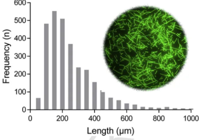
The lengths of the fibers ranged between 40 and 4400μm. The av- erage length was 236 ± 176 (SD) μm, the median length was 189μm (Fig. 1), and the width of the fibers was 30–35μm. Due to electro- static adhesion and their high numbers, fibres could not be counted properly. Instead, the administered quantity in the experiments was expressed by mass. Fluorescent microbeads of polystyrene (PS, Flu
Fig. 1.Length distribution and appearance of fluorescent fibers after production and be- fore administration to the test organisms.
oro-MaxTMGreen Fluorescent Microspheres, 9.9μm diameter) were purchased from Thermo Fisher Scientific. They were delivered in a suspension of 1% solids in water at a concentration of 20,000 mi- croplastics (MP)μL−1. The fibres and the beads were observed under a fluorescence stereomicroscope (Nikon Instruments, SMZ25) with FITC fluorescence filter (ex: 494 nm; em: 518 nm). Photographs were taken with a Nikon camera DS-Ri1 and attendant software (NIS-El- ements Basic Research 4.20.01), which also included features for length measurements that we used for the fibres.
2.3. Experimental procedure
The animals were acclimated to room temperature (22 °C) which was similar to the ambient water temperatures at the time the shrimps were collected (22–24 °C). Prior to the experiments, the test animals were starving for 48 h to void their stomachs from remaining food. The water was exchanged daily to prevent coprophagia. The fullness of the stomachs of the translucent shrimps was visually estimated under the stereo microscope. Only shrimps with empty stomachs were used for the feeding experiments.
2.4. Ingestion of fibres, beads, and food (Exp.1)
Animals received 0.5 mg of plastic fibres and four quantities of flake food (NovoVert, JBL): 0 mg, 2.5 mg, 5 mg, and 10 mg (see also Table S1). The fibres and the food were suspended in 100 ml brack- ish water (15 PSU) which was previously filtered through a 0.1-μm membrane filter. Animals were kept individually in the experimental units (beakers). Ten replicates were run in each of the four feeding ex- periments, accounting for 40 animals in total. The shrimps were incu- bated for 3 h with the food and the fibres. Thereafter, the animals were frozen at −20 °C for 10 min. The wet weights and the lengths from the frontal edge of the eyes to the tip of the uropods were recorded. The stomach, the midgut gland, and the gut of each animal were screened with a fluorescence microscope for microplastic fibres. Subsequently, the organs were dissected and suspended in 100–200 ml of dist. water.
The fibres were counted and their lengths were measured under a flu- orescence binocular.
2.5. Stomach residence time of microplastic fibers and beads (Exp. 2) A preliminary feeding experiment showed that the digestive or- gans (stomach, midgut gland, gut) of P. varianswere void of mi- crofibers after 24 h. Therefore, we ran an experiment to estimate the
UNCORRECTED
PROOF
residence times of food, fibres, and beads. Animals were starved for 48 h prior to the experiment. The maintenance water was exchanged repeatedly and only shrimps with empty stomachs were selected for the experiments. Preliminary observations also showed thatP. varians ingested fibres more eagerly when they were attached to food items.
Therefore, a blend was prepared consisting of 5 mg of flake food, 1 mg of fibres, and 10μL of 1:10 diluted solution of beads per portion. The control food contained 5 mg of flakes and 10μl of demineralised wa- ter. The food preparations dried overnight at room temperature. Care was taken during the procedure to avoid contamination of the food.
The following day, 200-mL petri dishes were filled with 100 ml fil- tered brackish water (15 PSU) and the prepared food was added with metal forceps. Each of the petri dishes contained about 200,000 beads L−1and 5 mg of fibres as estimated from the amount of food blend added to the water. The animals were placed into the petri dishes and allowed to feed for 20 min. Subsequently, the animals were inspected under the fluorescent microscope for ingested fibres, beads, and food.
If the stomachs contained microplastics, the animals were transferred into dishes with 100 ml of fresh and filtered brackish water. Every 2 h, the animals were inspected again and the following parameters were recorded: a) presence of food and microplastics in the digestive or- gans, b) amount of feces in the petri dish, c) presence of fibres and beads in the feces, and d) presence of mucus plaques at the bottom of the petri dish. After each inspection, the water in the petri dishes was exchanged to enable observation of newly produced feces which was egested during the following 2 h. In total, 27 animals were tested: 9 control animals and 18 fed with beads and fibres. The residence time of fibres was estimated in all 18 animals and the residence time of beads and food in 9 of the 18 animals. See also Table S1.
2.6. Survival
To investigate whether microplastic ingestion had adverse effects onP. varians, the survival of the shrimps was recorded for another 48 h. After the experiments were terminated, the animals were contin- uously fed with flakes (without microplastics) to prevent fatal starva- tion. Signs of vitality were upright position of the animal, locomotion, and a translucent appearance of the body.
2.7. Stomach movement and regurgitation
Time-lapse recordings were carried out to observe the stomach peristaltic and the egestion of the fibres and beads. Shrimps were fed with food containing fibres and beads as described above. The lateral side of the cephalothorax of the animals was shortly blotted dry with tissue paper and the animals were fixed with a two-component epoxy resin adhesive (UHU, 2-komponenten Epoxidharzkleber) onto the bot- tom of a petri dish. The dish was filled with brackish water and the animals were observed for 10 h under the fluorescence stereo micro- scope. Photographs were taken every 10 s with the software program NIS Elements.
2.8. Data analysis
Statistical analysis of data and preparation of graphs was done with the software package GraphPad Prism 7.05 for Windows (Graph- Pad Software, La Jolla California USA, www.graphpad.com). Data sets were analyzed for normal distribution and equal variances. Out- liers were identified by the ROUT method. Since data sets were not normally distributed, comparison between groups was performed
with sqrt-transformed data, one way ANOVA, and Dunnett's multiple comparison post-hoc test.
The stomach residence times were fitted to the Boltzmann sig- moidal equation:
The half-times of the stomach evacuation (V50) and their stan- dard errors (SE) were calculated and compared between animals fed with regular food (control group), microplastic fibers, and microplas- tic beads, and a mix of microplastic fibres, beads, and food. Treat- ments with overlapping standard errors (V50± SE) were considered not significantly different from each other.
3. Data availability
All data supporting the finding of this study are deposited in the Pangea data library . Additionally, information is available from the corresponding author upon reasonable request.
4. Results
4.1. Acceptance of microplastic fibers and beads and their distribution in the digestive organs of shrimps
The length of the shrimps Palaemon variansused in this study ranged between 14 and 36 mm and their masses between 88 and 490 mg. Fluorescent acrylic fibers and microspheres were ingested by the shrimps (Fig. 2a). Fibers and microspheres were present in 75%
of the stomach from shrimps deployed in Exp. 1. The lengths of the fibers in the stomach ranged from 26 to 2088μm (236 ± 176 SD), re
Fig. 2.Dorsal view of a) the cephalothorax and b) the abdomen of the shrimpP. var- ians. The stomach within the cephalothorax is filled with fluorescent fibers and beads.
The midgut gland (MGG) posterior to the stomach appears dark and does neither con- tain fibers nor beads. The gut within the abdomen contains beads and very short frag- ments (<100μm) of the microplastic fibers.
UNCORRECTED
PROOF
flecting the size range of the administered fibers. No fibers and no microspheres were observed in the midgut glands. Only microspheres and very small fragments of fibers (<100μm) were present in the gut (Fig. 2b). After 24 h the stomachs were void of fibers and beads. Like- wise, no microplastics were present in the guts and in the midgut glands.
4.2. Ingestion of fibers at different food concentrations (Exp. 1) Animals, which received 0.5 mg of plastic fibers and no food, con- tained 0 to 8 particles (median 0.5 fibers per animal) in their stom- achs after 3 h of exposure (Fig. 3). Ingestion of fibers differed signif- icantly with food concentrations (ANOVA, F3,33= 4.733, p = 0.0074).
When animals were fed with 2.5 mg of commercial food containing microplastics, the uptake of fibers increased significantly to up to 47 fibers (median: 16.5, p = 0.0027). Further increase of food to 5 mg re- duced the uptake of fibers to maximally 32 items (median: 15) which, however, was still significantly higher than ingestion of fibers of an- imals without food (p = 0.0395). At 10 mg of food, up to 17 items were ingested (median: 8) which was not significantly different from the unfed animals (p = 0.1374). Three individuals ingested exceptional amounts of fibers of 146, 1176, and 1469 items per animal, respec- tively. These data were identified as outliers and not considered in the statistical comparison nor in the graphical presentation of the results.
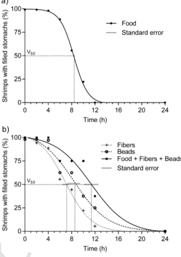
4.3. Stomach residence time of microplastic fibers and beads (Exp. 2) Gut evacuation of the shrimps followed a sigmoidal function. The coefficient of determination varied between R2= 0.9981 for food alone (Fig. 4a) and R2= 9.9883 for fibers, R2= 0.9714 for beads, and R2= 0.9519 for the entire mixture of food, fibers, and beads (Fig. 4b).
Food remained in all stomachs for the first 4 h but all stomachs were evacuated within 12 h. 50% of stomach evacuation was reached after 8.39 ± 0.13 h (Fig. 4a). Fibers were evacuated from the stomach most rapidly (Fig. 4b). Already after 2 h, the first stomachs were void of fibers. After 6.96 ± 0.55 h, 50% of the animals had no fibers in their stomachs. Thereafter, the animals continuously emptied their stom- achs within about 12 h. Beads were evacuated from the stomach at a similar rate as food. Half of the animals showed empty stomachs after
Fig. 3.Numbers of ingested plastic microfibers in stomachs ofPalaemon varians, which were offered 0.5 mg of microplastic fibres and variable amounts of commercial food (0, 2.5, 5, 10 mg) in 100 ml of water. Box plots show median, 25th and 75th per- centile, minimum and maximum values. Asterisks indicate significant differences be- tween animals, which were not fed and those which received 2.5 and 5 mg of food, re- spectively (ANOVA and Dunnett's multiple comparison on sqrt-transformed data, p <
0.05 (*), p < 0.01 (**), n = 8–10).
Fig. 4.Evacuation of a) food and b) plastics and food from shrimp stomachs. After feeding, animals were inspected for the presence of food and/or plastics in their stom- achs. The share (%) of animals with filled stomachs was plotted against the time. The times at which 50% of the stomachs were empty (V50), and their standard errors, served as measure to compare evacuation times. Overlapping standard error indicate no sig- nificant difference between V50of different treatments. The standard error bar in Fig.
4a is smaller than the size of the symbols in the graph. a) 9 shrimps were deployed and analyzed (n = 9). b) 18 shrimps were deployed and n = 18 were analyzed for fibers, n = 9 were analyzed for beads, n = 9 were analyzed for the entire stomach content (fibers, beads, and food).
8.38 ± 1.05 h but complete evacuation took more than 12 h. Total evac- uation of food, fibers and beads lasted longest. Half of the animals showed empty stomachs after 11.37 ± 1.22 h (Fig. 4b). Evacuation (V50± SE) of fibers was significantly faster than evacuation of food.
Evacuation of beads did not vary significantly from that of food. Evac- uation of the mix of food, fibers, and beads lasted significantly longer than the evacuation of food alone.
4.4. Survival
The ingestion of fibers and microspheres did not seem to enhance mortality of the shrimps. Only one out of 18 animals (i.e. 5.6%) died within 48 h of exposure.
4.5. Egestion and regurgitation
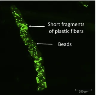
Most of the food remains and the microbeads were transferred from the stomach into the midgut and hindgut of the shrimp. Finally, they were egested as fecal strings, encased by a peritrophic mem
UNCORRECTED
PROOF
brane (Fig. 5). Besides microbeads, the fecal strings also contained some small fibers of about 100μm and less. The large fibers (>100μm) remained in the stomach but were cleared by regurgitation through the esophagus. Regurgitated material appeared as plaque-like mucous at the bottom of the aquaria (Fig. 6). It contained fluorescent fibers and undefined material, presumably food remains and gastric fluid. An example for a regurgitated plaque with various common air- borne microplastic pollutants is presented in the Supplementary mate- rial (Fig. S1).
Fig. 5.A fecal string ofPalaemon varianscontaining very short fragments of fluores- cent microplastic fibers and beads. The fecal string is coated with a chitinous peritrophic membrane.
Fig. 6.Mucus-like plaque of regurgitated material at the bottom of the aquarium of an- imals fed with experimentally administered green microplastic fibers (MPFs) and food.
(For interpretation of the references to color in this figure legend, the reader is referred to the Web version of this article.)
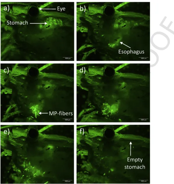
The process of regurgitation is shown in a time-lapse movie (Sup- plementary material). The fluorescent fibers were visible in the stom- ach (Fig. 7a). After contraction of the stomach, the fibers appeared in the esophagus (Fig. 7b). Fig. 7c and d show the release of the stomach content including fibers through the esophagus. Thereafter, the regur- gitated material dispersed (Fig. 7e). Finally, the stomach was empty and few fluorescent particles remained attached to the setae of the mouthparts (Fig. 7f). The process of stomach evacuation lasted less than 3 min.
Supplementary video related to this article can be found at https://
doi.org/10.1016/j.envpol.2019.113068.
5. Discussion
Fluorescent microplastic fibers and microspheres were ingested by the shrimps,Palaemon varians, preferably together with the offered food. The lengths of the fibers resemble those of fibers collected in the habitat of the shrimps, the brackish Baltic Sea waters off Sweden (290–2700μm, Gewert et al., 2017). The fibers were retained in the stomach while the microbeads were passed further into the gut. The latter were egested together with undigested food remains within fe- cal strings. After few hours, the fibers disappeared from the stomach as well and mucous plaques containing fluorescent fibers appeared at the bottoms of the aquaria in which the animals were maintained. The fibers were not egested through the gut, but left the stomach through the esophagus. This process has been reported previously as regurgi- tation.
Regurgitation is a common process in various invertebrates (Table 1). It is associated with e.g. defense behavior, food transfer among individuals, extracellular digestion, or defense suppression. In crus- taceans, it has been described as a mechanism to remove large and hardly digestible food particles from the stomach. Already Forster and Gabbott (1971) reported that regurgitation of parts of the indigestible food remains after the meal complicated the accurate determination of assimilation efficiencies in the prawnsPalaemon serratusand Pan- dalus platyceros. In the paddle crabOvalipes catharus (Brachyura) shell fragments were cleared from the foregut by regurgitation after virtually all soft parts had been digested (Haddon and Wear, 1987).
The speed of food procession and onset of regurgitation depended on temperature but not on animal size. At 20 °C, regurgitation happened within 3–6 h, at 11 °C within 11 h, and at 9 °C within 18 h.
Even the pelagic pluteus larvae of the Pacific Sand Dollar,Den- draster excentricus, are able to regurgitate too large or offending par- ticles from their esophagus by a reverse wave of peristaltic contrac- tions (Burke, 1981). Kaposi et al. (2014) observed that the number of plastic particles ingested by the pluteus larvae of the sea urchinTrip- neustes gratilladecreased with time. The authors suggested that the larvae recognized the low nutritional value of the plastic particles and rejected their ingestion or egested them over time. Another mecha- nism to select between digestible and indigestible food items is the formation of pseudofeces by certain bivalve species, such as the Blue musselMytilus edulis(Beninger and St-Jean, 1997; Beninger et al., 1999).
Apart from our laboratory experiments under controlled and clean conditions (Wesch et al., 2017) a few animals were left in an open uncontrolled area where they were exposed to the common airborne microplastics. These animals regurgitated plaque with various dark and blue-colored fibers (Fig. S1), which are most likely contaminants originating from clothes or other air-borne discharge. They resem- ble those fibers collected from the atmospheric fallout of urban envi- ronments or indoor facilities (Dris et al., 2016, 2017). This example,
UNCORRECTED
PROOF
Fig. 7.Sequence of time-lapse recordings showing regurgitation of fluorescent fibres and beads from a lateral view ofP. varianscephalothorax (a–d). The whole process of regurgi- tation lasted 160 s. Thereafter, the stomach of the shrimp was void of fibres and beads (e, f). MP-fibers = microplastic fibres.
however, clearly demonstrates that the shrimps are also capable of re- gurgitating the meanwhile ubiquitously occurring microplastic conta- minants.
The microplastic fibres used in our experiments had a similar shape as e.g. cellulose or lignin fibres. They were likely too large to be passed into the gut but were, instead, retained in the stomach and then regurgitated. However, when the animals received only food without microplastics, regurgitation also took place suggesting this behavior being no exclusive reaction to plastic contamination. An explanation for this behavior could be an adaptation ofP. variansto the omniv- orous, especially the herbivorous and detrivorous life style in parti- cle-rich estuarine waters. In this way, the numerous indigestible or- ganic and inorganic particles could be eliminated before injuring or clogging the digestive tract of the organism. Accordingly, the mortal- ity ofP. varianswas low in our experiments. Only one animal died for unknown reason.
Different toP. varians, the closely related speciesPalaemonetes pugio, was not able to clear its digestive tract from polypropylene fibres after a 3 h exposure to 34 and 96μm long fibres (Gray and
Weinstein, 2017). As a result, 100% (34μm) and 43% (93μm) of the shrimps died within 96 h.P. pugiolives in salt marshes at the Atlantic US coast of North America and, thus, encounters similar environmen- tal and trophic conditions asP. varians. However, the ability to re- gurgitate fibres seems not to have evolved in this species. Similarly, the Norway lobster Nephrops norvegicuswas shown to be unable to regurgitate indigestible items. Fibers accumulated in the stomach, formed small balls, and clogged the stomach. These balls could only be removed when the ectodermal structures of the stomach were shed off during moulting (Welden and Cowie, 2016a,b). Intermoult peri- ods, however, lasted for several months during which animals showed signs of starvation due to the accumulation of the indigestible mate- rial in their stomachs. Besides filling the stomach with indigestible material, the microplastic particles may cause inflammation reactions and oxidative stress as shown in mollusks, rotifers, and copepods (von Moos et al., 2012; Jeong et al., 2016, 2017).
Although regurgitation has been described for several crustacean species, the anatomical requirements, the physiological control mech- anisms, and the triggers for the regurgitation process are still un
UNCORRECTED
PROOF
Table 1
Crustaceans and other invertebrate species showing regurgitation.
Species Taxon
Regurgitated
material Reference
Cancer pagurus Crustacea– Brachyura
Exoskeletal fragments
Lawton (1989) Cancer gracilis Crustacea–
Brachyura
Food at low salinity McGaw (2006) Cancer productus Crustacea -
Brachyura
Not specified stomach content
McGaw (2007) Ovalipes catharus Crustacea–
Brachyura
Shell fragments Haddon &
Wear (1987) Scylla serrata Crustacea–
Brachyura
Shell fragments Hill (1976) Palaemon serratus Crustacea -
Caridea
Cellulose fibers Fair et al.
(1980) Palaemon serratus Crustacea -
Caridea Indigestible material Forster and Gabbott (1971) Pandalus
platyceros
Crustacea - Caridea
Cellulose, lignin, fragments of fish scales and bones
Forster and Gabbott (1971) Formica sanguinea Insecta–
Formicidae
Food exchange Wallis (1961) Lasius flavus Insecta–
Formicidae
Extracellular digestion, defense
Cammaerts (1995) Malacosoma
americanum
Insecta– Lepidoptera
Enteric fluid for defense
Peterson et al (1987) Spodoptera
littoralis Insecta– Lepidoptera
Suppression of plant
defense Vadassery et
al. (2012) Schistocera
emarginata
Insecta– Caelifera
Gastric fluid for defense
Sword (2001) Locusta migratoria Insecta–
Caelifera
Crop fluid for defense
Freeman (1968) Various species Instecta Suppression of plant
defense
Timisena and Mikó (2017) Dendraster
excentricus
Echinodermata, Echinoidea
Offending particles Burke (1981) Pleurobranchaea
californica
Mollusca– Gastropoda
Experimentally administered rotten squid or soap solution
McClellan (1982)
known. Sousa and Petriella (2006) suggested, that the lack of an oe- sophagial valve at the entrance of the pyloric stomach of the shrimp Palaemonetes argentinus may enable regurgitation. However, the mud crab,Scylla serratahas a tri-lobed valve between the short esoph- agus and the cardiac stomach (Barker and Gibson, 1978) but is able to regurgitate shell fragments (Hill, 1976). The regurgitation process starts with rapid waves of contraction in the posterior region of the cardiac stomach forcing food upward, while the simultaneous open- ing of the esophagus allows expulsion of the food (McGaw and Curtis, 2013). To facilitate food expulsion, it appears necessary to tighten the passage between stomach and midgut to avoid pressing the stom- ach content into the gut. Many crustaceans, like the brachyuran crabs Scylla serrataand Menippe rumphii,possess a cardio-pyloric valve separating the cardiac and pyloric regions of the stomach allowing the passage of only very small particles (Barker and Gibson, 1978;
Erri Babu et al., 1982). Additional comparative studies on crustaceans, which are capable of regurgitation (Table 1) and those which are not, like the Norway lobster,N. norvegicus, are required to understand the behavior and physiological mechanism of regurgitation.
Our experiments also indicate that shrimps are able to empty their stomach contents selectively. Indigestible plastic fibers were evac- uated faster than food. Apparently, the animals recognized that the plastic fibers did not provide nutrients and, thus, released the syn- thetic stomach content on average 1.4 h earlier than the food content.
A mixture of food and fibers, however, increased the stomach evacu
ation time by 3 h. We suggest, that the presence of plastic fibers mimics additional food, which, apparently, needs more time to be processed and digested. Consequently, the process of regurgitation seems to be linked to the sensory recognition for mechanical and chemical stimuli within the digestive organs as reported for e.g. in- sects (Park and Kwon, 2011).
The shrimps ingested most of the fibers along with food but only few when fibers were offered alone. This resembles natural feeding behavior where shrimps would avoid ingestion of indigestible mate- rial. However, indigestible or hardly digestible compounds of organ- isms such as e.g. silica shells of diatoms, chitin bristles of annelids, or calcareous shells of bivalves are inevitably ingested as part of the prey and need to be eliminated from the digestive tract. In addition to chem- ical and mechanical selection of food items and gastric filter-separa- tion of chyme and solids (Saborowski, 2015), regurgitation represents another sorting mechanism to efficiently separate assimilable material from indigestible remains.
6. Conclusions
Regurgitation reflects an evolutionary adaptation of crustaceans to remove large indigestible natural food items from the stomach.
Nowadays it may also serve as an efficient mechanism to get rid of ingested particulate anthropogenic pollutants, such as microplas- tic fibers. Species, which are able to extricate themselves from such a burden may have better risk scores and will be less affected by mi- croplastic pollution than others which are not able to do so. In turn, knowledge about biological features of species, such as regurgitation or other sorting mechanisms in the digestive tract, can help to esti- mate the vulnerability of species to ecotoxicological threats. This is- sue might be of interest for various stakeholders, including aquacul- ture farmers and environmental policy makes and should be addressed in future environmental risk assessment strategies.
Conflicts of interest
The authors declare no conflict of interest.
Funding
This work was performed within the research program PACES II of the Helmholtz Society. It did not receive any specific grant from funding agencies in the public, commercial, or not-for-profit sectors.
Acknowledgments
Dr. Ismeni Walter helped in creating the time-lapse video sequence of the regurgitation process.
Appendix A. Supplementary data
Supplementary data to this article can be found online at https://
doi.org/10.1016/j.envpol.2019.113068.
References
Andrady, A.L., 2011. Microplastics in the marine environment. Mar. Pollut. Bull. 62 (8), 1596–1605.
Au, S.Y., Bruce, T.F., Bridges, W.C., Klaine, S.J., 2015. Responses ofHyalella azteca to acute and chronic microplastic exposures. Environ. Toxicol. Chem. 34 (11), 2564–2572.
Barker, P.L., Gibson, R., 1978. Observations on the structure of the mouthparts, histol- ogy of the alimentary tract, and digestive physiology of the mud crabScylla ser- rata(Forskål) (Decapoda: Portunidae). J. Exp. Mar. Biol. Ecol. 32, 177–196.
UNCORRECTED
PROOF
Barnes, D.K.A., Galgani, F., Thompson, R.C., Barlaz, M., 2009. Accumulation and fragmentation of plastic debris in global environments. Philos. Trans. R. Soc. B 364, 1985–1998.
Beninger, P.G., St-Jean, S.D., 1997. Particle processing on the labial palps ofMytilus edulisandPlacopecten magellanicus(Mollusca: Bivalvia). Mar. Ecol. Prog. Ser.
147, 117–127.
Beninger, P.G., Veniot, A., Poussart, Y., 1999. Principles of pseudofeces rejection on the bivalve mantle: integration in particle processing. Mar. Ecol. Prog. Ser. 178, 259–269.
Bråte, I.L.N., Hurley, R., Iversen, K., Beyer, J., Thomas, K.V., Steindal, C.C., Green, N.W., Olsen, M., Lusher, A., 2018.Mytilusspp. as sentinels for monitoring mi- croplastic pollution in Norwegian coastal waters: a qualitative and quantitative study. Environ. Pollut. 243, 383–3993.
Browne, M.A., 2015. Sources and pathways of microplastics to habitats. In: Bergmann, M., Gutow, L., Klages, M. (Eds.), Marine Anthropogenic Litter. Springer, Cham, pp. 229–244.
Burke, R.D., 1981. Structure of the digestive tract of the pluteus larva ofDendraster excentricus(Echinodermata: Echinoida). Zoomorphology 98, 209–225.
Cammaerts, R., 1995. Regurgitation behaviours in theLasius flavusworker (Formici- dae) toward the myrmecophilous beetleClaviger testaceus(Pselaphidae) and other recipients. Behav. Process. 34, 241–264.
Cole, M., Lindeque, P., Halsband, C., Galloway, T.S., 2011. Microplastics as contami- nants in the marine environment: a review. Mar. Pollut. Bull. 62, 2588–2597.
De Falco, F., Gullo, M.P., Gentile, G., Di Pace, E., Cocca, M., Gelabert, L., Brouta-Agnésa, M., Rovira, A., Escudero, R., Villalba, R., Mossotti, R., Montar- solo, A., Gavignano, S., Tonin, C., Avella, M., 2018. Evaluation of microplastic release caused by textile washing processes of synthetic fabrics. Environ. Pollut.
236, 916–925.
Desforges, J.-P.W., Galbraith, M., Ross, P.S., 2015. Ingestion of microplastics by zoo- plankton in the northeast Pacific ocean. Arch. Environ. Contam. Pollut. 69, 320–330.
Devriese, L.I., van der Meulen, M.D., Maes, T., Bekaert, K., Paul-Pont, I., Frère, L., Robbens, J., Vethaak, A.D., 2015. Microplastic contamination in brown shrimp (Crangon crangon, Linnaeus 1758) from coastal waters of the southern North Sea channel area. Mar. Pollut. Bull. 98, 179–187.
Dris, R., Gasperi, J., Saad, M., Mirande, C., Tassin, B., 2016. Synthetic fibers in at- mospheric fallout: a source of microplastics in the environment. Mar. Pollut. Bull.
104, 290–293.
Dris, R., Gasperi, J., Mirande, C., Mandin, C., Guerrouache, M., Langlois, V., Tassin, B., 2017. A first overview of textile fibers, including microplastics, in indoor and outdoor environments. Environ. Pollut. 221, 453–458.
Erri Babu, D., Shyamasundrari, K., Hanumantha Rao, K., 1982. Studies on the diges- tive system of the crabMenippe rumphii(Fabricius) (Crustacea: Brachyura). J.
Exp. Mar. Biol. Ecol. 58, 175–191.
Fair, P.H., Fortner, A.R., Millikin, M.R., Sick, L.V., 1980. Effects of dietary fiber on growth, assimilation and cellulose activity of the prawn (Macrobrachium rosen- bergii). Proc. World Maricult. Soc. 11, 369–381.
Forster, R.M., Gabbott, P.A., 1971. The assimilation of nutrients from compounded di- ets by the prawnsPalaemon serratusandPandalus platyceros. J. Mar. Biol. As- soc. U. K. 51, 943–961.
Freeman, M.A., 1968. Pharmacological properties of the regurgitated crop fluid of the African migratory locust.Locusta migratoriaL. Comp. Biochem. Physiol. 26, 1041–1049.
Gewert, B., Ogonowski, M., Barth, A., MacLeod, M., 2017. Abundance and composi- tion of near surface microplastics and plastic debris in the Stockholm Archipelago, Baltic Sea. Mar. Pollut. Bull. 120, 292–302.
Gray, A.D., Weinstein, J.E., 2017. Size- and shape-dependent effects of microplastic particles on adult daggerblade grass shrimp (Palaemonetes pugio). Environ. Toxi- col. Chem. 36 (11), 3074–3080.
Haddon, M., Wear, R.G., 1987. Biology of feeding in the New Zealand paddle crab Ovalipes catharus(Crustacea, Portunidae). N. Z. J. Mar. Freshw. Res. 21, 55–64.
Hämer, J., Gutow, L., Köhler, A., Saborowski, R., 2014. Fate of microplastics in the marine isopodIdotea emarginata. Environ. Sci. Technol. 48 (22), 13451–13458.
Hill, B.J., 1976. Natural food, foregut clearance-rate and activity of the crabScylla ser- rata. Mar. Biol. 34, 109–116.
Jambeck, J.R., Geyer, R., Wilcox, C., Siegler, T.R., Perryman, M., Andrady, A., Narayan, R., Law, K.L., 2015. Plastic waste inputs from land into the ocean. Sci- ence 347 (6223), 768–771.
Jemec, A., Horvat, P., Kunej, U., Bele, M., Kržan, A., 2016. Uptake and effects of mi- croplastic textile fibres on freshwater crustaceanDaphnia magna. Environ. Pollut.
219, 201–209.
Jeong, C.-B., Won, E.-J., Kang, H.-M., Lee, M.-C., Hwang, D.-S., Hwang, U.-K., Zhou, B., Souissi, S., Lee, S.-J., Lee, J.S., 2016. Microplastic size-dependence tox- icity, oxidative stress induction, and p-JNK and p-p38 activation in the mono- gonont rotifer (Brachionus koreanus). Environ. Sci. Technol. 50 (16), 8849–8857.
Jeong, C.-B., Kang, H.-M., Lee, M.-C., Kim, D.-H., Han, J., Hwang, D.-S., Souissi, S., Lee, S.-J., Shin, K.-H., Park, H.G., Lee, J.-S., 2017. Adverse effects of microplas
tics and oxidative stress-induced MAPK/Nrf2 pathway mediated defense mecha- nisms in the marine copepodParacyclopina nana. Sci. Rep. 7, 41323.
Kaposi, K.L., Mos, B., Kelaher, B.P., Dworjanyn, S.A., 2014. Ingestion of microplas- tic has limited impact on a marina larva. Environ. Sci. Technol. 48 (3), 1638–1645.
Lawton, P., 1989. Predatory interaction between the brachyuran crabCancer pagurus and decapod crustacean prey. Mar. Ecol. Prog. Ser. 52, 169–179.
Macfadyen, G., Huntington, T., Cappell, R., 2009. Abandoned, lost or otherwise dis- carded fishing gear. UNEP Regional Seas Reports and Studies, No. 185. FAO Fisheries and Aquaculture Technical Paper, No. 523 UNEP/FAO, Rome, 115pp.
Mason, S.A., Garneau, D., Sutton, R., Chu, Y., Ehmann, K., Barnes, J., Fink, P., Pa- pazissimos, D., Rogers, D.L., 2016. Microplastic pollution is widely detected in US municipal wastewater treatment plant effluent. Environ. Pollut. 218, 1045–1054.
Mathalon, A., Hill, P., 2014. Microplastic fibers in the intertidal ecosystem surround- ing Halifax harbor, Nova Scotia. Mar. Pollut. Bull. 81, 69–79.
McClellan, A.D., 1982. Movement and motor patterns of the buccal mass ofPleuro- brancaeaduring feeding, regurgitation and rejection. J. Exp. Biol. 98, 195–211.
McGaw, I.J., 2006. Feeding and digestion in low salinity in an osmoconforming crab, Cancer gracilisII. Gastric evacuation and motility. J. Exp. Biol. 209, 3777–3785.
McGaw, I.J., 2007. Gastric processing and evacuation during emersion in red rock crab,Cancer productus. Mar. Freshw. Behav. Physiol. 40 (2), 117–131.
McGaw, I.J., Curtis, D.L., 2013. A review of gastric processing in decapod crus- taceans. J. Comp. Physiol. B 183, 443–465.
Murray, F., Cowie, P.R., 2011. Plastic contamination in the decapod crustacean Nephrops norvegicus(Linnaeus, 1758). Mar. Pollut. Bull. 62, 1207–1217.
Napper, I.E., Thompson, R.C., 2016. Release of synthetic plastic fibres from domestic washing machines: effects of fabric type and washing conditions. Mar. Pollut.
Bull. 112, 39–45.
Park, J.-H., Kwon, J.Y., 2011. Heterogeneous expression ofDrosophilagustatory re- ceptors in enteroendocrine cells. PLoS One 6 (12), e29022https://doi.org/10.1371/
journal.pone.0029022.
Petersen, S.C., Johnson, N.D., LeGuyader, J.L., 1987. Defense regurgitation of al- lochemcials derived from host cyanogenesis by eastern tent caterpillars. Ecology 68 (5), 1268–1272. https://doi.org/.
Rochman, C.M., Tahir, A., Williams, S.L., Baxa, D.V., Lam, R., Miller, J.T., The, F.-C., Werolilangi, S., The, S.J., 2015. Anthropogenic debris in seafood: plastic debris and fibers from textiles in fish and bivalves sold for human consumption.
Sci. Rep. 5, 14340.
Saborowski, R., 2015. Nutrition and digestion. In: Chang, E., Thiel, M. (Eds.), Natural History of the Crustacea. Vol IV Physiological Regulation. Oxford University Press, New York, pp. 285–319.
Salvador Cesa, F., Turra, A., Baruque-Ramos, J., 2017. Synthetic fibers as microplas- tics in the marine environment: a review from textile perspective with a focus on domestic washings. Sci. Total Environ. 598, 1116–1129.
Santana, M.F.M., Moreira, F.T., Pereira, C.D.S., Abessa, D.M.S., Turra, A., 2018.
Continuous exposure to microplastics does not cause physiological effects in the cultivated musselPerna perna. Arch. Environ. Contam. Toxicol. 74, 594–604.
Sousa, L., Petriella, A.M., 2006. Morphology and histology ofP. argentinus(Crus- tacea, Decapoda, Caridea) digestive tract. Biocell 30 (2), 287–294.
Sword, G.A., 2001. Tasty on the outside, but toxic in the middle: grasshopper regurgi- tation and host plant-mediated toxicity to a vertebrate predator. Oecologia 128, 416–421.
ter Halle, A., Ladirat, L., Gendre, X., Goudouneche, D., Pusineri, C., Routaboul, C., Tenailleau, C., Duployer, B., Perez, E., 2016. Understanding the fragmentation pattern of marine plastic debris. Environ. Sci. Technol. 50, 5668–5675.
Timilsena, B.P., Mikó, I., 2017. Know your insect: the structural backgrounds of regur- gitation, a case study onManduca sextaandHeliothis virescence(Lepidoptera : Sphingidae, Noctuidae). Res. Ideas Outcomes 3, e11997https://doi.org/10.3897/
rio.3.e11997.
Vadassery, J., Reichelt, M., Mithöfer, A.S., 2012. Direct proof of ingested food regur- gitation bySpodoptera littoraliscaterpillars during feeding onArabidopsis. J.
Chem. Ecol. 38, 865–872. https://doi.org/10.1007/s10886-012-0143-5.
von Moos, N., Burkhardt-Holm, P., Köhler, A., 2012. Uptake and effects of of mi- croplastics on cells and tissue of the Blue musselMytilus edulisL. after an experi- mental exposure. Environ. Sci. Technol. 46 (20), 11327–11335.
Wallis, D.I., 1961. Food-sharing behaviour of the antsFormica sanguineaand Formica fusca. Behaviour 17 (1), 17–47.
Weber, A., Scherer, C., Brennholt, N., Reifferscheid, G., Wagner, M., 2018. PET mi- croplastics do not negatively affect the survival, development, metabolism and feeding activity of the freshwater invertebrateGammarus pulex. Environ. Pollut.
234, 181–189.
Weinstein, J.E., Crocker, B.K., Gray, A.D., 2016. From macroplastics to microplastics:
degradation of high-density polyethylene, polypropylene, and polystyrene in a salt marsh habitat. Environ. Toxicol. Chem. 35 (7), 1632–1640.
Welden, N.A.C., Cowie, P.R., 2016a. Environment and gut morphology influence mi- croplastic retention in langoustine,Nephrops norvegicus. Environ. Pollut. 214, 859–865.
UNCORRECTED
PROOF
Welden, N.A.C., Cowie, P.R., 2016b. Long-term microplastic retention causes reduced body condition in the langoustine,Nephrops norvegicus. Environ. Pollut. 218, 895–900.
Wesch, C., Elert, A.M., Wörner, M., Braun, U., Klein, R., Paulus, M., 2017. Assuring quality in microplastic monitoring: about the value of clean-air devices as essen- tials for verified data. Sci. Rep. 7, 5424.
E T O C B L U R B
The evolutionary developed process of regurgitation in shrimps provides nowadays a suitable mechanism to eliminate ingested anthropogenic mi
croplastic fibers from the stomach, avoiding accumulation and, thus, health impairment.