M OLECULAR U NDERPINNINGS OF A NXIETY R EGULATION : N OVEL I NSIGHTS INTO THE R OLE
OF THE P URINERGIC AND O XYTOCINERGIC
S YSTEMS WITHIN THE P ARAVENTRICULAR
N UCLEUS OF THE H YPOTHALAMUS
DISSERTATION ZUR ERLANGUNG DES DOKTORGRADES DER NATURWISSENSCHAFTEN (DR. RER. NAT.) DER FAKULTÄT FÜR
BIOLOGIE UND VORKLINISCHE MEDIZIN DER UNIVERSITÄT REGENSBURG
vorgelegt von Stefanie Martinetz
aus München
im Jahr 2014
2
3 Das Promotionsgesuch wurde eingereicht am:
27. Mai 2014
Die Arbeit wurde angeleitet von:
Prof. Dr. rer. nat. Inga D. Neumann Unterschrift:
4
Content Overview
Introduction ... 11
Materials & Methods ... 37
Results ... 57
Discussion ... 90
Conclusions and perspectives ... 109
Summary ... 112
Deutsche Zusammenfassung ... 115
List of abbreviations ... 118
Bibliography ... 123
CV and list of publications ... 146
Danksagung ... 148
5
Content
Introduction ... 11
1. Positive and negative aspects of anxiety ... 11
2. Neurobiology of anxiety ... 12
3. Drug treatment of anxiety disorders ... 14
4. In vivo models of anxiety ... 16
5. Regulation of anxiety by neuropeptides ... 18
5.1. The neuropeptide oxytocin ... 18
5.2. Molecular mechanism of oxytocin’s effect on anxiety ... 19
5.3. Further involvement of neuropeptides in anxiety ... 20
6. ATP as a neurotransmitter ... 22
6.1. P2X4 receptor ... 25
7. Protein synthesis ... 26
8. MicroRNAs and the regulation of gene expression ... 29
8.1. General information... 29
8.2. MicroRNAs in brain and behaviour ... 30
8.3. Regulation of the microRNA pathway ... 32
9. Aims and outline of the present thesis ... 33
9.1. Aim 1: Identification of a novel anxiolytic factor within the PVN of rats ... 34
6
9.2. Aim 2: Elucidation of the role of de novo protein synthesis in the mediation of the
(long-term) anxiolytic effect of oxytocin ... 34
9.3. Aim 3: Determination of the control of microRNA expression levels by OT in the hypothalamus ... 35
Materials & Methods ... 37
1. Animals ... 37
2. Surgical Procedures ... 37
3. Cells ... 38
3.1. H32 cells ... 38
3.2. Be(2)-M17 cells ... 39
3.3. Primary hypothalamic neurons ... 39
3.4. Cell stimulation ... 40
4. Behavioural studies ... 40
5. RNA studies ... 42
5.1. RNA extraction ... 42
5.2. Microarray ... 42
5.3. Deep Sequencing ... 43
5.4. PCR and qPCR ... 44
5.5. Northern Blot ... 47
6. Protein studies... 48
6.1. Protein extraction ... 48
7
6.2. SDS-PAGE and Western Blot analysis ... 50
6.3. Labelling, tagging and affinity purification of newly synthesized proteins ... 52
6.4. Dot Blot ... 54
6.5. Immunohistochemistry ... 54
7. Statistics ... 55
Results ... 57
Part I: P2X4R as a regulator of anxiety in the PVN of Wistar rats ... 57
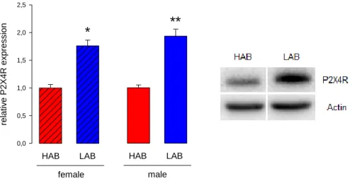
Experiment 1: Validation of a microarray study showing differences in gene expression profile in the PVN of female virgin HAB and LAB rats ... 57
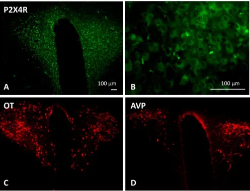
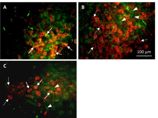
Experiment 2: Immunostaining of the ATP-receptor P2X4R in the hypothalamus of rats ... 59
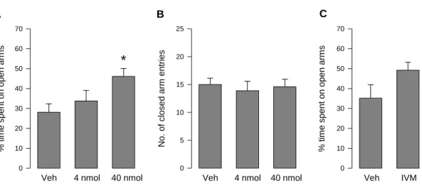
Experiment 3: Effects of the regulation of P2X4R within the PVN on anxiety-like behaviour in Wistar rats ... 61
Experiment 4: Effects of the regulation of P2X4R within the PVN on anxiety-like behaviour in HAB and LAB rats ... 65
Experiment 5: Elucidation of possible signalling cascades in hypothalamic cells following P2X4R activation ... 66
Part II: Oxytocin activates protein synthesis in the rat hypothalamus ... 72
Experiment 1: Effect of OT on key modulators of protein synthesis ... 72
Experiment 2: Elucidation of the signalling pathway for the OT-mediated eEF2- activation in the rat’s hypothalamus ... 74
8
Experiment 3: Verification of enhanced protein synthesis in the hypothalamus after OT-
treatment ... 76
Experiment 4: NPY5R as selected protein target for the regulatory properties of OT .... 78
Experiment 5: Inhibition of protein synthesis in the PVN and the effects on the anxiolytic property of OT ... 79
Part III: Oxytocin has regulatory effects on the microRNAome of the hypothalamus ... 82
Experiment 1: Determination of the expression of microRNAs in the hypothalamus with and without OT-treatment via Deep Sequencing ... 82
Experiment 2: Validation of the Deep Sequencing ... 85
Discussion ... 90
Part I: P2X4R as a regulator of anxiety in the PVN of Wistar rats ... 90
Part II: Oxytocin activates protein synthesis in the rat hypothalamus ... 97
Part III: Oxytocin has regulatory effects on the microRNAome of the hypothalamus ... 103
Conclusions and perspectives ... 109
Summary ... 112
Deutsche Zusammenfassung ... 115
List of abbreviations ... 118
Bibliography ... 123
CV and list of publications ... 146
Curriculum vitae ... 146
List of publications ... 147
9
Danksagung ... 148
10
INTRODUCTION
11
Introduction
1. Positive and negative aspects of anxiety
Feelings are what make life worth living. They can be beautiful and pleasant like love and joy, but there are also many situations throughout our life that make us angry, mad or anxious. When we encounter fire, our naturally occurring anxiety of the flames protects us from harm. Anxiety is described as a psychological, physiological, and behavioural state that is induced by a threat (Steimer, 2002). Thus, normal anxiety is healthy and extends our lifespan. Being afraid of something is not necessarily an inborn characteristic trait and can be reversed in many cases, like fear of a snake (Nili et al., 2010). On the other hand, phobias, such as arachnophobia (fear of spiders), claustrophobia (fear of having no escape and being closed in), or acrophobia (fear of heights) are examples of how exaggerated anxiety can be maladaptive, affecting the life of those suffering from it. Anxiety can also turn out as a serious threat for mental health, for example the everyday ongoing disturbing worry of panic disorder patients to have yet another panic attack. In the modern world, so-called anxiety disorders are becoming more and more problematic and the number of persons concerned rises dramatically (Wittchen et al., 2011).
The term “anxiety disorder” summarizes a whole group of psychiatric diseases, including phobia, panic disorder, generalized anxiety disorder (GAD), social anxiety disorder, obsessive-compulsive disorder (OCD), and posttraumatic stress disorder (PTSD). Phobias are characterised by a persistent fear of an object or situation, typically disproportionally high compared with the actual threat. Panic disorder is diagnosed when a person suffers from sudden and repeated attacks of fear that last for several minutes or longer, namely panic attacks. Panic attacks can occur at any time and are characterized by the fear of disasters or
12
losing control over a situation, even when there is no real danger. GAD causes the patient to have consistent worries about everyday life tasks, even when there is little or no reason for it, resulting in a permanent fear to fail or to not make it through the day. Social anxiety disorder or social phobia is the fear of being judged by others and of being embarrassed.
Affected persons feel extremely fearful and unsure around others. Even if they know that there is no need to, they cannot control their fear. Patients with OCD constantly feel the need to check and re-check things repeatedly, losing themselves in rituals by trying to gain control over their obsessions, which seriously affects their daily life. And, finally, PTSD is an anxiety disorder that might occur after seeing or living through a dangerous event. Persons concerned can be war veterans or survivors of physical or sexual assault, abuse, accidents, disasters and many other serious events, and even persons who suffer from a sudden, unexpected death of a loved one. The symptoms are flashbacks, bad dreams, feelings of strong guilt, depression, worry, or hyperarousal (NIMH).
In contrast to normal anxiety, the disordered variant lasts at least for months and can become more severe if no medication is applied. Although several treatment possibilities have already been discovered and are in use, the diversity of phenotypes of anxiety disorders demands for new therapeutic approaches incessantly.
2. Neurobiology of anxiety
Anxiety acts as a coping mechanism in dangerous situations and is therefore strongly associated with emotional processes as well as cognitive functions, such as learning and memory. Several neurotransmitter pathways are involved, including glutamate, - aminobutyric acid (GABA), serotonin, and norepinephrine. GABA is the primary inhibitory
13
neurotransmitter and its actions counterbalance those of the excitatory neurotransmitter glutamate. A down-regulation of the GABA system has been linked to the pathophysiology of anxiety disorders, and as such, GABA receptors are the known target of a number of pharmacological agents (Lydiard, 2003). In contrast, the excitatory glutamate neurotransmission is blocked by treatments to exert anxiolytic effects (Bergink et al., 2004).
Furthermore, alterations in serotonergic neurotransmission have been implicated in the development of mood and anxiety disorders as well (Ressler and Nemeroff, 2000). In particular, the inhibitory serotonergic receptor 1a (5-HT1A) was revealed to be important, since a 5-HT1A knock-out (KO) leads to increased anxiety and stress responsivity (Heisler et al., 1998). Activation of the 5-HT1A autoreceptor leads to hyperpolarization of the serotonergic cell and thus self-inhibition as a form of negative feedback regulation of transmitter release (Celada et al., 2004). The norepinephrinic neurons of the locus coeruleus project to the forebrain, and play a critical role in fear response, stress, and arousal (Bremner et al., 1996). The central effects of norepinephrine are mediated via post-synaptic
1 and 1 receptors and the pre-synaptic 2 receptor. The 2 receptor is an autoreceptor, similar to the 5-HT1A, and inhibits norepinephrine release pre-synaptically. Consequently,
2 receptor agonists reduce anxiety-like behaviour (Dell'Osso et al., 2010).
With the aid of functional magnetic resonance imaging, it was observed that distinct brain regions are activated in response to anxiety-inducing stimuli, including the amygdala, the hippocampus and the frontal cortex (Davidson et al., 1999). This confirmed earlier studies which had shown that fear and anxiety are mediated by several interrelated limbic structures: the amygdala, the septo-hippocampal system, and the hypothalamus (Charney and Deutch, 1996). The amygdala is composed of functionally and morphologically varying
14
subnuclei with complex connectivity. The basolateral amygdala (BLA) is primarily glutamatergic, whereas the central amygdala (CeA) mostly consists of GABAergic neurons (Tye et al., 2011). The BLA integrates processed information about the environment, is the locus for fear memory (LeDoux, 2000), and robustly projects to the CeA. CeA-projections to the hypothalamus and the brainstem mediate the anxiety response, such as avoidance of open spaces in rodents (Adhikari, 2014). The bed nucleus of the stria terminalis (BNST) modulates anxiety through processing of input from the amygdala and relaying the processed information to hypothalamic and brainstem structures. The BNST was therefore proposed to be part of the “extended amygdala” (Alheid et al., 1998). The regulation of anxiety by the amygdala is further expanded by interplay of BLA, medial pre-frontal cortex (mPFC) and ventral hippocampus (vHPC). It was suggested that contextual and sensory inputs from the mPFC and the vHPC are integrated by the BLA, which in turn regulates CeA and BNST, which activate downstream regions to control anxiety-related behaviour (Adhikari, 2014).
3. Drug treatment of anxiety disorders
Tricyclic antidepressants (TCAs) have been used in psychiatry since the 1950s and act by inhibiting the serotonin and norepinephrine reuptake from the synaptic cleft (Feighner, 1999; Ravindran and Stein, 2010). TCAs are able to reduce the number of panic attacks and decrease anticipatory anxiety and are therefore helpful in the acute treatment of panic disorder (Andersch et al., 1991). In addition, clomipramine, one of the most investigated TCAs, is regarded as the gold standard treatment for OCD. However, their side effects are multiple and diverse, including sedation, constipation, sexual dysfunction, and a high
15
possibility of a toxic overdose (Ravindran and Stein, 2010). These side effects have imposed the development for alternative drugs for the use of TCAs.
Other compounds that target the serotonin and norepinephrine pathways are monoamine oxidase (MAO) inhibitors and selective serotonin reuptake inhibitors (SSRIs) or serotonin- norepinephrine reuptake inhibitors (SNRIs). MAO is responsible for the degradation of serotonin and norepinephrine, and its inhibition leads to increased availability of both neurotransmitters in the synapse. SSRIs inhibit the reuptake of serotonin, whereas SNRIs inhibit the reuptake of both monoamines at the pre-synaptic site. SSRIs and SNRIs are considered as basic pharmacotherapy agents for each of the anxiety disorders (Ravindran and Stein, 2010). MAO inhibitors, SSRIs, and SNRIs have fewer side effects than TCAs.
However, it was observed that there is an increased risk for suicidal thinking and behaviour in adolescents, resulting in a “black-box-warning” by the U.S. Food and Drug Administration on the labelling of those drugs (US Food and Drug Administration, 2007).
Anticonvulsant drugs are used for the treatment of different psychiatric diseases. They can differ in their chemical structure, and their mechanism of anxiolytic action is not completely elucidated yet. They are thought to reduce anxiety by decreasing the excessive neuronal activation within defined fear circuits in the brain. This was especially evidenced for pregabalin, which was shown to increase GABAergic inhibitory activity and reduce the release of excitatory neurotransmitters such as glutamate in several brain regions including the cortex, hypothalamus, amygdala, and hippocampus (Mico and Prieto, 2012).
Benzodiazepines are among the most commonly used drugs for the treatment of anxiety disorder, because of their efficacy, rapid onset of effect, and favourable side effect profile (Stevens and Pollack, 2005). They are positive allosteric modulators of the GABAA receptor
16
and therefore potentiate the effect of the inhibitory neurotransmitter GABA by increasing the frequency of chloride channel opening (Study and Barker, 1981). This inhibitory effect of the benzodiazepines is a double-edged sword: while low doses have anxiolytic and anticonvulsive effects, higher doses produce sedation, amnesia, and even unconsciousness (Saari et al., 2011). However, benzodiazepines provide a rapid and effective relief of symptoms, but adverse effects are, unfortunately, significant. Patients discontinuing benzodiazepine use may even experience uncomfortable withdrawal symptoms, which make a benzodiazepine therapy unfitting for individuals with a history of substance abuse (Ravindran and Stein, 2010).
The diverse risks and side effects of the established anxiolytic drugs provide the impetus for the continuation of basic research and the development of new therapeutic strategies to treat anxiety disorders. Some of the strategies concern the potential use of endogenous and exogenous modulators of glutamate and neuropeptide signalling. In particular, the putative anxiolytic activity of corticotropin-releasing factor (CRF) receptor antagonists, glutamate receptor antagonists, as well as of the neuropeptides oxytocin (OT), neuropeptide Y (NPY), vasopressin (AVP), neuropeptide S (NPS), and cholecystokinin (CCK) is currently under intensive survey (Mathew et al., 2008).
4. In vivo models of anxiety
Animal models were and still are an important aid for the research on psychiatric disorders.
For anxiety research, many different KO models and animals selectively bred for extreme anxiolytic or anxiogenic phenotypes have been used. Consensus is that evidence from more than one behavioural test is required to make an animal model appropriate for the testing of
17
new anxiolytics, and, similarly, it is clear that no animal model will ever be able to combine all complex aspects of mood and anxiety disorders (Rotzinger et al., 2010).
One of the animal models for anxiety is represented by the high-anxiety behaviour (HAB) and low-anxiety behaviour (LAB) rats. These Wistar rats were selected for their anxiety levels, determined by their behaviour on the Elevated Plusmaze (EPM), and allocated to one of the two groups. Compared to LAB rats, HABs show high anxiety in a variety of tests, prefer passive coping strategies, and show signs of increased stress vulnerability (Landgraf and Wigger, 2003). Furthermore, female lactating HAB rats show a higher amount of maternal care and a heightened aggression towards a virgin intruder compared with LAB rats (Bosch, 2011). In contrast, male LAB rats display more aggressive behaviour towards an intruder than HAB males (Veenema and Neumann, 2007). Experimental evidence revealed overexpression and -release of AVP in the paraventricular nucleus (PVN) as underlying mechanism for the behavioural phenomena (Landgraf and Wigger, 2003).
Many behavioural tests of anxiety have been developed, so that researchers have the possibility to choose between tests for exploratory behaviour, social behaviour, reflexive fear responding, conflict behaviour as well as defensive behaviour (Rotzinger et al., 2010).
The EPM and the Light-Dark-Box (LDB) are both tests that utilize anxiogenic stimuli of open spaces. It should always be taken into account that such exploratory tests are sensitive to changes in locomotion, which therefore should be observed as well, in order to obtain trustworthy results.
18
5. Regulation of anxiety by neuropeptides
5.1. The neuropeptide oxytocin
OT is a long-known and well-studied neurohypophysial hormone of nine amino acids. It is named after the “quick birth” which reveals its main function in the mammalian periphery: it is released during labour, facilitating birth itself as well as maternal bonding and lactation.
Additionally, OT has a variety of central effects, including the regulation of anxiety-related behaviour, stress-coping, and multiple aspects of social behaviour (Neumann and Landgraf, 2012). OT, synthesised in two hypothalamic nuclei, the PVN and the supraoptic nuclei (SON), binds to one single OT receptor (OTR), which is a 389-amino acid polypeptide with seven transmembrane domains belonging to the class I G-protein-coupled receptor family (Gimpl and Fahrenholz, 2001). The receptor has relatively unselective binding capacities as the affinity for OT is only about tenfold higher than that for AVP – the second neurohypophysial hormone that differs from OT only in two amino acids. It was found that only two aromatic residues of the OTR need to be changed to allow full binding of AVP (Gimpl and Fahrenholz, 2001). OTR are functionally coupled to Gq/11 proteins and hence stimulate the activity of phospholipase C-PLC. Consequently, inositol triphosphate (IP3) and 1,2-diacylglycerol (DAG) are generated and trigger the release of calcium (Ca2+) from intracellular stores and protein kinase C (PKC) activation, respectively (Gimpl and Fahrenholz, 2001). Ca2+-induced processes can hereby include changes in gene transcription and protein synthesis. Strakova et al. showed that the rat OTR transfected into Chinese hamster ovary cells is coupled to both the Gq/11 and the Gi/o proteins (Strakova et al., 1998). Gi/o signalling generates an increase of intracellular Ca2+, independent of the IP3 pathway (Hoare et al., 1999). In addition, OT can couple to Gs proteins that activate adenylate cyclase and increase cyclic
19
adenosine monophosphate (cAMP) production which leads to a sodium-dependent inward current (Alberi et al., 1997). Possibly, these various signalling pathways are differentially expressed in neuronal and peripheral tissue (Stoop, 2012).
5.2. Molecular mechanism of oxytocin’s effect on anxiety
The axons of magnocellular OT neurons terminate in the posterior lobe of the pituitary gland, being part of the classic hypothalamic-neurohypophysial system (Brownstein et al., 1980). Vis-à-vis, parvocellular neurons of the PVN project to a series of brain regions.
Consistent with this, microdialysis studies revealed the release of OT in the olfactory bulb, the dorsal hippocampus, the CeA, the septum, the nucleus of the solitary tract, the SON, and the PVN itself (Landgraf and Neumann, 2004; Neumann, 2007). OT has anxiolytic properties when released in brain regions involved in stress and anxiety regulation, such as the CeA and the PVN (Bale et al., 2001; Blume et al., 2008; Neumann, 2001; Neumann and Landgraf, 2012). Axons of hypothalamic OT neurons that project to the CeA lead to the activation of local inhibiting GABAergic circuits and thereby attenuate the fear response (Knobloch et al., 2012). Within the PVN, exogenously applied OT reduces anxiety-like behaviour in rats within 10 min after the application (Blume et al., 2008). A series of behavioural tests confirmed the anxiolytic effect of OT, among them the EPM, the LDB (Blume et al., 2008), the four-plate test, and the elevated zero maze (Ring et al., 2006). Endogenously released OT after successful mating exerts anxiolytic effects to at least 4 hours after mating in male rats, showing that a relatively short OT surge can lead to long-term anxiolysis (Waldherr and Neumann, 2007). Interestingly, intranasal application of OT in humans suppresses anxiety as shown in studies using the Trier Social Stress- and the Simulated Public Speaking Test (de Oliveira et al., 2012; Heinrichs et al., 2003). Male participants who received intranasal OT 50
20
min before stressor exposure reported a lower post-stress anxiety level (evaluated by a self- reporting questionnaire) than participants receiving placebo. The effect was even increased when they received social support by their best friend (Heinrichs et al., 2003).
Studies that focused on the elucidation of the molecular mechanism behind anxiolysis identified the mitogen-activated protein (MAP) kinase MEK1/2 as an important regulator of anxiety-like behaviour in the PVN of rats (Blume et al., 2008; Jurek et al., 2012). Since the OTR, being a G-coupled receptor, is not classically linked to the MAP-kinase system, it was suggested that the OTR transactivates the tyrosine kinase epidermal growth factor (EGF) receptor to subsequently activate the MAP-kinase cascade (Blume et al., 2008). This hypothesis was confirmed in a rat hypothalamic cell line by blocking the EGF receptor with the specific inhibitor AG1478 (Blume et al., 2008).
5.3. Further involvement of neuropeptides in anxiety
Neuropeptides of the brain are important modulators of physiology and behaviour. Their functions are mediated either via dendritic release or release at axonal terminals (Ludwig and Leng, 2006). Several neuropeptides have been shown to modulate the regulation of anxiety behaviour.
CRF and AVP are both regulators of the hypothalamic pituitary adrenal (HPA) axis activity and mediate central effects on emotional and cognitive behaviours. CRF is generally considered to have anxiogenic effects, but many studies utilizing CRF receptor 1 (CRFR1) antagonists have demonstrated that a stressor is necessary in order to see an anxiolytic effect of the antagonist (Deak et al., 1999; Heinrichs et al., 2002; Heinrichs et al., 1994;
Schulz et al., 1996). The results of AVP administration are mixed. Septal injection of AVP leads to anxiolysis as determined on the EPM (Appenrodt et al., 1998). Injection of an V1b
21
receptor antagonist has anxiolytic properties on the EPM as well, although this observation was troubled by effects on locomotor activity (Liebsch et al., 1996). A review of the effects of SSR149415, a V1b antagonist, concluded that clear anxiolytic effects were only found in stressful situations (Griebel et al., 2003).
Additionally, the behavioural phenotype of HAB and LAB rats is correlated with AVP expression at the level of the PVN. Single nucleotide polymorphisms (SNPs) in regulatory structures of the AVP gene are the basis for the differential behavioural outcomes. Due to these SNPs, AVP is overexpressed and overreleased in the PVN of HAB rats (Landgraf et al., 2007b).
The most abundant neuropeptide in the brain, NPY, binds to five currently investigated receptors distributed in the central nervous system (CNS). Infusion of NPY, either icv or into the amygdala, has anxiolytic effects (Broqua et al., 1995; Heilig et al., 1989; Kokare et al., 2005). A specific NPY1 and 5 receptor agonist, given icv, showed dose-dependent anxiolytic effects as well, narrowing down the regulatory effect of NPY to those two receptor subtypes (Sorensen et al., 2004).
CCK, which primarily acts as a mediator of satiety, is another highly abundant neuropeptide in the brain and is found in cortex and limbic brain regions (Beinfeld et al., 1981). CCK was reported to induce anxiogenic effects, by activating the CCKB receptor in the BLA (Rotzinger and Vaccarino, 2003). CCKB agonists show anxiogenic effects in a number of tests. However, antagonists often have no effects on baseline anxiety behaviour, but instead modulate heightened states of anxiety (Rotzinger and Vaccarino, 2003; Wilson et al., 1998). The CCK fragment pentagastrin increases anxiety in the human social interaction test (McCann et al., 1994).
22
Only recently, NPS was discovered to increase wakefulness and arousal on the one hand, and to produce anxiolytic-like effects by reducing fear-responses on the other hand when administered centrally (Xu et al., 2004). NPS binds to a G-protein-coupled receptor and stimulates the mobilization of intracellular Ca2+ as well as activation of protein kinases (Pape et al., 2010). NPS receptor expression is found in brain regions involved in anxiety regulation, including the amygdala and the PVN. The neuropeptide is currently under intensive research and may provide new opportunities for clinical applications (Pape et al., 2010).
6. ATP as a neurotransmitter
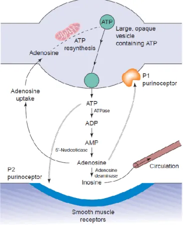
Adenosine-triphosphate (ATP) is the most important energy source in the intra- and extracellular space. It is involved in cellular respiration, cell division, muscle contraction, and almost every other important process needed for the cell’s wellbeing. In addition to this essential duty, ATP and its breakdown product adenosine were discovered to act as neurotransmitters in a wide variety of systems (Burnstock et al., 1970). ATP and adenosine bring about their effects through binding to two groups of receptors: P1 receptors for adenosine and P2 receptors, which have a high affinity for ATP (Figure 1) (Burnstock, 1978).
The neurotransmitter ATP is released by exocytosis from nerve terminals (Bodin and Burnstock, 2001) or astrocytes (Lalo et al., 2014), but other transport mechanisms have also been proposed, including ATP binding cassette transporters, connexin hemichannels, and voltage-dependent anion channels (Fields and Burnstock, 2006).
23
Figure 1: Purinergic transmission in a neuromuscular junction. Synthesis, storage, release, and inactivation of ATP are depicted (Burnstock, 2006a).
The P2 receptors are further divided into two subgroups: the ionotropic P2X receptors (P2XR) and the metabotropic P2Y receptors (P2YR) (Burnstock and Kennedy, 1985).
Additional subtypes of P2 receptors have been described, but will not be further discussed here: P2T receptors, selectively for ADP on platelets, P2Z receptors on macrophages, and P2U receptors that recognize pyrimidines (Gordon, 1986; O'Connor et al., 1991).
To date, eight G-protein-coupled P2Y receptor subtypes and seven P2X receptors have been defined (North, 2002). The responses to an ATP-stimulation can last from milliseconds to minutes, and even longer, since second messenger cascades can also induce changes in gene expression regulation (Khakh and North, 2012). The diversity of responses is further
24
increased by the fact that the receptors differ in their sensitivity to ATP. P2YR are already activated by ATP in the nanomolar range, but P2X7R requires hundreds of micromolar of ATP to open (Surprenant et al., 1996). Due to this diversity, purinergic signalling is very dynamic and accordingly, the expression of P2YR and P2XR throughout different tissues is variable.
P2XR are present in multiple species, even in simple organisms like the eukaryote green algae Ostreococcus tauri (Fountain et al., 2008), underlining the importance of the purinergic system.
The ionotropic P2XR are ligand-gated ion channels permeable for cations, with their highest permeability for Ca2+ (Khakh and North, 2012). However, there is one exception from the rule: P2X5R is permeable to Cl- (North, 2002). Each P2XR subunit assembles with two others into a trimeric channel, which can be either homomeric or heteromeric (Nicke et al., 1998).
The receptors are believed to have three classical agonist binding sites (Browne et al., 2010), and binding of ATP on these sites leads to the opening of the channel (gating). The gating is divided into three phases: the activation phase, the desensitization phase and the deactivation phase (Coddou et al., 2011). Differences in activation and desensitization rates are what characterize the different receptor subtypes.
Purinergic signalling is an important system in the brain’s neuroprotection, since it is involved in nervous tissue remodelling after trauma, stroke, ischaemia or neurodegenerative disorders (Burnstock, 2006a). In response to neuronal injuries, fibroblast growth factor, epidermal growth factor, and platelet-derived factor are released. Together with these factors, ATP stimulates astrocyte proliferation which leads to reactive astrogliosis (Burnstock, 2006a). In general, ATP acts as an important extracellular signalling molecule between neurons and glial cells in the CNS. Microglia, activated by ATP, release
25
inflammatory cytokines and tumour necrosis factor – an overstimulation of this system can thus accelerate the neuronal damage caused by ischaemia, trauma or neurodegenerative diseases (Burnstock, 2006a). One of the most studied purinergic receptors is the P2X4 receptor (P2X4R).
6.1. P2X4 receptor
The detailed atomic anatomy of the receptor is known from studies in zebrafish and was described in the review of Khakh and North in 2012 very plastically as a resemblance to “a dolphin rising from the ocean surface” (Khakh and North, 2012) (Figure 2). One subunit is composed of two hydrophobic membrane-spanning segments and an extracellular loop mostly formed of sheets. Three of those subunits curl around each other, building a trimeric channel that opens to let Ca2+ ions pass when ATP is bound (Khakh and North, 2012).
Figure 2: Crystal structure of the P2X4 receptor showing the two hydrophobic membrane-spanning segments (“fluke”) and the extracellular loop (Khakh and North, 2012)
The P2X4R is the most widely distributed purinergic receptor throughout the CNS (Buell et al., 1996). P2X4Rs play a role in the regulation of multiple nervous functions, including
26
neuropathic pain (Tsuda et al., 2003), neuropeptide release (Lemos et al., 2012), and hippocampal plasticity (Baxter et al., 2011). In addition, P2X4Rs have been shown to modulate the functions of other neurotransmitter systems, such as N-methyl-D-aspartate (NMDA) glutamate receptors (Baxter et al., 2011) and GABAA receptors (Jo et al., 2011). In detail, a facilitation of NMDA synaptic current by insertion of NR2B subunits cannot be seen in P2X4R KO mice, suggesting a changed NMDA receptor composition (Baxter et al., 2011).
Moreover, P2X4R directly interact with GABAA receptors via two residues in the C-terminus of the P2X4R. Electrophysiological experiments showed that the two receptors negatively interact with each other, which represents a form of short term synaptic plasticity (Jo et al., 2011). An involvement in the modulation of behaviour is therefore very likely. Indeed, P2X4R KO mice display less social interaction, and higher tactile sensitivity. In this study, no effect on anxiety-like behaviour was observed (Wyatt et al., 2013), which may be due to compensatory effects. In contrast, behavioural testing after P2X4R activation with the positive allosteric modulator ivermectin (IVM) produced anxiolytic-like effects on the EPM (Bortolato et al., 2013), indicating that P2X4R is involved in the modulation of anxiety-like behaviour. However, IVM has potentiating effects on GABAA receptors as well, so that further research on the role of P2X4R is necessary (Krusek and Zemkova, 1994).
7. Protein synthesis
One of the central dogmas of biology states that cellular processes, and therefore physiology and behaviour, directly or indirectly depend on the sequential transfer of biological information from DNA to RNA to protein via transcription and translation. Once the mRNA exits the cell nucleus, it is ready to be translated into a polypeptide chain by the ribosome in
27
the cell cytoplasm. This process requires the teamwork of non-coding RNAs (ribosomal RNAs and transfer RNAs (tRNAs)), the coding messenger RNA (mRNA) and a large number of proteins. These proteins are either ribosomal proteins or non-ribosomal proteins required for translation initiation (eIFs), for translation elongation (eEFs), or for translation termination (eTFs) (Meister, 2011).
The ribosome is responsible for the translation of the genetic code of the mRNA from 5’ to 3’
into the amino acid sequence and for the synthesis of the corresponding protein. It is composed of two subunits, in eukaryotes the large 60S and the small 40S ribosomal subunit.
Protein synthesis starts with a methionine, which is loaded onto the inititator tRNA. The ribosome has three tRNA binding sites that accept the incoming tRNAs loaded with amino acids corresponding to the presented codon of the mRNA. Every new amino acid is incorporated into the growing peptide chain where it is bound to the previous amino acid by a peptide bond. The process of elongation is GTP-dependent and requires several eEFs.
Among those, eEF2 is responsible for the release of the “old” tRNA, leaving a new space for the next tRNA (Figure 3). One mRNA is not only translated by one ribsosome but by several ribosomes (polyribosome) at the same time, allowing for fast and highly efficient protein synthesis (Meister, 2011). The translation is terminated by the presence of a stop codon, which leads to a release of the polypeptide from the ribosome.
28
Figure 3: Illustration of GTP-dependent peptide chain elongation (Steitz, 2008).
Translation is a highly critical process with a need for precise regulation to make appropriate cellular reactions to environmental challenges possible. All three stages – translation initiation, elongation and termination – are subject to a fine-tuning involving several factors.
Most prominently, proteins that interact with the initiation factor eIF4E, termed 4E binding proteins (4E-BPs), regulate the access of eIF4E and therefore translation initiation. eEF2, the important elongation factor, is part of the regulatory system of protein synthesis as well.
Phosphorylation of eEF2 slows down translation, as seen for example when a cell enters mitosis. eEF2 phosphorylation is also important for local inhibition of translation: In neurons, mRNAs are stalled at the polyribosomal stage at synapses and after arrival of an action potential at the synapse, protein synthesis is rapidly transformed (Meister, 2011).
The mammalian target of rapamycin (mTOR) controls several components involved in the initiation and elongation of translation. One of those is the aforementioned eIF4E, which is regulated by the 4E-BPs. mTOR phosphorylates several sites of 4E-BP and decreases thereby
29
their inhibiting effects on the initiation factor. Furthermore, mTOR inhibits eEF2 kinase activity with positive effects on eEF2 itself (Wang and Proud, 2006).
8. MicroRNAs and the regulation of gene expression
8.1. General information
MicroRNAs are endogenous, non-coding RNAs, approximately 22 nt in length. They were discovered in 1993 by Lee et al. (Lee et al., 1993) in Caenorhabditis elegans, and it was rapidly understood that they exhibit base complementarity to mRNAs. Binding of a microRNA to its respective target RNA leads to a double-stranded RNA molecule and thus to RNA interference (RNAi) (Fire et al., 1998).
MicroRNAs are endogenously expressed in almost all eukaryotes with the exception of Saccharomyces cerevisiae. MicroRNA genes are transcribed by RNA polymerase II to pri- microRNAs, which are then further processed by a large microprocessor protein complex containing the RNase III enzyme Drosha and its partner DiGeorge syndrome critical region 8.
The resulting product is a stem-loop-structured microRNA precursor (pre-miRNA) . The pre- miRNAs are transported from the nucleus into the cytoplasm via the export receptor exportin 5. In the cytoplasm, the mature microRNA is finalized by cleaving off the hairpin- loop of the pre-miRNA and unwinding of the microRNA duplex. The mature microRNA is incorporated into a microRNA-protein complex referred to as miRNP or RISC (RNA-induced silencing complex) (Meister, 2011). The miRNP includes the type III ribonuclease Dicer, the RNA-binding Argonaute proteins, and the adaptor protein transactivating response RNA- binding protein (O'Carroll and Schaefer, 2013).
30
The degree of complementarity between microRNAs and their target mRNAs determines the outcome of the binding of the miRNP to the target mRNA. A perfect or nearly perfect complementarity leads to the cleavage of the mRNA in an RNAi-like manner. This is the most common microRNA function in plants. In animals, however, a perfect complementarity is rare and microRNAs regulate the gene expression differently (Meister and Tuschl, 2004).
Here, microRNAs associate with sequence elements in the 3’ untranslated region (UTR) of the target mRNA. The target sites are only fully complementary to the nucleotides 2 – 8 of the microRNA, which is referred to as the seed sequence. The rest contacts only partially (Chen and Rajewsky, 2007). This way of binding does not result in RNA degradation but in the regulation of translation. Whether such regulation of translation affects translational elongation or initiation is still under debate (Meister, 2011). Either way, the level of total mRNA is not influenced by this interference, but rather the level of newly synthesised proteins. However, partial binding of a microRNA can also induce the deadenylation of the target RNA. The poly(A) tail is shortened, which leads to the initiation of RNA decay. Which pathway will be chosen is most likely dependent on the nature and sequence of the target mRNA, as well as the protein environment surrounding the microRNA target sites (Filipowicz et al., 2008; Meister, 2011).
8.2. MicroRNAs in brain and behaviour
The mammalian brain expresses almost 50 % of all identified microRNAs (Landgraf et al., 2007a), with specific expression profiles for specific brain regions. In addition, intraneuronal microRNA compartimentalization is common (O'Carroll and Schaefer, 2013). There are several microRNAs that are specialized on the expression in synapses or dendrites, like miR- 125b, miR-128, miR-132, and miR-134 (Edbauer et al., 2010; Schratt et al., 2006; Siegel et al.,
31
2009). This indicates their important role in the regulation of local protein synthesis involved in synapse maturation and function. Indeed, microRNAs are seen as crucial regulators of neurogenesis, neurite outgrowth, synaptogenesis, and neural plasticity (Zhou et al., 2009).
The expression level of certain microRNAs is significantly higher than that of others. Some microRNAs, such as let-7, miR-124, and miR-128 are very strongly expressed in neurons, whereas others are only present in 1 – 2 copies/cell (O'Carroll and Schaefer, 2013). This fact should be taken into consideration when analysing microRNA expression in neurons, since it may lead to false conclusions about the significance of some microRNA changes. This diversity might be, amongst other reasons, rooted in the multiplicity of genes encoding a single microRNA. In other words, many of the brain-enriched microRNAs are encoded by more than one gene (Griffiths-Jones, 2006). This could serve as a back-up mechanism that ensures the expression of those important microRNAs even in the case of mutation of one gene. Secondly, the multiplicity of microRNA-encoding genes could support the interneuronal diversity of microRNA expression (O'Carroll and Schaefer, 2013).
Brain microRNAs are strongly involved in the development of neurological and psychiatric disease, making them an even more interesting target of investigation. Abnormal microRNA expression patterns were shown for patients suffering from schizophrenia, autism, Huntington’s disease, Alzheimer’s disease, and others (Abu-Elneel et al., 2008; Beveridge and Cairns, 2012; Johnson et al., 2008; Lukiw, 2007). However, it is not clear if these changes are cause or consequence of the specific neuronal alterations that characterize those diseases. Recent findings depict the involvement of microRNAs also in behavioural matters.
Cocaine addiction (Hollander et al., 2010) as well as alcohol tolerance (Pietrzykowski et al., 2008) were shown to be controlled by specific microRNAs. Other studies observed a
32
regulation of fear extinction (Lin et al., 2011), or even the control of the circadian rhythm (Cheng et al., 2007). Most importantly for my studies, Haramati et al. found increased anxiety-like behaviour after Dicer ablation in the CeA. Furthermore, they identified miR-34c as a repressor of stress-induced anxiety (Haramati et al., 2011). Overexpression of this microRNA within the CeA induced anxiolytic behaviour after social defeat, thus marking microRNAs as promising targets for the development of novel treatments for anxiety disorders.
8.3. Regulation of the microRNA pathway
There are many ways how the microRNA pathway can be altered. Regulations can be of transcriptional, post-transcriptional or post-translational nature, and protein components of the pathway as well as the microRNAs themselves can be involved. Similar to genes that code for a protein, microRNA genes can be targets of gene expression regulation in response to developmental or environmental cues (O'Carroll and Schaefer, 2013). For example, regulation of gene expression in response to neuronal activity is an essential mechanism for neuronal adaptations like changes in synaptic strength and connectivity. miR-132 and miR- 134 are two typical neuronal microRNAs involved in activity-dependent dendritic remodelling (Christensen et al., 2010; Hansen et al., 2010). MicroRNA turnover is an important and strongly regulated event in neurons, and the highly differing half-life of specific neuronal microRNAs plays an essential role. Studies where Dicer was eliminated showed that numerous microRNAs remained expressed even for months after the deletion of Dicer, while others became undetectable within days (Schaefer et al., 2007). This differential regulation of their turnover rate may control distinct neuronal functions, appears
33
to be activity-dependent, and can be activated by stimulation with neurotransmitters like glutamate (Krol et al., 2010).
Another way to regulate microRNA stability is to interfere at the level of the pre-microRNA.
Enzymes that lead to a poly-uridylation of the pre-microRNA cause their degradation and thus the reduction of the corresponding mature microRNA (Heo et al., 2009).
Recently, a class of long noncoding RNAs was discovered that act as microRNA sponges in the cell and was termed “competing endogenous RNAs” (ceRNAs). ceRNAs can actively regulate microRNA activity, because they contain several consecutive microRNA binding sites (Cesana et al., 2011). In this way, it is believed that, especially in neurons where spatially separated zones of mRNA translation exist, local microRNA-mRNA-interactions are mediated. Thus, an activity-dependent induction of microRNA sponge expression could reduce target mRNA suppression, and hence increase their translation, because their inhibiting microRNAs would no longer be available (O'Carroll and Schaefer, 2013).
9. Aims and outline of the present thesis
The overall aim of my studies is to gather more fundamental knowledge about the molecular mechanisms that occur within the PVN controlling anxiety-related behaviour, supporting the role of this brain region for anxiety research. My experiments were carried out in the bigger context of developing new therapeutic approaches to help patients who suffer from anxiety disorders of any kind. In my studies, candidates for pharmacological treatment of anxiety disorders should give clear acute anxiolytic or anxiogenic effects in animal models of anxiety as well as in established behavioural tests in rodents. In addition, pharmacological
34
intervention should ideally lead to long-term effects to make treatments efficient. To achieve this, I addressed two main questions:
1. Can we determine a new target for future pharmacological intervention within the PVN that is anxiolytic with a rapid onset of effects?
2. Which intracellular processes are important to convert a rapid, acute anxiolytic effect into a sustained, long-term anxiolytic effect?
To answer these questions, three different studies were performed, each with a specific goal as described below.
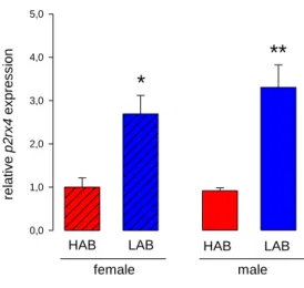
9.1. Aim 1: Identification of a novel anxiolytic factor within the PVN of rats To identify novel possible targets for elucidating the molecular mechanisms behind anxiety- related behaviour, we made use of a microarray that determined the differences of mRNA expression levels in an animal model for anxiety-related behaviour, the HAB and LAB rats.
Subsequent quantitative PCR and Western blot analysis identified the P2X4R as a promising factor that could be implicated in the control of anxiety. This was confirmed in two tests for anxiety-like behaviour by pharmacological activation or inhibition of P2X4R, as well as by biochemical analysis.
9.2. Aim 2: Elucidation of the role of de novo protein synthesis in the mediation of the (long-term) anxiolytic effect of oxytocin
A single OT surge in the PVN has previously been shown to be anxiolytic for at least 4 h (Waldherr and Neumann, 2007). Therefore, OT was used to determine whether protein synthesis is involved in long-term anxiolysis. To this end, the activation of key factors of protein synthesis signalling (eEF2 and mTOR) was analysed by Western blot in hypothalamic
35
cells and tissue. De novo protein synthesis, induced by OT, was demonstrated by incorporation of a synthetic amino acid and detection of the labelled proteins with Click- chemistry. Finally, the role of protein synthesis in the control of anxiety-like behaviour by OT was assessed in male Wistar rats in which protein synthesis was inhibited pharmacologically prior to OT infusion in the PVN. Anxiety was analysed in two independent tests, the LDB and EPM, 30 min and 3 h, respectively, following OT administration to determine whether protein synthesis within the PVN mediates the early onset or is rather a stable substrate for long-term anxiolysis.
9.3. Aim 3: Determination of the control of microRNA expression levels by OT in the hypothalamus
One way of controlling protein levels in cells concerns the regulation of the bioavailability of corresponding mRNA. In collaboration with Prof. Dr. Gunter Meister, we generated Deep Sequencing libraries to show the influence of OT on microRNA expression in hypothalamic cells. The results were validated by means of Northern blot and qPCR and revealed, for the first time, that microRNA expression can be controlled by a neuropeptide of the PVN. The potential role of some microRNAs in anxiety-related behaviour and stress will be discussed with respect to the possible mRNA targets in the PVN.
36
MATERIALS & METHODS
37
Materials & Methods
1. Animals
Adult female or male HAB and LAB (250 - 300 g; local breeding colony; for selection procedures see (Neumann et al., 2005)) as well as rats non-selected for anxiety-related behaviour (hereafter: Wistar rats; 250 - 300 g; Charles River Laboratories, Germany) were housed under standard laboratory conditions (12 h light : dark cycle, 22 - 24 °C, lights on at 06:00 h, food and water ad libitum). Wistar rats were allowed to habituate for one week after arrival. All experiments were performed between 08:00 - 11:00 h. The studies were conducted in accordance with the European Communities Council Directive (86/609/EEC) and were approved by the local government of the Upper Palatinate, Germany.
2. Surgical Procedures
All surgical stereotaxic procedures were performed under isoflurane anaesthesia and semi- sterile conditions. Following surgery, rats received a subcutaneous injection of enrofloxacin (2.5 mg Baytril; Bayer, Germany). Rats were single-housed after surgery, handled daily to habituate them to the respective central infusion procedure and allowed at least seven days of recovery before undergoing behavioural testing.
For analysis of local effects of pharmacological intervention within the PVN on anxiety- related behaviour, indwelling bilateral guide cannulas (stainless steel, 23 G, 12 mm long) were implanted 2 mm above both the left and right PVN (AP: -1.4 mm bregma, ML: -1.8 mm and +2.1 mm lateral, DV: +6 mm below the surface of the skull, angle 10 °) (Blume et al.,
38
2008; Jurek et al., 2012; Paxinos G, 1998) through holes drilled in the skull and attached to two stainless-steel screws using dental cement.
The position of the cannulas was verified after the experiments and post-mortem. Blue ink was infused through the guide cannulas and brains were collected. 40-µm cryosections of the brains were stained with Nissl and correct cannula placement was verified with the aid of a rat brain atlas (Paxinos G, 1998).
For the analysis of cell physiological responses to OT in the PVN, rats were implanted with guide cannulas (21 G, 12 mm long) 2 mm above the right lateral ventricle (AP: -1.0 mm bregma, ML: +1.6 mm lateral, DV: +1.8 mm below the surface of the skull) (Blume et al., 2008; Paxinos G, 1998; Slattery and Neumann, 2010). The guide cannulas were kept feasible with dummy cannulas, which were removed and cleaned every day during the handling procedure. Rats received an infusion of 1 nmol OT/5 µl.
3. Cells
3.1. H32 cells
The immortalized foetal rat hypothalamic cell line H32 (Mugele et al., 1993) was cultured at 37 °C and 5 % CO2 in Dulbecco’s modified Eagle’s medium (DMEM; Life Technologies, USA) containing 10 % foetal bovine serum (FBS), 10 % horse serum and 1 % penicillin/streptomycin (Life Technologies). For experiments, cells were seeded in 10 cm petri-dishes at a density of 3 x 10⁶ cells and grown to 80 % confluence overnight. Prior to stimulation, the medium was changed to serum-free DMEM containing 0.1 % bovine serum albumin (BSA; Sigma-Aldrich, Germany) and the cells were left undisturbed for 1 h.
39 3.2. Be(2)-M17 cells
The human neuroblastoma cell line Be(2)-M17 (European Collection of Cell Cultures,
#95011816, UK, (Jurek, 2014)) was cultured at 37 °C and 5 % CO2 in DMEM/F12 (1:1) (Life Technologies) containing 1.2 % L-glutamine, 15 % heat-inactivated FBS, 1.2 % non-essential amino acids, and 0.1 mg/ml gentamycin (Life Technologies). For experiments, 4 x 10⁶ cells were seeded per 10 cm petri-dish and treated with 5 µM retinoic acid (Sigma-Aldrich) to initiate differentiation into neurons. After three days at 37 °C and 5 % CO2, cells were ready for stimulation. On the day of the experiment, the cells were incubated in serum-free DMEM/F12 (+ 0.1 % BSA) for 1 h to reduce basal activation by any growth factors and steroids that might be present in the serum.
3.3. Primary hypothalamic neurons
Primary neurons were obtained from foetal Wistar rats on embryonic day 18. After decapitation of the foetuses, hypothalamic tissue was dissected and collected in ice-cold Hank’s balanced salt solution (HBSS; Life Technologies) containing 0.1 mg/ml gentamycin.
Hypothalami were digested for 45 min at 37 °C with 300 U/ml collagenase type 2 (Worthington, USA) diluted in HBSS supplemented with 4 mg/ml BSA (Sigma-Aldrich), 1 mg/ml glucose (Merck, Germany) and 0.2 mg/ml DNase (Sigma-Aldrich). The cell suspension was carefully filtered through a 40-μm cell strainer (BD Falcon, USA) and centrifuged at 200 x g for 10 min. The cell pellet was resuspended in DMEM/Ham’s F12 containing 0.1 mg/ml gentamycin and 10 % heat-inactivated FBS. Cells were plated at a density of 3 x 10⁶ cells/well in poly-D-lysine coated six-well plates (BD Falcon) and incubated for 24 h at 37 °C and 5 % CO2. Medium was then changed to neurobasal medium (Life Technologies) containing B27 supplement (Life Technologies), 2 mM L-glutamine, 0.1 mg/ml gentamycin and 5 µM
40
ascorbic acid (Sigma-Aldrich). Cytosine arabinoside (Sigma-Aldrich) was added to a final concentration of 5 μM from day 4 onwards to prevent glial cell proliferation. On day 10, cells were prepared for stimulation by replacing the growth medium with supplement-free neurobasal medium containing 0.1 % BSA, and incubated for 1 h before the start of the experiment.
3.4. Cell stimulation
To reveal the regulatory effects of OT at the cellular level, OT was added to the serum-free medium and the cells were left undisturbed at 37 °C and 5 % CO2 for different time periods as indicated in the results section. For signalling studies aimed to determine the pathway involved in OT-stimulated protein synthesis, the MEK1/2 inhibitor U0126 (final concentration 10 µM in 0.1 % DMSO) or the PKC inhibitor Gö6983 (final concentration 1 µM in 0.1 % DMSO; Sigma-Aldrich) were added to the cell medium 20 min before activating them with OT.
After incubation, cellular activity was stopped rapidly by cooling the petri-dishes on ice. Cells were lysed and total RNA or proteins were isolated as described below. RNA- and protein- expression levels were compared to respective vehicle-treated control groups and are shown as relative expression.
4. Behavioural studies
Rats received bilateral local infusions (0.5 µl/PVN) of the substance to be tested, and the effects of drug infusion were assessed on the EPM and in the LDB. The substances that were infused were OT (0.01 nmol in Ringer’s Solution), anisomycin (23.5 µmol in HCl, adjusted to
41
pH 7.4), cytidine triphosphate (CTP; 4 and 40 nmol in Ringer’s Solution) and 5-(3- Bromophenyl)-1,3-dihydro-2H-benzofuro[3,2-e]-1,4-diazepin-2-one (5-BDBD; 25 nmol in HEPES-buffered Ringer’s Solution). To exclude effects of the dissolvent on behaviour, every treatment group was compared with a control group, given only the respective vehicle. The time period between infusions, as well as that between the last infusion and behavioural testing, varied according to the experiment (please see results section).
The test protocols for the EPM and the LDB were performed similarly to those previously described (Bosch and Neumann, 2008; Neumann et al., 2000; Pellow et al., 1985; Slattery and Neumann, 2010; Waldherr and Neumann, 2007). Briefly, the plus-shaped maze is made of two open (50 x 10 cm, 80 lux) and two closed (50 x 10 x 30 cm, 10 lux) arms surrounding a neutral square-shaped central zone (10 x 10 cm, 65 lux), elevated 80 cm above the floor. The percentage of time spent on the open vs. time spent on all arms is indicative of anxiety- related behaviour, while the number of closed arm entries is used to assess locomotion (Neumann et al., 2000). The duration of the test was 5 min.
The LDB setup consisted of two boxes; one lit box (40 x 50 cm, 350 lux; light box) and one dark box (40 x 30 cm, 70 lux). Light conditions in the light box were changed for HAB (85 lux) and LAB (1000 lux) studies, considering their extreme phenotypes. The floors in each box were divided into squares (10 x 10 cm) and the boxes were connected by a small opening (7.5 x 7.5 cm) enabling transition between the boxes. Rats were placed in the light box and line-crossings, time spent in each box, rearing, latency to enter the dark box and the latency to first re-enter the light box were assessed during the 5-min test live via a camera located above the box.
42 5. RNA studies
5.1. RNA extraction
RNA was extracted from cultured cells and from PVN tissue that had been dissected from the hypothalamus. The dissection was done in brains that were snap-frozen on dry-ice, and cut on a cryostat in 250 µm thick slices. The PVN was then punched out with the aid of a tissue puncher of 2 mm in diameter (Fine Science Tools, Germany), and expelled into an Eppendorf tube. To collect most of the PVN from a single animal, two consecutive punches were pooled. The punches were either kept frozen on dry ice or were directly lysed in the appropriate buffer for RNA-extraction.
Punches and cells were lysed in 1 ml of peqGold TriFast (peqLab, Germany) and kept on ice for the whole procedure to prevent degradation of RNA. The lysate was mixed with chloroform (200 µl in 1 ml lysate) and centrifuged for 20 min at 12000 x g and 4 °C. The upper aqueous phase containing the RNA was collected and transferred to a fresh cup. Next, the RNA was concentrated by precipitation in isopropanol overnight at -20 °C. Following centrifugation at 16000 x g, the RNA pellet was washed twice with 80 % ethanol, air-dried, suspended in RNase-free water, and solved at 70 °C for 5 min at 1000 rpm. RNA quantity and quality were determined at 260/280 nm using a NanoDrop spectrophotometer (Thermo Scientific, USA).
5.2. Microarray
Differential gene expression in the PVN of HAB and LAB rats were assessed using the microarray technique in collaboration with Dr. David von Schack and Dr. Robert H. Ring, Pfizer Inc., New York, USA.
43
HAB and LAB virgin female rats were sacrificed and the PVN was punched out. The frozen material was sent to the collaborators who performed the microarray. This method allows for a comparison of the mRNA expression profile in different samples. The extracted, fluorescently labelled cDNA hybridizes to their complementary sequences on a solid surface and is then scanned with a laser. Differential gene expression is revealed with a simultaneous, two-colour hybridization scheme: fluorescent probes with different colours were prepared from the two mRNA sources, mixed together, hybridized to a single array, and scanned separately (Schena et al., 1995).
5.3. Deep Sequencing
Deep Sequencing libraries were generated in collaboration with Prof. Dr. Gunter Meister, Department of Biochemistry I, University of Regensburg. To study the effects of OT on microRNA expression in rat hypothalamic cells, H32 and primary hypothalamic cells were stimulated with 250 nM OT for 30 min and 3 h, and total RNA was extracted according to the protocol described above (5.1). Subsequent steps were performed as described previously (Dueck et al., 2012). Briefly, total RNA was separated by size on a 12 % urea-polyacrylamide (PAA) gel and short RNAs (between 10 and 20 bp) were cut out and extracted from the gel.
Isolated small RNA was ligated to a bar-coded, adenylated 3’ adapter by a truncated T4 RNA ligase 2. Additionally, the RNA was enlarged by a 5’ RNA adapter that was added in a second ligation step by T4 RNA ligase 1. The product was reverse transcribed and amplified by PCR (see below). The samples were run on a 6 % urea-PAA gel and the bands containing the ligation product were cut out of the gel and eluted overnight in 300 mM NaCl, 2 mM EDTA.
After precipitation with ethanol overnight at -20 °C, samples were collected by centrifugation and dissolved in H2O. The libraries were analysed on a Genome Analyzer GAIIx
44
(Illumina, USA) (Dueck et al., 2012), with the minimal length of a read set to 18 nucleotides, no mismatch was allowed. The reads for each microRNA were normalized against the total read number of the respective library (Dueck et al., 2014).
5.4. PCR and qPCR
Isolated RNA was reverse transcribed into cDNA. Random primers (3 µg/µl) and dNTPs (final concentration 0.5 mM; Life Technologies) were mixed with 1 µg total RNA and the mix was incubated for 5 min at 65 °C to anneal the primers. To start reverse transcription, FirstStrandBuffer, dithiothreitol (DTT; final concentration 5 mM), RNase OUT (40 U/µl) and the reverse transcriptase Super Script III (200 U/µl; Life Technologies) were added to a final volume of 20 µl. cDNA synthesis was performed at 42 °C for 50 min. The reaction was stopped by degradation of the enzyme at 70 °C for 15 min.
Primers were created with the open-source application PerlPrimer (Marshall, 2004).
Wherever possible, primers were designed to span an intron/exon boundary to assure that no genomic DNA was amplified.
A regular reverse transcription PCR (RT-PCR) was always performed to test the specificity and efficacy of the primers, and to determine whether a particular gene product is expressed in hypothalamic tissue or cells. Following validation of the quality of the primers, cDNA (50 ng), 2 pmol forward and reverse primers (Metabion, Germany) and water were added to DreamTaq Master Mix (Thermo Scientific, Germany), containing dNTPs (final concentration 0.2 mM each) and DreamTaq™ polymerase, to a final reaction volume of 25 µl. Negative controls consisted of reactions where cDNA was substituted by H2O, or samples where the reverse transcription had been omitted. The PCR was run for 40 amplification cycles with an initiating denaturation step at 95 °C for 5 min. Primer-annealing was
45
performed at 60 °C and elongation at 72 °C. The blockcycler (C1000 Thermal Cycler; BioRad, Germany) was programmed to run a final elongation for 10 additional min and to cool the reaction down to 4 °C. The PCR-products were then loaded onto a 1.5 % agarose gel. After electrophoresis, cDNA bands were detected with DNA Stain G (SERVA electrophoresis, Germany) and visualized with UV-light in the ChemiDoc XRS+ Imager (BioRad).
The method of real-time or quantitative PCR (qPCR) is essentially similar to that of RT-PCR.
Differences are found in the detection method of the amplified products, and the analysis.
qPCR was carried out with the 7500 Fast Real-Time PCR System (Applied Biosystems GmbH, Germany) and as detection dye SYBR Green I was used. SYBR Green binds to double stranded DNA and emits green light at 522 nm. The reaction mixture consisted of 10 µl QuantiFast SYBR Green PCR MasterMix (QIAGEN, the Netherlands), 4 µl RNAse-free water and each 2 µl of reverse and forward primers (4 pmol) and cDNA. Following amplification, a melting curve was made by slowly heating the sample from 60 °C to 95 °C, while constantly measuring the green fluorescence. This procedure gives the melting point of the amplified DNA. The detection of more than one melting point indicates the amplification of unspecific products, which was always verified on an agarose gel. Gene expression was quantified relative to the expression of a housekeeping gene (glyceraldehyde-3-phosphate dehydrogenase (GAPDH) or ribosomal protein L13a (Rpl) (Bonefeld et al., 2008)).
For validation of the Deep Sequencing on microRNAs in primary hypothalamic neurons, qPCR was used, with minor changes compared to the conventional qPCR described above. Mature microRNAs obtained from primary neurons stimulated with 250 nM OT or control cells were first modified by the addition of a polyA tail. To this end, RNA was treated with E-PAP buffer, MnCl (25 mM), ATP solution (10 mM) and E. coli Poly (A) Polymerase I (E-PAP; Poly (A) Tailing
46
Kit, Ambion/Life Technologies) (Hurteau et al., 2006). Next, the E-PAP treated total RNA was incubated with a dNTP mix and 100 µM Universal Reverse Transcription primer (URT-primer) – a primer that is composed of a complementary part to the added polyA tail, a polyT- sequence, and a universal sequence. After annealing for 5 min at 65 °C, the sample was reversely transcribed under the same conditions as described above.
The PCR was performed with a primer that is exactly complementary to the desired microRNA and a universal PCR primer that is complementary to the unique sequence of the URT primer. Quantitation of the reaction products is achieved using the intercalating dye SYBR Green with 18s ribosomal RNA as an internal standard (Hurteau et al., 2006).
P2X4R studies
target forward-primer 5'-3' reverse primer 5'-3'
Casq2 CTC TGT CTC TAC TAC CAC GA ATA CAG GCT TCC TTC TTC AC Cckbr TCC CTT CTC AAC AGC AGT AG GCC AAC ACT CAT CAG AAA GA
Hcrt CTC CAG ACA CCA TGA ACC TT GTG CAA CAG TTC GTA GAG AC Nrg1 GTC ATT ACA CTT CCA CAG CC ATC ATA TTT CTT CTC CCG TAG CC P2rx4 GTC CAG AGA TTC CTG ATA AGA C GTA CCA GAT GTT GTT CTT TAC C Trpm7 GCT GAA ATG TCC CAT ATC CC GTA TGC CAA TGT GTT AAA CCA G
Trpv2 GAA ATC CTC TTT CTC CTT CAG TGT GTA GTA AAG CAG GTT CAG
VIP CGC CCT ATT ATG ATG TGT CC CTG ATT CGT TTG CCA ATG AG
Rpl13A ACA AGA AAA AGC GGA TGG TG TTC CGG TAA TGG ATC TTT GC Gapdh TGA TGA CAT CAA GAA GGT GG CAT TGT CAT ACC AGG AAA TGA G protein synthesis studies
target forward-primer 5'-3' reverse primer 5'-3'
OTR rat CAG TAG TGT CAA GCT TAT CTC CA AAG AGC ATG TAG ATC CAC GG OTR human AAG AGC AAC TCG TCC TCC TTT ACA AAC ATA CGC CAT CAC CT


![P r g p a r a t i v e , l 1 B - , 9 3 N b - N M R - s p e k t r o s k o p i s c h e u n d s t r u k t u r e l l e U n t e r s u c h u n g e n a n C p , N b B H , - u n d [ C p N b ( B , H , ) ] , - K o m p l e x e n](data:image/gif;base64,R0lGODlhAQABAIAAAP///wAAACH5BAEAAAAALAAAAAABAAEAAAICRAEAOw==)