Structural aspects of systems with spin–orbit coupling
Tobias Fröhlich
Structural aspects of systems with spin–orbit coupling
I n a u g u r a l - D i s s e r t a t i o n zur
Erlangung des Doktorgrades
der Mathematisch-Naturwissenschaftlichen Fakultät der Universität zu Köln
vorgelegt von Tobias Fröhlich
aus Münster
Köln, 2020
Prof. Dr. Thomas Lorenz Vorsitzende der Prüfungskommision: Prof. Dr. Petra Becker
Tag der mündlichen Prüfung: 2020-02-03
Contents
Introduction 1
1 Theory 3
1.1 Band models and topology . . . . 3
1.1.1 Tight binding model . . . . 3
1.1.2 Nearly free electron model . . . . 3
1.1.3 Comparison of the two models . . . . 3
1.1.4 Topological insulators . . . . 4
1.1.5 Topological superconductors . . . . 5
1.2 Frustrated systems . . . . 6
1.2.1 Classical spin liquid . . . . 6
1.2.2 Quantum spin liquid . . . . 6
1.2.3 Quantum spin-orbital liquid . . . . 7
1.3 Dzyaloshinskii-Moriya interaction . . . . 8
2 Fundamental principles of diffraction 11
2.1 Group theory . . . 11
2.2 Scattering of neutrons by matter . . . 11
2.2.1 Scattering of neutrons by nuclei . . . 11
2.2.2 Scattering of neutrons at electrons . . . 18
2.3 Neutron diffraction . . . 19
2.3.1 Nuclear neutron diffraction . . . 19
2.3.2 Magnetic neutron diffraction . . . 20
2.3.3 Unpolarized neutron diffraction . . . 25
2.3.4 Polarized neutron diffraction . . . 26
2.4 Structural refinements . . . 31
2.4.1 The algorithm used in refinement software . . . 31
2.4.2
Rvalues and goodness of fit . . . 32
2.4.3 The physical meaning of structural results . . . 34
3 Instruments 37
3.1 X-ray diffraction instruments . . . 37
3.1.1 Bruker AXS Kappa APEX II . . . 37
3.1.2 D5000 . . . 39
3.2 Neutron diffraction instruments . . . 44
3.2.1 6T2 . . . 44
3.2.2 5C2 . . . 44
3.2.3 HEiDi . . . 44
3.2.4 D9 . . . 45
3.2.5 D3 . . . 45
3.3 Further instruments . . . 46
3.3.1 DiMoS . . . 46
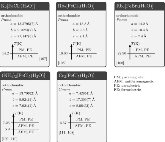
4 Erythrosiderite-type compounds 47
4.1 The family of erythrosiderite-type compounds . . . 47
4.1.1 Crystal structures of erythrosiderite-type compounds . . . 47
4.1.2 Magnetic and ferroelectric properties of erythrosiderite-type com- pounds . . . 49
4.2 (NH
4)
2[FeCl
5(H
2O)] and (ND
4)
2[FeCl
5(D
2O)] . . . 51
4.2.1 Sample characterization and experimental setup . . . 51
4.2.2 Results and discussion . . . 52
4.2.3 Conclusion . . . 61
4.3 Cs
2[FeCl
5(H
2O)] . . . 61
4.3.1 Crystal growth and experimental setup . . . 61
4.3.2 Results and discussion . . . 63
4.3.3 Conclusion and comparison to existing results . . . 72
5 Cs3Fe2Br9 75
5.1 Phase diagram . . . 75
5.2 Sample characterization and experimental setup . . . 75
5.3 Results and discussion . . . 77
5.3.1 Investigation of the crystal structure . . . 79
5.3.2 Investigation of the magnetic structure . . . 81
6 Compounds of the form Ba3XIr2O9 93
6.1 Latest state of research . . . 93
6.2 Ba
3InIr
2O
9. . . 95
6.2.1 Samples and experimental setup . . . 95
6.2.2 Occupation ordering and non-stoichiometry . . . 96
6.2.3 Breaking of symmetries . . . 98
6.2.4 Conclusion . . . 101
6.3 Ba
3NbIr
2O
9. . . 104
6.3.1 Samples and experimental setup . . . 104
6.3.2 Occupation ordering and non-stoichiometry . . . 104
6.3.3 Breaking of symmetries . . . 104
6.3.4 Conclusion . . . 109
6.4 Ba
3CeIr
2O
9. . . 109
6.4.1 Samples and experimental setup . . . 109
6.4.2 Results and discussion . . . 110
6.4.3 Conclusion . . . 113
6.5 Comparison of Ba
3XIr
2O
9compounds . . . 113
6.6 Conclusion . . . 117
Contents
7 Double-perovskites of the form Ba2XIrO6 119
7.1 Ba
2CeIrO
6. . . 120
7.1.1 Latest state of research . . . 120
7.1.2 Samples and experimental setup . . . 121
7.1.3 Room temperature crystal structure . . . 122
7.1.4 Low temperature crystal structure . . . 129
7.1.5 Conclusion . . . 134
7.2 Ba
2PrIrO
6. . . 134
7.2.1 Latest state of research . . . 134
7.2.2 Samples and experimental setup . . . 134
7.2.3 Breaking of translational symmetries . . . 135
7.2.4 Occupation disorder . . . 136
7.2.5 Breaking of non-translational symmetries . . . 137
7.2.6 Conclusion . . . 140
8 Cu-intercalated Bi2Se3 141
8.1 Latest state of research . . . 141
8.2 Sample characterization end experimental setup . . . 144
8.2.1 Single crystal neutron diffraction . . . 144
8.2.2 Polarized single crystal neutron diffraction . . . 145
8.2.3 Single-crystal X-ray diffraction . . . 146
8.3 Results and discussion . . . 147
8.3.1 Vacancies . . . 147
8.3.2 The Cu doping . . . 148
8.3.3 Structural phase transition . . . 151
8.3.4 Breaking of symmetry . . . 153
8.3.5 Magnetic properties . . . 164
8.3.6 Impurity phase . . . 168
8.4 Conclusion . . . 171
A Appendix 173
Bibliography 223
Danksagung 237
Abstract 239
Kurzzusammenfassung 241
Offizielle Erklärung 243
Introduction
Spin-orbit coupling gained great attention [1] in solid state physics after experimental results from 1983 [2, 3] were explained one year later [4] using the Rashba term [5]. Also the Kugel-Khomskii model [6] depends on spin-orbit coupling. Spin-orbit coupling plays an important role for the physics of many compounds investigated in the present work.
Pristine Bi
2Se
3is a topological insulator [7]. Its band structure is characterized by strong spin-orbit coupling: The Hamiltonian contains terms that combine orbital states with the electronic spin [7]. This gives rise to a spin texture on the Fermi surface, where the spin depends on the orbital and on k [7]. In the present work, the doped compound Cu
xBi
2Se
3is investigated. The doping induces charge carriers that give rise to topological superconductivity [8, 7].
The compounds Ba
3CuSb
2O
9[9] and Ba
3IrTi
2O
9[10] were proposed as spin-liquid can- didates. The idea behind this is the Kitaev model [11], where the coupling of spins on a honeycomb lattice depends on the bond-direction between them. The realization of this model needs a system with spin-orbit coupling [12]. Related compounds of the form Ba
3XIr
2O
9were investigated in this work.
The superexchange interaction derived by H. A. Kramers [13] and P. W. Anderson [14] was extended by T. Moriya [15] who added spin-orbit coupling. This results in an antisymmetric term that was introduced before by I. Dzyaloshinsky [16]. This so- called Dzyaloshinsky-Moriya interaction gives rise to weak ferromagnetism driven by magnetic ordering in multiferroic compounds like (NH
4)
2[FeCl
5(H
2O)] as investigated in the present work.
After W. C. Röntgen published the discovery of X-rays in 1896 [17], M. von Laue reported the first X-ray diffraction experiment in 1914 [18]. This technique allows investigations of bulk crystal structures on the atomic scale. Several decades later, also neutron radiation was used for diffraction experiments. The neutron was discovered by J. Chadwick in 1932 [19]. The first neutron diffraction experiments were carried out in the 1940s in the context of the Manhattan Project. An overview of these early experiments is given in [20].
Since then, diffraction experiments have become an important tool for crystallography and solid-state physics. Despite the fact that polarized synchrotron radiation can be used for the investigation of magnetic structures (e.g. [21]), neutron diffraction is usually the first choice for this issue. For the determination of crystal structures, both neutron and X-ray diffraction can be used.
The geometry of crystal structures plays an important role for the understanding of inter-
atomic coupling mechanisms like in the Kitaev model [12] or for the Dzaloshinsky-Moriya interaction [15], as introduced above. In the present work, small distortions of crystal structures are studied by means of elastic X-ray and neutron diffraction techniques.
Magnetic structures are investigated using neutron diffraction.
The work is structured as follows: The first chapters contain basics of theory and ex- periment. Chapter 1 gives an introduction to the mechanisms that are relevant for the physical properties of the investigated materials. In Chapter 2, concepts of diffraction are elucidated with the focus on neutron diffraction. While Chapter 2 is very general, Chapter 3 introduces the instruments used in the present work. The next chapters present and discuss the results of the work. In Chapter 4, the erythrosiderite-type com- pounds (NH
4)
2[FeCl
5(H
2O)] (including the deuterated compound) and Cs
2[FeCl
5(H
2O)]
are treated. The former compound is multiferroic while the latter is magneto-electric.
For both compounds, a structural and a magnetic phase transition are investigated.
Chapter 5 deals with the compound Cs
3Fe
2Br
9, which is related to the erythrosiderite-
type compounds. It exhibits a very rich phase diagram with a plethora of magnetic
phases. For one of these phases, the magnetic structure is determined. Chapters 6 and
7 present structural investigations of the compounds Ba
3XIr
2O
9with X = In, Nb, Ce
and Ba
2X IrO
6with X = Ce, Pr. Occupation disorder and breaking of symmetry are
discussed. In Chapter 8, Cu doped Bi
2Se
3is investigated. It focuses on the search for
Cu atoms because it is still an open question which sites they occupy. Furthermore,
structural distortions of this compound are discussed and an impurity phase is charac-
terized.
1 Theory
This section gives a short overview of the theoretical concepts that are used in the context of the investigated compounds.
1.1 Band models and topology
1.1.1 Tight binding model
The tight binding model describes electronic wavefunctions in a periodic potential as linear combination of atomic orbitals. In the limit where all atoms are separated, there exists only one energy level for all the electrons, namely the energy of the atomic orbital
atomic. However, if the atoms are closer together, these atomic orbitals are no longer eigenstates of the Hamiltonian. The deviation from the limit where all atoms are seper- ated is expressed as the hopping term t, which equals zero if the atoms are separated and increases as the atoms approach each other. A finite value for t has the effect, that the eigenstates are no longer degenerate, but cover an energy interval, which is called band.
If there are several non-degenerate orbitals for each atom, there exist several bands. If the hopping term t is large enough, these bands are broad and might overlap. If they do not overlap, the material exhibits a band gap [22].
1.1.2 Nearly free electron model
Another way of treating this problem is the nearly free electron model. Here, the de- scription starts with a free electron gas and the periodic potential caused by the atoms is treated via perturbation theory. While the free electron gas has a dispersion that covers all energies above a minimum energy, the periodic potential makes band gaps open up at the Brillouin zone boundaries [22].
1.1.3 Comparison of the two models
The tight binding model and the nearly free electron model result in very similar dis-
persions: In the vicinity of momentum
k=
0and at the Brillouin zone, the dispersion
relation is parabolic. In both cases, band gaps appear or might be avoided by overlapping
bands.
A
1v B
1w A
2v B
2w A
3v B
3w A
4v B
4w A
5v B
5l 2l
Figure 1.1:Su-Schrieffer-Heeger model for2N atoms withN = 5. The atoms are alternatingly spaced with a distance ofl+δandl−δ. Between them, there are alternating hopping amplitudes v andw. In the general case, the unit cell has length2l.
1.1.4 Topological insulators
Topological insulators of dimension 3 (2, 1) exhibit an insulating topological band struc- ture in the bulk and conducting states at the surface (edge, ends) [23]. This concept is rather old and for simple cases well understood. Perhaps the simplest model with a topological band structure is the Su-Schrieffer-Heeger model [24]: It describes the band structure of polyacetylene, which is in principle a one-dimensional chain of C atoms, which are bound by alternating single and double bounds. This results in staggered hopping amplitudes, as shown in Figure 1.1. 2N atoms with distance l are alternatingly coupled with hopping amplitudes v and w. A detailed derivation of the electronic bands can be found in [25].
For v = w
6= 0, the chain is conducting with parabolic bands and the band gap is closed at the edges of the Brillouin zone. For v = 1 and w = 0, the electronic wave functions are localized and the bands are flat with a band gap [25].Assuming periodic boundary conditions, this case is physically equivalent to v = 0 and w = 1 . However, the equations look very different: The eigen energy of states with different wave vectors k can be identified with the magnitude of the vector on the circle with center v + 0i and radius w in the complex plane. Depending on the parameters v and w , this loop might enclose the origin or not. The number of times the loop winds around the origin (in this case 0 or 1) is called winding number and is characteristic for the topology of the system.
Systems with different winding numbers are called adiabatically distinct: They cannot be transformed into each other by an adiabatic deformation
1. For experimental and technological purposes, it is important that the system cannot change between states with different topology by small perturbations like thermal activation. An intuitively accessible explanation in the case of the Su-Schrieffer-Heeger model is that it is not possible to flip the coupling parameters one by one using a small energy, but instead all of them must be flipped at once. This protects topological states against e.g. thermal activation. The topologically non-trivial band structures of topological insulators lead to the effect that there must be gapless states at interfaces with topologically trivial materials [23].
The compound Bi
2Se
3, which is of importance for the present work, has a band structure that exhibits two orbitals which can be ascribed to the two symmetrically not equivalent Se positions. The Hamiltonian contains a strong spin-orbit-coupling term [7] so that the
1For a definition of ”adiabatic deformation” cf. [25]
1.1 Band models and topology spin orientation at the Fermi surface depends on the wave vector and the orbital. States in different orbitals with the same wave vector have opposite spin direction [7].
1.1.5 Topological superconductors
Topological superconductors are adiabatically distinct from conventional superconduc- tors that exhibit a topologically trivial band structure [7]. In general, two classes of topological superconductors are distinguished: For ”weak topological superconductors”
it is sufficient to exhibit a topologically non-trivial band structure. If additionally the gap is fully open, the superconductivity is called ”strong” [7]. There are several ways for realizing a topological superconductor. A material is said to show ”intrinsic topological superconductivity” if the pristine sample is topologically superconducting. So far, there are not many candidates for this and only superfluid
3He is a verified intrinsic topological superconductor [26]. Other materials are topological insulators or topological semimet- als and become superconducting when high pressure is applied [26]. Recently, a further method was discovered that turns topological semimetals into superconductors by apply- ing a hard point contact. The physics of these so called ”tip-induced superconductors”
is not well understood yet [26]. Furthermore, there are many different setups in order to induce topological superconductivity by arranging materials in artificial structures.
With some of these approaches, superconductivity can be tuned by a voltage in a struc- ture similar to a field-effect transistor. Others use the proximity effect in order to induce topological superconductivity at interfaces between different materials [26]. A further possibility is doping of topological insulators or topological crystalline insulators. Since in this work, the compound Cu
xBi
2Se
3is investigated, the next paragraph pays closer attention to doping induced topological superconductivity in topological insulators.
As stated above, a topological insulator has an insulating band structure in the bulk where the topology of the bands is non-trivial. By doping the material appropriately, it is possible to induce charge carriers that are at the Fermi surface [7]. The pristine material exhibits a spin texture due to strong spin-orbit coupling: The spin of an electron depends on the wave number and this dependence is different for different orbitals [7].
This results in two possibilities for forming Cooper pairs. If cooper-pairs consist of electrons in the same orbital, these electrons will have opposite spin and form a spin singlet state. If the electrons of a Cooper pair come from different orbitals, they have parallel spins and form a spin triplet state. The way the electrons couple and form Cooper pairs is predicted by the form of the pair potential ∆. Let P be the inversion operator, then the pair potential is called even if P ∆P
t= ∆ and odd if P ∆P
t=
−∆. Possible coupling mechanisms can be categorized by the parity of the pair potential and the multiplet of the spins. In the case of Cu
xBi
2Se
3, four combinations are possible.
Calculations revealed that an odd-parity spin-triplet state is realized.
1.2 Frustrated systems
When it is not possible to satisfy all competing forces in a system simultaneously, the system is called frustrated [27]. Some possible states for such a system are presented in this section.
1.2.1 Classical spin liquid
A spin liquid is a system of spins which are frustrated and fluctuate classically [27]. The simplest example for a frustrated system consists of three S = 1/2 Ising spins which are placed at the corners of an equilateral triangle and which are coupled antiferromagneti- cally [27]. The system is frustrated because not all three spins can be pairwise oriented in opposite direction. At first glance it seems obvious that frustration can occur only in antiferromagnetically coupled systems because for ferromagnetic coupling all forces can be satisfied by orienting all spins in the same direction. However, when the spins exhibit Ising anisotropy, frustration is also possible for ferromagnetically coupled spins. This is the case in Ho
2Ti
2O
7[28] with strong easy-axis anisotropy, which is the first realization of a spin ice material [27]. Both the system investigated in [29] and spin ice are classical spin liquids.
1.2.2 Quantum spin liquid
A quantum spin liquid state is a state where the entropy at low energy is released by quantum effects [30]. There are two models for describing such an effect in certain systems: The resonant valence bond state and the Kitaev model.
The resonant valence bond state describes a system in which spin dimers fluctuate. If the interactions between spins in a lattice is antiferromagnetic, it is possible that in the the ground state the spins couple to ordered singlet dimers. An example for such a material is SrCu
2(BO
3)
2[31]. Since every singlet has the total spin quantum number J = 0 , the ground state is not magnetic [27]. Such a system is called valence bond solid.
It is also possible that the system fluctuates between different dimer configurations. The state of the system is then a linear combination of wave functions that correspond to these dimer configurations [32]. This is called resonant valence bond state and realizes a quantum spin liquid. The word ”resonant” stems from the pictorial image that the system is resonating between the different configurations [32]. The resonant valence bond state is not exactly solved [30]. It is predicted for a triangular antiferromagnet, however next-nearest-neighbor interactions are supposed to destroy this state [32].
Another kind of quantum spin liquid is the Kitaev model. It consists of a honeycomb
lattice of S = 1/2 spins where each spin couples to three nearest neighbors. The coupling
between each pair of neighbored spins is ferromagnetic and possesses a strong Ising
anisotropy which depends on the spatial orientation of the connecting line of these spins
[11][30]. The model is solved exactly in a formalism which represents spins by Majorana
1.2 Frustrated systems operators [11].
While classical spin liquids occur for large spin quantum numbers, quantum spin liquids are expected for systems with S = 1/2 because there is no energy barrier [27]. To the knowledge of the author, no such material could be unambiguously identified so far. The difficulty in searching for such materials experimentally lies in the fact that it is not characterized by a single experiment. There are rather indirect hints, which however do not exclude other explanations. One indication for a quantum spin liquid is the absence of magnetic ordering down to low temperatures despite a strong magnetic interaction. A measure for this is the frustration parameter f =
|ΘCW|/Tc, where Θ
CWis the Curie-Weiss temperature determined from the temperature dependent susceptibility and T
cis the temperature at which magnetic ordering sets in. For quantum spin liquids, a frustration parameter of at least 100 is expected [27]. Using muon spin resonance or nuclear magnetic resonance experiments, also disordered magnetic moments can be detected, which must be absent in quantum spin liquids. By specific heat or thermal transport measurement or by neutron scattering experiments, excitations can be detected which are characteristic for quantum spin liquids. A candidate was Ba
3CuSb
2O
9[9], which exhibits single- and double-octahedrons. The single octahedrons are occupied by Sb while one site of the the double octahedrons is occupied by Sb and the other site by Cu. The occupation is nearly ordered [9], so that effectively a triangular lattice of Cu
2+ions in isolated octahedrons is formed [9]. The 3d electrons of Cu
2+were expected to form a quantum spin liquid. However, later it turned out to be rather a random singlet state than a quantum spin orbital liquid [33]. Crystallizing with a very similar structure, Ba
3TiIr
2O
9was proposed to be a quantum spin liquid [10]. Instead of 3d electrons, Ir
4+exhibits 5d electrons.
1.2.3 Quantum spin-orbital liquid
If the spin and orbital degrees of freedom in a compound are coupled, the Kugel-Khomskii model [6] is used. The basis states for a site are the pairs of a spin and an orbital state [34]. These combinations are called colors, so that e.g. two orbitals and two spin states give four different colors. This formalism is applied to Mott insulators [34]. If the spins in the formalism of quantum spin liquids are replaced by colors, this state is called quantum spin-orbital liquid [34].
Candidates for such a state are LaTiO
3[35], FeCs
2S
4[36]. The crystal structure of LaTiO
3consists of corner-sharing TiO
6octahedrons [37], while FeCs
2S
4consists of iso- lated FeS
4tetrahedrons [38].
The compound Ba
3ZnIr
2O
9exhibits a crystal structure similar to that of the previously mentioned Ba
3CuSb
2O
9, but both sites of the double octahedrons are occupied by Ir
5+ions with only 5 % disorder [39]. The magnetic moment of an Ir
5+ion is expected to be
J = 0 due to spin orbit coupling [39]. However, a small momentum remains [39], which
is supposed to form a quantum spin-orbital liquid [39].
a)
M
+nM
+n+1X
−nSn Sn+1
un en,n+1
b)
Figure 1.2:Dzyaloshinskii-Moriya interaction. a) A non-magnetic negatively charged ion X−n is placed between two positively charged magnetic ions M+n and M+n+1with magnetic momentsSn
and Sn+1, respectively. The negatively charged ion Xn− can be shifted along the displacement vectorun. The total energy depends on this displacement. The vectoren,n+1 is the unit vector with the direction of the connecting line from M+n to M+n+1. b) It can be shown that in the case of a spin cycloide the total energy is minimized when all X− ions are shifted in the same direction, resulting in a non-zero polarization.
1.3 Dzyaloshinskii-Moriya interaction
In 1960, T. Moriya [15] developed an explanation for weak ferrimagnetism in α
−Fe
2O
3, MnCO
3and CrF
3based on symmetry arguments by I. Dzyaloshinskii [16] in 1957. This model uses an antisymmetric magnetic contribution to the Hamiltonian. In 2006, this so called Dzyaloshinskii-Moriya interaction was used to explain the strong coupling between ferroelectricity and antiferromagnetism in RMnO
3(R = Gd, Tb, Dy) [40].
The modell is based on an alternating chain of positively charged magnetic ions M
+and negatively charged non-magnetic ions X
−, as depicted in Figure 1.2a. The negatively charged ion X
−nis allowed to shift away from the connecting line of the magnetic ions by a displacement vector
un. The magnetic ions carry magnetic moments
Snand
Sn+1, respectively.
Dependent on the orientation of the magnetic moments
Sn,
Sn+1and the displacement vector
un, antisymmetric contribution to the Hamiltonian can be written as
H
DM=
Xn
Dn,n+1·
(S
n×Sn+1) , (1.1)
where the Dzyaloshinskii vector
Dn,n+1can be written as
Dn,n+1
= γ(u
n×en,n+1) (1.2)
with the unit vector
en,n+1along the connecting line from M
+nto M
+n+1and a constant
of proportionality γ [40, 41].
1.3 Dzyaloshinskii-Moriya interaction Furthermore, since in the antiferromagnetic phase, the ion X
n−is shifted away from its equilibrium position of the paramagnetic phase, we have to take into account an elastic energy H
el=
Pn
κ/2|u
n|2with stiffness κ > 0 [40]. Thus, the total energy can be written as
H = H
DM+ H
el(1.3)
=
Xn
Dn,n+1·
(S
n×Sn+1) + κ 2
|un|2(1.4)
=
Xn
γ(u
n×en,n+1)
·(S
n×Sn+1) + κ 2
|un|2(1.5)
=
Xn
γu
n·(e
n,n+1×(S
n×Sn+1)) + κ 2
|un|2. (1.6)
We use a Cartesian coordinate system and differentiate with respect to the coordinates (u
n)
ifor i
∈ {1,2, 3} :
∂H
∂(u
n)
i= γ(e
n,n+1×(S
n×Sn+1))
i+ κ(u
n)
i(1.7) The equilibrium condition ∂H /∂(u
n)
i= 0 implies (u
n)
i=
−γ/κ(en,n+1×(S
n×Sn+1))
iand thus
un∝en,n+1×
(S
n×Sn+1) . (1.8)
Let us now analyze this result for some exemplary magnetic structures. For a ferromag- netic or a collinear antiferromagnetic structure, the cross product
Sn×Sn+1is zero, thus no effect occurs. For a spiral magnetic structure, neighbored magnetic moments are canted by a certain angle, so the cross product is non-zero. Let us furthermore assume, all magnetic moments are oriented within a common plane A and can be described by a propagation vector
q. We consider two cases: If the propagation vectorqis orthogonal to the plane A , we have a helical structure. The cross product is non-zero only for
en,n+1 k q. But Sn×Sn+1 k en,n+1, so
en,n+1 ×(S
n×Sn+1) = 0. Thus, a helical structure does not induce a shift of the X
−ions neither. The second case is a cycloidal structure where
qlies within the plane A . In this case,
Sn×Sn+1 ⊥en,n+1, so
unis non-zero. Furthermore, the angle between two neighbored magnetic moments with sites along the
qdirection is the same. This implies that all X
−ions are shifted in the same direction, resulting in a non-zero polarization. The polarization is perpendicular to
qand lies within the plane A, see Figure 1.2b.
We have seen, that Dzyaloshinskii-Moriya interaction induces a polarization for cycloidal
magnetic structures. This is a type II magneto-electric multiferroic effect.
2 Fundamental principles of diffraction
2.1 Group theory
Basic concepts of group theory like the definition of a group, coset decomposition or homomorphisms can be found in many text books like e.g. [42]. The application to crystallographic symmetry groups is described in [43], [44] and [45]. In [46], symmetry groups are considered in the context of quantum mechanics.
Representation theory is introduced [47] or alternatively in [42]. Note that the terms
”projective representation” [44] and ”loaded representation” [48] are used synonymously.
A recipe for the construction of any irreducible representation of a space group is given in [44]. A more general mathematical derivation of this can be found in [49].
Space groups with its representation theory do not cover cases where magnetic moments are included. For this purpose, magnetic space groups are needed and instead of rep- resentations one deals with corepresentations. The reason for this is that symmetry elements are quantum mechanical operators that can be unitary or anti-unitary. In [50] this is discussed in detail. The construction of corepresentations of magnetic space groups is derived in [46] and useful formulas for practical calculations can be found in [48]. Ibidem, also a draft about the application of corepresentation theory for the analysis of magnetic structures is given.
2.2 Scattering of neutrons by matter
2.2.1 Scattering of neutrons by nuclei
The scatter process of neutrons on a nucleus is a nuclear process. It consists of elastic scattering and nuclear reactions. A part of the elastic scattering is coherent so that it can be exploited for crystallographic investigations as Bragg reflections. All other processes absorb a neutron or scatter in an incoherent way and thus give not rise to Bragg reflections. They have to be taken into account for absorption and extinction correction.
The following interactions of neutrons with nuclei take place:
• Potential scattering
–
elastic potential scattering
–inelastic scattering
–
resonant absorption
• spin dependent scattering
• magnetic scattering by the nuclear moment
The mechanisms for inelastic scattering are direct processes and processes which contain a compound nucleus [51]. Inelastic scattering does not occur for thermal neutrons
1. Magnetic scattering by the magnetic moment of the nucleus is very small and in many cases neglectable.
2.2.1.1 Potential scattering
The term ”potential scattering” is not consistently used in literature. Some authors (i.e. [52]) use it for the part of the scattering with an A
1/3dependence, where A is the mass number of the nucleus. Other authors (i.e. [53]) label a quantity by ”potential scattering” which shows a complicated isotope dependence. An explanation for this inconsistency is that there are different models that use different potentials and explain the experimental data more or less precisely. In this text, the term is used for the part of the nuclear scattering that does not depend on the spins or the magnetic moments of the neutron and the nucleus.
The potential scattering of a neutron on a nucleus is described by a central potential acting on a plane wave. The plane wave can be developed by a series using partial wave expansion: Every summand is a spherical wave (Bessel function) multiplied with a term that depends on the scattering angle (Legendre polynomial) [51]. The summands are numerated by the order 0
≤l <
∞. The zeroth order (l = 0) is a purely spherical wave without scattering angle dependence. Fortunately, below a neutron energy of about 20 MeV, only the l = 0 term must be taken into account [51]. This can be explained by the fact that the nucleus is small compared to the neutron wavelength. Using this approximation, the problem can be described one-dimensional. A basic quantum me- chanical calculation yields the result that the scattered wave possesses the same wave length
2as the incoming wave and a phase shift δ that depends on the potential. An attractive square-well potential shifts the scattered wave towards the origin ( δ > 0 ), whereas a repulsive square-well potential shifts the scattered wave away from the origin (δ < 0) [51]. For more complicated potentials, this must not be true [54].
It has been found that for neutron energies below 20 MeV, the scattering process does
1We talk about nuclear processes which are not to be confused with inelastic neutron scattering where the neutron interacts with phonons of a crystal.
2Formally, the two particles are described as one particle with reduced mass. In this calculation, the wave length does not change. When the neutron is scattered at a free nucleus, the wavelength of the neutron in the two-particle description can change.
2.2 Scattering of neutrons by matter not depend on the details of the potential, but instead can be described by two param- eters: The scattering length a and the effective range r
e. They determine the phase shift δ by the equation k cot(δ) =
−1/a+ 1/2r
ek
2which is called the ”Effective Range Approximation” [55][56]. The sign conventions may vary in literature.
For very low energies, the Effective Range Approximation can be further simplified to k cot(δ) =
−1/aor even kδ =
−a[51]. This is the case for neutron diffraction used in solid state physics, so we can deal with this approximation and describe the elastic scattering process solely by the scattering length a.
If the scattered current density is evaluated from the wave function, one yields for the scattering cross section σ = π/k
21
−e
i2δ2
. For a real scattering length, this can be simplified to σ = 4π/k
2sin(δ)
2[51]. Using the approximation kδ =
−1/a, one getsσ = 4πa
2.
The phase shift δ gives for the scattered wave a factor e
2iδ. Often in literature, the scattering length is given as a complex quantity [57][56]. A non-zero imaginary part of the scattering length is equivalent to a non-zero imaginary part of the phase shift, thus reducing the amplitude of the scattered wave because the factor e
2iδnot only changes the phase but also the amplitude. This can be illustrated by an opaque glass sphere that scatters light, which is why in nuclear physics this model is called the ”optical model” [51]. This absorption is called ”resonant absorption” because it happens when the energy of the neutron is suitable to form a compound nucleus. Physically, this is neutron capture. One might think that this resonant absorption is strongly wavelength- dependent. However, the energy that has to be considered is not only the kinetic energy of the neutron, but also the energy that is gained by the capture. For thermal neutrons, this energy is much higher than the kinetic energy, so we do not see resonant peaks.
Using a non-real scattering length a (including absorption processes in the imaginary part) the calculation becomes more complicated. The elastic scattering cross section amounts to π/k
21−
e
i2δ2
and the absorption cross section to π/k
21
−e
i2δ2
, so that the total scattering cross section is 2π/k
21
−Re e
i2δ[51]. For low energies, one gets a scattering cross section of approximately 4π|a|
2and an absorption cross section of approximately 4πIm(a)/k [56]. This is often called the 1/v law where v
∝k is the velocity of the neutron.
The scattering process of a neutron by a free nucleus differs from that by a bound nucleus. A scattering process with two free particles of masses m
1and m
2is described in the center-of-mass frame as a one-particle scattering process by a central potential using the reduced mass m
1m
2/(m
1+m
2). In contrast to that, if the particle is kept fixed, its frame and the mass of the other particle are used. Neutron scattering by a nucleus in a crystal corresponds to the latter case
3. The free scattering length a of a neutron scattering by a free nucleus with atomic mass number A and the bound scattering length b of the same nucleus bound in a crystal relate to each other by b = (A + 1)/A a [56].
For neutron diffraction with crystals, the bound scattering length b must be used.
3As long as inelastic scattering (in the sense of interaction with phonons) is neglected.
This scattering is not only dependent on the element, but also on the isotope.
2.2.1.2 Spin dependence of nuclear scattering
Nuclear forces are strongly spin-dependent [51]. The nuclear scattering of a neutron depends on the orientation of the spins of the neutron and of the nucleus. The system of a nucleus with spin I and a neutron with spin 1/2 can exhibit the total quantum numbers I + 1/2 and I
−1/2 if I > 0. For I = 0, only the total quantum number 1/2 is possible. For I > 0 , the two different states have different bound scattering length b
+for total spin I + 1/2 and b
−for total spin I
−1/2. Since each spin state has the same probability, the probability for total spin I + 1/2 is (I + 1)/(2I + 1), and the probability for total spin I
−1/2 is I/(2I + 1) . In the next paragraph, we will use these probabilities in order to derive mean values that are used to describe diffraction experiments.
2.2.1.3 Coherent and incoherent scattering
Because isotopes and nuclear spins are not ordered in most cases, the scattered neutron waves interfere coherently on the common parts and incoherently on the deviations. A detailed derivation of this effect is done in [58]. The derivation given here is strongly simplified but follows the ideas of that publication that are relevant for this work.
Consider a crystal that consists of a very large number of N unit cells. Each unit cell contains J atoms 1
≤j
≤J. Different isotopes randomly occupy the lattice sites.
Often, all isotopes of one lattice site will belong to the same element, but the following calculation works also for lattice sites shared by different elements.
For a given distribution of isotopes on the lattice sites, consider an ensemble. The crystals of this ensemble differ in the orientation of the nuclear spins. The orientations of the spins of different lattice sites are independent from each other. We will name this ensemble ”spin ensemble”. The crystals of the spin ensemble contain the same occupation by isotopes but differ in spin orientations.
Consider an ensemble of spin ensembles: The spin ensembles differ in the distribution of the isotopes on the lattice sites. The occupations of different sites are independent from each other. We will call this ensemble of spin ensembles ”isotope ensemble”.
We enumerate the isotope ensembles by 1
≤γ
≤Γ. We enumerate the spin ensembles (of the isotope ensemble γ ) by 1
≤ξ
≤Ξ(γ). The atom of isotope ensemble γ and spin ensemble ξ in unit cell n on site j has the bound scattering length b
γξnj. Independent on the ensembles, the atom in unit cell n on site j has the position r
nj.
According to [58], we use different symbols for averaging over the different ensembles,
2.2 Scattering of neutrons by matter
average over isotope ensembles:
hxi:=
Γ1 PΓ γ=1x(γ)
average over spin ensembles:
{y(γ)}:=
Ξ(γ)1Ξ(γ)
P
ξ=1
y(γ, ξ) ,
(2.1)
where x(γ) is a quantity that depends on the isotope ensemble and y(γ, ξ) is a quantity that depends on both ensembles.
The partial scattering cross section in dependence of wave vector ~ k for a given isotope and spin ensemble (γ, ξ) is given by
4:
dσ
dΩ ( ~ k, γ , ξ) =
X
nj
b
γξnje
i2π~k·~rnj2
=
Xnjn0j0
b
γξnjb
γξn0j0e
i2π~k·(~rnj−~rn0j0),
where the limits of the sums are 1
≤n , n
0 ≤N and 1
≤j , j
0 ≤J . We are interested on the average over the ensembles:
4The scattering lengths are allowed to be complex. This is justified, because the formalism does not change if the interfering waves have attenuated amplitudes
d σ dΩ ( ~ k)
=
*
X
njn0j0
b
njb
n0j0e
i2π~k·(~rnj−~rn0j0)
+
(1)
=
* X
njn0j0
b
njb
n0j0e
i2π~k·(~rnj−~rn0j0) +(2)
=
* X
njn0j0
{bnj}
b
n0j0e
i2π~k·(~rnj−~rn0j0)+
Xnj
n|bnj|2o
− |{bnj}|2 +
(3)
=
Xnjn0j0
{bnj}
b
n0j0e
i2π~k·(~rnj−~rn0j0)+
Xnj
Dn
|bnj|2oE
−D
|{bnj}|2E
(4)
=
Xnjn0j0
h{bnj}i
b
n0j0e
i2π~k·(~rnj−~rn0j0)+
Xnj
Dn|bnj|2oE
−D
|{bnj}|2E
+
D|{bnj}|2E
− |h{bnj}i|2
=
Xnjn0j0
h{bnj}i
b
n0j0e
i2π~k·(~rnj−~rn0j0)+
Xnj
Dn|bnj|2oE
− |h{bnj}i|2
= dσ
cohdΩ ( ~ k) + dσ
incdΩ
In steps (1) and (3), we used linearity of the average. In steps (2) and (4) we added terms because for (n, j) = (n
0, j
0), the scattering lengths are not independent. The two components of the partial scattering cross section are called coherent and incoherent partial scattering cross sections (the subscript n can be ommited for the averaged values because it does not depend on the cell):
dσ
cohdΩ ( ~ k) =
Xnjn0j0
h{bj}i
b
j0e
i2π~k·(~rnj−~rn0j0)dσ
incdΩ =
Xnj
Dn
|bj|2oE
− |h{bj}i|2
(2.2)
The coherent part depends on the wave vector ~ k and gives rise to Bragg reflections. The incoherent part does not depend on the structure or on ~ k and gives rise to background.
The mean values can be computed as the average over the ensembles using Equation
2.1, where the choice of n is arbitrary:
2.2 Scattering of neutrons by matter
h{bj}i
=
h{bnj}i= 1 Γ
Γ
X
γ=1
1 Ξ(γ)
Ξ(γ)
X
ξ=1
b
γξnjDn
|bj|2oE
=
Dn|bnj|2oE
= 1 Γ
Γ
X
γ=1
1 Ξ(γ)
Ξ(γ)
X
ξ=1
b
γξnj
2
(2.3)
If for each lattice site j, the isotope distribution is known, we can easily calculate the averages. Let c
j,ibe the concentration of isotope i on lattice site j, such that
Pi
c
j,i= 1 for all sites j. The probabilities for the nuclear spin orientation are given in the previous chapter and depend on the spin quantum number I
iof the isotope i. We thus yield the well known formula [54]:
h{bj}i
=
Xi
c
j,i2I
i+ 1 (I
i+ 1)b
+i+ I
ib
−i Dn|bj|2oE
=
Xi
c
j,i2I
i+ 1
(I
i+ 1) b
+i
2
+ I
i
b
−i
2
The ensembles were defined in the way that this equations are consistent with Equations 2.3.
Additionally, it is possible to define scattering cross sections for coherent and incoherent scattering by a site j in the following way [54]:
σ
cohj= 4π
|h{bj}i|2σ
jinc= 4π
Dn|bj|2oE
− |h{bj}i|2
(2.4) It is possible to define coherent and incoherent scattering lengths b
cohand b
inc. They are constructed such that Equation 2.4 reads like
σ
coh= 4π
b
coh
2
σ
inc= 4π b
inc
2
.
For a single isotope with spin I and spin-dependent scattering length b
+, b
−, we can
calculate them:
b
coh
2
=
|h{b}i|2=
1
2I + 1 (I + 1)b
++ Ib
−2
binc
2
=
Dn|b|2oE
− |h{b}i|2
= I + 1 2I + 1
b
+
2
+ I
2I + 1
b
−
2−
1
2I + 1 (I + 1)b
+−Ib
−2
= I(I + 1) (2I + 1)
2
b
+−b
−2
Since b
cohand b
incdo not have any physical meaning, we can choose the phases arbitrarily and define [56]:
b
coh= 1
2I + 1 (I + 1)b
++ Ib
−b
inc=
p
I (I + 1)
2I + 1 b
+−b
−2.2.2 Scattering of neutrons at electrons
There are the following interactions between a neutron at the electrons of an atom:
• Electrostatic interactions
• Magnetic scattering
2.2.2.1 Electromagnetic scattering
The neutron does not carry any electric charge
5, so electrostatic interactions are small.
However, the neutron is electrically polarizable, so the scattering of neutrons at the electrons has a small electrostatic contribution [57]. This effect is smaller than 0 . 5 % of the nuclear interactions [57] and depends on the scattering vector because the electron density of an atom has extensions on the length scale of the neutron wave length. For crystallographic or magnetic investigations like in this work, it can be neglected.
2.2.2.2 Magnetic scattering
Neutrons scatter at the magnetic moments of unpaired electrons. The scattering lengths are similar to those of nuclear scattering [60]. It derives from electromagnetic interactions
5Measurements in 2018 have found an electric charge compatible with zero in the order of magnitude 10−23electron charges [59].
2.3 Neutron diffraction of the magnetic moment ~ µ
nof the neutron with the magnetic field H ~ caused by the unpaired electrons of the sample, so that the energy of the neutron at position ~ r reads as follows [61]:
V
M(~ r ) =
−~µ
n·H(~ ~ r )
However, for calculating the scattering amplitude, it is sufficient to know the magnetic moment density distribution. If some atom has magnetic moment S ~ and the density distribution ~ r
7→d(~ r) , then the moment density is
M ~ (~ r) = S d ~
j(~ r) .
Because the moment density distribution varies on the length scale which is comparable of the neutron wave length, the scattering length depends on the scattering vector. This will later be taken into account by the form factor f
j( ~ k) in Equation 2.5 in section 2.3.2.
For practical use, these form factors are tabulated in section 4.4.5 of [57] for different ions. Furthermore, only the component of the magnetic moment perpendicular to the scattering vector takes effect. This will be considered in Equation 2.6 in this work.
2.3 Neutron diffraction
Neutrons interact with a crystal in two different ways: With the nuclei and with the magnetic moments of unpaired electrons. The interaction with the magnetic moment of the nuclei is very small and can be neglected in this work.
In this section, the following symbols are used for the distinct spaces:
Pis the affine space of spatial positions.
Vis the spatial vector space of differences of positions.
Ais the vector space of axial vectors like magnetic moments.
V∗is the reciprocal space, so that a dot product
·:
V∗×V→R: ( ~ k, ~ v)
7→~ k
·~ v is defined. As usual,
Ris the field of real numbers and
Cis the field of complex numbers.
2.3.1 Nuclear neutron diffraction
Usual, the energy of neutrons does not suffice to excite a nucleus, so nuclear scattering can be considered elastic. Since nuclear forces are of short range compared to the wavelength of the neutron, we can describe the scattering density by δ functions [61]:
N :
P→R: O ~
a+ ~ r
7→ X~p∈P
X
j∈J
b
jδ(~ r
−~ r
j −~ p)
P
=
{n1~a
1+ n
2~a
2+ n
3~a
3 |(n
1, n
2, n
3)
∈Z3} ⊂Vis the position of the unit cells with
respect to the origin O ~
ausing the basis vectors (~a
1, ~a
2, ~a
3)
∈V3of a unit cell a . The set
J
contains all atoms within the zeroth unit cell, und ~ r
j ∈Vis the position of the atom j
∈ Jwith respect to the origin O ~
a. b
jis the scattering length of atom j
∈ J, which does not depend on the wave vector.
Diffraction processes can be described with the aid of the Fourier transform of the scattering density. In order to yield finite values, we carry out the integration over some subset V
⊂Vof the direct space and normalize by its volume. The limit of this volume covering the whole space
Vyields the normalized Fourier transform (
|V|is the volume of V ):
N
∗:
V∗→C: ~ k
7→lim
V→V
1
|V| Z
V
N ( O ~
a+ ~ r)e
2πi~k·~rd~ r
The scattering amplitude a
Nof one unit cell at a scattering vector ~ k
∈ T∗=
{h~a1+k~a
2+ l~a
3 |(h , k , l)
∈ Z3} ⊂ V∗is identical with the Fourier transform N
∗of the scattering density N :
a
N:
V∗ →C: ~ k
7→a
N( ~ k) = N
∗( ~ k) =
1 Va
P
j∈J
b
je
2πi~k·~rjfor ~ k
∈ T∗0 otherwise
V
ais the volume of the unit cell spanned by the basis vectors ~a
1,~a
2,~a
3. Equation 2.3.1 is (apart from the factor 1/V
a) the well known structure that is derived in any text book about solid state physics e.g. in [61] or [62] for neutron diffraction and in [63] for X-ray diffraction.
2.3.2 Magnetic neutron diffraction
Magnetic neutron diffraction arises from the interaction of the magnetic moment of the neutron with the magnetic moment density of unpaired electrons in the crystal.
We start with a quite general description of a magnetic structure. A magnetic structure is the moment density, i.e. a function from the affine space of positions to the vector space of axial vectors
M ~ :
P7→A: O ~
a+ ~ r
7→ Xj∈J,~p∈P
![Figure 4.4: (a) (NH 4 ) 2 [FeCl 5 (H 2 O)] sample LB004-S002 used for single crystal neutron diffrac- diffrac-tion on 6T2](https://thumb-eu.123doks.com/thumbv2/1library_info/3698364.1505901/60.892.169.736.116.304/figure-fecl-sample-single-crystal-neutron-diffrac-diffrac.webp)
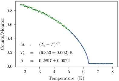
![Figure 4.7: Temperature-dependent ω scan of the (0 4 0) reflection in (ND 4 ) 2 [FeCl 5 (H 2 O)] by single crystal neutron diffraction on HEiDi](https://thumb-eu.123doks.com/thumbv2/1library_info/3698364.1505901/64.892.173.781.133.440/figure-temperature-dependent-reflection-single-crystal-neutron-diffraction.webp)
![Figure 4.8: Single crystal neutron diffraction ω scans of (ND 4 ) 2 [FeCl 5 (D 2 O)] at 100 K (red) and 10 K (blue)](https://thumb-eu.123doks.com/thumbv2/1library_info/3698364.1505901/65.892.130.756.241.943/figure-single-crystal-neutron-diffraction-scans-fecl-blue.webp)
![Figure 4.11: Crystals of the compound Cs 2 [FeCl 5 (H 2 O)]. (a) As-grown crystal. (b) Sam- Sam-ple LB007-S002 for neutron diffraction](https://thumb-eu.123doks.com/thumbv2/1library_info/3698364.1505901/70.892.176.760.119.353/figure-crystals-compound-fecl-grown-crystal-neutron-diffraction.webp)

![Figure 5.1: Phase diagram of Cs 3 Fe 2 Br 9 with the magnetic field aligned along the c direction [129]](https://thumb-eu.123doks.com/thumbv2/1library_info/3698364.1505901/84.892.172.765.163.527/figure-phase-diagram-cs-magnetic-field-aligned-direction.webp)
![Figure 5.5: A Bärnighausen diagram (cf. [132]) shows how the crystal structure of Cs 3 Fe 2 Br 9](https://thumb-eu.123doks.com/thumbv2/1library_info/3698364.1505901/91.892.120.629.180.438/figure-bärnighausen-diagram-shows-crystal-structure-cs-fe.webp)
