characterization of BICD2 mutations causing SMALED2, a congenital dominant form of spinal muscular
atrophy
Inaugural-Dissertation zur
Erlangung des Doktorgrades
der Mathematisch-Naturwissenschaftlichen Fakultät der Universität zu Köln
vorgelegt von
Lilian Andrea Martínez Carrera aus Guatemala Stadt, Guatemala
Köln 2018
performed at the Institute of Human Genetics, Institute of Genetics and Center for Molecular Medicine Cologne (CMMC) of the University of Cologne from January 2012
to April 2017.
Berichterstatter: Prof. Dr. Brunhilde Wirth
Prof. Dr. Elena Rugarli
Tag der mündlichen Prüfung: 26.06.2017
Acknowledgements
This thesis would not be possible without the collaboration and support of many people.
First, I would like to express my gratitude to Prof. Brunhilde Wirth for believing in me and confide such an amazing project to me. I thank for sharing of her time, and enthusiasm about science during all these years. I saw her support and guidance in each plan, in each decision to try a new technique or animal model, and not only when celebrating the nice results but also when the results were not as expected.
I would like to thank to Prof. Elena Rugarli, for each meeting and scientific advice, also for taking the time to review my work.
I am glad to thank Prof. Ulrich Baumann for his time and kind help in taking the chair in my defense.
I thank Dr. Isabell Witt, for her kind counseling and advice during my studies.
Many thanks to our collaborators, RG Gopalakrishnan and RG Uhlirova:
To Dr. Jay Gopalakrishnan for enriching this project with his experience in microtubules and cytoskeleton. I thank for your time and energy in many discussions and experimental design. Also, I thank to all the kind people of his group, especially to Dr.
Elke Gabriel for introducing me into the beauty of confocal imaging and for being at my side in the “painful but gratifying” processing of the images. Also, I thank to Arpit Wason and Dr. Arul Mariappan for our “trips” to the Pathology department (S2 lab), and always being kind to me even when following my very precise instructions and time schedule.
To Prof. Mirka Uhlirova for helping us to establish for the first time, the Drosophila melanogaster model system in our lab. I thank for her time, knowledge and dedication in this project. I thank Dr. Colin Donohoe for his patience in introducing me into a new “tiny”
world, and for taking care of me in the fly lab. Many thanks to Merve Kilinc for sharing the protocol and tips for NMJ preparation.
At the beginning of my PhD studies, in the RG Wirth, during the transition “Occludin- BICD2), I had the opportunity to learn and to get acquainted with the “sequencing era”
and functional analysis from Dr. Lutz Garbes. I thank you a lot, Lutzi .
Later on, another member joined into the BICD2 team, yes I am talking about Irmgard Hölker. I thank you for your excellent technical work, commitment and honesty, also for your wise words.
I thank as well for the technical support in the mice work from Andrea Hoffmann and Vanessa Grysko. I also thank to Kristina Hupperich for taking care of me almost seven years ago, when I was the new one, and for later on letting me be part of the “last lab”.
A big thank you to all the members of the RG Wirth and of the RG Kye: Janine Milbradt (“Bones”), Mert Karakaya, Eike Strathmann, Christian Hoffmann, Aaradhita Upadhyay, Svenja Schneider, Andrea Delle Vedove, Eva Janzen, Wiebke Rehorst, Max Thelen. I thank Seyyedmohsen Hosseinibarkooie for introducing me into the “protein world”, even when I came from the “DNA and RNA world”. I thank to Natalia Mendoza for sharing moments of joy and not so much joy, far away of home but doing what we like, and together with Laura Torres-Benito and Inês do Carmo Gil Gonçalves “gracias guapas”.
I also thank to former members especially to Ludwig Heesen.
I thank to Dr. Min Kye for sharing with me her protocols and advices. To Inês do Carmo Gil Gonçalves for teaching me how to isolate MN and make them happy.
Besides the scientific background, this work was accomplished with the support and motivation of my family and friends, to whom I dedicate this thesis.
To my parents, my brothers Willis and Pablo, and my niece Daniela, for their constant love and support, even when miles and miles separate us.
To the ones that have made me feel like home, Family Storbeck: Ihr seid großartig!
Danke, dass Ihr so viel Gutes für uns tut. Family Arnold-Aguayo, thank you for all your unconditional help and loving care during these years. Son muy lindos.
To Martina Karsubke, for helping me since the first day at University, welcoming me to Germany, and becoming my friend.
To Jared Afre Martinez, the person that inspires me to be better and not give up. Thank you for sharing your mom with her dreams. Our dreams come true, with work and learning from our own mistakes. Please, remember that.
At last, but not least, I would like to thank to Dr. Markus Storbeck for his beautiful
company, and unshakable calmness through the moments of struggle. Thank you for
making me stronger. Without you, everything would be even more difficult - if not
impossible. You remind me every day what is the most important part in life.
Table of Contents
ACKNOWLEDGEMENTS... I TABLE OF CONTENTS... III DIRECTORY OF FIGURES ...VI DIRECTORY OF TABLES...VI
1. AIM, THESIS STRUCTURE AND MAJOR FINDINGS ... 1
2. INTRODUCTION... 3
2.1. S
PINAL MUSCULAR ATROPHIES... 3
2.1.1. Autosomal recessive and X-linked forms of SMA ... 3
2.1.2. Autosomal dominant SMAs ... 4
2.1.2.1. Autosomal dominant SMAs that affect lower and upper extremities ... 4
2.1.2.2. Autosomal dominant SMA that affects lower extremities predominantly, type 1 (SMALED1) ... 7
2.1.2.3. Autosomal dominant SMA that affects lower limbs predominantly, type 2 (SMALED2). 7
2.2. BICD2:
STRUCTURE AND FUNCTION... 8
2.2.1. BICD2 is an adaptor of dynein and functions in different cellular processes... 9
2.2.2. COPI/independent Golgi transport ... 10
2.2.3. Centrosome and nuclear positioning during mitotic entry ... 11
2.2.4. mRNA localization ... 11
2.2.5. Lipid droplet transport... 11
2.2.6. Bidirectional transport... 12
2.3. I
MPORTANCE OFBICD2
IN NEURONAL TISSUE DEVELOPMENT... 12
2.3.1. Cerebellar development ... 12
2.3.2. Synaptic vesicle recycling ... 13
3. PUBLICATIONS ... 15
3.1. T
HE DISCOVERY OFBICD2
AS THE CAUSING GENE OFSMALED2 ... 17
3.1.1. Publications ... 17
3.1.2. Description... 17
3.1.3. Own contributions... 18
3.2. F
UNCTIONAL ANALYSES AND DISEASE MODEL... 19
3.2.1. Publications ... 19
3.2.2. Description of the studies ... 19
3.2.3. Own contributions... 20
3.3. O
THER DISORDERS CAUSED BY VARIANTS INBICD2 ... 22
3.3.1. Publications ... 22
3.3.2. Description of the studies ... 22
3.3.2.1. Chronic myopathy... 22
3.3.2.2. Congenital arthrogryposis multiplex, respiratory failure and early lethality... 23
3.3.3. Own contributions... 23
4. UNPUBLISHED FINDINGS ... 25
4.1. I
MPACT OFBICD2
MUTATIONS ON ENDOCYTOSIS... 25
4.2. I
MPACT OF P.T
HR703M
ET MUTATION ON CENTROSOMES AND CELL CYCLE... 27
4.3. I
MPLICATIONS OF P.A
RG747C
YS MUTATION IN PROTEIN STRUCTURE AND AGGREGATION... 29
4.4. S
UMMARY OF RESULTS OBTAINED FROM FUNCTIONAL CHARACTERIZATION OFBICD2
MUTATIONS... 30
4.5. M
ETHODS(
UNPUBLISHED DATA) ... 31
4.5.1. Eukaryotic cell culture... 31
4.5.2. Endocytosis assay using FITC-dextran... 31
4.5.3. Analysis of centrosomes in primary fibroblasts ... 31
4.5.4. Generation of a mutant BICD2 p.Arg747Cys expression vector... 32
4.5.5. Solubility assay for mutant BICD2 protein... 32
4.5.6. Statistical analyses ... 33
5. DISCUSSION ... 34
5.1. A
LTERATIONS EXERTED BY MUTATIONS INBICD2 ... 35
5.2. P
ATHOLOGICAL CONSEQUENCES OF ALTERATIONS IN DIFFERENT CELLULAR PROCESSES DUE TOBICD2
MUTATIONS... 37
5.3. SMALED2-
ASSOCIATEDBICD2
MUTATIONS STABILIZE MICROTUBULES IN FIBROBLAST CELLS38 5.4. BICD2
MUTATIONS SHARE A COMMON EFFECT ON MOTOR NEURONS... 38
5.5. SMALED2 D
ROSOPHILAMODEL: BICD2
MUTATIONS CAUSE LOCOMOTION IMPAIRMENT WITH DEFECTS INNMJ
DEVELOPMENT... 39
5.6. BICD2
MUTATIONS ARE ASSOCIATED WITH OTHER DISORDERS BESIDESSMALED2... 40
5.6.1. Chronic myopathy... 40
5.6.2. Congenital arthrogryposis multiplex, respiratory failure and early lethality ... 40
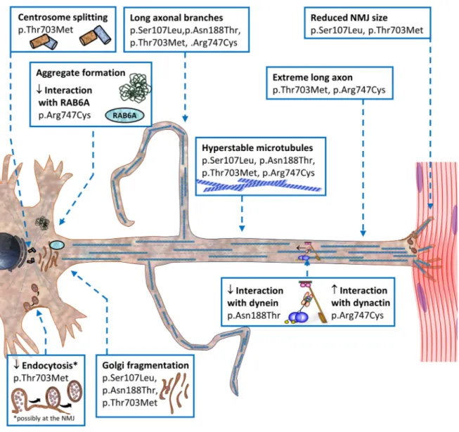
5.7. S
YNOPSIS OF ALTERATIONS TO DUE TOSMALED2-
CAUSINGBICD2
MUTATIONS... 42
6. SUMMARY ... 43
7. ZUSAMMENFASSUNG ... 45
8. ADDITIONAL INVESTIGATIONS DURING THE PHD THESIS... 47
8.1. P
UBLICATION... 47
8.2. A
BSTRACT OF THE PUBLICATION: ... 47
8.3. O
WN CONTRIBUTIONS... 47
9. REFERENCES ... 48
APPENDIX ... XIV
P
UBLICATIONS... XIV
I. Neveling, K.*, Martinez-Carrera, L. A.* et al. (2013). "Mutations in BICD2, which Encodes a Golgin and Important Motor Adaptor, Cause Congenital Autosomal-Dominant Spinal Muscular Atrophy." Am J Hum Genet 96(6): 946-954. (*These authors contributed equally to this work) ...XIV II. Synofzik, M., Martinez-Carrera, L. A. et al. (2014). "Dominant spinal muscular atrophy due to BICD2: a novel mutation refines the phenotype." J Neurol Neurosurg Psychiatry 85(5): 590-592.
XIV
III. Martinez-Carrera, L. A. and Wirth, B. (2015). "Dominant spinal muscular atrophy is caused by mutations in BICD2, an important golgin protein." Front Neurosci 9: 401...XIV IV. Martinez Carrera, L. A., Gabriel, E. et al. (2018). "Novel insights into SMALED2: BICD2 mutations increase microtubule stability and cause defects in axonal and NMJ development." Hum Mol Genet (Epub ahead of print) ...XIV V. Unger, A., Dekomien, G. et al. (2016). "Expanding the phenotype of BICD2 mutations toward skeletal muscle involvement." Neurology 87(21): 2235-2243...XIV VI. Storbeck, M., Eriksen, B. et al. (2017). "Phenotypic extremes of BICD2-opathies: from lethal, congenital muscular atrophy with arthrogryposis to asymptomatic with subclinical features."
Eur J Hum Genet 25(9): 1040-1048 ...XIV VII. Komlosi, K., Hadzsiev, K. et al. (2014). “Exome sequencing identifies Laing distal
myopathy MYH7 mutation in a Roma family previously diagnosed with distal neuronopathy.”
Neuromuscul Disord. 24(2): 156-61 ...XIV
E
RKLÄRUNGDirectory of figures
Figure 1. Cartoon of dynein-dynactin-BICD2 complex. ... 9 Figure 2. Functions of BICD2 in the cell. ... 10 Figure 3. Cerebellar development and granule cell migration. ... 13 Figure 4. FITC-Dextran uptake in fibroblasts derived from affected individuals carrying the p.Thr703Met and p.Arg747Cys mutations in BICD2. ... 26 Figure 5. Centrosome splitting in primary fibroblasts carrying the p.Thr703Met mutation.
... 28 Figure 6. The BICD2 p.Arg747Cys mutation forms insoluble aggregates. ... 29 Figure 7. Schematic synopsis of alterations due to SMALED2- causing BICD2 mutations.
... 42
Directory of tables
Table 1. Current known disease-causing genes for autosomal dominant spinal muscular
atrophies. ... 6
Table 2. Overview of functionally assessed consequences of disease-causing BICD2
mutations... 30
1. Aim, thesis structure and major findings
Homozygous alterations in SMN1 are the most common genetic cause of spinal muscular atrophy (SMA). However, the causing gene of many other cases with SMA remain unknown.
My PhD project aimed to identify and analyze functionally novel genes causing motor neuropathies. To this end, the current work led to the discovery of BICD2 as the causing gene of spinal muscular atrophy, lower extremity-predominant, autosomal dominant, type 2 (SMALED2 MIM 615290).
This PhD thesis starts with an introduction (Section 2) about the different SMA forms, with emphasis on autosomal dominant SMA and focus on the lower extremity- predominant form (SMALED). In the last part of the introduction, the cellular functions of BICD2 are described including the role of this protein in neuronal tissue.
The major findings of my work are presented in Section 3, in conjunction with my individual contributions for each scientific publication.
I summarized in Section 3.1 the discovery of BICD2 variants in affected individuals with SMALED2 (Neveling; Martinez-Carrera et al. 2013, Synofzik et al. 2014). In addition, a review is included where I elaborated on the clinical features of the affected individuals, the locations of the mutations within the protein domains, and possible consequences at the molecular level (Martinez-Carrera and Wirth 2015). Reading this review is highly recommendable for a better overview of this part of the work.
Section 3.2 describes the pathological consequences that the different mutations exert at cellular level: These were Golgi fragmentation in some cases (Neveling; Martinez- Carrera et al. 2013) and alterations in the interaction with the dynein-dynactin complex in others (Martinez-Carrera et al. 2018) . However, the striking finding was that regardless of where the mutation is located in the protein, all of them altered the microtubule array (arrangement), which led to aberrations in axon development in motor neurons, the disease relevant cell type in SMALED2 (Martinez-Carrera et al. 2018).
Furthermore, I also include in this section, the findings from the characterization of the
first in vivo Drosophila model for SMALED2. I generated transgenic Drosophila lines that
carry the BICD2 variants found in individuals with SMALED2. By using the UAS-GAL4, I
was able to allow the expression of the transgenic construct (either wild type or mutant)
in a tissue-restricted manner. When expressed in neuronal tissue, but not in muscle, the
BICD2 mutations caused impaired locomotion with reduced neuromuscular junction size, a hallmark of developmental impairment (Martinez-Carrera et al. 2018).
In Section 3.3, I expand the knowledge about the spectrum of disorders that are associated with variants in BICD2. The clinical presentation in such disorders differed from the typical SMALED2. Two of the BICD2 variants that cause SMALED2, were also identified in affected individuals with chronic myopathy (Unger et al. 2016), and novel variants were associated with congenital arthrogryposis multiplex with respiratory failure and early lethality, the most severe clinical presentation observed in association with BICD2 (Storbeck et al. 2017).
In the next part of my thesis (Section 4), I include unpublished findings obtained during functional investigation of endocytosis, centrosomes, and aggregate formation.
At last, I discuss the compilation of findings of my work (Section 5), correlate with the
findings of others, underline the novel insights into the disease and suggest possible
molecular mechanisms. Sections 6 and 7 include the summary and highlights of this
work. Section 8 includes an additional contribution (Komlosi et al. 2014)
2. Introduction
2.1. Spinal muscular atrophies
Spinal muscular atrophies (SMAs) comprise a group of genetic disorders characterized by aberrant development and/or loss of spinal motor neurons. The clinical features of spinal muscular atrophy are wasting and weakness of muscles supplied by the affected motor neurons (Emery 1971).
SMAs present a broad clinical spectrum, and differential diagnosis from other disorders can be challenging due to the overlapping of symptoms. However, SMAs are often classified based on the mode of inheritance (autosomal recessive, autosomal dominant, X-linked), age of onset, and pattern of muscle weakness (i.e. proximal or distal).
2.1.1. Autosomal recessive and X-linked forms of SMA
The majority of the SMA cases are autosomal recessive linked to chromosome 5 (5q-SMA), and caused by homozygous deletion/mutation of the survival motor neuron 1 (SMN1) gene, localized on chromosome 5q12-q13 (Lefebvre et al. 1995, Wirth et al.
1999, Wirth 2000). The muscle weakness in 5q-SMA is usually symmetrical, more proximal than distal, the lower limbs are more affected than upper. The 5q-SMA is classified into four subtypes based on age of onset and severity of symptoms (MIM 253300, 253550, 253400, 271150).
X-linked forms of SMA (SMAX) affect mainly men and are considered of rare incidence.
The X-linked type I (SMAX1, MIM 313200) is characterized by adult onset of weakness and atrophy of the limb and bulbar muscles, with fasciculations, and an increase in the size of breast tissue (Harding et al. 1982). SMAX1 is associated with trinucleotide repeat expansion CAG(n) in the androgen receptor gene (La Spada et al. 1991, Lund et al.
2001). In contrast to SMAX1, cases with X-linked SMA type II (SMAX2, MIM 301830) show a neonatal onset of severe hypotonia, areflexia, contractures (muscle shortening) and/or fractures. Death occurs in infancy due to respiratory failure (Ramser et al. 2008).
Variants in the gene encoding ubiquitin-activating enzyme-1 (UBA1) have been described as causative of SMAX2 (Ramser et al. 2008). A less frequent X-linked SMA type III (SMAX3, MIM 300489) affects distal muscles of lower limbs and later of upper limbs. The symptoms appear in the first decade of life and are slowly progressive.
Variants in the ATP7A gene, which encodes a transmembrane copper-transporting
ATPase, have been identified in individuals with SMAX3 (Kennerson et al. 2010).
2.1.2. Autosomal dominant SMAs
Autosomal dominant forms of SMA are considered highly heterogeneous and show high variability of clinical features. It is estimated that less than 2% of the total cases of SMAs are dominantly inherited (Farrar and Kiernan 2015).
In contrast to 5q-SMA, autosomal dominant SMAs are considered milder and without or slow progression. Most of the dominant SMAs affect lower and upper limbs, with proximal and/or distal pattern of muscle weakness and the age of onset ranges from congenital to late adulthood (Table 1).
In the great majority of individuals with autosomal dominant SMA, the genetic causes are unknown. However, the rapid advance in genetic screening due to massive parallel sequencing and improvement in phenotypical classification have contributed remarkably to the discovery and characterization of novel causative genes (Table 1).
2.1.2.1. Autosomal dominant SMAs that affect lower and upper extremities
f
This group of autosomal dominant SMAs that affects lower and upper limbs includes:
The scapuloperoneal SMA (SPSMA MIM 181405) is characterized by weakness of the scapular (shoulder blade) and peroneal (lower leg) muscles. The onset of the symptoms is congenital or early childhood. Variants in TRPV4, have been described as the genetic cause of SPSMA (Auer-Grumbach et al. 2010). TRPV4 is a transient receptor potential cation channel that plays a role in neuronal signalling (Liedtke 2008). Variants in TRPV4 have been also associated with Charcot-Marie-Tooth type 2C (CMT2C MIM 606071) (Landoure et al. 2010).
The Finkel SMA with late adult onset (SMAFK MIM 182980), presents proximal muscle weakness and fasciculations. Variants in VAPB are associated to SMAFK (Nishimura et al. 2004). VAPB encodes a vesicle-associated membrane protein that plays a role in the unfolded protein response (UPR) and has been associated with amyotrophic lateral sclerosis type 8 (ALS8 MIM 608627) (Kanekura et al. 2006).
The distal SMAs, also known as distal hereditary motor neuropathies (dHMN), cause
weakness and atrophy of lower and upper limbs with distal predominance. dHMN are
clinically and genetically diverse. The phenotypes of various dHMN overlap with other
disorders such as Charcot-Marie-Tooth and amyotrophic lateral sclerosis. Several types
and subtypes of dHMN have been associated with variants in genes that encode for
heat-shock proteins like HSPB8 (Irobi et al. 2004), HSPB1 (Houlden et al. 2008) and
HSPB3 (Kolb et al. 2010), also the choline transporter SLC6A7 (Barwick et al. 2012) and dynactin-1 (Puls et al. 2003), among others (Table 1).
Other two dominant SMAs with distal predominance are the hereditary neuropathy with or without age-related macular degeneration (HNARMD MIM 608895) and the peripheral neuropathy, myopathy, hoarseness, and hearing loss (PNMHH MIM 614369). HNARMD has been associated with variants in the FBLN5 gene, which encodes the fibulin-5 that might promote the deposit formation in macular degeneration (Mullins et al. 2007).
Regarding PNMHH, the identified causing-gene MYH14 encodes a member of the
myosin II family, which interact with cytoskeletal actin (Choi et al. 2011).
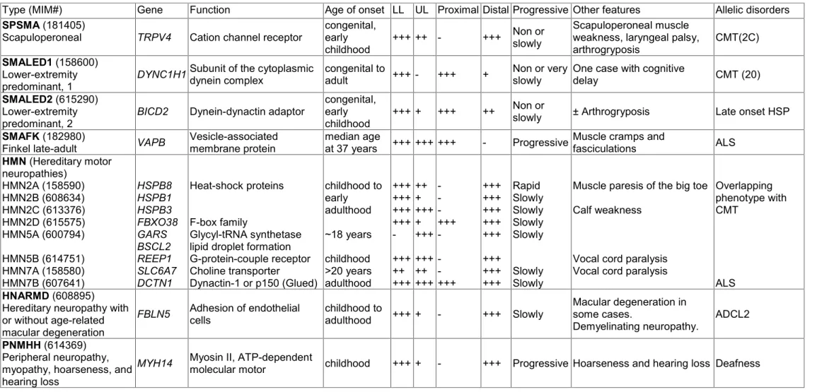
Table 1. Current known disease-causing genes for autosomal dominant spinal muscular atrophies.
Limbs affected LL: lower limbs; UL: upper limbs; +++: majority of the cases; ++: some of the cases; +: few cases; -: no cases; CMT: Charcot-Marie-Tooth; HSP: Hereditary spastic paraplegia; ALS: Amyotrophic lateral sclerosis; ADCL2: autosomal dominant cutis laxa-2
Type (MIM#) Gene Function Age of onset LL UL Proximal Distal Progressive Other features Allelic disorders
SPSMA (181405)
Scapuloperoneal TRPV4 Cation channel receptor congenital, early
childhood +++ ++ - +++ Non or slowly
Scapuloperoneal muscle weakness, laryngeal palsy,
arthrogryposis CMT(2C)
SMALED1 (158600)
Lower-extremity
predominant, 1 DYNC1H1 Subunit of the cytoplasmic
dynein complex congenital to
adult +++ - +++ + Non or very
slowly One case with cognitive
delay CMT (20)
SMALED2 (615290)
Lower-extremity
predominant, 2 BICD2 Dynein-dynactin adaptor congenital,
early
childhood +++ + +++ ++ Non or
slowly ± Arthrogryposis Late onset HSP
SMAFK (182980)
Finkel late-adult VAPB Vesicle-associated
membrane protein median age
at 37 years +++ +++ +++ - Progressive Muscle cramps and fasciculations ALS HMN (Hereditary motor
neuropathies) HMN2A (158590) HMN2B (608634) HMN2C (613376) HMN2D (615575) HMN5A (600794) HMN5B (614751) HMN7A (158580) HMN7B (607641)
HSPB8 HSPB1 HSPB3 FBXO38 GARS BSCL2 REEP1 SLC6A7 DCTN1
Heat-shock proteins
F-box family
Glycyl-tRNA synthetase lipid droplet formation G-protein-couple receptor Choline transporter
Dynactin-1 or p150 (Glued)
childhood to early adulthood
~18 years childhood
>20 years adulthood
+++ +++
+++ +++
- +++ ++
+++
++ + +++ + +++
+++ ++
+++
- - - +++
- - - +++
+++ +++
+++ +++
+++
+++ +++
+++
Rapid Slowly Slowly Slowly Slowly
Slowly Slowly
Muscle paresis of the big toe Calf weakness
Vocal cord paralysis Vocal cord paralysis
Overlapping phenotype with CMT
HNARMD (608895) ALS Hereditary neuropathy with or without age-related macular degeneration
FBLN5 Adhesion of endothelial
cells childhood to
adulthood +++ + - +++ Slowly Macular degeneration in
some cases.
Demyelinating neuropathy. ADCL2
PNMHH (614369)
Peripheral neuropathy, myopathy, hoarseness, and hearing loss
MYH14 Myosin II, ATP-dependent
molecular motor childhood +++ + - +++ Progressive Hoarseness and hearing loss Deafness
2.1.2.2. Autosomal dominant SMA that affects lower extremities predominantly, type 1 (SMALED1)
The term SMALED was first used by Harms and colleagues, to distinguish a dominant SMA with clear lower limb predominance from other dominant SMA forms with upper extremity involvement (Harms et al. 2010). The first gene to be identified as causative of SMALED was DYNC1H1 (SMALED1 MIM 158600).
Individuals with SMALED1 present difficulties to walk and show proximal leg weakness with muscular atrophy without sensory involvement. The weakness and atrophy are prominent in quadriceps muscles, and mild atrophy of distal leg muscles has been also reported. In very few cases, the upper limbs are mildly affected. Reduced or absent reflexes in the lower limbs are also reported. The symptoms appear in early childhood and the course of the disease is non-progressive or very slowly progressive (Harms et al. 2010, Harms et al. 2012, Tsurusaki et al. 2012). In many cases of SMALED1, electromyography (EMG) and skeletal muscle biopsy show signs of chronic denervation (Harms et al. 2010, Harms et al. 2012). Very few individuals with SMALED1 present contractures.
Regarding the molecular basis of SMALED1, several heterozygous mutations in the tail domain of the heavy chain of cytoplasmic dynein (DYNC1H1) have been described, and experimental evidence suggested that these mutations disrupt dynein complex stability, and/or affect its function (Harms et al. 2012, Hoang et al. 2017). Interestingly, it was recently reported that the majority of the mutations do not affect binding of dynein to dynactin and to its cargo adaptor BICD2 (Hoang et al. 2017). However, those mutations decrease the travel distances of moving dynein-dynactin complex, presumably by changes in the microtubule-binding domain of dynein (Hoang et al. 2017). Mutations in the motor domain of DYNC1H1 are described as to possess the strongest effects on dynein motility in vitro, and have been associated with malformations of cortical development (MCD, MIM 614563) (Schiavo et al. 2013, Hoang et al. 2017).
2.1.2.3. Autosomal dominant SMA that affects lower limbs predominantly, type 2 (SMALED2)
In 1994, Frijns et al. described a family of Dutch origin, in which the affected individuals exhibited congenital nonprogressive atrophy and weakness predominantly of lower limb muscles, in association with contractures in ankles and feet (Frijns et al.
1994). Tendon reflexes were reduced or absent. The affected individuals presented
difficulties when walking, such as the ability to walk only on toes and waddling gait.
Histological studies of the affected muscles revealed evidence of a neurogenic disorder, and signs of denervation and reinnervation (Frijns et al. 1994).
The pattern of inheritance was dominant and linkage to the 5q12-q13 region was excluded. The phenotype of the disease observed in family 1 was described as dominant non-5q SMA with lower limb predominance. However, the genetic cause was unknown.
This family described by (Frijns et al. 1994), constituted the starting point of my project.
We identified variants in the BICD2 gene as disease-causing (Section 3.1). The next section includes an overview of the cellular functions of BICD2, which are widely investigated.
2.2. BICD2: structure and function
Bicaudal-D (BicD) was initially identified in Drosophila melanogaster through a characterization of two dominant lethal maternal mutations, which disrupt the establishment of anterior and posterior polarity giving rise to bicaudal (two tails) embryos (Mohler and Wieschaus 1986, Steward 1987, Schupbach and Wieschaus 1991).
Only one BicD gene is present in invertebrates, while mammals have two homologs BICD1 and BICD2. In humans, BICD1 is localized in the chromosomal region 12p11.2- p11.1 and is mainly expressed in brain, skeletal muscle and heart (Baens and Marynen 1997), while BICD2 localizes in the chromosomal region 9q22.3 and is ubiquitously expressed.
Structural analysis in Drosophila revealed that more than half of the BicD protein consists of heptad repeats. A heptad repeat is defined as a repeating pattern of seven amino acids of which hydrophobic residues are preferentially located at positions 1 and 4 (McLachlan and Karn 1983). These heptad repeats are responsible of mediating the packaging of one helix against another, resulting in the formation of coiled-coil (CC) structures (Bruccoleri et al. 1986). The N-terminal domain contains the coiled-coil segment 1 (CC1) and the coiled-coil segment 2 (CC2), and the C-terminal domain contains the coiled-coil segment 3 (CC3).
In the case of BICD2, experimental evidence supports that the CC1 binds to CC3 and undergo intramolecular interactions forming homodimers (Hoogenraad et al. 2001). It is further proposed that once the CC3 engages in an interaction with other proteins, the CC1 becomes available for interaction with other proteins. These protein interactions seem to determine the cellular localization and function of BICD2 (Hoogenraad et al.
2001).
2.2.1. BICD2 is an adaptor of dynein and functions in different cellular processes
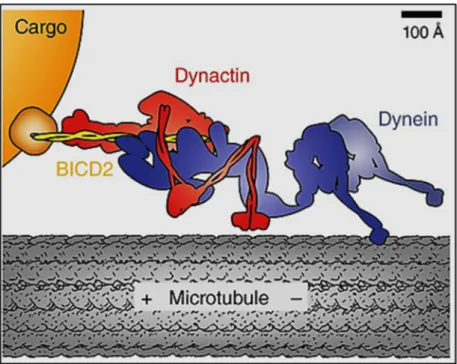
Motor adaptor proteins that link motors to cargo are commonly implicated in controlling motor coordination and cargo movement (Schlager and Hoogenraad 2009, Akhmanova and Hammer 2010, Jolly and Gelfand 2011, Fu and Holzbaur 2014). BICD2 is a widely studied cargo adaptor of cytoplasmic dynein, the major contributor to minus end-directed microtubule transport (Figure 1) (Hoogenraad et al. 2001, Matanis et al.
2002, Splinter et al. 2010, Splinter et al. 2012).
Figure 1. Cartoon of dynein-dynactin-BICD2 complex.
Dynein (blue) moves toward the minus end of the microtubule. Dynein associates with dynactin (red) and with BICD2 (yellow) to link to cargo (orange). Source: (Reck-Peterson 2015).
Dynein requires dynactin (name is derived from dynein activator) for nearly all of its cellular functions (Holleran et al. 1998, Karki and Holzbaur 1999, Schroer 2004). Dynein and dynactin bind to each other via the interaction of dynein intermediate chain and the dynactin subunit p150 (Vaughan et al. 1995, Karki and Holzbaur 1999, King et al. 2003).
Several studies in vitro described that these two complexes exist as separate pools that
bind transiently, suggesting that additional factors must be present in cells to strengthen
this association in order to achieve long distance transport (Quintyne et al. 1999,
Habermann et al. 2001, Quintyne and Schroer 2002). Studies have suggested that the
N-terminal domain of BICD2 binds to the dynein-dynactin complex, not only to function
as a linker for cargos, but also to promote a stable interaction between dynein and
dynactin (Splinter et al. 2012).
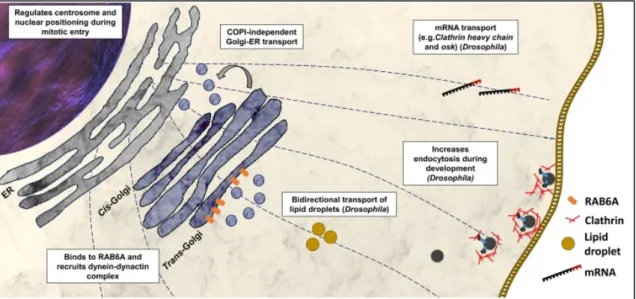
The C-terminal domain of BICD2 shows the highest degree of conservation among metazoans and is the cargo-binding domain (Hoogenraad et al. 2001, Terenzio and Schiavo 2010). BICD2 links the dynein-dynactin complex to cargos that are involved in different dynein-mediated processes such as retrograde COPI-independent Golgi transport, centrosome and nuclear positioning during mitotic entry, mRNA localization, lipid droplet transport, endocytosis, and microtubule organization (Figure 2).
Figure 2. Functions of BICD2 in the cell.
Studies in mammals and/orDrosophila melanogasterhave shown that BICD2 is involved in several cellular processes. Source: (Martinez-Carrera and Wirth 2015).
2.2.2. COPI/independent Golgi transport
The C-terminal domain of BICD2 binds to the small GTPase RAB6A, which coordinates the retrograde COPI-independent Golgi-ER pathway, a recycling route of Golgi-resident membrane proteins that allows the assembly of functional Golgi stacks (Hoogenraad et al. 2001, Young et al. 2005). The active form of RAB6A (GTP-bound) is associated with the trans-Golgi membrane and recruits BICD2 via interaction with the C-terminal domain. The N-terminal domain of BICD2 becomes available and recruits the dynein-dynactin complex to transport RAB6A vesicles from the trans-Golgi to the ER (Matanis et al. 2002).
BICD2 is considered a golgin due to its abundant coiled coil structure, localization at the
Golgi, and interaction with a member of the RAB family of GTPases (Barr and Short
2003, Short et al. 2005, Goud and Gleeson 2010). Golgins are proteins associated with
the Golgi apparatus and contribute to maintain its organization.
2.2.3. Centrosome and nuclear positioning during mitotic entry
In early G2 phase, the C-terminal domain of BICD2 switches from interacting with RAB6A to interact with RANBP2, a component of the nuclear pore (Splinter et al. 2010).
BICD2 in turn, recruits the dynein-dynactin complex to the nuclear envelope to facilitate a proper positioning of the nucleus relative to centrosomes prior mitosis. However, the mechanism that controls the shift of interacting partners of BICD2 during the cell cycle is unknown.
2.2.4. mRNA localization
Studies in Drosophila have shown that the C-terminal domain of BicD interacts with Egalitarian (Egl), a RNA-binding protein, whose human ortholog is EXD1 (exonuclease 3´-5´Domain Containing 1) (Bullock and Ish-Horowicz 2001, Delanoue and Davis 2005).
BicD recruits the dynein-dynactin complex to target the mRNA cargo to distinct cellular compartments. This complex comprising BicD-Egl-dynein-dynactin is thought to associate with further proteins that regulate translation and stability of transported mRNA. During Drosophila development, the correct mRNA localization is crucial for specifying anteroposterior and dorsoventral axes of oocytes and embryos (Weil 2014).
The identification of the mRNAs associated with BicD-Egl has been subject of investigation. Clathrin heavy chain (Chc) mRNA has been identified as a cargo of BicD.
Chc mRNA requires BicD to be transported into the oocyte where is presumably needed to establish microtubule polarity, an endocytosis-independent role of Chc (Vazquez- Pianzola et al. 2014).
Even though, mRNA transport is an important process in mammals and BICD is highly conserved among metazoans, the role of BICD in mRNA transport during development in mammals has not been investigated. However, to study this possibility would be of special interest not only in the field of developmental science but also for the understanding of early developmental pathologies.
2.2.5. Lipid droplet transport
Lipid droplets are present at all developmental stages of Drosophila and are
particularly important as energy sources (Kuhnlein 2012). It has been described that lipid
droplets are moved by the dynein complex (Gross et al. 2000). Based on observations
in the composition and motion of lipid droplets, it has been suggested that BicD binds to
lipid droplets and recruits the dynein complex to transport those droplets in the forming
embryo (Larsen et al. 2008).
In humans, increasing evidence suggests that disrupted lipid droplet function/localization may contribute to neurodegenerative disorders, for example Huntington’s disease (Martinez-Vicente et al. 2010), Parkinson´s disease (Cole et al. 2002), and hereditary spastic paraplegias (Welte 2015). The role of BICD proteins in lipid droplet transport in mammals and defects in lipid metabolism has not been explored.
2.2.6. Bidirectional transport
A binding between BICD2 and the tail domain of kinesin-1 has been previously described (Grigoriev et al. 2007). However, this binding was suggested to be weaker than the association of BICD2 with dynein-dynactin complex. By using yeast two-hybrid system, the interaction between the middle part of BICD2 (Coiled coil 2 domain) and the tail of kinesin-1 (KIF5A) has been confirmed. However, this interaction seems to be strongly suppressed in the full-length BICD2, possibly by self inactivation due to interaction of the N-terminal domain of BICD2 (Coiled coil 1 domain) with its C-terminal domain (Coiled coil 3). Studies in Drosophila have suggested that BicD plays a role in plus end-directed microtubule transport, and in balancing plus end (kinesin mediated) versus minus end motion (dynein mediated) (Larsen et al. 2008). Recent studies have shown that dynein-dynactin complex and kinesin are opponents in a tug-war competition along microtubules, and BICD2 increases the force of dynein to successfully resist against kinesin (Belyy et al. 2016).
2.3. Importance of BICD2 in neuronal tissue development 2.3.1. Cerebellar development
In vivo studies in a homozygous Bicd2 knockout mouse have provided further
insights into BICD2 function (Jaarsma et al. 2014). Bicd2-deficient mice show cerebellar
defects with severe hydrocephalus, and all died before postnatal day 30. Histological
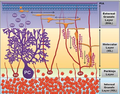
examination has revealed deficits in granule cell migration in the cerebellum. It is known
that during cerebellar development, the granule cells (postmitotic neurons) migrate from
the external granular layer (EGL) toward their final destination within the internal granular
layer (IGL) (Figure 3) (Komuro and Yacubova 2003). The granule cells migrate along
Bergmann glia cells, which determine their correct position from the molecular layer (ML)
to the IGL (Komuro et al. 2001).
Figure 3. Cerebellar development and granule cell migration.
Precursor granule cells (blue spheres, top) proliferate in response to growth signals including sonic hedgehog (shh) released by Purkinje cells (PC, purple). Postmitotic granule cells (orange sphere) start to migrate and attach to Bergamnn glia cells (BG, red cells) to finally arrive at the internal granule layer. Source:
(Tharmalingam and Hampson 2016).
Further histological studies have shown that BICD2 is highly expressed in Bergmann cells (Jaarsma et al. 2014). Moreover, conditional knockout mice, with Bicd2 depleted in Bergmann cells, show decreased levels of Tenascin C, an extracellular matrix protein produced by Bergmann glia cells, possibly by defects in secretion due to the BICD2 deficiency. Tenascin C is known to mediate the attachment of neurons to Bergmann cells and stimulate migration (Bartsch et al. 1992, Chiquet-Ehrismann and Tucker 2011).
Importantly, besides the cerebellar defects, the homozygous Bicd2 knockout mice do not show alterations in any tissue including spinal cord, and the heterozygous knockout mice do not display a pathological phenotype (Jaarsma et al. 2014). Individuals with SMALED2 do not show any pathological changes in the cerebellum. These differences in phenotypes between Bicd2-deficient mice and individuals with SMALED2 indicate that the missense BICD2 mutations exert gain-of-function effect rather than a loss-of-function effect.
2.3.2. Synaptic vesicle recycling
In Drosophila, the complete absence of BicD expression leads to decreased larval
locomotion, and lethality is reported during pupa stage due to inability to eclose (Ran et
al. 1994, Li et al. 2010). However, the transgenic expression of BicD in pan-neuronal
tissue has been proven to fully rescue the larval locomotion and lethality, whereas
expression in the muscles does not (Li et al. 2010). These observations suggest that despite its widespread expression, BicD function is only obligatory in the nervous system during development.
Subsequent studies have shown that the C-terminal domain of BicD binds to Clathrin
heavy chain (Chc), while the N-terminal domain recruits the dynein complex to transport
clathrin-associated synaptic vesicles for their efficient recycling (Li et al. 2010). More
studies seemed to be needed to investigate the extent of BicD participation in
endocytosis and neuromuscular junction morphology.
3. Publications
The discovery of BICD2 as the causing gene of SMALED2
Neveling, K.*, Martinez-Carrera, L. A.*, Holker, I., Heister, A., Verrips, A., Hosseini- Barkooie, S. M., Gilissen, C., Vermeer, S., Pennings, M., Meijer, R., Te Riele, M., Frijns, C. J., Suchowersky, O., Maclaren, L., Rudnik-Schoneborn, S., Sinke, R. J., Zerres, K., Lowry, R. B., Lemmink, H. H., Garbes, L., Veltman, J. A., Schelhaas, H. J., Scheffer, H.
and Wirth, B. (2013). "Mutations in BICD2, which Encodes a Golgin and Important Motor Adaptor, Cause Congenital Autosomal-Dominant Spinal Muscular Atrophy." Am J Hum Genet 96(6): 946-954.
*These authors contributed equally to this work.
Synofzik, M., Martinez-Carrera, L. A., Lindig, T., Schols, L. and Wirth, B. (2014).
"Dominant spinal muscular atrophy due to BICD2: a novel mutation refines the phenotype." J Neurol Neurosurg Psychiatry 85(5): 590-592.
Martinez-Carrera, L. A. and Wirth, B. (2015). "Dominant spinal muscular atrophy is caused by mutations in BICD2, an important golgin protein." Front Neurosci 9: 401.
Functional analysis and disease model
Neveling, K.*, Martinez-Carrera, L. A.*, Holker, I., Heister, A., Verrips, A., Hosseini- Barkooie, S. M., Gilissen, C., Vermeer, S., Pennings, M., Meijer, R., Te Riele, M., Frijns, C. J., Suchowersky, O., Maclaren, L., Rudnik-Schoneborn, S., Sinke, R. J., Zerres, K., Lowry, R. B., Lemmink, H. H., Garbes, L., Veltman, J. A., Schelhaas, H. J., Scheffer, H.
and Wirth, B. (2013). "Mutations in BICD2, which Encodes a Golgin and Important Motor Adaptor, Cause Congenital Autosomal-Dominant Spinal Muscular Atrophy." Am J Hum Genet 96(6): 946-954.
*These authors contributed equally to this work.
Martinez Carrera, L. A., Gabriel, E., Donohoe, C., Hölker, I., Wason, A., Storbeck, M., Uhlirova, M., Gopalakrishnan, J. and Wirth, B. (2018). "Novel insights into SMALED2:
BICD2 mutations increase microtubule stability and cause defects in axonal and NMJ
development." Hum Mol Genet (Epub ahead of print).
Other disorders caused by variants in BICD2
Unger, A., Dekomien, G., Guttsches, A., Dreps, T., Kley, R., Tegenthoff, M., Ferbert, A., Weis, J., Heyer, C., Linke, W. A., Martinez-Carrera, L.A., Storbeck, M., Wirth, B., Hoffjan, S. and Vorgerd, M. (2016). "Expanding the phenotype of BICD2 mutations toward skeletal muscle involvement." Neurology 87(21): 2235-2243.
Storbeck, M., Eriksen, B., Unger, A., Hölker, I., Aukrust, I., Martinez-Carrera, L.A., Linke, W. A., Ferbert, A., Heller, R., Vorgerd, M., Houge, G. and Wirth, B. (2017).
"Phenotypic extremes of BICD2-opathies: from lethal, congenital muscular atrophy with
arthrogryposis to asymptomatic with subclinical features." Eur J Hum Genet 25(9): 1040-
1048.
3.1. The discovery of BICD2 as the causing gene of SMALED2 3.1.1. Publications
Neveling, K.
*, Martinez-Carrera, L. A.
*, Holker, I., Heister, A., Verrips, A., Hosseini- Barkooie, S. M., Gilissen, C., Vermeer, S., Pennings, M., Meijer, R., Te Riele, M., Frijns, C. J., Suchowersky, O., Maclaren, L., Rudnik-Schoneborn, S., Sinke, R. J., Zerres, K., Lowry, R. B., Lemmink, H. H., Garbes, L., Veltman, J. A., Schelhaas, H. J., Scheffer, H.
and Wirth, B. (2013). "Mutations in BICD2, which Encodes a Golgin and Important Motor Adaptor, Cause Congenital Autosomal-Dominant Spinal Muscular Atrophy." Am J Hum Genet 96(6): 946-954.
*These authors contributed equally to this work.
Synofzik, M., Martinez-Carrera, L. A., Lindig, T., Schols, L. and Wirth, B. (2014).
"Dominant spinal muscular atrophy due to BICD2: a novel mutation refines the phenotype." J Neurol Neurosurg Psychiatry 85(5): 590-592.
Martinez-Carrera, L. A. and Wirth, B. (2015). "Dominant spinal muscular atrophy is caused by mutations in BICD2, an important golgin protein." Front Neurosci 9: 401.
3.1.2. Description
Almost 20 years after Frijns and colleagues described family 1, and thanks to the advances in next generation sequencing, we were able to identify the genetic cause of the dominant non-5q SMA in family 1 (Neveling; Martinez-Carrera et al. 2013). To identify the chromosomal location of the disease-causing mutations, linkage analysis was performed in nine members of family 1, which identified a locus on chromosome 9 (chr9:
94,4440,951-104,432,543). Exome sequencing was performed in a single affected individual, and variants were filtered based on location on chromosome 9, nonsynonymous exonic and splice-sites. After excluding known SNPs and previously identified variants, just five variants in five different genes were found. Of those five genes, only BICD2 was located within the linkage region. The heterozygous variant in BICD2 (c.320C>T) results in the amino acid change p.Ser107Leu (Neveling; Martinez- Carrera et al. 2013).
To search for further BICD2 variants that might cause dominant SMA, the seven exons of BICD2 were analyzed by Sanger sequencing in twenty-three additional families with dominant non-5q-SMA. Further two heterozygous variants (c.563A>C [p.Asn188Thr]
and c.2108C>T [p.Thr703Met]) were identified (Neveling; Martinez-Carrera et al. 2013).
A rare SNV (c.269A>G [p.Lys90Arg]) was identified in two additional families. However,
some affected individuals in these families did not carry this SNV, which discards this
variant as disease-causing.
A fourth heterozygous variant (c.2239C>T [p.Arg747Cys]) was identified in all the affected individuals with dominant SMA, in a three-generation family (Synofzik et al.
2014).
Affected individuals with dominant SMA that carry BICD2 variants, present weakness and atrophy predominantly affecting the proximal and distal muscles of the lower extremities. For this reason, the pathology was named spinal muscular atrophy, lower extremity predominant, autosomal dominant, type 2 (SMALED2 MIM 615290).
The symptoms are congenital or appear in early childhood, but few cases with late onset have been also described. The course of the disease is slowly progressive or nonprogressive. Tendon reflexes are reduced or absent. In the majority of the cases, the SMALED2 phenotype is very mild, and during clinical examination may be first-glance diagnosed as SMA type IV, the mildest form of the classical 5q-SMA. Delayed motor milestones, waddling gait, and chronic walking on toes are commonly reported. It seems very characteristic that many individuals with SMALED2 show evident wasting of the lower limbs and a very broad upper body, which resembles a bodybuilder-like shape.
Another differential feature in many individuals with SMALED2 is the presence of contractures in knee, feet, and/or hip, which correlates with congenital onset, possibly due to decreased intrauterine movements (Neveling; Martinez-Carrera et al. 2013). In contrast, very few individuals with SMALED1 (Harms et al. 2010, Harms et al. 2012) and only very severe 5q-SMA cases present contractures (Rudnik-Schoneborn et al. 2008).
In this Section, I include a review about all the individuals with SMALED2 that have been reported (Martinez-Carrera and Wirth 2015). This review provides a summary of the clinical presentation including age of onset, presence of contractures, and pattern of muscular weakness/atrophy. Importantly, we show an overview about the location and possible molecular mechanism of the different mutations.
3.1.3. Own contributions
My contributions to (Neveling; Martinez-Carrera et al. 2013) are split into two parts. Here I include the part of the gene discovery and Section 3.2 includes the part of functional analysis for this paper.
In the gene discovery part of the study (Neveling; Martinez-Carrera et al. 2013), I
designed, carried out and analyzed the exon sequencing of BICD2 in all twenty three
additional families. Subsequently to the variant identification, I performed the
corresponding segregation analysis. For the manuscript, I contributed with the design of
the figure 1, writing and reviewing.
In (Synofzik et al. 2014), I performed the Sanger sequencing, identified the variant in BICD2, and continued with segregation analysis. For the publication, I contributed with the preparation of the figure, writing my corresponding part and reviewing the draft.
I wrote the review (Martinez-Carrera and Wirth 2015).
3.2. Functional analyses and disease model 3.2.1. Publications
Neveling, K.
*, Martinez-Carrera, L. A.
*, Holker, I., Heister, A., Verrips, A., Hosseini- Barkooie, S. M., Gilissen, C., Vermeer, S., Pennings, M., Meijer, R., Te Riele, M., Frijns, C. J., Suchowersky, O., Maclaren, L., Rudnik-Schoneborn, S., Sinke, R. J., Zerres, K., Lowry, R. B., Lemmink, H. H., Garbes, L., Veltman, J. A., Schelhaas, H. J., Scheffer, H.
and Wirth, B. (2013). "Mutations in BICD2, which Encodes a Golgin and Important Motor Adaptor, Cause Congenital Autosomal-Dominant Spinal Muscular Atrophy." Am J Hum Genet 96(6): 946-954.
*These authors contributed equally to this work.
Martinez Carrera, L. A., Gabriel, E., Donohoe, C., Hölker, I., Wason, A., Storbeck, M., Uhlirova, M., Gopalakrishnan, J. and Wirth, B. (2018). "Novel insights into SMALED2:
BICD2 mutations increase microtubule stability and cause defects in axonal and NMJ development." Hum Mol Genet (Epub ahead of print).
3.2.2. Description of the studies
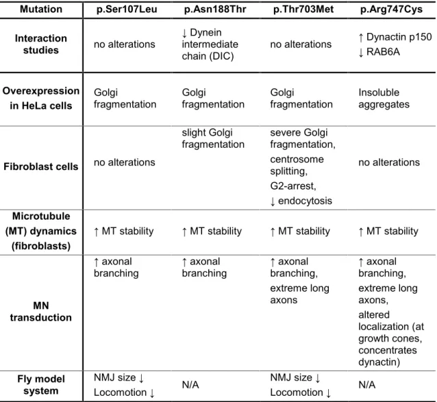
We proceeded to examine the effects of the four mutations described by us, on cellular processes involving BICD2 and that have been previously described as affected in neurodegenerative disorders, such as Golgi structure and interactions with dynein- dynactin complex and RAB6A.
In vitro overexpression experiments and studies of fibroblasts derived from individuals with SMALED2 showed that p.Thr703Met and p.Asn188Thr mutations cause Golgi fragmentation (Neveling; Martinez-Carrera et al. 2013).
Interaction studies revealed that two mutations change the binding of BICD2 with the dynein-dynactin complex. The p.Asn188Thr mutation slightly decreased the binding to dynein. The p.Arg747Cys mutation increases the interaction of BICD2 with the p150 subunit of dynactin and decreases the interaction with RAB6A (Martinez-Carrera et al.
2018).
We focused on the observations that some of the BICD2 mutations alter interactions with the dynein-dynactin complex and/or cause Golgi fragmentation. We investigated whether the BICD2 mutations impair microtubule organization. The fibroblast derived from individuals with SMALED2 exhibit longer and hyperstable microtubules, in comparison with controls. This effect was observed regardless of where the mutation is located, which may constitute a common cellular mechanism affecting microtubules (Martinez- Carrera et al. 2018).
The next step in our investigation was to determine the effect of BICD2 mutations on motor neurons, the disease relevant cell type in SMALED2. Motor neurons overexpressing BICD2 mutations developed axonal aberrations such as collateral branching and overgrowth, possibly by alterations in the microtubule array (arrangement) (Martinez-Carrera et al. 2018).
To study the in vivo consequences of BICD2 mutations, we generated a Drosophila model for SMALED2. The specific expression of BICD2 mutations in neuronal tissue, but not in muscle, led to impaired locomotion and reduced neuromuscular junction size (Martinez-Carrera et al. 2018).
3.2.3. Own contributions
In the functional analysis part of (Neveling; Martinez-Carrera et al. 2013), I performed the overexpression studies carrying out the site directed mutagenesis for each of the constructs used, high scale plasmid preparation of the constructs, transfection, imaging, analysis, and interpretation of the results. I conducted the expression analysis and Golgi studies in primary fibroblast cells derived from individuals with SMALED2. I wrote the corresponding part of functional analysis in the manuscript, and prepared the figures 3, 4 and S1.
In (Martinez-Carrera et al. 2018), I conducted the microtubule experiments in fibroblast cells, imaged them, analyzed and interpreted results. I cloned, purified and high scale produced the constructs for lentiviral transduction, isolated motor neurons and cultured them. I stained the transduced motor neurons and imaged them using confocal microscopy. I performed the quantification analyses, statistics, and interpretation.
Regarding the part of Drosophila melanogaster, I designed and established the construct
to be used for the generation of the transgenic flies, performed each cross to generate
the SMALED2 flies and controls, validated them by PCR and western blot, carried out
the locomotion tests. I dissected the NMJs from the larvae, stained, and imaged them
using confocal microscope. I analyzed the data, applied statistics, and interpreted the
results. Regarding the interaction studies, I performed and analyzed all the experiments.
For the expression analysis of BICD2 in murine tissues, I dissected the tissues,
statistically analyzed and interpreted. I wrote the manuscript and prepared the figures 1,
2, 3, 4, 5, S1, S2, S3, S4, S5, S6, S7, S8, S9.
3.3. Other disorders caused by variants in BICD2 3.3.1. Publications
Unger, A., Dekomien, G., Guttsches, A., Dreps, T., Kley, R., Tegenthoff, M., Ferbert, A., Weis, J., Heyer, C., Linke, W. A., Martinez-Carrera, L.A., Storbeck, M., Wirth, B., Hoffjan, S. and Vorgerd, M. (2016). "Expanding the phenotype of BICD2 mutations toward skeletal muscle involvement." Neurology 87(21): 2235-2243.
Storbeck, M., Eriksen, B., Unger, A., Hölker, I., Aukrust, I., Martinez-Carrera, L.A., Linke, W. A., Ferbert, A., Heller, R., Vorgerd, M., Houge, G. and Wirth, B. (2017).
"Phenotypic extremes of BICD2-opathies: from lethal, congenital muscular atrophy with arthrogryposis to asymptomatic with subclinical features." Eur J Hum Genet 25(9): 1040- 1048
3.3.2. Description of the studies 3.3.2.1. Chronic myopathy
The p.Ser107Leu and p.Thr703Met mutations that we previously described in individuals with SMALED2, were also identified in two families where affected individuals present muscle weakness due to chronic myopathic alterations of skeletal muscle fibers with minor neurogenic changes (Unger et al. 2016). Muscle studies in affected individuals showed impairment of Golgi integrity, increased Golgi-derived vesicles in muscle fibers, and abnormal BICD2 localization. Two proteins, dysferlin and caveolin-3, were reported as strongly reduced at the sarcolemma (cell membrane of muscle fiber cell) in almost all muscle fibers. It is known that vesicular dysferlin is transported via kinesin in muscle cells and is important for muscle membrane repair, and caveolin-3 has been implicated in the trafficking of dysferlin to the plasma membrane (Cai et al. 2009, McDade and Michele 2014). Even though, the role of BICD2 in muscle has not been explored, it has been speculated that BICD2 mutations affect the transport of exocytic cargoes containing dysferlin and caveolin-3 leading to impaired muscle integrity (Unger et al. 2016).
Interestingly, segregation analysis of the individual with chronic myopathy that carries
the p.Thr703Met showed that his unaffected mother carries the same heterozygous
mutation. The mutant allele was confirmed to be expressed in the unaffected mother
(Storbeck et al. 2017). The clinical features in SMALED2 are known to be variable, for
example the presence of contractures and the onset of disease even in individuals
harbouring the same mutation (Martinez-Carrera and Wirth 2015). However, it was not
reported before an asymptomatic carrier, which suggest the existence of modifying
elements (Storbeck et al. 2017).
3.3.2.2. Congenital arthrogryposis multiplex, respiratory failure and early lethality
This study identified three variants in BICD2 (c.581A>G [p.Gln194Arg], c.1626C>G [p.Cys542Trp] and c.2080C>T [p.Arg694Cys]), in cases with an extremely severe form of congenital muscular atrophy with multiple joint contractures (arthrogryposis) affecting the whole body, respiratory insufficiency and death within four months after birth (Storbeck et al. 2017). Reduced fetal movement was reported. Two of the three mutations were confirmed to be de novo.
The variant c.581A>G (p.Arg694Cys) has been previously described in two additional families (Ravenscroft et al. 2016). In both cases the variant arose de novo, which suggested this is a recurrent de novo variation in BICD2. Affected individuals presented similar phenotypes as described by (Storbeck et al. 2017), but in addition, brain alterations including microcephaly and bilateral perisylvian polymicrogyria (Ravenscroft et al. 2016).
Muscle biopsy in individuals with congenital arthrogryposis multiplex revealed few muscle fibers, atrophy, and in some very little muscle tissue comprising mostly fat (Ravenscroft et al. 2016, Storbeck et al. 2017). Electromyography showed in two cases neurogenic pathology with signs of denervation (Storbeck et al. 2017).
Importantly, none of these three arthrogryposis multiplex-associated mutations has been found in individuals with SMALED2. In some cases of SMALED2 congenital contractures have been reported. However, the multiple contractures and the lethality in these severe cases, have never been seen before in typical cases of SMALED2.
Our study suggests that these mutations may have a more deleterious effect on BICD2 function, with an earlier in utero onset leading to reduced fetal movement and muscle shortening (contracture). However, the exact mechanism how these mutations affect so dramatically the early development of neurons and/or muscles is unknown.
These differences in the spectrum of disorders associated to BICD2 mutations, open the possibility to investigate further pathological consequences and perhaps to elucidate novel functions of BICD2.
3.3.3. Own contributions
In the paper about chronic myopathy (Unger et al. 2016), I contributed with the constructs
used in the overexpression experiments performed in muscle. I contributed with the
preparation and revision of the manuscript.
My contribution in (Storbeck et al. 2017) was to design and validate the PCR primers for
RNA expression analysis. I provided the protocol and advised about the BICD2
sequencing. For the manuscript, I prepared the table 1 to summarize the phenotypical
features, and contributed with the writing and revision of the draft.
4. Unpublished findings
4.1. Impact of BICD2 mutations on endocytosis
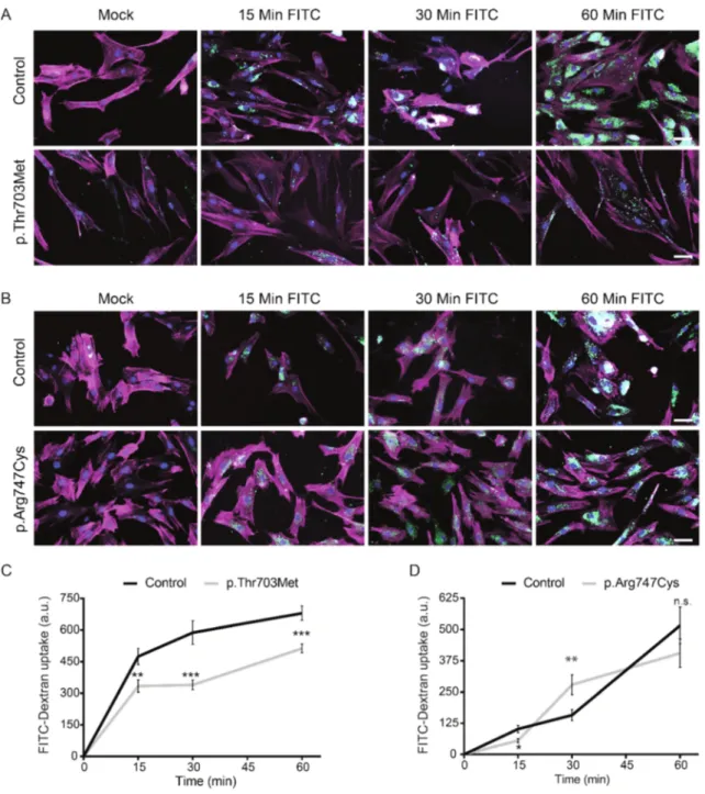
The BICD2 disease-causing mutations, p.Thr703Met (Neveling; Martinez- Carrera et al. 2013) and p.Arg747Cys (Synofzik et al. 2014), localize to the C-terminal end of the BICD2 protein, which has been described in Drosophila melanogaster to interact with clathrin heavy chain (Li et al. 2010). The function of clathrin is important for the process of endocytosis and defects in endocytosis have emerged as a central pathological hallmark of autosomal recessive SMA (Hosseinibarkooie et al. 2016, Riessland et al. 2017). To test whether these BICD2 mutations have a direct impact on endocytosis, the cellular uptake of Fluorescein isothiocyanate (FITC)-labelled dextran was assayed in fibroblasts derived from affected individuals carrying the p.Thr703Met and p.Arg747Cys mutations. Fibroblasts from control individual and affected individuals were serum starved and subsequently treated with FITC-dextran for 15, 30, or 60 minutes (Figure 4A and 4B). FITC-dextran uptake was measured microscopically. The experiment was performed in triplicate.
Fibroblasts harboring the p.Thr703Met mutation displayed a significantly reduced
FITC-dextran uptake at all assayed time points (Figure 4C). Even though mutant cells
maintained dextran uptake over time, fluorescence levels were constantly lower as
compared to control fibroblasts. This may indicate diminished endocytosis caused by
this mutant BICD2 protein. However, cells carrying the p.Arg747Cys mutation
showed only minor deviations from control fibroblasts (Figure 4D). This suggests that
this mutation likely does not have any impact on the process of endocytosis in
fibroblasts. Thus, this assay pointed out possible alterations in endocytosis caused
by p.Thr703Met. However, FITC-dextran is in addition taken up by cells via pathways
other than clathrin-mediated endocytosis, for example fluid-phase endocytosis
(Pustylnikov et al. 2014).
Figure 4. FITC-Dextran uptake in fibroblasts derived from affected individuals carrying the p.Thr703Met and p.Arg747Cys mutations in BICD2.
Fibroblasts were serum starved and treated with FITC-dextran for 15, 30 and 60 minutes. FITC-dextran uptake was analyzed microscopically. (A) and (B): Representative images of control and mutant fibroblasts at the indicated time points. Red channel: Phalloidin-AlexaFluor568; blue channel: DAPI; green channel:
FITC-dextran. Scale bar corresponds to 50 µm. (C) and (D): Quantification of fluorescence intensity of FITC- dextran. The experiments were performed in triplicate. 50 cells were counted per time point. Error bars correspond to mean ± SEM. *P<0.05, **P<0.01, ***P<0.001 in unpaired two-tailedt-test.
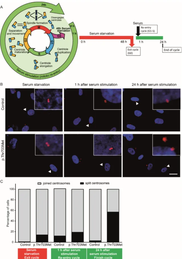
4.2. Impact of p.Thr703Met mutation on centrosomes and cell cycle
Previous analyses have described a G2-phase-specific role of BICD2 in the regulation of centrosome positioning prior mitosis (Splinter et al. 2010). To initiate mitosis, centrosomes split at the end of G2-phase to later allow spindle formation (Wang et al. 2014) (Figure 5A, left panel). The fibroblasts derived from an individual with SMALED2 that carries the p.Thr703Met mutation showed particularly slow proliferation rates in cell culture as compared to control and other BICD2 mutant cells. Mutant and control cells were initially synchronized to G0 phase by serum starvation for 48 hours (Figure 5A, right panel). Cell cycle was resumed by addition of serum. After 1 h of serum addition, the cells are expected to be in G1/S phase transition and at 24 h to complete the cell cycle (Gabriel et al. 2016). At the indicated time points, the cells were fixed and centrosomes were immunostained using an antibody against -tubulin and confocal imaged (Figure 5B). Joined centrosomes are visible as two nearby localized dots close to the nucleus. Split centrosome are visible as apart dots.
Quantification of mutant vs. control cells showed only a slight increase of mutant cells
with split centrosomes directly after serum starvation and 1 hour after re-entry of cell
cycle (Figure 5C). Strikingly, almost 60 % of fibroblasts harboring the p.Thr703Met
mutation showed split centrosomes 24 hours after cycle re-entry, while only single
control cells displayed this phenomenon. This strongly suggests that almost all control
cells have completed cell cycle after 24 hours, but a large fraction of mutant cells failed
to enter mitosis. Evaluating the sole centrosome status without considering other
indicators, implies that many mutant cells are retained in G2-phase. The slow
proliferation might be attributable to aberrant cell cycle progression and/or pre-mitotic
cell cycle arrest.
Figure 5. Centrosome splitting in primary fibroblasts carrying the p.Thr703Met mutation.
(A) Cell cycle and centrosome status. Left: scheme was modified based on the review by Wang et al. (Wang et al. 2014). Right: experimental plan. (B) Representative confocal images of synchronized control fibroblasts and fibroblasts harboring the p.Thr703Met mutation at 0, 1, and 24 hours after cell cycle re-entry. Red channel:-tubulin (centrosomes); blue channel: DAPI (nuclei). Arrowheads point to areas that are magnified in the squares. Scale bar corresponds to 20 µm. (C) Quantification of cells with joined and split centrosomes (N= 150). The experiment was performed once.
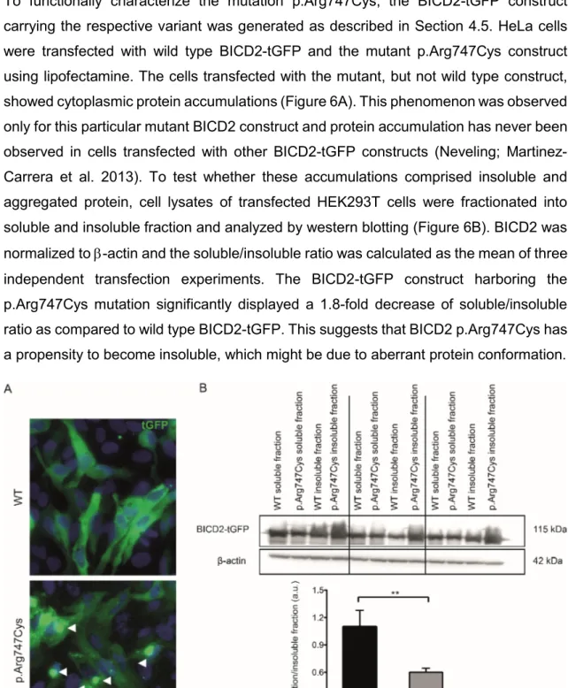
4.3. Implications of p.Arg747Cys mutation in protein structure and aggregation
To functionally characterize the mutation p.Arg747Cys, the BICD2-tGFP construct carrying the respective variant was generated as described in Section 4.5. HeLa cells were transfected with wild type BICD2-tGFP and the mutant p.Arg747Cys construct using lipofectamine. The cells transfected with the mutant, but not wild type construct, showed cytoplasmic protein accumulations (Figure 6A). This phenomenon was observed only for this particular mutant BICD2 construct and protein accumulation has never been observed in cells transfected with other BICD2-tGFP constructs (Neveling; Martinez- Carrera et al. 2013). To test whether these accumulations comprised insoluble and aggregated protein, cell lysates of transfected HEK293T cells were fractionated into soluble and insoluble fraction and analyzed by western blotting (Figure 6B). BICD2 was normalized to -actin and the soluble/insoluble ratio was calculated as the mean of three independent transfection experiments. The BICD2-tGFP construct harboring the p.Arg747Cys mutation significantly displayed a 1.8-fold decrease of soluble/insoluble ratio as compared to wild type BICD2-tGFP. This suggests that BICD2 p.Arg747Cys has a propensity to become insoluble, which might be due to aberrant protein conformation.
Figure 6. The BICD2 p.Arg747Cys mutation forms insoluble aggregates.
(A) Representative images of HeLa cells after 48h of transfection with p.CMV-BICD2-tGFP constructs (wild type and mutant p.Arg747Cys). Note that the mutant p.Arg747Cys forms aggregates, which are indicated by arrows.
(B) Upper panel: western blot of soluble and insoluble fractions from lysate of HEK293T cells transfected with control and mutant constructs. For BICD2-tGFP detection, antibody against BICD2 was used. All BICD2 bands were normalized to -actin expression. Lower panel: western blot quantification. The ratio of soluble/insoluble BICD2 was calculated for each transfection experiment. Bars represent the mean ratios of 3 transfection experiments. Error bars denote standard deviation. ** p<0.01 in unpaired two-tailedt-test.