Original article:
WOUND HEALING POTENTIAL OF SPIRULINA PLATENSIS EXTRACTS ON HUMAN DERMAL FIBROBLAST CELLS Pauzi Nur Aimi Syarina1, Govindarajan Karthivashan1, Faridah Abas2,
Palanisamy Arulselvan1, Sharida Fakurazi1,3
Universiti Putra Malaysia, 43400 UPM Serdang, Selangor, Malaysia
1 Laboratory of Vaccines and Immunotherapeutics, Institute of Bioscience
2 Department of Food Science, Faculty of Food Science and Technology
3 Department of Human Anatomy, Faculty of Medicine and Health Sciences
* Corresponding author: Dr. Sharida Fakurazi, Ph.D., Associate Professor and Head of Laboratory, Department of Human Anatomy, Faculty of Medicine and Health Sciences, and Laboratory of Vaccines and Immunotherapeutics, Institute of Bioscience, Universiti Putra Malaysia, 43400 UPM Serdang, Selangor, Malaysia; Tel: +603 8947 2117;
Fax: +603 8942 2341. E-mail: sharida@upm.edu.my; sharida.fakurazi@gmail.com
http://dx.doi.org/10.17179/excli2014-697
This is an Open Access article distributed under the terms of the Creative Commons Attribution License (http://creativecommons.org/licenses/by/4.0/).
ABSTRACT
Blue-green alga (Spirulina platensis) is a well renowned nutri-supplement due to its high nutritional and medici- nal properties. The aim of this study was to examine the wound healing efficiency of Spirulina platensis at vari- ous solvent extracts using in vitro scratch assay on human dermal fibroblast cells (HDF). Various gradient sol- vent extracts (50 μg/ml of methanolic, ethanolic and aqueous extracts) from Spirulina platensis were treated on HDF cells to acquire its wound healing properties through scratch assay and in this investigation we have used allantoin, as a positive control to compare efficacy among the phytoextracts. Interestingly, aqueous extract were found to stimulate proliferation and migration of HDF cells at given concentrations and enhanced closure rate of wound area within 24 hours after treatment. Methanolic and ethanolic extracts have shown proliferative effect, however these extracts did not aid in the migration and closure of wound area when compared to aqueous ex- tract. Based on phytochemical profile of the plant extracts analyzed by LC-MS/MS, it was shown that com- pounds supposedly involved in accelerating wound healing are cinnamic acid, narigenin, kaempferol, temsiroli- mus, phosphatidylserine isomeric derivatives and sulphoquinovosyl diacylglycerol. Our findings concluded that blue-green algae may pose potential biomedical application to treat various chronic wounds especially in diabe- tes mellitus patients.
Keywords: Spirulina platensis, wound healing activity, aqueous extract, LC-MS/MS, wound scratch assay
INTRODUCTION
Wound healing is a dynamic process in- volving complex interactions between cellu- lar, molecular, biochemical and physiologi- cal activities which result in the regeneration and replacement of injured connective tissue
at the wound site (Velnar et al., 2009). Nor- mal wound undergoes series of event that in- volve in wound healing process. Initial stag- es of wound healing engaged the formation of a blood clot and inflammation, which oc- cur immediately upon injury. Following 4
days injury, the inflammatory response is followed by proliferation and migration of dermal and epidermal cells, and matrix syn- thesis, in order to fill the wound gap and re- establish skin barrier (Hackam and Ford, 2002; Werner et al., 2007). Finally, tissue remodeling and maturation enable full re- covery of the skin tissue which sometimes leaving no remaining trace of the previous skin damage (Diegelmann and Evans, 2004;
Harding et al., 2005).
The increasing popularity of natural ther- apy has led podiatrist hope of finding a more effective and cost effective product for heal- ing of chronic wounds (Ananda, 2012).
Since, ancient years people have been using plants as treatments to accelerate wound healing process though without any scientific evidence of the plant efficiency and very lit- tle knowledge about the compounds and mode of actions. Plants are believed to have natural therapeutic effects against endocrine disorders and other diseases (Arulselvan et al., 2014) and can be treated for any kind of injury such as cut or burned, with simple formulation procedures to make them the best candidate as natural remedies.
Algae have expanded growing interest in their values as medicine and functional foods (Barrow and Shahidi, 2007) as they provide harmless and nutritious product for healthcare supplement market (Dominguez, 2013). Spirulina platensis is a filamentous blue-green alga originates in many alkaline lakes with a high pH. It contains approxi- mately 70 % easily digestible protein where 18 out of 22 amino acids and all of the essen- tial amino acids are available, making it a unique vegetarian source of complete protein (Somchit et al., 2007).
Since, wound healing is a complex bio- logical process, there are a few methods of in vitro and in vivo assays are available to study the first insight of how plant preparation can possibly influence the regeneration of new tissue and enhance the wound. However, among all the methods present, in vitro scratch assay has been proven a valuable and
inexpensive tool to study wound healing po- tential of drugs (Liang et al., 2007).
The objective of present study was to evaluate the efficacy of different extracts of Spirulina platensis in accelerating the prolif- eration and migration into the monolayer of human dermal fibroblast cells (HDF) and to determine the bioactive compounds by sec- ondary metabolite profiling responsible for wound healing potential.
MATERIALS AND METHODS Preparation of extract
The Spirulina platensis (SP) were pur- chased from Algaetech International Sdn.
Bhd. The Spirulina platensis extract were prepared by maceration of 10 g of freeze- dried powder in 500 ml of different solvents (methanol, ethanol and ultrapure water) for 24 h at room temperature. The mixture was then centrifuged at 3000 rpm for 10 min (4 °C) and the supernatant was filtered (Whatman No. 1) to remove cell debris. All samples were then evaporated at 40 °C by vacuum rotary evaporator. The dried extracts from each solvent were stored at 4 °C until further experiments.
Cell culture and plant extract preparation for treatment
Normal Adult Human Primary Dermal Fibroblast cell (HDF) was purchased from ATCC (American Type Culture Collection, USA). Cells were maintained in complete DMEM high glucose media with 5 % FBS and 1 % of penicillin-streptomycin in a hu- midified 5 % CO2 incubator at 37ºC.
Methanolic, ethanolic and aqueous ex- tracts solution were freshly prepared for each experiment by dissolving each dried sample in cell culture medium and sterilized by sy- ringe filter (0.22 µm). Concentration used in the experiment was based on dry weight of the extract (mg/ml).
Cytotoxicity assay
Cytotoxicity assay was conducted to de- termine the range of concentrations of ex-
in vitro analysis. HDF cells were cultured in 96 well culture plates at density 1 × 10⁶ cells/ml with DMEM complete media for 24 h. The medium was replaced with 100 µl of methanolic, ethanolic and aqueous extract solution with different concentration (50, 100, 150, 200, 250, 300 µg/ml) for 24 h. The cell viability was assessed using 3-[4,5-Di- methylthiazol-2-yl]-2,5-diphenyltetrazolium bromide (MTT) solution and MTT powder was dissolved in phosphate-buffered saline (PBS) at a concentration of 5 mg/ml. MTT was added to each well (20 μl) and plates were incubated at 37 °C for 3 hrs. The medi- um was replaced with 100 μl DMSO and the absorbance for each well was measured at 570 nm on a microplate reader.
In vitro scratch assay
HDF cells were seeded in 24-well plates at a density of 3×10⁶ cells/ml and allowed to grow for 24 h at 37 °C and 5 % CO2. A small linear scratch was created in the confluent monolayer by gently scraping with sterile p200 pipette tips (care was taken during scratching process to ensure universal size and distant was made for all samples). Cells were extensively rinsed with PBS to remove cellular debris before adding the media with different treatment solution (methanolic, eth- anolic and aqueous extracts) at concentration of 50 µg/ml. Allantoin (Sigma Aldrich, Germany), was used as a positive control drug and cells without treatment was used as negative control. After 24 h, images of mi- grated cells were taken using digital camera connected to inverted microscope to observe the closure of wound area. All scratch assays were performed in quadruplicate.
LCMS/MS analysis
Mass spectra were acquired using AB Sciex 3200 QTrap LCMS/MS with Perkin Elmer FX 15 UHPLC system (MA, USA).
The negative ion mass spectra were obtained from the LC QTrap MS/MS detector on full ion scan mode (100 to 1200 m/z for full scan and 50–1200 m/z for MS/MS scan) at a scan
rate of 0.5 Hz. The hyphenated system was supported with a mass spectrometry software and spectral library provided by ACD labs (TO, CA). Analyte separation was carried out on a pre-packed C18 (4 × 250 mm, 5 μm, Phenomenex) column with a gradient mobile phase comprising water (solvent A) and methanol with 1 % acetonitrile (solvent B), each containing 0.1 % formic acid and 5mM ammonium formate. The gradient program commenced with 80 % to 90 % solvent B from 0.01 to 11.00 min with a flow rate of 1.0 ml/min. The injection volume was set to 20 µL. All chromatographic procedures were performed at ambient temperature and the corresponding peaks from QTrap LCMS/MS analysis of the aqueous extracts of Spirulina platensis were identified by comparison with the literature data / ACD labs mass spectral library.
Statistical analysis
All experimental values are presented as mean ± SD. The data were analyzed using ANOVA, and then paired t-test was used to analyze the difference between the groups.
P-values at less than 0.05 were considered as statistically significant.
RESULTS
Effect of Spirulina platensis extracts on HDF cells viability
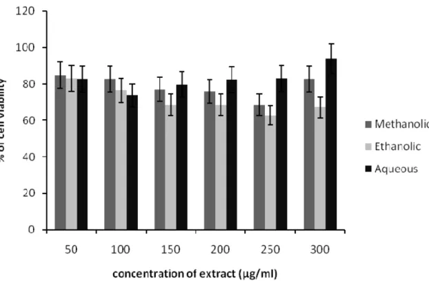
The viability of HDF cells was reduced by the treatment of methanolic, ethanolic and aqueous Spirulina extract up to 300 μg/ml (Figure 1). The concentration of ethanolic extract at 150 – 300 μg/ml showed 30–40 % cyto-toxicity after 24 h incubation. At a dose of 50 μg/ml of each extract, the HDF cells have shown more than 80 % of cell viability.
Further, increase in extract concentration led to gradual decrease in cell viability as higher concentrations were found to be cytotoxic to the cells. Therefore, the non-toxic concentra- tion of extract was used for further testing in scratch assay.
Figure 1: Effects of methanolic, ethanolic and aqueous extracts of Spirulina platensis on the viability of HDF cells. The cells were incubated for 24 h before being assessed for their viability. Data present- ed as mean ± SD.
Effect of Spirulina platensis extracts on in vitro scratch assay
Figure 2 shows the images of scratch as- says on HDF cells at 0 and 24 h post injury time without treatment (control) and with treatment. All the images are shown progres- sion of wound closure on scratch wounded HDF cells. Enhanced migration and wound closure were observed in aqueous extract treated cells as compared with those of un- treated cells in which the degree of cell mi- gration and cell proliferation are slow. Rapid cell migration and wound closure rate of aqueous extract treated HDF cells were ob- served and these effects were comparable with positive control drug, allantoin. Wound area at 24 h post injury remained open for methanolic and ethanolic Spirulina extract on scratch wounded HDF cells.
LCMS/MS analysis of Spirulina platensis extracts
Based on in-vitro scratch assay which used methanol, ethanol and aqueous extracts
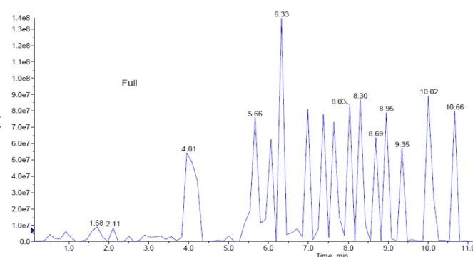
of S. platensis for treatment, it was found that aqueous extract has shown significant wound healing activity on HDF cells. Fur- ther analysis of S. platensis aqueous extract through LCMS/MS QTrap with an adaptive data dependent gradient program, the com- pounds were revealed to be phenolic com- pounds and fatty acid derivatives (Figure 3).
Tentatively, few compounds have been iden- tified and enlisted in Table 1 based on the literature data and Advanced Chemistry De- velopment (ACD) labs based mass spectral library.
Peak 1 which was released at 0.93 min was identified as cinnamic acid based on its parent ion of m/z 147 (Wang et al., 2008).
Peak 2 with m/z 271 was identified as nari- genin (Sanchez-Rabaneda et al., 2003), peak 3 gave molecular ion peak at m/z 285 was recognized as kaempferol (Sanchez-Rabane- da et al., 2003) whereas peak 4 was found to be temsirolimus express significant parent ions at m/z 1046 (Cai et al., 2007). Peaks 5- 12 which were released between 5.6 to
Figure 2: In vitro scratch assay (x4 magnification). HDF cells were injured and cell migration assay with and without treatment was performed. Aqueous extract treated fibroblasts showed faster cell pro- liferation and migration as compared with those untreated cells as well as those cells treated with methanolic and ethanolic extract.
Figure 3: This figure represents peaks extracted from the aqueous extract from 0.0 to 11.0 min. Fur- ther analysis of LCMS/MS QTrap with an adaptive data dependent gradient program, it was revealed to be phenolic compounds and fatty acid derivatives.
Table 1: Retention times, MS and MS2values of the major bioactive constituents present in S. platen- sis aqueous extract by HPLC–DAD–ESI–MS/MS.
Peak Retention time (RT)
Molecular ion
peak (M-H)- MS2 fragment ions intensity Tentative Compounds Identified
1 0.93 147 119 (100), 117, 103 Cinnamic acid
2 1.68 271 227, 185 (100), 130, 116 Narigenin
3 2.11 285 241, 199 (100), 182, 130 Kaempferol
4 4.01 1046 1029 (100), 986, 532, 365 Temsirolimus
5 5.66 834 554, 279, 255 (100), 241
Phosphatidylserine
6 6.33 834 554, 279, 255 (100), 241
7 7.00 834 554, 279, 255 (100), 241
8 7.38 835 554, 391, 297, 279 (100), 241
9 8.03 835 554, 391, 279, 241(100),
10 8.30 834 554, 391, 279, 255 (100), 241
11 8.69 835 554, 391, 279 (100), 255, 241
12 8.95 835 554, 391, 279, 255 (100), 241
13 9.35 816 537, 255, 225 (100) Unknown
14 10.02 817 537, 255, 225 (100) Sulphoquinovosyl diacyl-
glycerol (SQDG)
15 10.66 817 537, 255, 225 (100)
8.95 min were identified as phosphatidylser- ine isomeric derivatives which revealed a same parent ion at m/z 834 and 835 as well as the same MS2 fragment ions at m/z 554, 279, 391, 255 and 241, which made them as isomers (Tyurin et al., 2008). Peaks 14 and 15 produced molecular ion peak at m/z 817 and MS2 fragment ions at m/z 537, 255 as well as 225. Based on this significant frag- mentation pattern and comparison with pre- viously reported data, the peaks 14 and 15 were found to be sulphoquinovosyl diacyl- glycerol (SQDG) (Mizushina et al., 2003a).
In the present study, tentative active compounds such as hydroxylated cinnamic acid, narigenin, kaempferol, phosphatidylser- ine isomeric derivatives and sulphoquinovo- syl diacylglycerol (SQDG) have identified from S. platensis aqueous extract by LC- MS/MS analysis. Until now, the most gen- eral compounds that have been reported in S.
platensis extract are phenolic compound, ca- rotenoids, phycobiliproteins, chlorophyll,
polyunsaturated fatty acids, sulphated poly- saccharides and sterols (Herrero et al., 2007).
DISCUSSION
The skin provides protection and acts as external barrier of body cells and tissues against microbial infection from external en- vironment. Any damage to skin barrier must be rapidly and effectively restored via wound healing process. Dermal fibroblast is the first line defense which responses to injury and is essential for cutaneous wound repair by pro- liferation and migration process into the wound site. It differentiates into myofibro- blasts as a response to macrophage-derived cytokines such as TGF-β1, which is reliant on their contact with fibronectin (Serini et al., 1998). Proliferation and migration of the cells are important events in wound healing process, therefore, the study of natural prod- ucts and their active compounds which influ- ence the migration of fibroblast may help in
improving the cutaneous wound healing pro- gression.
To make sure that effect of extract treat- ment on HDF proliferation and migration were not interfere by any toxicity; we deter- mined the viability of fibroblast after 24 hours in different extracts concentrations.
Based on cell viability assay, our results demonstrated that low concentration of methanolic, ethanolic and aqueous extract did not give any cytotoxicity effects on HDF cells after 24 hours of treatment. These find- ings indicated that 50ug/ml was non-toxic concentration of Spirulina platensis crude extracts and this concentration was used for further treatment.
Scratch assay is a suitable and inexpen- sive screening method to differentiate and verify natural products for their in vitro wound healing activity. This assay is highly related to the second phase of wound healing process characterized by proliferation and migration of the keratinocytes and fibro- blasts (Schafer and Werner, 2007; Liu et al., 2013). In this study, the crude extracts from Spirulina platensis were used as treatment during scratch assay, aqueous extract in- creases the population of HDF cells in the
‘wounded’ or scratched area due the migra- tion of cells and also by proliferation of the migrated cells. Conversely, methanolic and ethanolic extracts from Spirulina platensis showed some increment in the abundance cells yet they did not aid in cell migration.
This may explain the non-healing activity of HDF cells when treated with those extracts.
Our results are in agreement with that of a previous study which explained the wound healing potential of methanolic extract of Moringa oleifera leaves against stimulated HDF cells based on its wound closure activi- ty (Muhammad et al., 2013).
In the present study, LC-MS/MS analysis on S. platensis aqueous extract compounds revealed as hydroxylated cinnamic acid, nar- igenin, kaempferol, phosphatidylserine iso- meric derivatives and sulphoquinovosyl di- acylglycerol (SQDG) compounds. Until now, the most general compounds that have
been reported in S. platensis extract are phe- nolic compound, carotenoids, phycobilipro- teins, chlorophyll, polyunsaturated fatty ac- ids, sulphated polysaccharides and sterols (Chojnacka et al., 2012; Lordan et al., 2011;
Pumas et al., 2011). It has been previously reported that hydroxyl cinnamic acid and their derivatives inhibit diabetic complica- tions mediated by advanced glycation end products formation and moderately induce corneal epithelial wound healing by inhibit- ing protein tyrosine phosphatases (PTP)-1β gene (Adisakwattana et al., 2013). Narigenin is an effective inhibitor of the pro-inflam- matory cytokine (IL-1β, IL-6, IL-8 and TNF- α) response stimulated by lipopolysaccharide in both macrophages and in whole blood (Bodet et al., 2008). Kaempferol has been reported to possess anti-oxidant (Karthi- vashan et al., 2013) and anti-inflammatory activity via its inhibition of aldosterone sig- naling and aldosterone-induced gene expres- sion in HUVECs (Liu et al., 2000). Ram- strom et al. in (2003) reported that phospha- tidylserine plays an essential role in wound healing as a pro-coagulant in platelet for- mation process. It has also been reported that collagen-adherent platelets activate blood coagulation by exposing phosphatidylserine (PS) at wound site (Heemskerk et al., 2002).
In general, fatty acid and its derivatives pro- mote wound healing by angiogenic activity, Mizushina et al. (2003b) reported that sulphoquinovosyl diacylglycerol (SQDG) as a selective inhibitor of eukaryotic DNA pol- ymerases a and b and also an immunosup- pressive agent. These reports strongly sug- gest that bioactive compounds such as, hy- droxylated cinnamic acid, phosphatidylserine isomeric derivatives, sulphoquinovosyl di- acylglycerol (SQDG), present in S. platensis aqueous extract are virtuously responsible for its enhanced wound healing activity.
CONCLUSION
Our findings suggested that Spirulina platensis, aqueous extract showed highest wound healing activity and might be consid- ered as a potential source of therapeutic
agent for chronic wound and its associated complications. In addition, this study demonstrated that scratch assay is a suitable and economical method that gives dependa- ble results for the proliferation as well as mi- gration of the HDF cells in an artificial wounded model. However, further study need to be conducted to confirm their biolog- ical activity more specially in experimentally induced diabetic wound model. Further, iso- lation, identification and purification active compounds those are possess wound healing potential are in progress in our research group.
Acknowledgements
This research work was supported by Re- search University Grant Scheme (RUGS) of Universiti Putra Malaysia (05-02-11- 1419RU and 04-02-12-2089RU).
Conflict of interest
The authors have declared no conflict of interest.
REFERENCES
Adisakwattana S, Pongsuwan J, Wungcharoen C, Yibchok-anun S. In-vitro effects of cinnamic acid de- rivatives on protein tyrosine phosphatise 1B. J. En- zyme Inhib Med Chem. 2013;28:1067-72.
Ananda AD. Wound care with traditional, comple- mentary and alternative medicine. Indian J Plast Surg.
2012;45:418–24.
Arulselvan P, Ghofar HAA, Karthivashan G, Halim MFA, Ghafar MSA, Fakurazi S. Antidiabetic thera- peutics from natural source: A systematic review. Bi- omed Prev Nutr. 2014;4:607-17.
Barrow C, Shahidi F. Marine nutraceuticals and func- tional foods. Boca Raton, FL: CRC Press, 2007.
Bodet C, La VD, Epifano F, Grenier D. Narigenin has anti-inflammatory properties in macrophage and ex vivo human whole-blood models. J Periodontal Res.
2008;43:400-7.
Cai P, Tsao R, Ruppen ME. In vitro metabolic study of temsirolimus: preparation, isolation and identifica- tion of the metabolites. Drug Metab Dispos. 2007;35:
1554-63.
Chojnacka K, Saeid A, Witkowska Z, Tuhy L. Bio- logically active compounds in seaweed extracts - the prospects for the application. The Open Conference Proceedings Journal. 2012;S1- M4(3):20-8.
Diegelmann RF, Evans MC. Wound healing: an over- view of acute, fibrotic and delayed healing. Front Bio- sci. 2004;9:283–9.
Dominguez H (ed). Functional ingredients from algae for foods and nutraceuticals. Amsterdam: Woodhead Publishing, 2013.
Hackam DJ, Ford HR. Cellular, biochemical, and clinical aspects of wound healing. Surg Infect (Larchmt). 2002;3(Suppl 1):S23–35.
Harding KG, Moore K, Phillips TJ. Wound chronicity and fibroblast senescence–implications for treatment.
Int Wound J. 2005;2:364–8.
Heemskerk JW, Bevers EM, Lindhout T. Platelet ac- tivation and blood coagulation. Thromb Haemost.
2002;88:186-93.
Herrero M, Vicente MJ, Cifuentes A, Ibanez E. Char- acterization by high-performance liquid chromatog- raphy/electrospray ionization quadrupole time-of- flight mass spectrometry of the lipid fraction of Spir- ulina platensis pressurized ethanol extract. Rapid Commun Mass Spectrom. 2007;21:1729-38.
Karthivashan G, Tangestani Fard M, Arulselvan P, Abas F, Fakurazi S. Identification of bioactive candi- date compounds responsible for oxidative challenge from hydro‐ethanolic extract of Moringa oleifera leaves. J Food Sci. 2013;78:C1368–C75.
Liang CC, Park AY, Guan JL. In vitro scratch assay: a convenient and inexpensive method for analysis of cell migration in vitro. Nat Protoc. 2007;2:329-33.
Liu XJ, Kong FZ, Wang YH, Zheng JH, Wan WD.
Lumican accelerates wound healing by enhancing α2β1 integrin-mediated fibroblast contractility. PLoS ONE. 2013;8:e67124.
Liu Y, Xu L, Cheng N, Lin L, Zhang C. Inhibitory ef- fect of phycocyanin from Spirulina platensis onthe growth of human leukemia K562 cells. J Appl Phycol.
2000;12:125-30.
Lordan S, Ross RP, Stanton C. Marine bioactives as functional food ingredients: potential to reduce the in- cidence of chronic diseases. Mar Drugs. 2011;9:
1056–100.
Mizushina Y, Maeda N, Kawasaki M, Ichikawa H, Murakami C, Takemura M, et al. Inhibitory action of emulsified sulfoquinovosyl acylglycerol on mamma-
Mizushina Y, Xu X, Asahara H, Takeuchi R, Oshige M, Shimazaki N, et al. A sulphoquinovosyl diacyl- glycerol is a DNA polymerase epsilon inhibitor. Bio- chem. J. 2003b;370:299-305.
Muhammad AA, Pauzi NAS, Arulselvan P, Abas, Fakurazi S. In vitro wound healing potential and iden- tification of bioactive compounds from moringa oleif- era lam. Biomed Res Int. 2013;2013:974580.
Pumas C, Vacharapiyasophon P, Peerapornpisal Y, Leelapornpisid P, Boonchum W, Ishii M, et al. Ther- mostablility of phycobiliproteins and antioxidant ac- tivity from four thermotolerant cyanobacteria. Phyco- logical Res. 2011;59:166–74.
Ramstrom S, Ranby M, Lindahl TL. Platelet phospha- tidylserine exposure and procoagulant activity in clot- ting whole blood-different effects of collagen, TRAP and calcium ionphore A23187. Thromb Haemost.
2003;89:132-41.
Sanchez-Rabaneda F, Jauregui O, Casals I, Andrés- Lacueva C, Izquierdo-Pulido M, Lamuela-Raventos RM. Liquid chromatographic/electrospray ionization tandem mass spectrometric study of the phenolic composition of cocoa (Theobroma cacao). J Mass Spectrom. 2003;38:35–42.
Schafer M, Werner S. Transcriptional control of wound repair. Annu Rev Cell Dev Biol. 2007;23:69- 92.
Serini G, Bochaton-Piallat ML, Ropraz P, Geinoz A, Borsi L, Zardi L, et al. The Fibronectin domain ED-A is crucial for myofibroblastic phenotype induction by transforming growth factor-β1. J Cell Biol. 1998;142:
873–81.
Somchit MN, Siti Rahmah S, Zuraini A, Ahmad Bustamam A, Zakaria ZA, Somchit N, et al. Gastro- protective activity of Spirulina platensis in acetic acid and ethanol induced ulcers in rats. J Natl Remedies.
2007;7:37-42.
Tyurin VA, Tyurina YY, Feng W, Mnuskin A, Jiang J, Tang M, et al. Mass-spectrometric characterization of phospholipids and their primary peroxidation prod- ucts in rat cortical neurons during staurosporine- induced apoptosis. J. Neurochem. 2008;107:1614-33.
Velnar T, Bailet T, Smrkol V. The wound healing process: an overview of the cellular and molecular mechanisms. J Med Int Med Res. 2009;37:1528-42.
Wang SJ, Ruan JX, Zhao YH, Zhang ZQ. Simultane- ous determination of harpagoside and cinnamic acid in rat plasma by liquid chromatography electrospray ionization mass spectrometry and its application to pharmacokinetic studies. Biomed Chromatorgr. 2008;
22:50-7.
Werner S, Krieg T, Smola H. Keratinocyte–fibroblast interactions in wound healing. J Invest Dermatol.
2007;127:998–1008.