Original article:
THERAPEUTIC EFFECT OF
BACCHARIS ANOMALA
DC.EXTRACTS ON ACTIVATED HEPATIC STELLATE CELLS Bruno de Souza Basso1, *, Fernanda Cristina de Mesquita1,2, Henrique Bregolin Dias1, Gabriele Catyana Krause1, Matheus Scherer1, Eliane Romanato Santarém3,
Jarbas Rodrigues de Oliveira1
1 PUCRS, Escola de Ciências, Laboratório de Pesquisa em Biofísica Celular e Inflamação, Porto Alegre, Brazil
2 Graduate Program in Neuroengineering, Edmond and Lily Safra International Institute of Neuroscience, Santos Dumont Institute, Macaiba, Brazil
3 PUCRS, Escola de Ciências, Laboratório de Biotecnologia Vegetal, Porto Alegre, Brazil
* Corresponding author: Bruno de Souza Basso, Laboratório de Pesquisa em Biofísica Celular e Inflamação, Pontifícia Universidade Católica do Rio Grande do Sul, Av.
Ipiranga, 6681, Sala 221, bloco C, prédio 12, Porto Alegre, RS, Brazil.
Tel.: 00+55+51 3320.4147; Fax: 00+55+51 3320.3500, E-mail: bruno.basso.001@acad.pucrs.br
http://dx.doi.org/10.17179/excli2018-1696
This is an Open Access article distributed under the terms of the Creative Commons Attribution License (http://creativecommons.org/licenses/by/4.0/).
ABSTRACT
The therapeutic potential of Baccharis anomala DC. extracts was evaluated through its cytotoxic and antiprolifer- ative effect and their phenotypic reversion property in activated hepatic stellate cells (HSCs). Baccharis anomala is distributed in Brazil (southeastern and south regions) and used for diuretic effect in folk medicine. Four fractions were obtained from the fractionation of the methanolic extract. Fractions III and IV decreased cell proliferation without increasing cell necrosis markers levels and induced cell cycle arrest in G1 phase. Fraction III induced phenotypic reversion through PPAR-γ activation pathway, while fraction IV did not alter PPAR-α/γ expression levels, suggesting that there is an independent PPAR-α/γ pathway involved. Hydroxybenzoic, chlorogenic and coumaric acids were identified. Fractions III and IV showed antiproliferative effect and ability to induce reversion of activated phenotype of HSCs.
Keywords: Hepatic fibrosis, HSCs, Baccharis, PPAR-γ, phenolic compounds
INTRODUCTION
Hepatic fibrosis is associated with chronic liver damage and can be caused by different factors such as chronic viral hepatitis, alcohol abuse, toxins, metal accumulation and hered- itary diseases (Iredale, 2008). In response to liver injuries, the hepatic stellate cells (HSC) gradually transdifferentiate to an activated
phenotype (aHSC) and consequently liver fi- brosis. aHSC lose the ability to store vitamin A, increase expression of a-smooth muscle actin (α-SMA) and profibrotic genes, and ac- quired a larger and polygonal shape. In addi- tion, aHSC synthesize collagen, a component of the extracellular matrix, and produce con- tractile movements after stimulation, as oc- curs in wound healing. These characteristics
may lead to the destruction of hepatic tissue architecture and, in severe cases, to the devel- opment of cirrhosis and liver failure (Fried- man, 2008; Gressner, 1998; Wynn and Rama- lingam, 2012). Therefore, it is widely ac- cepted that activation of HSC is a key factor in the pathogenesis of liver fibrosis.
Under normal conditions, the quiescent phenotype of the HSC is maintained by the transcription factors peroxisome proliferator- activated receptors (PPAR) such as PPAR-α, PPAR-β/δ and PPAR-γ. The PPAR-γ tran- scription factor has been considered as one of the most important factors for the regulation of adipogenesis in HSCs (Guimaraes et al., 2007; Tsukamoto et al., 2006) and mainte- nance of quiescent phenotype (Moraes et al., 2014; Mesquita et al., 2013). Moreover, stud- ies also revealed that modulation of lipid me- tabolism could occur via PPAR-α (Chen et al., 2015). Therefore, the inhibition of HSC acti- vation seems to be an effective strategy for therapy of liver fibrosis.
GRX cell line is an immortalized lineage of liver cells obtained from hepatic granulo- mas of mice infected with Schistosoma man- soni and it is considered an excellent model for representation of liver fibrosis in vitro (Borojevic et al., 1985). GRX lineage nor- mally presents the myofibroblast transitional phenotype, being between the quiescent and activated stages (Herrmann et al., 2007). In order to activate myofibroblasts the presence of profibrogenic cytokines and oxidative stress are necessary that cause cytoskeleton reorganization as well as an increase in colla- gen production. The quiescent phenotype ex- pression can be induced when the transitional myofibroblast is exposed either to retinol, in- domethacin or β-carotene (Guimaraes et al., 2006).
The N-acetylcysteine, a drug mainly known for its antioxidant effect, can be high- lighted amongst the most used drugs for con- trolling fibrosis, and it is often used in cases of paracetamol intoxication (Vargha et al., 2014; Heard, 2008; Lauterburg et al., 1983).
Plants have been pointed out as natural re- sources for traditional medicine and for the
modern pharmaceutical industry. Besides, the use of plants able to inhibit the proliferation of activated HSCs has a great potential in re- versing fibrosis. The beneficial effects are due to compounds produced by the plant, known as secondary metabolites, and their produc- tion and accumulation by the plant are related to the interaction of the plant with the envi- ronment. Factors such as temperature, preda- tion and luminosity can substantially influ- ence the phytochemical profile (Sartor et al., 2013). Phenolic compounds represent one of the main studied classes of secondary metab- olites, mainly due to their antioxidant activity (Balasundram et al., 2006; Neuhouser, 2004).
The genus Baccharis has approximately 500 known species (Abad and Bermejo, 2007; Bu- del et al., 2005) and many species are used in folk medicine in form of teas, alcoholic bev- erages or macerated, for the treatment of var- ious diseases, such as gastrointestinal dis- eases, liver diseases, inflammation, diarrhea, fever, infections and diabetes (Campos et al., 2016; Hocayen et al., 2016). Among the known species of the genus, about 90 % can be found in South America, being more com- mon in Brazil, Argentina, Uruguay, Chile, Colombia and Mexico. The main constituents found in the Baccharis genus are phenolic compounds and terpenoids (Campos et al., 2016). The species B. megapotamica, B. in- carum, B. trimera, B. trinervis, B. salicifolia, B. uncinella, B. coridifolia, B. dracunculifo- lia, B. grisebachii and B. tricuneata have been extensively studied for their chemical compo- sition (Verdi, 2005).
Baccharis anomala DC., also known as
“cambará-de-cipó” or “parreirinha”, is geo- graphically distributed in the southeastern (Minas Gerais, São Paulo) and south (Paraná, Santa Catarina, Rio Grande do Sul) regions of Brazil (Heiden and Schneider, 2010), alt- hough it is also found in Argentina, Uruguay, and Paraguay. The aerial parts of B. anomala are mainly used for its diuretic effect in folk medicine (Budel et al., 2005; Alice et al., 1985; Garlet, 2000). Since many species of the Baccharis genus have beneficial effects on
liver diseases and also in inflammatory pro- cesses, the identification of the therapeutic ac- tivity of other species of this genus can be promising. As few studies on the phytochem- ical composition and medicinal properties of this species are currently available, the aim of this study was to evaluate the cytotoxicity, an- tiproliferative and deactivation effects of the methanolic extract and fractions of B.
anomala on activated hepatic stellate cell line GRX.
MATERIAL AND METHODS Plant material and extraction
Leaves from Baccharis anomala DC.
were collected in São Francisco de Paula in the state of Rio Grande do Sul (Southern Bra- zil; latitude, 29◦29S; longitude, 50◦11 W; 950 m). The specimen was deposited in the Her- barium of the Science and Technology Mu- seum of the Pontifícia Universidade Católica do Rio Grande do Sul (Herbarium MPUC) under the voucher specimen number Herbar- ium 3354. Plant material was dried, powdered and then stored at -20 ºC until use. Extracts were prepared by combining 3 g of dried plant material with either 100 mL of distilled water (aqueous extract), 100 mL of ethanol 96 % (ethanolic extract) or 100 mL 80 % aqueous methanolic solution (80:20; methanolic ex- tract). Aqueous and ethanolic extracts were agitated for 5 and 72 h at room temperature, respectively. Methanolic extract was kept in ultrasonic bath for 15 min at room tempera- ture. Extracts were then centrifuged for 10 min at 4000 g, the supernatant was collected and dried in a rotary evaporator. For the ex- periments with extracts and fractions, the dry material resulting from evaporation of the re- spective solvents in the rotary evaporator was resuspended in culture Dulbecco’s Modified Eagle’s Medium (DMEM). The negative con- trol consisted of culture medium and GRX cells with no extract added.
HSC culture
Immortalized hepatic stellate cell GRX was obtained from the Rio de Janeiro Cell
Bank and presents a myofibroblast transit- ional phenotype. Cells were maintained in culture medium (DMEM) supplemented with 5 % fetal bovine serum (FBS) (GIBCO), 1 % penicillin and streptomycin (Invitrogen) and pH 7.4. HSC were incubated with Baccharis anomala extracts for 72 hours at 37 °C in a humidified atmosphere of 5 % CO2. Results using GRX derived from at least three repli- cates per experimental condition.
Antiproliferative and cytotoxicity evaluation of aqueous, ethanolic and methanolic extracts
Proliferation and cell viability was as- sessed determining living cell numbers by cell count with Trypan blue exclusion method.
GRX cells were seeded in a 24-well tissue culture plate (3x10³ cells/well) and incubated with B. anomala crude extracts at concentra- tions of 25, 50, 500 µg/mL. N-acetylcysteine (NAC; 400 µg/mL), a well-known medicine used in the treatment of hepatic fibrosis, was used as a positive control. For all experi- ments, the negative control consisted of GRX cells on culture medium. Methanolic extract was chosen to conduct the experiments, due to the most significant antiproliferative effect.
Fractionation of methanolic extract by chromatography column
Methanolic extract was fractionated by chromatography on a silica column, using sil- ica gel 60 (Merk) as stationary phase and the mobile phase consisted in eluents of increas- ing polarity (v/v): dichloromethane (100:0), dichloromethane: methanol (95:5), dichloro- methane: methanol (90:10), methanol (100:0) and methanol: water (80:20). Four different fractions were obtained, therein named FI, FII, FIII and FIV. Fractions were filtered, dried and weighed for their use in the experi- ments.
Evaluation of antioxidant activity of the fractions from methanolic extract
Fractions from the methanolic extract were analyzed for its antioxidant activity. The
reduction of DPPH (2,2-diphenyl-1-picrylhy- drazyl) through electron transfer by the action of an antioxidant was measured by spectro- photometry in an ELISA reader at the wave- length of 515 nm. All samples analyzed were dissolved in methanol 100 %. Ascorbic acid (550 μg/mL) was used as a positive control for antioxidant activity.
Antiproliferative effect and cytotoxicity of FIII and FIV on GRX cells
In order to evaluate the cytotoxity of frac- tions and their effect on cell proliferation of GRX cells, cells were seeded in a 24-wells tis- sue culture plate (3x10³ cells/well) and incu- bated with FIII and FIV at concentrations of 1.25, 2.5, 5, 50, and 100 µg/mL. NAC (400 µg/mL) was used as positive control. Prolifer- ation was assessed by cell count with Trypan blue exclusion method. The evaluation of cy- totoxicity was performed using a lactate de- hydrogenase (LDH) kit (LabTest, Brazil) in the culture supernatants. As LDH is a known enzyme related to membrane cell damage, its activity was measured by a colorimetric assay at 420 nm and compared with the negative control. For the control of total cell lysis, 5 % Tween was used. Analyses were performed after 72 h of treatment with fractions.
Cell cycle evaluation
Cell cycle arrest was evaluated using 7- AAD staining. GRX cells were seeded into 24-well plates at 3x10³ cells/well and treated either with FIII at concentration of 50 µg/mL, FIV at 5 µg/mL or NAC at 400 μg/mL. Cells were collected by trypsinization, washed twice with PBS and then, while vortexing, 2.5 mL of ethanol 70 % was added per sample.
Cells were incubated at -20 ºC for 2 h and then washed twice with PBS solution to remove ethanol. Cells were centrifuged and resus- pended in 100 µL of stain buffer and 4 µL of 7-AAD, and incubated for 15 min at room temperature. Samples were analyzed by flow cytometry to identify the cell cycle phases.
Data were analyzed using FlowJo 7.6.5 soft- ware (Tree Star). Analysis allows discrimina- tion among Sub-G1, Go/G1, S, G2/M.
Detection of lipid droplets in aHSC
Phenotypic reversion in hepatic stellate cells was observed using Oil Red assay. Cells were plated in a 24-well tissue culture plate (3x10³ cells/well), and 72 h after treatment with FIII and FIV, cells were fixed with 10 % formaldehyde for 1 h and stained with Oil Red-O (ORO; Sigma Chemical). Intracellular lipid accumulation was observed after 30 min, using an inverted light microscope at magni- fication of 400x. For estimation of lipid accu- mulation, the ORO within the lipid droplets was extracted using isopropanol and the ab- sorbance was read at 492 nm using ELISA plate reader. Specific lipid content was calcu- lated as the ratio of absorbance value obtained for ORO and number of cell count.
Morphological analysis of aHSC nucleus for apoptosis detection
The nuclear morphology of cells was evaluated using the cell-permeable DNA dye 6-diamidino-2-phenylindole (DAPI). Cells were seeded at a cell density of 3x10³ cells/well in 24-well plates and incubated for 24 h at 37 ºC in a 5 % CO2 incubator. Cells were then treated with FIII and FIV fractions, NAC at 400 μg/mL and Cisplatin at 2.5 µM (positive control of apoptosis). After 72 h in- cubation, cells were fixed with 4 % paraform- aldehyde for 2 h. Fixed cells were permea- bilized with 0.3 % Triton X-100 in phosphate buffered saline (PBS) for 30 min and subse- quently stained with a solution containing 300 nM of DAPI for 2 min. Incubation was carried out at room temperature and cells were pho- tographed using a fluorescence microscope (IX71, Olympus).
RNA extraction and real time quantitative PCR
Total RNA was extracted from cells using TRIzol reagent (Invitrogen). RNA was re- versely transcribed into cDNA, using Super- script III First-Strand Synthesis SuperMix (Invitrogen) according to the manufacturer's instructions. Relative expression levels of α- SMA, PPAR-α and PPAR-γ with respect to glyceraldehyde 3-phosphate dehydrogenase
(GAPDH) was quantified by qRT-PCR, con- ducted on Step One Applied Biosystems. The reaction was catalyzed by SYBR Green I (Ap- plied Biosystems- Thermo Fisher Scientific) kit according to the manufacturer´s instruc- tions.
Assessment of aHSC cell contraction by collagen gel analysis
Collagen from rat tail tendon was ex- tracted and prepared as described by Rajan et al. (2006). Collagen gels (125 μl of 4x DMEM and 125 μl of 4 mg/mL Rat Tail Ten- don Collagen) were impregnated with 1x10⁵ cells resuspended in 250 μl of PBS. Gels were added to a 24-well plate, left to polymerize for 30 min at 37 °C, detached and suspended in 600 μL of DMEM with 5 % fetal bovine se- rum (FBS) alone or with FIII, FIV and NAC.
Images were made after 24 h and the surface of the area of each gel was determined as per- centage of well area, using ImageJ (http://rsb.info.nih.gov/ij).
Determination of phenolic compounds Phenolic compounds were analyzed in FIII and FIV by HPLC. Chromatographic analysis was performed using a Sykam Sys- tem S600 equipped with a MetaSil ODS re- verse phase column (5 μm, 150 x 4.6 mm) and DAD UV-VIS detector set at 280 nm). The column was maintained at 40 °C, and the in- jected sample volume was 20 μL. Chromato- graphic data were obtained and processed by the Clarity Chromatography Software® sys- tem. The following eluents were used: 2 % phosphoric acid in water (eluent A) and meth- anol (eluent B). The gradient elution program was 10 % eluent B from 0 to 10 min, 20 % to 80 % B from 10 to 25 min, 80 % to 100 % B from 25 min to 32 min and 100 % B from 32
to 35 min. The flow rate was maintained at 1 mL/min. Compounds were identified based on the retention time of pure standard and quantified by reference to peak areas of the standard curves.
Statistical analysis
Data are reported as mean ± SD. Data were analyzed by one-way analysis of vari- ance (ANOVA) followed by Tukey multiple comparison test at a significance level of p <
0.05. The statistical program used was GraphPad Prism Version 5.00.
RESULTS Effect of aqueous, ethanolic and
methanolic crude extracts of B. anomala on cell number
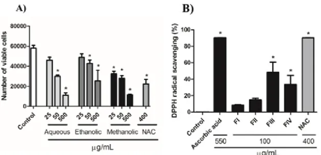
Results showed that all extracts reduced cell proliferation within 72 h of treatment at 50 and 500 µg/mL. However, only the meth- anolic extract decreased the cell number at 25 µg/mL when compared with control group (Figure 1A). As the methanolic extract pre- sented the most significant result, it was se- lected for the next experiments.
Identification of antioxidant activity by DPPH in fractions obtained by column chromatography
The antioxidant potential of each fraction obtained in chromatography column was de- termined with the DPPH assay. At concentra- tion of 100 µg/mL, fractions FIII and FIV showed significant antioxidant activity when compared to control group, while no signifi- cant activity was observed with FI and FII.
NAC exhibited a great antioxidant activity reaching the same level of activity as ascorbic acid (Figure 1B).
Figure 1: A) Effect of crude extracts of Baccharis anomala on cellular proliferation of GRX cells during 72 h of treatment. Cellular proliferation and cell viability was assessed determining the number of viable cells by Trypan blue exclusion method. Data represent the mean ± SD (n = 3). Results were expressed as cell number. * P<0.05 compared with negative control. B) Antioxidant activity of the fractions obtained from the methanolic extract at 100 µg/mL. The antioxidant activity of NAC was evaluated at 400 μg/mL.
Ascorbic acid was used as the control for antioxidant activity at 550 µg/mL. The results are presented as percentage of DPPH reduction in relation to the control group. Data represent the mean ± SD (n = 3). * P < 0.05 compared with negative control
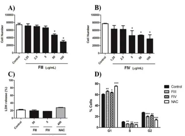
FIII and FIV decrease cell proliferation without cytotoxicity
Fractions FIII and FIV were selected for evaluating their effect on GRX cell prolifera- tion based on their high antioxidant activity.
FIII significantly decreased cell number com- pared to negative control at concentration of 50 and 100 µg/Ml (Figure 2A). Moreover, treatment with FIV resulted in reduction of proliferation when 5, 50 and 100 µg/mL were used (Figure 2B), evidencing a stronger effect of FIV on GRX cell line when compared to FIII. Thus, based on the minimum concentra- tion for a significant reduction of cell number, fractions were used at 50 µg/mL and 5 µg/mL for FIII and FIV, respectively. The superna- tant of cell culture was used to evaluate the
cytotoxicity of the fractions FIII and FIV through LDH assay 72 h after treatment. Re- sults showed that the fractions were capable to reduce cell number without causing cell damage, indicating that both fractions show no cytotoxicity on GRX cells at the concen- trations tested (Figure 2C). In an attempt to understand the decrease in cell number after treatment with FIII and FIV, evaluation of the cell cycle was carried out. Both fractions and NAC significantly induced GRX cell cycle ar- rest in G1-phase when compared to the nega- tive control. Indeed, FIII promoted a signifi- cant increase in cycle arrest when compared to FIV (Figure 2D).
Figure 2: Effect of fractions on GRX cells proliferation during 72 h of treatment. (A) FIII; (B) FIV at concentrations of 1.25, 2.5, 5, 50, and 100 µg/mL. Cellular proliferation assessed by Trypan blue exclu- sion method. Data represent the mean ± SD (n = 3). Results were expressed as cell number.* P < 0.05 compared with control. (C) Cytotoxicity of fractions III and IV was evaluated by measuring LDH release levels in the supernatant after 72 h of treatment. Data represent the mean ± SD (n = 3). The results were presented as percentage of LDH release in the supernatant in relation to the total content of LDH culture obtained by cell lysis. (D) Cell cycle arrest in GRX cells were evaluated by 7-AAD. FIII at 50 µg/mL, FIV at 5 µg/mL and NAC at 400 µg/mL. Samples were analyzed by flow cytometry to identify cell cycle phases. Results were presented as percentage of cells at each phase of the cell cycle. Data were analyzed using FlowJo 7.6.5 software (n=3). ***P < 0.0001, **P < 0.001 and * P < 0.05 compared with negative control.
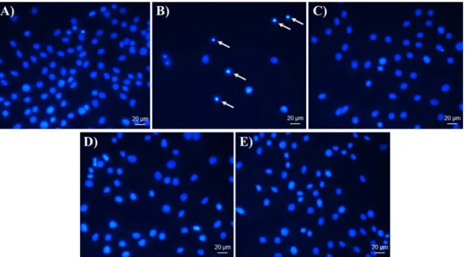
Visualization of apoptotic cells by DAPI In order to investigate whether the de- crease in cell proliferation caused by FIII and FIV was due to apoptosis, the nuclei were stained with DAPI. The morphological pro- file indicated nuclear condensation and frag- mentation suggesting a pro-apoptotic effect of Cisplatin on GRX cells (Figure 3B). How- ever, cells treated with NAC as well with FIII and FIV showed no alteration on morpholog- ical profile (Figure 2,C,D,E).
FIII and FIV induce phenotypic reversion on aHSC
The ability of fractions to revert GRX cell phenotype by inducing the accumulation of lipids in the cytoplasm was investigated.
Compared to the negative control, GRX cells treated with the fractions showed increased ability of storing fat in the cytoplasm (Figure 4B, C). The NAC treatment also induced the accumulation of lipids in the cytoplasm (Fig- ure 4D). Lipid accumulation was quantified by absorbance at 492 nm and confirmed the positive result of the fractions on storing li- pids by GRX cells (Figure 4E). Furthermore,
GRX cells treated with FIII, FIV and NAC lost their elongated and parallel strand ap- pearance and acquired a larger and polygonal
shape, whereas control cells maintained their myofibroblast-like morphology.
Figure 3: DAPI nuclei staining. Effect of FIII and FIV, NAC and Cisplatin on apoptosis of GRX cells analyzed by nuclear morphology. (A) Negative control group, (B) Cisplatin at 2.5 µM, (C) NAC at 400 µg/mL, (D) FIII at 50 µg/mL and (E) FIV at 5 µg/mL. Cells were treated for 72 h. Cisplatin was used as positive control of apoptotic inducer; apoptotic cells demonstrated nuclear condensation (arrows).
Figure 4: Oil Red-O (ORO) staining and lipid quantification of GRX cells at 72 h. (A) Negative control, (B) FIII at 50 µg/mL, (C) FIV at 5 µg/mL and (D) NAC at 400 µg/mL, 400x magnification. Lipid droplets indicated by arrows. (E) Lipid quantification. Results are shown as the absorbance value obtained for ORO adjusted for number of 5x10⁴ cells. Results are expressed as mean ± SD (n=3). * P < 0.05 com- pared with negative control
Expression of α-SMA, PPAR-α and PPAR-γ
The relative expression of α-SMA de- creased in all treatments in relation to nega- tive control group (Figure 5A). As ORO staining revealed the formation of fat drop- lets, the differential expression of PPAR-α and PPAR-γ was assessed in order to eluci- date the pathway of the phenotypic reversion mechanism (Figure 5B, C). Although GRX cells treated with NAC showed a significant increase in PPAR-α expression, treatment with FIII and FIV did not show significant re- sult. No changes were observed in PPAR-γ expression in treated groups with FIV and NAC, only treatment with FIII increased the relative expression of PPAR-γ when com- pared to negative control (Figure 5B), sug- gesting that another pathway might be in- volved in accumulation of lipids droplets on cytoplasm of GRX cells treated with FIV.
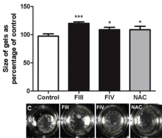
Collagen gel contraction assay
After 24 h of treatment, it was possible to observe that the area occupied by the collagen gel that received treatment with the fractions was larger in relation to the area of the control group. Therefore, it indicates that both frac- tions (FIII and FIV) were able to decrease cell contraction, whereas a greater contraction was detected in the control group cells likely due to its activated phenotype (Figure 6).
Figure 6: Cell contraction assessed by collagen gel assay in GRX cells. Mean ± SD are shown. n=
4 per group. ***P < 0.001 vs.*P < 0.05 vs. nega- tive control, by one-way ANOVA and Tukey test
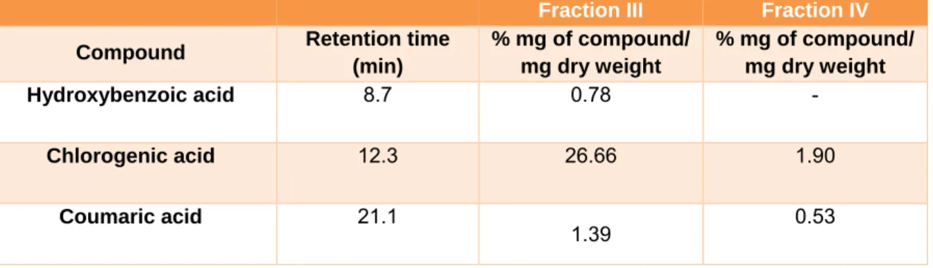
Determination of phenolic compounds Identification and quantification of se- lected phenolic compounds in FIII and FIV were performed by HPLC analysis. In FIII, chlorogenic acid (26.66 %), coumaric acid (1.39 %) and hydroxybenzoic acid (0.78 %) were the identified phenolic compounds. In FIV, only chlorogenicacid (1.9 %) and cou- maric acid (0.53 %) were detected. The reten- tion times and percentages of each compound identified in FIII and FIV are shown in Table 1.
Figure 5: Effect of FIII (5 µg/mL), FIV (50 µg/mL) and NAC (400 µg/mL) on (A) α-SMA, (B) PPAR-ɣ and (C) PPAR-α expression of GRX cells treated for 72 h. Results are presented as relative expression of GAPDH. Data are expressed as mean ± SD (n=3). *P < 0.05 vs. negative control
Table 1: HPLC analysis of phenolic compounds in B. anomala obtained fractions. Results expressed as mean ± standard deviations (SD) of three samples
DISCUSSION
The major finding of the current study is that Baccharis anomala DC. promotes deac- tivating effect in the phenotype of activated HSC evidenced by the reduction of prolifera- tion, induction of lipid drops accumulation and decrease in α-SMA expression.
Studies have shown that the consumption of foods rich in phenolic compounds can pre- vent disease development (Neuhouser, 2004).
It has been also demonstrated the health ben- efits and the applicability of these compounds to the development of new therapies, indicat- ing that the search for natural sources of phe- nolic compounds is a promising alternative.
Some groups of plants have gained visibility due to the high production and accumulation of phenolics, such as the genus Baccharis (Budel et al., 2005; Campos et al., 2016). This genus comprises many species used in the popular medicine by several cultures of South America. Out of the 500 known species ap- proximately 30 % have the phytochemical composition determined (Abad and Bermejo, 2007). Thereby, the search for species with potential for the development of new applica- bility and therapies is worthwhile.
The present study investigated the cyto- toxicity and antiproliferative effect of leaf ex- tract from Baccharis anomala DC. on hepatic stellate cells, as well as the ability of revert the phenotype of such cell. In addition, se- lected phenolic compounds were identified in the extract and their possible relation to the biological effects of the extract is suggested.
Amongst the B. anomala extracts tested, the methanolic showed a significant reduc- tion of cell proliferation in the lowest concen- tration tested (25 µg/mL). This result led us to look for the main compounds present in the methanolic extract. To this end, the extract was fractionated by column chromatography and four fractions were obtained. Methanolic fractions (FIII and FIV) showed high antiox- idant activity, a chemical characteristic that seems to be promising for treatment of he- patic fibrosis (Francia et al., 2016)
FIV fraction was able to significantly de- crease cell proliferation at 5 µg/mL, while FIII was efficient at 50 µg/mL. These doses were selected for cytotoxicity assessment by LDH test, because the activity of LDH in cell culture supernatant is a marker of cellular in- jury. Cytotoxicity was not observed from FIII, FIV and NAC, showing that the de- crease in proliferation was not consequence of necrosis.
A study with Baccharis articulata re- vealed the ability of aqueous extract to induce apoptosis in human peripheral blood mono- nuclear cells (Cariddi et al., 2012). Moreover, a study conducted with the polyphenol resveratrol on activated hepatic stellate cells demonstrated the ability to induce apoptosis through the evaluation performed with DAPI staining (Souza et al., 2008). To verify a pos- sible pro-apoptotic mechanism action of the methanolic fractions that could lead to a de- crease in proliferation of GRX cells, nuclear morphology was visualized through the
Fraction III Fraction IV Compound Retention time
(min)
% mg of compound/
mg dry weight
% mg of compound/
mg dry weight
Hydroxybenzoic acid 8.7 0.78 -
Chlorogenic acid 12.3 26.66 1.90
Coumaric acid 21.1
1.39 0.53
DAPI staining. As positive control of apopto- sis cisplatin was used, which is a well-known chemotherapeutic drug used for treatment of numerous human cancers (Dasari and Tchounwou, 2014). Cisplatin form adducts on DNA, causing damage and subsequently, inducing apoptosis (Siddik, 2003). The re- sults showed that the cells treated with FIII, FIV and NAC showed no alteration in their nuclear morphology in relation to the control group. On the other hand, in addition to sig- nificantly reducing cell proliferation, cispla- tin has shown nuclei with a condensed mor- phology, indicating the formation of apop- totic bodies. This led us to believe that the de- crease in cell proliferation was not due to death by apoptosis, driven our investigation to the action of FIII and FIV methanolic frac- tions on the cell cycle.
Several studies have reported the action of phenolic compounds on the cell cycle (Jafari et al., 2014). Cell cycle is modulated and controlled by complexes of cyclins and cyclin-dependent kinase enzymes (CDKs).
The expression of these enzymes can be reg- ulated by simple phenols or complex poly- meric structures, such as tannins (Jafari et al., 2014). Treatment of HL-60 cells with gallic acid resulted in G0/G1 phase arrest, which was associated with up-regulation of p21 and p27 (Yeh et al., 2011). Thus, the investigation of the cell cycle seems to be necessary to un- derstand the activity of these compounds on cell proliferation. Our results showed that both fractions and NAC induces cell cycle ar- rest in G1-phase in GRX cells. Cell cycle ar- rest in G1-phase observed in NAC treatment is in agreement with the literature, since NAC mediates cell cycle arrest through a Sp1-de- pendent mechanism (Kim et al., 2001). The fundamental point of this evaluation is the stop of the cycle in phase G1, since all the proliferation is suffering a delay. It is note- worthy that in G1 there was a significant in- crease in the number of cells in relation to the control (p <0.001 and 0.05). We believe that it represents a significant decrease in cell pro- liferation, although it is a small difference.
Quiescent state HSCs accumulate retinol (vitamin A) in their cytoplasm, therefore they are also known as lipocytes, functioning in the maintenance of hepatic tissue, through synthesis of proteins responsible for for- mation and degradation of ECM components (Iredale, 2008; Borojevic et al., 1985). This study showed lipid deposits in intracellular content of GRX cells as well as changes in cell morphology observed in phase contrast microscopy. The GRX cells treated with FIII, FIV and NAC lost their elongated and paral- lel strand appearance and acquired a larger and polygonal shape, while control cells pre- served their myofibroblast-like morphology, devoid of large lipid droplets. Quantification of the total lipid content was possible through the absorbance corrected by number of 5x10⁴ cells, which confirmed the significant in- crease in lipid production in cells treated with the fractions and NAC during 72 h. Our re- sults show that FIII and FIV obtained from the methanolic extract showed antiprolifera- tive effect and the ability to deactivate HSCs, transforming the fibroblastic phenotype into quiescent cells.
There are three peroxisome proliferator- activated receptors (PPARs) members of the nuclear hormone receptor family of ligand- activated transcription factors and consisting of three different isoforms: PPAR-α, PPAR- β/δ and PPAR-γ, involved in the regulation of lipid synthesis. PPAR-γ is a transcription fac- tor that is related to induction of quiescent phenotype in HSCs (Guimaraes et al., 2007;
Tsukamoto et al., 2006). Currently, there are studies on molecules capable of inducing the expression of PPAR-α, leading to deactiva- tion of HSCs through pathways independent of PPAR-γ expression (Chen et al., 2015). To investigate the mechanism involved in restor- ing the quiescent phenotype, the PPAR-α and PPAR-γ expression was evaluated by qPCR.
The results showed that expression of PPAR- γ was increased with FIII treatment but did not alter by treatment with FIV and NAC.
The non-alteration of PPAR-γ expression in NAC treatment appears to be in agreement with the literature and the obtained results,
since its effect is mainly related to the expres- sion of PPAR-α (Paintlia et al., 2008). The in- crease in PPAR-α expression was only statis- tically significant in the treatment with NAC.
As treatment with FIII increased the expres- sion of PPAR-γ we believe that might be the mechanism by which induction of quiescent phenotype occurs in HSCs. Thus, it is possi- ble that the restoration of the quiescent phe- notype of the HSCs treated with the FIV could be occurring by a PPAR- α/γ-independ- ent pathway.
Analysis by HPLC performed in FIII al- lowed the identification of the major phenolic compounds, using internal phenolic stand- ards. It was possible to identify in FIII the presence of chlorogenic, hydroxybezoic and coumaric acids. The chlorogenic acid was the most abundant phenolic in FIII. We cannot ignore the possibility of presence of others compounds, however they were not identified by the used method. Some of the phenolic compounds identified by HPLC analysis are in agreement with the phytochemical compo- sition of species found in the genus (Campos et al., 2016). Species that have several studies regarding their chemical composition, such as, B. dracucunlifolia, are known to produce a large amount of phenolic compounds, such as caffeic acid, cinnamic acid, drupanin, bac- charin, artepillin C and p-coumaric acid (Campos et al., 2016; Hocayen et al., 2016).
Studies have demonstrated the antiviral, anti- biotic, antidiabetic, antimicrobial, hepatopro- tective properties and antiproliferative activ- ity, in vitro and in vivo models, using crude or fractionated extracts of this species (Cam- pos et al., 2016; Hocayen et al., 2016; Paintlia et al., 2008; Pereira et al., 2016). Similarly, aqueous and ethanolic extracts of B. trimera showed antiulcerogenic activity, anti-inflam- matory and anthelmintic activity (Oliveira et al., 2014; Dos Reis Liveiro et al., 2016;
Menezes et al., 2016; Nogueira et al., 2011) due to the presence of phenolic compounds such as chlorogenic acid, rutin, ellagic acid, rosmarinic acid, luteolin and quercetin (Menezes et al., 2016). B. uncinella is often used as anti-inflammatory in folk medicine
(Zalewski et al., 2011). Studies on the chem- ical composition of this species have revealed the presence of hispidulin, caffeic acid, chlorogenic acid and pectolinarigenin (Cam- pos et al., 2016). Moreover, benzoic acid and it derivatives, such as p-hydroxybenzoic acid, found in both fractions, are phenolic com- pounds that exhibit a well-known antioxidant activity and are widely used as food preserv- atives (Ding et al., 2015). Chlorogenic acid belongs to the group of hydroxycinnamates, being the most abundant element in human diet from this group. Chlorogenic acid is the main phenolic compound in coffee and it con- sists of the conjugate ester of caffeic, ferulic or p-coumaric acids with quinic acid. The molecule shows a strong antioxidant activity (Zhao and Moghadasian, 2010) and it is the third most abundant compound in FIII. Thus, we believe that at least in part, the antioxi- dant, antiproliferative and the reversion of phenotype effects observed in the treatments with the methanolic fractions are due to the presence of hydrobenzoic acid and chloro- genic acid. In the FIV, chlorogenic and cou- maric acids were detected in smaller propor- tions than in the FIII fraction. For this reason, we believe that there is a molecule or syner- gism of molecules which could be responsi- ble for the strong antiproliferative effect ob- served with FIV. Further investigations are necessary to identify these molecules.
During the development of liver fibrosis occurs the process of transformation HSCs quiescent (lipocytes) into myofibroblasts and this process can result in alterations in con- traction capacity of cells and, therefore, lead to a physiological effect in the liver, an effect that can result in a more serious problem, such as portal hypertension (Iredale, 2008).
The deactivation of the HSCs cells is an im- portant therapeutic target. With the deactiva- tion of the HSCs, the production of compo- nents of extracellular matrix decreases, re- sulting in reduction of contraction. In our work, this reduction was observed in collagen gel with cells treated with FIII and FIV of methanolic extracts of B. anomala.
Notwithstanding FIII and FIV presented some components in common, they vary greatly in quantity. FIV was able to decrease proliferation and reverse cell phenotype with a concentration ten-fold lower than FIII.
However, FIII presented better results in cell cycle arrest and antioxidant activity, showing that both fractions have potential for the treat- ment of liver fibrosis, even though they own distinctive features. FIV appears to be more purified than FIII, since it was the last frac- tion to be collected in column chromatog- raphy. Thereby, we believe that the predomi- nance of a certain compound in this fraction may be responsible for the effect observed in the HSCs in a lower concentration.
Further studies will be necessary to com- pare the effect of the fractions with the iso- lated molecules, in order to better understand the effect that each one exerts on HSCs and how they could act concomitantly and thus search for mechanisms of action. Phenolic ac- ids are natural compounds that can act syner- gistically with other molecules present in the plant extract. Therefore, their effects can complement each other acting more effective on a biological system and likely do not ex- hibit as significant biological activity when used isolated as they do in synergism (Vaz et al., 2012). This study was able to evaluate the cytotoxicity, phenotypic reversion and anti- proliferative effect of the fractions obtained from methanolic extract of Baccharis anomala and showed the potential for the treatment of liver fibrosis.
Acknowledgements
Authors are thankful to the Institute of Toxicology and Pharmacology (INTOX) for providing help and technical support. This study was financed in part by the Coorden- ação de Aperfeiçoamento de Pessoal de Nível Superior – Brasil (CAPES) – Finance Code 001. License for research on Brazil’s biodi- versity, CNPq # 010852/2014-0.
Conflict of interest
We confirm that there are no known con- flicts of interest associated with this publica- tion and there has been no significant finan- cial support for this work that could have in- fluenced its outcome.
REFERENCES
Abad MJ, Bermejo P. Baccharis (Compositae): a review update. ARKIVOC. 2007;(vii):76-96.
Alice CB, Silva GA, Siqueira NC, Mentz LA. Phyto- chemical screening of some plants used in popular medicine of Rio Grande do Sul: Part I. Cad Farm.
1985;1(2):83-94.
Balasundram N, Sundram K, Samman S. Phenolic compounds in plants and agri-industrial by-products:
Antioxidant activity, occurrence, and potential uses.
Food Chem. 2006;99:191-203.
Borojevic R, Monteiro AN, Vinhas SA, Domont GB, Mourao PA, Emonard H, et al. Establishment of a con- tinuous cell line from fibrotic schistosomal granulo- mas in mice livers. In Vitro Cell Dev Biol.
1985;21:382-90.
Budel JM, Duarte MR, Santos CAM, Farago PV, Matzenbacher NI. O progresso da pesquisa sobre o gênero Baccharis, Asteraceae: I - Estudos botânicos.
Rev Brasil Farmacogn. 2005;15:268-71.
Campos FR, Bressan J, Godoy Jasinski VC, Zuccolotto T, da Silva LE, Bonancio Cerqueira L.
Baccharis (Asteraceae): Chemical constituents and biological activities. Chem Biodivers. 2016;13:1-17.
Cariddi L, Escobar F, Sabini C, Torres C, Reinoso E, Cristofolini A, et al. Apoptosis and mutagenicity in- duction by a characterized aqueous extract of Baccha- ris articulata (Lam.) Pers. (Asteraceae) on normal cells. Food Chem Toxicol. 2012;50:155-161.
Chen L, Li L, Chen J, Li L, Zheng Z, Ren J, et al.
Oleoylethanolamide, an endogenous PPAR-alpha lig- and, attenuates liver fibrosis targeting hepatic stellate cells. Oncotarget. 2015;6:42530-40.
Dasari S, Tchounwou PB. Cisplatin in cancer therapy:
molecular mechanisms of action. Eur J Pharmacol.
2014;740:364-78.
Ding M, Peng J, Ma S, Zhang Y. An environment- friendly procedure for the high performance liquid chromatography determination of benzoic acid and sorbic acid in soy sauce. Food Chem. 2015;183:26-9.
Dos Reis Livero FA, da Silva LM, Ferreira DM, Galuppo LF, Borato DG, Prando TB, et al. Hydroeth- anolic extract of Baccharis trimera promotes gastro- protection and healing of acute and chronic gastric ul- cers induced by ethanol and acetic acid. Naunyn Schmiedebergs Arch Pharmacol. 2016;389: 985-98.
Francia R, Rinaldi L, Cillo M, Varriale E, Facchini G, D'Aniello C, et al. Antioxidant diet and genotyping as tools for the prevention of liver disease. Eur Rev Med Pharmacol Sci. 2016;20:5155-63.
Friedman SL. Mechanisms of hepatic fibrogenesis.
Gastroenterology. 2008;134:1655-69.
Garlet TMB. Levantamento das plantas medicinais utilizadas no municı́pio de Cruz Alta, RS, Brazil.
M.Sc. Thesis. Porto Alegre: Universidade Federal do Rio Grande do Sul, 2000.
Guimaraes EL, Franceschi MF, Grivicich I, Dal-Pizzol F, Moreira JC, Guaragna RM, et al. Relationship be- tween oxidative stress levels and activation state on a hepatic stellate cell line. Liver Int. 2006;26:477-85.
Guimaraes EL, Franceschi MF, Andrade CM, Guaragna RM, Borojevic R, Margis R, et al. Hepatic stellate cell line modulates lipogenic transcription fac- tors. Liver Int. 2007;27:1255-64.
Gressner AM. The cell biology of liver fibrogenesis - an imbalance of proliferation, growth arrest and apop- tosis of myofibroblasts. Cell Tissue Res. 1998;292:
447-52.
Heard KJ. Acetylcysteine for acetaminophen poison- ing. N Engl J Med. 2008;359:285–92.
Heiden G, Schneider A. Baccharis in lista de espécies da flora do Brasil. Jardim Botânico do Rio de Janeiro.
2010.http://dipeq.jbrj.gov.br/conservacao/lista-da- flora-do-brasil-reflora/.
Herrmann J, Gressner AM, Weiskirchen R. Immortal hepatic stellate cell lines: useful tools to study hepatic stellate cell biology and function? J Cell Mol Med.
2007;11:704-22.
Hocayen A, Grassiolli S, Leite NC, Pochapski MT, Pereira RA, da Silva LA, et al. Baccharis dracunculi- folia methanol extract enhances glucose-stimulated in- sulin secretion in pancreatic islets of monosodium glu- tamate induced-obesity model rats. Pharm Biol.
2016;54: 1263-71.
Iredale J. Defining therapeutic targets for liver fibrosis:
exploiting the biology of inflammation and repair.
Pharmacol Res. 2008;58:129-36.
Jafari S, Saeidnia S, Abdollahi M. Role of natural phe- nolic compounds in cancer chemoprevention via regu- lation of the cell cycle. Curr Pharm Biotechnol. 2014;
15:409-21.
Kim KY, Rhim T, Choi I, Kim SS. N-acetylcysteine induces cell cycle arrest in hepatic stellate cells through its reducing activity. J Biol Chem. 2001;276:
40591-8.
Lauterburg B, Corcoran G, Mitchell JR. Mechanism of action of N-acetylcysteine in the protection against the hepatotoxicity of acetaminophen in rats in vivo. J Clin Invest. 1983;71:980-91.
Menezes AP, da Silva J, Fisher C, da Silva FR, Reyes JM, Picada JN, et al. Chemical and toxicological ef- fects of medicinal Baccharis trimera extract from coal burning area. Chemosphere. 2016;146:396-404.
Mesquita FC, Bitencourt S, Caberlon E, da Silva GV, Basso BS, Schmid J, et al. Fructose-1,6-bisphosphate induces phenotypic reversion of activated hepatic stel- late cell. Eur J Pharmacol. 2013; 720:320-5.
Moraes CM, Bitencourt S, de Mesquita FC, Mello D, de Oliveira LP, da Silva GV, et al. (+)-Catechin atten- uates activation of hepatic stellate cells. Cell Biol Int.
2014;38:526-30.
Neuhouser ML. Dietary flavonoids and cancer risk:
evidence from human population studies. Nutr Cancer.
2004;50:1-7.
Nogueira NP, Reis PA, Laranja GA, Pinto AC, Aiub CA, Felzenszwalb I, et al. In vitro and in vivo toxico- logical evaluation of extract and fractions from Bac- charis trimera with anti-inflammatory activity. J Ethnopharmacol. 2011;138:513-22.
Oliveira RN, Rehder VL, Oliveira AS, Jeraldo Vde L, Linhares AX, Allegretti SM. Anthelmintic activity in vitro and in vivo of Baccharis trimera (Less) DC against immature and adult worms of Schistosoma mansoni. Exp Parasitol. 2014;139:63-72.
Paintlia MK, Paintlia AS, Khan M, Singh I, Singh AK.
Modulation of peroxisome proliferator-activated re- ceptor-alpha activity by N-acetyl cysteine attenuates inhibition of oligodendrocyte development in lipopol- ysaccharide stimulated mixed glial cultures. J Neurochem. 2008;105:956-70.
Pereira CA, Costa AC, Liporoni PC, Rego MA, Jorge AO. Antibacterial activity of Baccharis dracunculifo- lia in planktonic cultures and biofilms of Streptococ- cus mutans. J Infect Public Health.. 2016;9:324-30.
Rajan N, Habermehl J, Cote MF, Doillon CJ, Manto- vani D. Preparation of ready-to-use, storable and re- constituted type I collagen from rat tail tendon for tis- sue engineering applications. Nat Protoc. 2006;1(6):
2753-8.
Sartor T, Falcão XVB, Mondin CA, dos Santos MA, Cassel E, Astarita LV, et al. Seasonal changes in phe- nolic compounds and in the biological activities of Baccharis dentata (Vell.) G.M. Barroso. Ind Crops Prod. 2013;51:355–9.
Siddik ZH. Cisplatin: mode of cytotoxic action and molecular basis of resistance. Oncogene. 2003;22:
7265-79.
Souza IC, Martins LA, Coelho BP, Grivicich I, Guaragna RM, Gottfried C, et al. Resveratrol inhibits cell growth by inducing cell cycle arrest in activated hepatic stellate cells. Mol Cell Biochem. 2008;315:1- 7.
Tsukamoto H, She H, Hazra S, Cheng J, Miyahara T.
Anti-adipogenic regulation underlies hepatic stellate cell transdifferentiation. J Gastroenterol Hepatol.
2006;21(Suppl 3):S102-.
Vargha R, Mostafa G, Burda G, Hermon M, Tritten- wein G, Golej J. Treatment with N-acetylcystein and total plasma exchange for extracorporeal liver support in children with paracetamol intoxication. Klin Pädiatr. 2014;226:84-5.
Vaz JA, Almeida GM, Ferreira IC, Martins A, Vasconcelos MH. Clitocybe alexandri extract induces cell cycle arrest and apoptosis in a lung cancer cell line: Identification of phenolic acids with cytotoxic po- tential. Food Chem. 2012;132:482-6.
Verdi LG. Gênero Baccharis (ASTERACEAE):
Aspectos químicos, econômicos e biológicos. Quim Nova. 2005;28:85-94.
Wynn TA, Ramalingam TR. Mechanisms of fibrosis:
therapeutic translation for fibrotic disease. Nat Med.
2012;18:1028-40.
Yeh RD, Chen JC, Lai TY, Yang JS, Yu CS, Chiang JH, et al. Gallic acid induces G(0)/G(1) phase arrest and apoptosis in human leukemia HL-60 cells through inhibiting cyclin D and E, and activating mitochon- dria-dependent pathway. Anticancer Res. 2011;31:
2821-32.
Zalewski CA, Passero LF, Melo AS, Corbett CE, Lau- renti MD, Toyama MH, et al. Evaluation of anti-in- flammatory activity of derivatives from aerial parts of Baccharis uncinella. Pharm Biol. 2011;49:602-7.
Zhao ZH, Moghadasian MH. Bioavailability of hy- droxycinnamates: a brief review of in vivo and in vitro studies. Phytochem Rev. 2010;9:133–45.