Cyclase associated protein 2:
Roles in heart physiology and wound healing
INAUGURAL-DISSERTATION zur
Erlangung des Doktorgrades
der Mathematischen-Naturwissenschaftlichen Fakultät der Universität zu Köln
vorgelegt von Kosmas Kosmas
aus
Ioannina, Griechenland Köln, 2014
Referees/Berichterstatter: Prof. Dr. Angelika A. Noegel BProf. Dr. Jürgen Dohmen
Date of oral examination: 23/06/2014
Tag der mündlichen Prüfung
The present research work was carried out under the supervision of Prof. Angelika Noegel and Dr. Vivek Peche, in the Institute of Biochemistry I, Medical Faculty, University of Cologne, Cologne, Germany, from April 2011 to April 2014.
Diese Arbeit wurde von April 2011 bis April 2014 am Institut für Biochemie I der Medizinischen Fakultät der Universität zu Köln unter der Leitung von Prof. Angelika Noegel und Dr. Vivek Peche durchgeführt.
“Δεν μπορώ να διδάξω σε κανένα τίποτα. Μπορώ μόνο να τον κάνω να σκέφτεται.”
Σωκράτης
“I cannot teach anybody anything. I can only make them think.”
Socrates
Acknowledgments
The present thesis was carried out in the research group of Prof. Dr. Angelika A. Noegel in the Institute of Biochemistry I, Medical Faculty, University of Cologne under the supervision of Dr. Vivek Peche.
First of all, I would like to thank my boss, Dr. Vivek Peche, for introducing me to the laboratory methods and the real scientific way of thinking, for supervising my experiments, the nice working conditions in the lab and for making me think and work totally independently.
My thanks also go to the director of the institute Prof. Dr. Angelika A. Noegel for the chance she gave me to work in her institute, for her interest in the development of my work and my skills, and the critical corrections of the manuscripts.
In addition, I would like to thank my 2nd referee Prof. Dr. Jürgen Dohmen and the chair of my committee Prof. Dr. Peter Kloppenburg for their time and effort spent on my thesis.
I would also like to thank all the members of the Biochemistry I and II for their useful tips throughout my work, their assistance and the nice moments we had all these years. I will not list the names because I will definitely need 10 pages. I feel grateful for all the people including the professors, the employees, the students, the secretary, the lab assistants, the technical assistants and the animal care takers.
Special thanks to the IGSDHD for the funding, the support and for making the official work easy.
Last but not least, my heartiest gratitude goes to my family and friends for their constant support throughout my studies.
Abbreviations
aa amino acids
ATP Adenosine 5’-triphosPhate
bp base pair(s)
cDNA complementary DNA
DMEM Dulbecco’s Modified Eagle’s Medium
DMSO Dimethylsulphoxide
DNA Deoxyribonucleic Acid DTT 1,4-dithiothreitol E. coli Escherichia coli
EDTA Ethylenediaminetetraacetic acid
EGTA Ethyleneglycol-bis (2-amino-ethylene)N,N,N,N-tetraacetic acid
ES Embryonic stem
FITC Fluorescein-5-isothiocyanat
GAPDH Glyceraldehyd-3-phosphat Dehydrogenase GFP Green Fluorescent Protein
GST Glutathion-S-Transferase
HEK Human Embryonic Kidney
IPTG iso-propylthio-galactopyranoside
M Molar
MW Molecular Weight
NP Nonyl Phenoxypolyethoxylethanol
PAGE Polyacrylamide Gel Electrophoresis
PBS Phosphate Buffered Saline
PCR Polymerase Chain Reaction
PIPES Piperazine-N,N’-bis [2-ethanesulphonic acid]
PMSF Phenylmethylsulphonylfluoride
RNA Ribonucleic Acid
SDS Sodium Dodecyl Sulphate
Tris Tris –(hydroxymethyl)-aminomethane TRITC Tetramethylrhodamine Isothiocyanate Units of Measure
D Dalton
g gram
h hour
l litre
m meter
min minute
s sec
Prefixes
k kilo (103)
c centi (10-2)
m milli (10-3)
μ micro (10-6)
n nano (10-9)
Table of contents
1. Introduction 1
1.1 The Cytoskeleton 1
1.2 Types of the cytoskeleton 1
1.3 Actin filaments 2
1.4 Acting binding proteins 3
1.5 CAP2 4
1.6 CAP2 and cardiomyopathy 6
1.7 Cell migration and actin cytoskeleton 7
1.8. Wound healing 10
1.8.1 Phases of wound healing 10
A. Inflammation 10
B. Repair 11
C. Remodeling 12
1.9 Aim of the research 14
2. Materials and methods 16
2.1 Generation of Cap2gt/gt 16
2.2 Skin wounding 16
2.3 Preparation of tissue 17
2.4 Immunohistochemistry, antibodies and histology 17
2.5 Cell culture and cell scratch assay 18
2.6 Western blot analyses 18
2.7 Focal adhesion assay 19
2.8 Disruption of actin cytoskeleton and recovery 19
2.9 RNA isolation 19
2.10 Expression of CAP2 domains and in vitro assays 20
2.11 DNA transfection 20
2.12 Recombinant protein expression 20
3. Results 21
3.1 Generation of a CAP2 knockout mouse 21
3.2 Characterization of CAP2 monoclonal antibodies 23 3.3 CAP2 deletion leads to weight loss and is lethal in
postnatal stages of mice 23
3.4 Cardiac and skeletal muscle phenotype of Cap2gt/gt mice 25 3.4.1 Cap2gt/gt mice develop dilated cardiomyopathy 25 3.4.2 CAP2 is required for proper sarcomeric organization in cardiac
and skeletal muscle 28
3.4.3 CAP2 deletion may lead to sarcopenia 30
3.5 Roles of CAP2 in wound healing 31
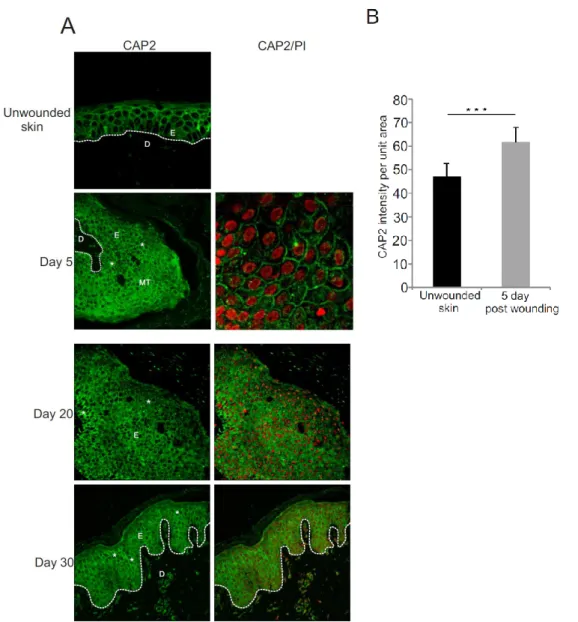
3.5.1 Expression of CAP2 in human wounds 31
3.5.2 Loss of CAP2 results in delayed wound repair 33 3.5.3 Histological analysis with Masson’s trichrome staining 34 3.5.4 Proliferation is reduced in Cap2gt/gt mice 35 3.5.5 Delayed wound contraction in Cap2gt/gt mice 36 3.5.6 Cap2gt/gt mice show decreased macrophage infiltration 37 3.5.7 Slower neovascularization in Cap2gt/gt mice 38 3.5.8 Increase in apoptosis in Cap2gt/gt wounds 39 3.6 Cell migration defects in Cap2gt/gt fibroblasts 40
3.6.1 Cap2gt/gt fibroblasts show reduced velocity 40 3.6.2 Cap2gt/gt fibroblasts develop long filopodia 42 3.6.3 Focal adhesions are altered in Cap2gt/gt fibroblasts 42 3.6.4 G-/F-actin ratio is altered in Cap2gt/gt fibroblasts 44 3.6.5 Recovery of the actin cytoskeleton is faster in mutant fibroblasts 45 3.7 Identification of CAP2 interacting partners 46
3.7.1 CAP2 interacts with CPT1B 50
3.8 CAP in cancer 51
4. Discussion 53
4.1 CAP2 in the cardiovascular system and in skeletal muscle 53
4.2 Role of CAP2 in wound healing 56
Summary / Zusammenfassung 64
Bibliography 66
Erklärung 78
Curriculum Vitae / Lebenslauf 79
1. Intoduction 1
1. Introduction
1.1 The Cytoskeleton
A vital need for the survival of eukaryotic cells is to adapt to a variety of shapes and to carry out coordinated and directed movements. This is carried out by the cytoskeleton which is a complex network of protein filaments that extends throughout the cytoplasm. This network is a highly dynamic protein mosaic that dynamically coordinates cytoplasmic biochemistry and is reorganized continuously as the cell changes shape, divides and responds to its environment. It is also essential for intracellular transport of vesicles and organelles in the cytoplasm and the segregation of chromosomes at mitosis (Peters, 1929; Alberts et al., 2007, Molecular Biology of the Cell, 5th Edition).
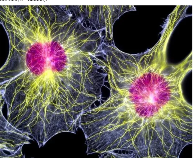
Figure 1.1: Fluorescent light micrograph of two fibroblast cells, showing their nuclei (purple) and cytoskeleton. The cytoskeleton is made up of microtubules (yellow) and actin filaments (white) (Image adopted by Google).
1.2 Types of the cytoskeleton
The diverse activities of the cytoskeleton depend on three types of protein filaments, actin filaments, microtubules, and intermediate filaments. Each filament type is
formed from a different protein subunit: actin for actin filaments, tubulin for microtubules, and a family of related fibrous proteins, such as vimentin or lamin, for intermediate filaments.
Intermediate filaments have a function in providing cells with mechanical strength. In vertebrate cells they can be grouped into three classes: (1) keratin filaments, (2) vimentin and vimentin-related filaments and (3) neurofilaments, each formed by polymerization of their corresponding subunit proteins (Alberts et al., 2007, Molecular Biology of the Cell, 5th Edition).
Microtubules together with actin filaments are the primary organizers of the cytoskeleton. They usually have one end anchored in the centrosome and the other free in the cytoplasm. In many cells microtubules are highly dynamic structures that alternately grow and shrink by the addition and loss of tubulin subunits. Motor proteins move in one direction or the other along microtubules, carrying specific membrane-bound organelles to desired locations in the cell.
Actin filaments are essential for many movements of the cell. They are also dynamic structures, but they normally exist in bundles or networks rather than as single filaments. A layer called the cortex is formed just beneath the plasma membrane from actin filaments and a variety of actin-binding proteins. This actin-rich layer controls the shape and movements of most animal cells (Alberts et al., 2007, Molecular Biology of the Cell, 5th Edition).
1.3 Actin filaments
All eukaryotic species contain actin. It is the most abundant protein in many eukaryotic cells, often constituting 5% or more of the total cell protein. It exists in a monomeric or G actin state (G for globular) and a polymeric state, F-actin or filamentous actin. Actin filaments can form both stable and labile structures in cells.
Stable actin filaments form the core of microvilli and are a crucial component of the contractile apparatus of muscle cells. Cell movements, however, depend on labile structures constructed from actin filaments. Actin filaments appear in electron micrographs as threads about 8 nm wide. They consist of a tight helix of uniformly oriented actin molecules. Like a microtubule, an actin filament is a polar structure, with two structurally and functionally different ends - a relatively inert and slow growing minus or pointed end and a faster growing plus or barbed end.
1. Intoduction 3 Actin is an ATPase. ATP-bound actin is polymerization proficient and is found in newly polymerized filaments. The ATP molecule hydrolyzes and “older” filaments contain ADP actin. ADP actin is released from the pointed end and needs to be recharged with ATP for new polymerization. This ADP/ATP exchange requires several actin-binding proteins like cofilin, profilin and CAP (cyclase associated protein).
1.4 Acting binding proteins
Actin filaments are organized in two general types: bundles and networks that are essential for cell migration, division and intracellular transport. These structures are formed by actin-binding proteins that cross link actin filaments, motor proteins, branching proteins, severing proteins, polymerization factors, and capping proteins.
Sets of actin-binding proteins are thought to act cooperatively in generating the movements of cells and inside cells as during endo- and exocytosis or phagocytosis, in cytokinesis and cell locomotion. One family of proteins also called G-actin binding proteins or G-actin sequestering proteins can bind to G-actin and thus is involved in controlling F-actin formation. The binding of these proteins is reversible and through certain extracellular signals they can release G-actin to allow formation of F-actin.
Typical members of this family are profilin, cofilin, thymosin and CAP (Carlier and Pantaloni 1994; Gottwald et al., 1996).
Another group is collectively called “capping proteins”. They inhibit further addition of monomers, thus keeping filaments short. By binding to the plus ends of actin filaments, capping proteins slow the rate of filament growth. Even at the minus end the actin filament may be capped by minus end capping proteins. The association of capping proteins with actin filament ends is regulated by various localized intracellular signals. Uncapping of actin filaments makes the plus ends available for elongation, thereby promoting actin filament polymerization near the cell cortex. An example of this category of proteins is the cap32/34 (CapZ) as plus end capping protein that is regulated by PIP2 (Hartmann et al., 1990; Haus et al., 1991) or tropomodulin as minus end capping protein (Yamashiro et al., 2012). Severing proteins on the other hand fragment F-actin. A representative member of this group is the Ca2+ activated severin which in addition can also nucleate actin assembly (Eichinger et al., 1991).
The third category of actin binding proteins are the F-actin crosslinking proteins, which can either stabilize the filament itself or crosslink filaments to form bundles as well as three-dimensional networks. Prototypes of this class are tropomyosin, which can stabilize the filament (Lehmann et al., 1994), α-actinin and filamin, which bundle and crosslink the filaments (Noegel et al., 1987; Stossel et al., 2001).
1.5 CAP2
Proteins essential for maintaining the equilibrium between G- and F-actin form the monomer actin binding or G-actin sequestering protein family. CAP belongs to this family and its homologs in yeast and mammals have been shown to sequester G-actin through their C-terminal domain and prevent them from polymerization in vitro (Hubberstey and Mottillo, 2002). In addition, recent biochemical studies have revealed new biochemical functions of CAP apart from the actin-monomer- sequestering function involved in actin reorganization (Balcer et al., 2003; Bertling et al., 2004; Freeman and Field, 2000; Peche et al., 2013). It promotes actin filament dynamics closely cooperating with ADF (actin depolymerizing factor)/cofilin in vitro and in vivo (Moriyama and Yahara, 2002), and self-oligomerization of CAP enhances its activities (Quintero-Monzon et al., 2009). Furthermore, the conservation of CAPs among eukaryotes suggests that CAP is a fundamentally important actin regulator.
CAP/Srv2 was originally identified in budding yeast by biochemical means as a protein associated with adenylyl cyclase and also genetically as a suppressor of adenylyl-cyclase in conjunction with hyperactive RAS2(V19), thus explaining the yeast name Srv2 (Field et al., 1990; Fedor-Chaiken et al., 1990). The N-terminal region of Srv2 interacts with adenylyl cyclase; whereas the C-terminal region binds monomeric actin with high affinity (Gerst et al., 1991; Freeman et al., 1995; Mattila et al., 2004). Subsequent studies showed that the ability to interact with actin in vitro and regulate actin dynamics in vivo are conserved functions of CAPs in all eukaryotes (Hubberstey and Mottillo, 2002). In a screen to identify genes required for Drosophila oocyte polarity , a Drosophila homologue was found which was allelic to capulet and act up (Baum et al., 2000; Wills et al., 2002; Benlali et al., 2000). Caenorhabditis elegans CAP genes were named cas-1 and cas-2. The amino acid sequence of CAS-1 shows a 37% sequence identity with human CAP1. In addition, CAS-2, a second CAP isoform in C. elegans, attenuates the actin-monomer-sequestering effect of ADF/cofilin to increase the steady-state levels of actin filaments in an ATP-dependent
1. Intoduction 5 manner (Nomura et al., 2012; Nomura et al., 2013). ASP-56 has been isolated from pig platelets and characterized by actin-binding assays as the first mammalian orthologue of CAP (Gieselmann and Mann, 1992). Furthermore, rat MCHI cDNA encodes a protein of 474 amino acids that is 36% identical to S. cerevisiue CAP and is capable of suppressing the loss of the COOH-terminal functions of CAP when expressed in yeast. (Zelicof et al., 1993). Except for these, “cyclase-associated protein” or CAP is used as a common name in most of the literature. CAPs play a major role in various cellular activities. Knockout of CAP in Dictyostelium discoideum and in yeast has revealed many important functions of CAP like in cell polarity and cell migration (Noegel et al., 2004; Vojtek et al., 1991). Although oocyst development is not considered as an actin-dependent process, inactivation of the CAP homologue from Plasmodium berghei demonstrated that this protein is essential for malaria parasite oocyst development in the mosquito midgut .The direct role of CAP is this process needs to be elucidated (Hliscs et al., 2010). Inactivation of CAP in C., Drosophila, and plant cells results in severe defects in the organization of the actin cytoskeleton, abnormal accumulation of filamentous actin, and consequently problems in many actin-dependent processes (Nomura et al., 2012; Baum et al., 2000;
Benlali et al., 2000; Barrero et al., 2002; Effendi et al., 2013).
Mammals have two CAP genes encoding the related CAP1 and CAP2. CAP1 has been well studied. It is expressed in nearly all cells and organs of the mouse and is highly abundant. At the subcellular level, it is present in regions with high actin dynamics (Bertling et al., 2004). CAP2 shows a more restricted distribution and is significantly expressed only in brain, heart and skeletal muscle, and in skin. CAP2 is found in the nucleus in undifferentiated myoblasts and at the M-line of differentiated myotubes. During myogenesis, CAP2 is mainly a nuclear protein; in the adult muscle it is an M-band protein. In skin-derived cell lines, CAP2 is primarily a nuclear protein, in skin it is a nuclear protein and also present at cell borders (Peche et al., 2007).
A comparison of the CAP1 and CAP2 amino acid sequences shows that mammalian CAP1 and CAP2 are highly related proteins. Mouse CAP2 shares 62 % identity and 76 % similarity with mouse CAP1. CAP1 and CAP2 from various mammalian species are 93 – 96 % (CAP1) and 88 – 93 % (CAP2) identical among each other and are equally distant to CAPs from non-mammalian species showing 33 – 34% identity each to CAP/Srv2 from S. cerevisiae. The degree of homology between mouse CAP1
and CAP2 varies within the domains. It is slightly higher in the C-terminal domain than in the proline-rich central domain and the N-terminal domain. Loss of CAP results in defects in cell morphology, migration, endocytosis and development in S.
cerervisiae, D. discoideum and Drosophila (Hubberstay and Motillo, 2002).
In Pam212, a mouse keratinocyte cell line, CAP2 is enriched in the nucleus and less prominent in the cytosol whereas CAP1 localizes to the cytoplasm in these cells. In human skin, CAP2 is present in all living layers of the epidermis where it localizes to the nuclei and the cell periphery. In biochemical studies it was shown that a C- terminal fragment of CAP2 interacted with actin, indicating that CAP2 has the capacity to bind to actin. CAP2 is also strongly enriched in the nucleus in developing cardiomyocytes. It changes its localization in the adult cardiomyocyte and is then observed at the M-band. The M-band is an important element of the sarcomere, the elementary contractile unit of striated muscle. It maintains the thick filament lattice through interactions of the prominent M-band component myomesin, which links the thick filaments (Peche et al., 2007).
CAP2 is up-regulated in hepatocellular carcinoma (HCC) when compared with noncancerous and precancerous lesions. That indicates that CAP2 is up-regulated in human cancers. Since it is possibly related to multistage hepatocarcinogenesis, it has been suggested as a ‘potential biomarker’ for pathological diagnosis (Shibata et al., 2006).
1.6 CAP2 and cardiomyopathy
Cardiomyopathy is a disease characterized by either thickening or thinning of the heart muscle, and both conditions, hypertrophic cardiomyopathy (HCM) and dilated cardiomyopathy (DCM), lead to inefficient functioning of the heart muscle and can cause sudden cardiac death. DCM is the most common cardiomyopathy and many studies point out the importance of left ventricular pathophysiology in congestive heart diseases, whereas right ventricular DCM, in which the right ventricle is dilated with thinning of the ventricular wall, is less frequently observed than left ventricular cardiomyopathy and is therefore not extensively studied (Jefferies and Towbin, 2010).
At the structural level, DCM is associated with a loss of myofibrils and sarcomeric disorganization (Mann et al., 1991; Schaper et al., 1991). The inherited forms of DCM are associated with mutations in genes that generally encode cytoskeletal and sarcomeric proteins (Jefferies and Towbin, 2010; Harvey and Leinwand, 2011).
1. Intoduction 7 In sarcomeres, the precise control of actin filament length contributes to the proper function of the contractile apparatus. This control appears to occur at the barbed and pointed ends of the filament as actin is incorporated at the Z-disc and in the middle of the sarcomere (A-band region) where it depends upon effective termination of polymerization by capZ and tropomodulin, respectively (Sussman et al., 1998;
Littlefield and Fowler, 2008). Filament growth is also affected by the G-actin/F-actin equilibrium, which is regulated by G-actin sequestering proteins. Recent studies demonstrated that actin filaments in sarcomeres of actively contracting cells undergo rapid turnover in which actin depolymerizing factors cofilin 1 and 2 are involved promoting rapid actin dynamics (Skwarek-Maruszewska et al., 2009). Importantly, the Cap2 gene is expressed at early to late developmental stages during cardiogenesis of mice embryos (Christoforou et al., 2008).
Although various studies implicate CAPs in the organization of the actin cytoskeleton, a detailed analysis of the in vivo function of CAP in mammals is still lacking. We have generated a mouse in which the Cap2 gene is inactivated by a gene- trap approach. Our results show that ablation of CAP2 leads to severe cardiac defects marked by dilated cardiomyopathy associated with a drastic reduction in basal heart rate and prolongations in atrial as well as ventricular conduction times. Moreover, we found alterations in the mechanical properties of the CAP2-deficient myofibrils with a significantly reduced Hill coefficient and severe changes in the structure of the sarcomere. As the underlying mechanism, we proposed a misregulation of actin filament assembly near the M-line due to the absence of CAP2 (Peche et al., 2013).
1.7 Cell migration and actin cytoskeleton
Cells can sense and respond to environmental signals such as mechanical forces that act as critical regulators of physiological processes including embryogenesis and wound healing (Janmey and McCulloch, 2007; Parsons et al., 2010; Geiger et al., 2009). These mechanical forces have to be transmitted across the cell membrane in both directions through cell adhesions that are coupled by the actin cytoskeleton. Cell adhesions are large macromolecular assemblies that form cell-extracellular matrix contacts (hemidesmosomes and focal adhesions) or cell-cell contacts (adherence junctions) (Geiger et al., 2009; Geiger et al., 2001). Members of this group include vinculin, talin, zyxin, FAK, and paxilin that are organized at the basal surface of adherent cells. Focal adhesions have long been speculated to play a critical role in
many cell functions, in particular, cell migration (Ridley et al., 2003). For example, rapidly moving cells, such as D. discoideum and neutrophils exhibit negligible small focal adhesions and seem to glide over the substratum (Nagasaki et al., 2009), while slow-moving cells such as fibroblasts display prominent focal adhesions and seem to crawl over the substratum.
A functional relationship between focal adhesions (surface density, size, shape, number, turnover dynamics, etc.) and cell migration is largely till now missing (Kim and Wirtz, 2013). Cell migration can be easily considered as a highly integrated cyclic process (Lauffenburger and Horwitz, 1996). Initially, cells migrate through polarizing and extending protrusions in the direction of migration. These protrusive structures can be large ruffling veil-like lamellipodia or thin, spike-like filopodia. Lamellipodia are composed of orthogonal arrays of actin filaments with branched actin filaments close to the leading edge of plasma membrane, whereas filopodia consist of parallel bundles of actin filaments (Chhabra and Higgs, 2007). Elongation at the barbed ends of actin in lamellipodia and filopodia is thought to form the protrusive machinery for generating force for leading edge advancement (Pollard and Borisy, 2003; Bugyi and Carlier, 2010).
This membrane protrusion is driven by the polarity of actin filaments through fast- growing “barbed” ends and slow-growing “pointed” ends (Welch and Mullins, 2000).
Binding of Arp2/3 complex on the already existing actin filament enables the formation of a new daughter filament that branches off the mother filament. Arp2/3 complex is activated by WASP (Wiskott - Aldrich syndrome Protein)/WAVE (WASP family Verprolin-homologous protein) family members. Actin polymerization is regulated by several actin-binding proteins that affect the pool of available monomers and free ends (Pollard and Borisy, 2003; Dos Remedios, 2003). Profilin, for instance, binds to actin monomers, blocks self-nucleation and also targets monomers to barbed ends. Capping proteins terminate filament elongation, while minimizing polymerization to new filaments close to the plasma membrane. Besides, members of the ADF/cofilin family sever and disassemble already stable filaments at the pointed end, a process that is essential for the replenishment of the actin monomer pool needed for polymerization at the front end. Additional proteins stabilize actin filament like Cortactin, Filamin and α-actinin (Welch and Mullins, 2000). The pivotal mechanism for the formation of the filopodial protrusion is the filament treadmilling, via which actin filaments elongate at their barbed ends and release actin monomers
1. Intoduction 9 from their pointed ends (Welch and Mullins, 2000). Ena/VASP proteins bind at the barbed ends allowing continuous elongation of the actin filaments while antagonizing both capping and branching. Furthermore, stiffness that is required for efficient pushing of the plasma membrane in filopodia is achieved by fascin which is an F- actin bundling protein (Welch and Mullins, 2000).
The formation of lamellipodia and filopodia as well as the adhesion organization are mainly controlled by small guanosine triphosphate (GTP)–binding proteins (GTPases) that belong to the Rho family. In their active form (bound to GTP) these regulators interact with a variety of downstream target proteins including protein kinases, lipid- modifying enzymes and activators of the Arp2/3 complex (Etienne-Manneville and Hall, 2002). Guanine nucleotide exchange factors (GEFs) activate Rho GTPases and GTPase activating proteins (GAPs) terminate the signaling event. The most essential Rho GTPases for the formation of stress fibers, lamellipodia and filopodia are RhoA, Rac and Cdc42 respectively (Ridley et al., 2003; Zhou et al., 2014). Cdc42 and Rac mediate actin polymerization in protrusions via the WASP/WAVE family of Arp2/3 complex activators. Cdc42 stimulates the Arp2/3 complex through binding to WASP proteins in order to induce dendritic actin polymerization (Welch and Mullins, 2000).
Besides, Rac stimulates lamellipodial extension by activating WAVE proteins (Gory and Ridley, 2002).
CAP2 contains also a WASP homology (WH2) domain which is responsible for its actin-sequestering activity (Peche et al., 2013). This motif, consisting of 25 amino acids is found in proteins such as WASP, thymosin, Spire, Cordon-bleu, Leiomodin, and JMY. Thymosin mediates sequestration of monomeric actin and inhibition of actin polymerization. On the other hand, Spire, Cordon-bleu, and JMY nucleate actin assembly (Ducka et al., 2010).
The WH2 domain of CAP2 was identified in a comparison with N-WASP and thymosin β4. It is located at position 247–310 and contains the essential LRHV motif and a N-terminal helix preceding this motif (Chereau et al., 2005). CAP2 influences actin dynamics by binding to G-actin through its WH2 domain, preventing polymerization and can also sever filaments thereby affecting filament stability (Peche et al., 2013). These activities might be vital for the organization of lamellipodia that provide a veil-like structure that is able to push the plasma membrane. The lamellipodium could then grow in a particular direction, providing the basis for directional migration.
1.8 Wound healing
Skin is the largest organ in the human body and serves as the interface between the organism and the environment, and protects against infection and excessive water loss. Furthermore, multiple components in the skin, such as the sebaceous and sweat glands, hair follicles, blood capillaries, and nerve endings, confer secondary properties that are essential to everyday function (Almine et al., 2012; Seeley et al., 2005).
Cutaneous trauma disrupts skin architecture and integrity, which elicits a highly regulated localized response that cleans, debrides, and heals the site of injury. Trauma to skin can arise from abrasions, lacerations, and thermal, electrical, or chemical burns (Almine et al., 2012; Trott, 1988).
Cutaneous wound healing is a complex and dynamic process involving soluble mediators, blood cells, extracellular matrix (ECM), and parenchymal cells (Singer and Clark, 1999). This phenomenon is characterized by an attenuated inflammatory response to tissue injury, which involves differential expression of signaling factors, and regeneration of normal skin architecture (Almine et al., 2012). The process of wound healing normally proceeds from coagulation and inflammation through fibroplasia, matrix deposition, angiogenesis, epithelialization, collagen maturation and finally wound contraction (Schäfer and Werner, 2008). These processes compose three different overlapping phases: (1) inflammation, (2) repair, and (3) remodeling (Fig. 2).
1.8.1 Phases of wound healing A. Inflammation
Skin injury causes cell damage and injury of blood vessels. A wound must stop bleeding in order to heal and for the injured host to survive. Blood vessels constrict within seconds after injury to prevent blood loss and afterwards platelets are activated by thrombin, they aggregate and clotting occurs (Mahdavian Delavary et al., 2011).
Together, these events are responsible for the formation of a haemostatic blood clot, mainly composed of complement cascades which are activated and crosslinked like fibrin, fibronectin, vitronectin, thrombospondin, as well as erythrocytes and platelets (Midwood et al., 2004; Metcalfe and Ferguson, 2007; Krafts, 2010). Immediately after wounding insulin like growth factor 𝛼 (IGF-𝛼), transforming growth factor 𝛽 (TGF-𝛽), platelet-derived growth factor (PDGF) and vascular endothelial growth
1. Intoduction 11 factor (VEGF) are released from the platelets, a process that attracts leukocytes and fibroblasts into the wound area.
Mast cells are migrating first to the wound site assisting the recruitment of neutrophils to protect against infectious agents and initiate the removal of debris from damaged cells and ECM (Egozi et al., 2003). The influx of neutrophils peaks in the first 48 hr after injury. The neutrophils are eventually replaced by monocytes, which will subsequently differentiate into macrophages (Almine et al., 2012).
One of the primary roles of macrophages in the inflammatory stage is to complete the removal of debris and foreign material through their phagocytic function and through their capacity to secrete toxic mediators. In addition they recruit fibroblasts, keratinocytes and endothelial cells through secreting growth factors (Krafts, 2010).
Furthermore, macrophages participate in the remodeling of the extracellular matrix for the formation of the scar. Macrophages also assist with the transition of the wound site from inflammation to repair. (Fig. 1.2 A).
B. Repair
The repair stage is characterized by active fibroplasia granulation tissue formation, wound contraction, re-epithelialization, and angiogenesis (Grinnell, 1982).
Keratinocytes migrate from the epidermis at the wound edge and express various proteases allowing the degradation of the connective tissue (Martin, 1997). This process is followed by active fibroplasia in which fibroblasts migrate, proliferate and deposit extracellular matrix forming the granulation tissue. Granulation tissue is an amorphous structure composed of blood vessels, extracellular matrix (ECM) (collagen, fibronectin), and fibroblasts, replacing the fibrin eschar (scab) as a scaffold for cell infiltration (Almine et al., 2012).
Some fibroblasts differentiate into myofibroblasts, a contractile cell that expresses smooth muscle actin, and is active in the repair stage of wound healing (Werner et al., 2007). The myofibroblast phenotype is induced by mechanical tension and TGF-β.
The formation and function of myofibroblasts are essential for drawing the margins of the wound edge together, facilitating the physical closure of the wound site (Tomasek et al., 2002; Hinz and Gabbiani, 2003).
Concurrently, re-epithelialization of the epidermis occurs, where undamaged basal keratinocyte epithelial cells migrate and proliferate to the wound edge providing cover for the formation of the neoepidermis. Epidermal stem cells resting in the hair
follicle bulge can replenish the pool of proliferative keratinocytes (Gurtner et al., 2008). Consequently, the basement membrane which anchors the epithelium to the dermis underneath through substrate adhesion molecules (SAMs) is re-established by extracellular components secreted and deposited by keratinocytes. This process is also enhanced by fibroblasts. Finally a stratified, keratinized epithelium is formed, due to differentiation of basal keratinocytes caused by contact inhibition (Smola et al., 1993).
In angiogenesis, which is vital for the transport of oxygen, nutrients, and cells, new blood vessels are formed. This step is promoted by the fibrin plug, platelets, and endothelial cells (Singer and Clark, 1999) (Fig. 1.2 B).
C. Remodeling
The remodeling of the mature scar, which is the final and longest stage of wound healing, can last for weeks to months. The acellular, fibrous scar is mainly composed of ECM components (Broughton et al., 2006). During this stage processes like cell proliferation and protein synthesis are slowed down and formation of collagen fibrils takes place (Mahdavian Delavary et al., 2011). The synthesis of collagen I and III increases dramatically to form the central core of the mature scar (Lovvorn et al., 1999). Collagen is remodeled and realigned along tension lines and cells that are no longer needed are removed by apoptosis.
Fibroblasts, macrophages and endothelial cells secrete matrix metalloproteases (MMPs) that contribute in strengthening the repaired tissue (Lovvorn et al., 1999).
The collagen-based scar recuperates the rigidity of skin, but exhibits a lower tensile strength, which is due to a deviation in matrix composition and organization compared with uninjured skin (Levenson et al., 1965). Furthermore, the peripheral functions of skin are diminished because skin components, such as hair follicles, sebaceous and sweat glands, are not regenerated (Almine et al., 2012) (Fig. 1.2 C).
1. Intoduction 13
Figure 1.2: The three classical phases in adult skin wound healing. A) A fibrin clot is formed and inflammatory cells enter the wound site. B) Re-epithelialization and angiogenesis of a provisional matrix occurs. C) Remodeling is the final stage of wound healing. ECM remodeling factors modulate and revise the scar tissue (taken from Gurtner et al., 2008).
1.9 Aim of the research
Despite the fact that CAP proteins have been studied for more than 20 years and are present in all organisms, many issues remain to be addressed about CAP function in higher eukaryotes. The role of mammalian CAP2 in actin cytoskeleton organization has not been yet studied extensively. For this aim, we generated a whole body knock- out mouse in which the CAP2 gene was inactivated by a gene-trap approach. Cap2 deletion led to weight loss and here I will study whether it is associated with muscular atrophy or even sarcopenia.
The observation that Cap2gt/gt mice died earlier and had enlarged hearts in contrast to the WT, prompted us to characterize the cardiac phenotype of the mutant animals, since we already know that CAP2 is highly expressed in the cardiac tissue. The severe cardiac defects can be easily marked by dilated cardiomyopathy (DCM) or hypertrophy. The atrial and ventricular conduction times will also been addressed in order to fully characterize the cardiac defects. At tissue level we will investigate the organization of the sarcomeres.
CAP2 is strongly expressed in skin. We plan to focus on this organ in our further analysis of CAP2 knockout animals. Our major plan is to perform in vivo wound healing experiments with which we are going to probe CAP2’s role in cell proliferation, differentiation and migration in wound healing, something that is still unknown. The role of CAP2 in regulating wound healing will be characterized by evaluating its effect on neo-epidermis formation, fibroblast myofibroblast transition, and cellular proliferation and apoptosis in the wounds. Additional parameters of wound healing process such as macrophage infiltration and neovascularization will also be extensively investigated.
The formation of filopodia and possible difference in the formation of focal adhesions will come under investigation in terms of cell migration. In addition, analysis of the effect of CAP2 on the subcellular G-/F-actin ratio will take place.
1. Intoduction 15 Apart from these, we will also try to further characterize the molecular function of CAP2 through identifying its interacting partners. We will generate and characterize monoclonal antibodies against CAP2.
In addition to this, we will focus on the possible role of CAP proteins in cancer and specifically the role of CAP1 and CAP2 in cancer.
2. Materials and methods
2.1 Generation of Cap2gt/gt
Clone D07 was obtained from the EUCOMM consortium, Helmholtz Zentrum München, Munich, Germany. ES cells were microinjected into blastocysts and chimeras were produced. The generated chimeric males were then intercrossed with C57BL/6N females to generate F1 offspring. The Cap2gt allele was detected by BamHI digestion of genomic DNA and Southern blotting by using probes generated by using primers pairs;
Forward-probe 5’-GGAAAACCTGTTGAAGGCAG-3’and Reverse-probe 5’-CCCTGAACTG AGAATGTTCC-3’
PCR primers for genotyping were:
Forward-Cap2: 5’GTGCTTCACTGATGGGCTTG3’
Reverse-Cap2: 5’TCACCCCACATTTACGATGG3’
Forward-neo: 5’GCCGCTCCCGATTCGCAG3’
Additionally, heterozygous Cap2 gene-trap mice were obtained from the EUCOMM consortium, Helmholtz Zentrum München, Munich, Germany. These mice were maintained in the C57BL/6 background.
All animals (C57Bl6) used in these studies were between E13.5 and 1 year of age; age and sex-matched littermates were used as controls. Animals were housed in specific pathogen-free facilities and all animal protocols were approved by the local veterinary authorities.
2.2 Skin wounding
For wounding healing experiments mice were first anesthetized, backs were shaved by a hair shaver, cleaned with ethanol, and four circular wounds of 5 mm diameter were generated at the dorsal site by excising skin ,the subcutaneous fat and muscle panniculus carnosus using a punch (pfm medical ag, Köln Germany). 4 mice per genotype per time point were used in these studies. Wounds were left uncovered, digitally photographed at the indicated time points and harvested at days 3, 7 and 10 after wounding. Following tracing, the ImageJ software calculated the open wound area. Animals were housed in specific pathogen-free facilities and all animal protocols were approved by the local veterinary authorities.
2. Materials and methods 17
2.3 Preparation of tissue
Wound exudate was obtained from patients with normally healing cutaneous wounds from Prof. Eming, Institute of Dermatology, Medical Faculty, University of Cologne).
For histology, the complete wounds were excised with a small margin of surrounding skin. Tissues were fixed for 2 h in 4% paraformaldehyde before paraffin embedding or frozen unfixed in optimal cutting temperature compound (OCT, Sakura, Torrance, CA). The paraffin embedded and cryopreserved wounds were cut in serial sections from the surrounding wound margin across the center of the wound towards the opposite wound edge in the caudocranial direction.
Cardiac and skeletal muscle tissue was fixed for 2 h in 4 % paraformaldehyde, embedded in paraffin, and sectioned (6-9 µm).
2.4 Immunohistochemistry, antibodies and histology
For general histology, the samples (paraffin sections of 7 µm) were stained with hematoxylin and eosin (H&E) according to standard procedures. For immunofluorescence, paraffin sections were deparaffinised in 2 changes of xylene and rehydrated through a graded ethanol series, which was then followed by antigen retrieval and antibody incubation. For heart and skeletal muscle incubation was done with primary mouse monoclonal antibodies (mAb) specific for desmin, alpha-actinin, troponin-I, connexin 43, rabbit polyclonal antibodies (pAb) specific for myomesin (all from Sigma), rabbit mAb antibodies against cleaved caspase 3 (Cell Signaling Technology, Beverly, MA, USA). For skin samples incubation was done with rabbit pAbs specific for CAP2, cleaved caspase 3 (Cell signaling), Ki-67 (Abcam), rabbit pAbs specific for CD31 (Abcam), mouse mAbs specific for α-SMA (Sigma), vinculin (Sigma) and rat mAb F4/80 (Molecular Probes). Appropriate secondary antibodies were conjugated with Alexa Fluor 488 and 568 (Molecular Probes). Nuclei were visualized with 4',6-diamidino-2-phenylindole (DAPI) or propidium iodide (PI).
Sections were incubated with primary and secondary antibodies for 1 h at room temperature each and then mounted and imaged with a Leica confocal microscope.
Masson’s trichrome staining to detect fibrosis was performed according to the manufacturer’s protocol (Sigma).
Cultured cells were fixed with paraformaldehyde and processed for immunofluorescence analysis for detecting CAP2 with mAb K82–381-1 which had been generated against bacterially expressed N-terminal domain of CAP2 (aa 1-310).
F-actin was visualized with FITC or TRITC Phalloidin. Nuclei were stained with DAPI. Cells were incubated with primary and secondary antibodies for 1 h at room temperature each.
2.5 Cell culture and cell scratch assay
Fibroblasts were isolated from both WT and Cap2gt/gt mice and were cultured in DMEM medium with 10% fetal bovine serum (FBS), L-Glutamine and antibiotics like Penicillin and Streptomycin in 5% CO2 in a 37°C incubator. For observing cell spreading and morphology, WT and Cap2gt/gt fibroblasts were trypsinized with 0.05%
trypsin/EDTA for 5 min and centrifuged. The cells were resuspended in medium as mentioned above and placed in a 10 cm petri-dish overnight.
For the cell scratch assays, mouse primary fibroblasts from Cap2gt/gt and WT were cultured in medium as described above and placed in a 15µ-slide 8 well dish (Ibidi) attached to a culture insert (Ibidi). Fibroblasts were trypsinized with 0.05% trypsin for 5 min, centrifuged, and resuspended in medium as mentioned before. 25 × 103 cells were seeded and cultured overnight at 37°C with 5% CO2. The next day, the culture insert was removed to create the scratch and cells were rinsed with fresh medium once and fed with culture medium supplemented with 10% FBS. Migration of wild type and mutant fibroblasts after creating the scratch was analyzed by time lapse video microscopy (37°C, 5% CO2) using a Leica CTR 7000 HS microscope (LAS AF-AF6000 software) equipped with a Hamamatsu A3472-07 camera and a Plan- Neofluar 10x/0.30 Ph1 objective. For the cell-tracking analysis movies were made for 24 h with frames taken every 15 min and quantification of cell migration speed was done using ImageJ tool.
2.6 Western blot analyses
Tissues and cells were homogenized and lysed (1% Triton X-100, 0.1 M NaCl, 0.05 M Tris-HCl, pH 7.5, 0.01 M EDTA, freshly added 1x protease inhibitor coctail (PIC), 0.5 mM PMSF, 0.01 M DTT) and proteins were resolved on polyacrylamide SDS gels, transferred to nitrocellulose membranes, and then subjected to immunolabeling.
Primary antibodies used were rabbit pAb against CAP1 and CAP2 (Peche et al., 2007). Horseradish peroxidase conjugated secondary antibodies were used for detection. mAb against GAPDH conjugated with horseradish peroxidase (Sigma, St.
Louis, MO, USA) was used as a loading control. For G/F actin ratio, cells were
2. Materials and methods 19 washed once in ice-cold PBS and lysed with actin stabilization buffer (0.1 M PIPES, pH 6.9, 30% glycerol, 5% DMSO, 1 mM MgSO4, 1 mM EGTA, 1% TX-100, 1 mM ATP, and PIC) on ice for 10 minutes. Cells were dislodged by scraping and centrifuged at 4°C for 75 minutes at 16,000 g. The supernatant (G-actin) and the pellet (F-actin) fraction were resolved on 12% SDS-PAGE gels and then western blotted with monoclonal anti-β-actin antibody (Sigma). Densitometric analysis was done using Image J for estimation of the cellular G/F-actin ratio.
2.7 Focal adhesion assay
Focal adhesion assay was carried out as described by Taranum et al., 2012. Briefly, trypsinised cells were seeded on coverslips in culture dishes with an initial cell number of 1 × 10³ and subjected to immunofluorescence as described above by staining for vinculin. Analysis was carried out with a confocal laser scanning microscope TCS-SP5 (Leica) equipped with TCSNT software. The individual immunofluorescences shown have the same magnification and were taken in the same z-plane so that the spreading of focal adhesions on the surface of the coverslip is comparable. LAS-AF Lite Application Suite software from Leica was used to quantify the spreading area in µm2.
2.8 Disruption of actin cytoskeleton and recovery
WT and Cap2gt/gt fibroblasts were plated on coverslips overnight in 24 well plates in normal growth medium. Next day cells were washed three times with PBS and 500 µl of DMEM medium containing latrunculin B at a concentration of 2.5 µM (without FBS and antibiotics) were added. For control, on a separate coverslip medium containing 2.5 µl DMSO was added. After 30 minutes incubation in a humidified chamber (5% CO2, 37°C), the medium containing latrunculin B was removed and cells were washed three time with PBS to remove any traces of the drug. Normal growth medium was added for cell recovery. Cells were fixed at various time points (10, 20, 30 and 60 min). After permeabilization cells were stained with TRITC- Phalloidin to visualize F-actin. Nuclei were visualized using DAPI. Coverslips were mounted and processed for confocal microscopy.
2.9 RNA isolation
Hearts were dissected from 6 to 8-week-old WT and Cap2gt/gt mice (n = 5 for each group) and immediately frozen in liquid nitrogen. Tissues were homogenized with an ULTRA TURRAX (IKA Labortechnik, Staufen, Germany) and RNA was isolated using Qiagen RNA isolation kit (Qiagen, Hilden, Germany). Quantity and quality of RNA was analyzed on an Agilent Bioanalyser (Agilent Technologies). Northern blot analysis was done as previously described (Peche et al., 2007).
2.10 Expression of CAP2 domains and in vitro assays
N-CAP2-WH2 (aa 1–310) and WH2-C-CAP2 (aa 247–476) encoding sequences were cloned into pGEX 4T-3 expression vector (GE Healthcare), proteins were expressed in E. coli BL21, purified and the GST moiety was removed by thrombin cleavage.
2.11 DNA transfection
For experiments involving transfection two methods were followed. For immunofluorescence, lipofectamine 2000 (Invitrogen) was used to transfect HEK293 cells in a 24-well plate to overexpress GFP-CAP2 and FLAG-CPT1B, cloned in vectors pEGFP-C1 (Clontech Laboratories) and pCMV-3tag-6 (Agilent Technologies), respectively. Samples were fixed 24 h after transfection.
For pulldown, electroporation (single cuvette electroporator Biorad) was applied to transfect COS7 cells in a 15 cm dish to overexpress FLAG-CPT1B cloned in vector pCMV-3tag-6. Samples were harvested 24 h after transfection and lysates were prepared as mentioned above.
2.12 Recombinant protein expression
GST pull-down assays were performed using GST-N-CAP2 and GST-C-CAP2 fusion proteins and GST control, which were extracted from E. coli BL21 with Bacterial Protein Extraction Reagent (50 mM Tris-HCl, pH:8.0, 300 mM NaCl, 0.05 % NP40) and then purified using Glutathione Agarose 4B (Protino Macherey Nagel).
3. Results 21
3. Results
3.1 Generation of a CAP2 knockout mouse (Peche et al., 2013)
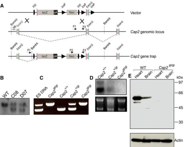
To generate mice lacking the Cap2 gene, we used targeted ES cells (JM8.N4) containing an insertional gene trap, which were obtained from the EUCOMM consortium, Helmholtz Zentrum München, Munich, Germany. ES cell clone D07, which was used in this study, represented a gene-trap that could terminate the transcription of the endogenous gene through altered splicing (Fig. 3.1 A). The gene- trap (gt) cassette was inserted in intron 2 of the mouse Cap2 gene on chromosome 13.
Alternative splicing of the Cap2gt allele generates a new transcript that is a fusion of exon 2 and the LacZ reporter. The fusion protein encodes the first 40 amino acids of CAP2, which are unlikely to show any function mediated by full-length CAP2. We confirmed clones carrying the homologous recombination event with Southern blot analysis in which we detected an additional band of 8 kb representing the mutant allele (Fig. 3.1 B).
We obtained Cap2gt/g mice by mating Cap2+/gt male and Cap2+/gt female from clone D07. Additionally, we also obtained Cap2+/gt male and Cap2+/gt female, which were generated from a different gene-trap clone (B08) at EUCOMM, Munich, Germany.
PCR on genomic DNA from tail biopsies was performed with animals, which confirmed the genotype of Cap2gt/gt mice showing a single band of 800 bp (Fig. 3.1 C).
All phenotypes were confirmed with both mouse lines obtained from the two independent clones. We also carried out Northern blot analysis to confirm the mutant and to rule out any possibility of generation of aberrant transcripts. An N-terminal probe (1–671 bp of Cap2 cDNA) showed the expected transcripts at 3.6 and 3.2 kb in WT as previously reported (Peche et al., 2007). The amounts were reduced in Cap2gt/+
mice and no transcripts were observed in Cap2gt/gt mice (Fig. 3.1 D). The successful inactivation of the Cap2 gene was confirmed by Western blot analysis where we probed heart and brain lysates obtained from Cap2gt/gt mice and their wild-type (WT) littermates with CAP2-specific polyclonal antibodies (Peche et al., 2007). In lysates from WT brain and heart, a signal at ~56 kDa was detected; no protein was seen in lysates from Cap2gt/gt mice (Fig. 3.1 E). When we probed the blot for expression of CAP1, we did not detect significant up-regulation upon loss of CAP2 excluding the possibility that CAP1 compensates for the deficiency (data not shown) (Peche et al., 2013).
Figure 3.1: Targeting strategy for Cap2gt/gt generation. Schematic representation of CAP2 targeting. A) The knockout vector consists of the lacZ gene as a reporter and the neomycin phosphotransferase gene. Genomic locus of the Cap2 gene depicting exon 2, 3, and 4. Transcripts initiated at the endogenous promoter are spliced from the splice donor (green) of an endogenous exon (exon 2 and exon 3) to the splice acceptor (purple) of endogenous exons (exon 3 and exon 4). Homologous recombination gave rise to a gene trap of CAP2 (3′ LoxP missed). Transcripts shown as gray dotted line initiated at the endogenous promoter are spliced from the splice donor of endogenous exon 2 and the splice acceptor of lacZ cassette (diagram not drawn to scale). P1, P2, and P3 are the primers used for genotyping of mice. B) Southern blot analysis of Bam HI digested genomic DNA. Hybridization of radioactively labeled CAP2 probe results in detection of the 10-kb fragment of the WT genomic locus. After the homologous recombination event, restriction with Bam H1 enzyme gave rise to an additional fragment of 8 kb. C) PCR analysis for genotyping. PCR was performed using primers mentioned in the Materials and methods section for genotyping the animals. The WT allele gave a product of ~550 bp (P1 and P3) while the mutant allele gave a product of ~800 bp (P2 and P3). D) Northern blot analysis. 10 μg of RNA from hearts of WT, Cap2gt/+ and Cap2gt/gt was separated on a 1 % agarose gel in the presence of formaldehyde (6 %). The resulting blot was probed with a probe corresponding to nucleotides 1–671 of the mouse CAP2 cDNA. E) Western blot analysis using WT and
Cap2gt/gt heart and brain lysates. Proteins of heart and brain lysates were separated on
SDS-PAGE (10 % acrylamide) and transferred onto a nitrocellulose membrane. The blots were probed with anti-CAP2 polyclonal antibodies. No protein was detected in
Cap2gt/gt,whereas in WT lysates the protein was detected at ~56 kDa. Actin was used
as a control (taken from Peche et al., 2013).
3. Results 23
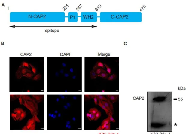
3.2 Characterization of CAP2 monoclonal antibodies
For a further analysis of CAP2 distribution at the protein level, apart from the polyclonal antibodies that we have previously used, we generated monoclonal antibodies using a bacterially expressed polypeptide corresponding to the N-terminal domain of CAP2 (amino acids 1-310) as an antigen (Fig. 3.2 A). mAb K82-381-1 detected the protein in immunofluorescence analysis in nuclei of HaCaT cells and as an
~56 kDa protein in western blots (Fig. 3.2 B,C). This part of work was done with Dr.
med. dent, Ali Eskandarnaz and Arya B Khorsandi.
Figure 3.2: Characterization of CAP2 monoclonal antibody K82-381-1. A) Schematic representation of CAP2 protein domains depicting the polypeptide against which monoclonal antibody K82-381-1 was raised. B) HaCaT cells were stained with mAb K82-381-1, which recognizes nuclear and cytosolic CAP2. Nuclei were stained with DAPI. Scale bars, 10 µm. C) Homogenate of adult mouse heart muscle was separated by SDS-PAGE (10% acrylamide) and the blot probed for CAP2 presence with K82-381-1. The asterisk indicates a degradation product.
3.3 CAP2 deletion leads to weight loss and is lethal in postnatal stages of mice The notion that the size of Cap2gt/gt mice at birth appeared smaller prompted us to follow the body weight. An average weight reduction of approximately 30–40% was consistently observed in mutant females (Fig. 3.3 A, B). CAP2 deficiency appeared to manifest shortly after birth, as during development there was no significant difference
in the size of the embryos (data not shown). For male mice, we also noted a lower body weight with an average weight reduction of 40–45% compared to their WT littermates (Fig. 3.3 C, 40 days of age, n = 8). The survival rates in the Cap2gt/gt mice differed from the one in WT mice and Cap2gt/gt died earlier. This phenotype was more drastic in males compared to females as 25 out of 40 Cap2gt/gt males died between 1 and 70 days after birth. The remaining 15 animals were still alive after 70 days (Fig. 3.3 D).
Analyses of Cap2gt/gt embryonic stages revealed that mutant mice did not die during embryogenesis. This was also underlined by the Mendelian ratio in which the animals were born (25% WT, 50% Cap2+/gt, 25% Cap2gt/gt) (Peche et al., 2013).
Figure 3.3: Inactivation of Cap2 leads to weight loss and reduced survival. A) Overall appearance of WT and Cap2gt/gt mice aged 40 days. Reduced body length and leanness can be seen in Cap2gt/gt mice. B, C) Body weight of mice of different genotypes and gender shows a reduction for Cap2gt/gt mice (WT/Cap2gt/gt females: n = 5/8, WT/Cap2gt/gt males: n = 7/7). D) Percent survival versus age in days for WT (male + female, n = 86) versus Cap2gt/gt female (n = 47) and Cap2gt/gt male (n = 32)
3. Results 25 mice. 70+ days survival was monitored in Cap2gt/gt female and Cap2gt/gt male. Only 40 % Cap2gt/gt male and 87 % of Cap2gt/gt female survived over 70 days in comparison to WT animals (99% survival) (from Peche et al., 2013).
3.4 Cardiac and skeletal muscle phenotype of Cap2gt/gt mice 3.4.1 Cap2gt/gt mice develop dilated cardiomyopathy
In the following we examined the consequences of the deletion of CAP2 in detail. This work was done with Dr. Vivek Peche. For both sexes, we observed a reduction in weight at any given time point. The animals were fertile and female mice showed a life span up to 12–14 months (n = 24) after which survival decreased rapidly. Autopsy revealed gross morphological differences between Cap2gt/gt and their control littermates. Cap2gt/gt male and female hearts were characterized by drastic enlargement of ventricles, which was consistently observed in all mice from 40 days onwards.
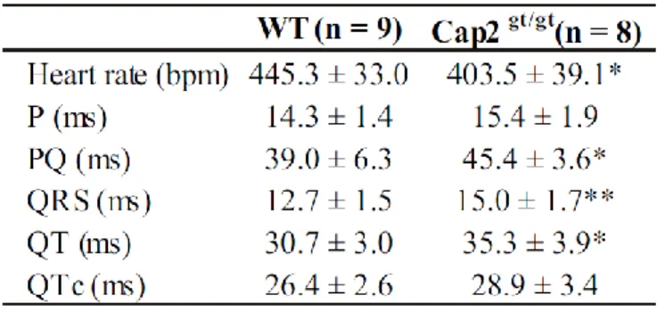
Interestingly, all of the Cap2gt/gt mice that died between P1 and P70 also showed an enlarged right and left ventricle. H & E staining, a two-stage stain for cells in which hematoxylin is followed by a counterstain of red eosin so that the nuclei stain a deep blue-black and the cytoplasm stains pink, was applied in cardiac sections and confirmed the dilation of the ventricles (Fig. 3.4 A-D; Table 3.1). Consequently, the total area of the right ventricular chamber was also increased significantly in Cap2gt/gt mice (Fig. 3.4 E). In addition, we noticed a thinning of the ventricular myocardium compared to the total area (Fig. 3.4 F). Dilated cardiomyopathy is often associated with abnormalities in electrical conductivity of the heart. To check conductivities in mutant hearts, we performed surface electrocardiography (Table 3.2). Nine WT (five male, four female) and eight mutant (four male, four female) animals were used in surface ECG recordings. The surface ECG showed a significantly decreased heart rate in
Cap2gt/gt . With decelerated heart rate, we also observed a significantly prolonged PQ
interval at equal P-wave length in Cap2gt/gt mice, which can be attributed to negative dromotropic effects correlated with slower heart rate. In the Cap2gt/gt mice, the parameters for atrio-ventricular conduction time (PQ time) as well as intraventricular conduction times (QRS time and QT time) showed marked prolongations compared to WT (Table 3.2; Fig. 3.4 G). After correction for the heart rate, the QTc did not differ between the groups.
Proliferation of interstitial fibroblasts and biosynthesis of extracellular matrix components in the heart are defined as cardiac fibrosis. It is a consequence of remodeling processes initiated by pathologic events associated with a variety of
cardiovascular disorders, which leads to abnormal myocardial stiffness and, ultimately, ventricular dysfunction (Tamura et al., 2000). Staining with Masson’s trichrome on transverse cardiac sections of 2-month-old mice revealed no symptoms of fibrosis in Cap2gt/gt mice (data not shown), but at the age of 6 months we could clearly observe fibrosis in the ventricles of Cap2gt/gt mice whereas this was not the case in their WT littermates (Fig. 3.4 H). As an increase in fibrosis might be associated with increased apoptosis, we performed caspase 3 staining on cardiac sections (three male, one female; 2–6 months old) and found that mutant myocardium had significantly higher numbers of apoptotic cells than WT (WT, 0.12% cells; Cap2gt/gt , 0.94% cells;
p < 0.0005). Also, caspase 3-positive cells were not restricted to any particular region of the myocardium (Fig. 3.4 K). In general, apoptosis was more prominent in failing hearts.
To investigate embryonal heart development and the possibility of development of cardiomyopathy/cardiac defects during embryogenesis, embryos between E11-E15 were studied. Whole-mount analysis revealed that embryos did not show obvious external abnormalities. Similar to their WT littermates, at E13.5 cardiac chamber formation was observed in Cap2gt/gt mice (Fig. 3.4 I). The cardiac ventricular walls of the Cap2gt/gt were slightly thinner than those of the control embryos; the ventricular myocardium of control and Cap2gt/gt appeared normal (Fig. 3.4 I). Thus, overall heart development appeared to be not severely affected during embryogenesis of Cap2gt/gt mice. At age P4, mutant hearts exhibited dilated atria and mildly dilated ventricles (Fig. 3.4 J). This underlines our previous finding that CAP2 is expressed in all four chambers and is responsible for physiological functioning of the atria and ventricles, which ultimately govern the heart performance (Peche et al., 2013).
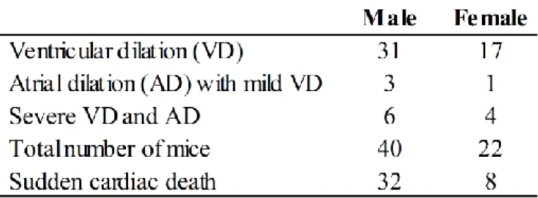
Table 3.1: Gross morphological cardiac defects observed in Cap2gt/gt mice.
3. Results 27 Table 3.2: Surface ECG parameters.
QTc rate corrected QT time
Figure 3.4: Characterization of cardiac phenotypes of Cap2gt/gt mice. Histological analyses of 2-month-old mice. Representative images of transverse (A,B) and longitudinal (C,D) sections of WT and Cap2gt/gt mice stained with H&E. The mutant exhibited an enlarged ventricular chamber. Scale bars, 1 mm. E) The relative right ventricular area was also increased in Cap2gt/gt mice as compared to their WT