roles in cell differentiation, proliferation and chromatin association
Inaugural-Dissertation zur
Erlangung des Doktorgrades
der Mathematisch-Naturwissenschaftlichen Fakultät der Universität zu Köln
vorgelegt von
Rashmi Rudrappa Nanjundappa aus Birur, Indien
2011
Berichterstatter: Prof. Dr. Angelika A. Noegel
Berichterstatter: Prof. Dr. Siegfried Roth
Tag der mündlichen Prüfung: 13. 07. 2010
Acknowledgment III
List of Figures VI
List of Tables VII
List of Abbreviations XI
1 Introduction 1
1.1 Nucleus and Nuclear envelope . . . . 1 1.2 Nesprins - their roles in human disease . . . . 3
1.2.1 Structural composition, binding partners and role of Nesprin- 2 at the nuclear envelope . . . . 5 1.3 Cell migration and wound healing . . . . 8 1.3.1 Phases of wound healing . . . . 8 1.4 Role of nuclear envelope proteins in transcription regulation and cell
proliferation . . . . 12 1.5 Aim of the research . . . . 13
2 Results 15
2.1 Transcriptional profiling in Nesprin-2 knockout fibroblasts . . . . 15 2.2 In vivo wound healing study in Nesprin-2 knock out mice . . . . 20
2.2.1 Macroscopic observation showed a delay in wound healing in Nesprin-2 KO mice as compared to wild type . . . . 20 2.2.2 The distance between the epidermal tips is altered in Nesprin-
2 KO wounds . . . . 21 2.2.3 Inflammatory phase-
study of Macrophages . . . . 25 2.2.4 New tissue formation and tissue remodelling phase -
Study of keratinocytes migration, proliferation and fibroblast differentiation . . . . 29 2.3 The transcriptional regulation of genes involved in wound healing is
altered in Nespin-2 Giant knock out mice . . . . 41
2.4 c-Fos localization in Nesprin-2 KO and WT epidermis . . . . 44
2.5 Nesprin-2 regulates the localization of c-Fos in human keratinocytes 46
2.6 Localization of c-Fos in cell scratch assay . . . . 47
2.7 Apoptosis in Nesprin-2 KO cultured fibroblasts . . . . 49
2.8 Caspase-3 in Nesprin-2 KO fibroblasts . . . . 51
2.9 The Localization of SAFB1 has changed in Nesprin-2 KO fibroblasts 51 2.10 Nesprin-2 Giant binds to heterochromatic DNA . . . . 54
3 Discussion 59 3.1 Transcriptional profiling in Nesprin-2 KO and WT fibroblasts . . . . 59
3.2 The wound healing process is delayed in Nesprin-2 KO mice . . . . . 60
3.3 Nesprin-2 regulates the cell proliferation and differentiation under the wound healing situation by affecting transcription factors . . . . 62
3.4 A disturbed F-actin network can cause the impaired myofibroblast differentiation in Nesprin-2 KO mice . . . . 66
3.5 Fate of the cells which showed nuclear deformation in Nesprin-2 KO mice (an apoptotic approach) . . . . 67
3.6 Nesprin-2 interacts with heterochromatin and associates with chro- matin . . . . 68
3.7 Model for Nesprin-2 Giant function in wound healing . . . . 69
4 Materials and Methods 72 4.1 Kits and Reagents . . . . 72
4.2 Oligonucleotides . . . . 75
4.3 Enzymes and Antibodies . . . . 76
4.4 Media, Buffers and solutions . . . . 77
4.5 Nesprin-2 Giant knock out mice . . . . 79
4.6 Isolation of primary fibroblasts . . . . 80
4.7 Wounding . . . . 80
4.8 Wound tissue harvesting and sectioning . . . . 80
4.9 Histological staining . . . . 81
4.10 Immunohistochemistry and Immunofluorescence . . . . 82
4.11 Western blotting . . . . 82
4.12 RNA isolation and RT-PCR analysis . . . . 83
4.13 Cell culture and transfection . . . . 85
4.14 Chromatin immunoprecipitation assay (ChiP) . . . . 85
4.15 Microarray analysis . . . . 86
Bibliography 91
Abstract i
Zusammenfassung iii
Erklärung v
‘Gratitude is the memory of the heart’
I would like to express my deep and sincere gratitude to my supervisor, Prof. Dr.
A. A. Noegel. Her wide knowledge and her logical way of thinking have been of great value for me. Her understanding, encouraging and belief in my strength have provided a good basis for the successful completion of PhD thesis.
I avail this opportunity to express my deep sense of gratitude to other members of my thesis committee Prof. Dr. S. Roth, Institute of Developmental Biology and Prof. Dr. T. Wiehe, Institute of Genetics, University of Cologne.
I wish to express my warm and sincere thanks to Prof. Dr. Ludwig Eichinger, Dr. Iakowos Karakesisoglou, Dr. Beate Eckes (Dept. of Dermatology, Cologne), Dr. Gernot Glöckner (Berlin Center for Genomics in Biodiversity Research, Berlin) and Dr. Marco Groth (Leibniz Institute for Age Research - Fritz Lipmann Insti- tute, Jena) for their detailed and constructive comments, for the valuable advice and friendly help.
I am very much thankful to Rosi, Maria, Martina, Berthold, Rolf,Sonya, Brigitte and Bärbel for their valuable help and co-operation in the lab. My special thanks to Dörte Püsche who helped me all the time to complete and solve official matters.
I also thank Budi, Gudrun and Roman who helped me to solve computer related problems whenever I needed.
I am privileged for having Anja, Bhagyashri, Eva, Georgia, Janbo, Karl-Heinz,
Karthik I, Karthik II, Liu, Margit, Martin, Raphael,Sandra, Sascha, Shahrzad,
Surayya, Vivek, Tanja, Xin and Yvonne as my colleagues who have provided me great help and company during all these years in Biochemistry Institute, Cologne.
I also thank all the members of Ludwig’s group for making me to feel comfortable while doing my experiments in their lab. I should not forget to thank Ralf Hallinger who helped me a lot during wound healing analysis, thanks a lot Ralf. I am very much thankful to Alex Friede for her great help in using the Latex programme for writing my thesis. I also take this opportunity to thank all the members of AG.
Huelskamp who were always very friendly and made me feel as a part of their group.
I thank all my Kannadiga friends in Germany, I thank Nikki, Manoj and Ashish and other friends with whom I had wonderful time. I also thank my all other friends in India and other parts of the world.
My parents deserve special mention for their constant support, encouragement and selfless sacrifice. Without their love and blessings I would not have been what I am today. Thank you Amma and Appa for everything you have done for me. I dedi- cate all my success to you both. I remember with great respect and love my sister and her family and also my brother. My in laws deserve special place in my heart for their invaluable inspiration and love. Without their support this study would have scarcely been accomplished.
One more important person in my life is my husband Rachu, who is guiding and inspiring me all the time. I cannot imagine my personal and professional life with- out his love,care and encouragement.Thank you rachi.
Last but not the least I thank all the people who directly or indirectly helped me during my PhD work.
Vielen Dank an alle für alle
1.1 Schematic representation of a eukaryotic cell . . . . 2 1.2 Nesprin-2 localization and its interaction partners in mammalin cells 5 1.3 Structural features of Nesprin-2 Giant . . . . 8 1.4 Phases of wound healing . . . . 10 2.1 Significance analysis of microarray (SAM) plot . . . . 16 2.2 Classification of differentially regulated genes based on GOAT . . . . 19 2.3 Macroscopic analysis of wound closure. . . . 22 2.4 Measurement of distance between the migrating tips. . . . 24 2.5 Comparison of macrophage population in WT Nesprin-2KO mice. . . 26 2.6 The expression of the inflammation related gene SAA3 is increased
in Nesprin-2 KO wounds. . . . . 27 2.7 The expression of chemokine MCP1 (CCL2) in the wounds of WT and
Nesprin-2 KO. . . . 28 2.8 The keratinocyte migration pattern during wound healing process. . 31 2.9 Observation of keratinocyte proliferation in the wounds. . . . 33 2.10 Differentiation of fibroblasts into myofibroblasts during the wound
healing process. . . . 35 2.11 Study of F-actin in primary dermal fibroblasts. . . . . 37 2.12 Nesprin-2 Giant maintains actin fibers around the nucleus in pri-
mary fibroblasts. . . . . 39 2.13 F-actin distribution and localization in shRNA knock down HaCaT
cells. . . . . 40 2.14 Sirius red staining showing the granulation tissue area. . . . . 41 2.15 Transcription factors are differentially regulated in Nesprin-2 KO
wounds. . . . 43 2.16 c-Fos localization at various stages of wound healing in Nesprin-2
KO and WT mice. . . . 45 2.17 The localization and expression of c-Fos in knock down HaCaT cells. 48 2.18 c-Fos localization in control and Nesprin-2 KD HaCaT cells after
scratching. . . . 50 2.19 Apoptosis study in Nesprin-2 KO and WT fibroblasts. . . . 52 2.20 Caspase-3 in WT and Nesprin-2 KO fibroblasts. . . . . 53 2.21 Scaffold attachment factor-B1 (SAFB1) in WT and Nesprin-2 KO
fibroblasts. . . . 55
2.22 Heterochromatin HP1𝛽 distribution in WT and Nesprin-2 KO fibrob-
lasts. . . . 58
3.1 Model for Nesprin-2 Giant function in wound healing . . . . 70
4.1 Principle of microarray . . . . 90
4.2 Overview of the research findings . . . . ii
2.1 List of differentially regulated genes from microarray analysis . . . . 16 2.2 List of up-regulated inflammation related genes from microarray
analysis . . . . 26 2.3 Chip-assay data showing the presence of centromere sequences in
the ChIP carried out with mAb K20-478 . . . . 57
2.4 Quantification of the number of HP1 𝛽 stained speckles. . . . . 58
𝛼-SMA alpha smooth muscle actin ABD actin binding domain
AD-EDMD autosomal dominant EDMD
AO acridine orange
AP1 activating protein 1
BAF barrier to autointegration factor BMP bone morphogenic protein
BSA bovine serum albumin
BTF BCL2 the associated transcription factor cDNA complementary deoxynucleiacid
CH calponin homology
ChIP chromatin immunoprecipitation DAPI 4’,6-diamidino-2-phenylindole DAW / D days after wounding
DEPC diethylpyrocarbonate DMSO dimethyl sulfoxide DNA deoxyribonucleic acid
DR death receptor
DTT 1, 4-dithiothreitol
EB ethidium bromide
ECL enhanced chemiluminescence
EDMD emery Dreifuss muscular dystrophy
EDTA ethylenediaminetetraacetic acid Egr-1 early growth response factor 1
EGTA ethylene-glycol-bis(2-aminoethylether)-N, N, N, N-tetraessigsacid EMR EGF module containing mucin like hormone receptor
ER endoplasmic reticulum
ERK extracellular signal-regulated kinases
EtOH ethanol
FasL fas ligand
FBS fetal bovine serum
FN fibronectin
GCL germ-cell-less
GO gene Ontology
GOAT gene Ontology Annotation Tool
GT granulation tissue
H&E haematoxylin and Eosin HaCaT human keratinocyte HDL high-density lipoprotein
HEPES N-(2-hydroxethyl)piperazine-N´-2-ethanesulphonic acid HMW high molecular weight marker
HP1𝛽 heterochromatin protein 1 beta
HRP horse peroxidase
IF intermediate filament INM inner nuclear membrane
IPTG isopropyl 𝛽-D-thiogalactopyranoside
K14 keratin 14
KASH Klarsicht ANC-1 SYNE homology
KD knock down
kDa kilodalton
KO knock out
LAP lamina associated proteins
LINC linker of nucleoplasm and cytoplasm LMW low molecular weight marker
MAPK mitogen-activated protein kinase MCP1 monocyte chemoattract protein 1 MEFs mouse embryonic fibroblasts
MOPS 3-(N-morpholino)propanesulfonic acid MTOC microtubule organizing center
NE nuclear envelope
NETs nuclear envelope transmembrane proteins NPC nuclear pore complexes
OCT optimum cutting temperature ONM outer nuclear membrane
PBG Phosphate buffer with fish gelatin PBS Phosphate buffer solution
PC Panniculus carnosus
PDGF platelet-derived growth factor
PFA paraformaldehyde
PKC Ca
2+induced protein kinase C PMSF phenylmethylsulphonylfluoride
PPAR𝛽/𝛿, Peroxisome proliferator-activator receptor 𝛽/𝛿, q-PCR Quantitative polymerase chain reaction
RNA Ribonucleic acid
SAA3 Serum Amyloid A3
SAFB Scaffold attachment factor-B
SAM Significance analysis of microarray SDS sodium dodecylsulfate
TAFII68 TATA Element-binding Protein-associated Factor TBP TATA box binding protein
TEMED Tetramethylethylenediamine TGF transforming growth factor
TRAIL TNF-related apoptosis-inducing ligand
WT Wild type
1.1 Nucleus and Nuclear envelope
The eukaryotic cell is enclosed by a plasma membrane and contains several
membrane-bound organelles with specific functions. The nucleus is the largest
membrane bound organelle encompassing about 10% of the cell volume. It is
sometimes referred to as the control centre of the cell as it harbors most of the
genetic material of the cell in the form of chromosomes formed as a result of
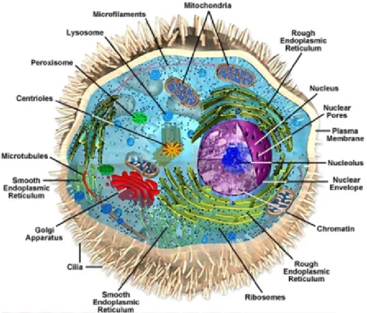
condensation of the DNA in complex with histone proteins (Figure 1.1). The
nucleus is physically separated from the cytoplasm by a membrane barrier called
nuclear envelope NEnuclear enevelope. The NE is a double lipid bilayer forming an
inner (INM) and outer nuclear (ONM) membrane the later of which is continuous
with the endoplasmic reticulum (ER). Nuclear pore complexes (NPC)consisting of
approximately 30 different proteins called nucleoporins are inserted into the NE
and serve as channels for the trafficking of proteins, RNA and ribonucleoprotein
complexes between nucleoplasm and cytoplasm (Hetzer et al., 2005; Tran and
Went, 2006; D’Angelo and Hetzer, 2008). Nuclear pore channels typically have
a ∼100 nm outer diameter and ∼40 nm as central transport channel (Beck et
al., 2004; Beck et al., 2007; Teryy et al., 2007). The nuclear membranes contain
specific proteins, the nuclear envelope proteins. Furthermore, certain proteins are
specifically localized either to the INM or ONM. Some well known proteins residing
in the INM are Lamina associated proteins (LAP1, LAP2), Emerin, SUN domain
Figure 1.1: Schematic representation of a eukaryotic cell. Every cell is enclosed by a selectively permeable membrane called the plasma membrane. It contains ribosomes, centrioles, microtubules, microfilaments, and membrane bound organelles like the endo- plasmic reticulum, Golgi apparatus, lysosomes, the nucleus and mitochondria. These or- ganelles perform different functions with in the cell. Figure adopted from Google image (Glogster beta).
containing proteins and MAN1 (Akhtar and Gasser, 2007; Dorner et al., 2007;
Schirmer and Foisner, 2007) whereas Nesprin-1 and -2 are localized at the ONM and INM. INM specific proteins have important roles in gene expression, chromatin organization and DNA metabolism (Mattout et al., 2006; Heessen and Fornerod, 2007; Reddy et al., 2008), while ONM specific proteins are involved in nuclear positioning which is important for processes like cell polarization, pronuclear migration, and syncitia organization (Fridkin et al., 2009).
The inner nuclear membrane (INM) contains a subset of integral membrane
proteins, termed nuclear envelope transmembrane proteins (NETs) (Schirmer et
al., 2003). ONM and INM are separated by a luminal space of ∼100 nm in width
and are fused through NPCs. Beneath the INM towards the nucleoplasm the nuclear lamina is located, composed of type V intermediate filament (IF) proteins called lamins. The proteins of the lamina are A, C and B type Lamins (Fisher et al., 1986; McKeon et al., 1986). The nuclear lamina is thought to provide a frame work for organizing the NE structure and providing an anchoring site at the nuclear periphery for interphase chromatin (Gerace et al., 1978; Hancock et al., 1982; Lebkowski and Laemmli 1982).Mutations in lamina associated proteins lead to a variety of diseases known as Laminopathies, indicating the importance of the lamina in the development of different tissues.
1.2 Nesprins - their roles in human disease
Nesprins (Nuclear envelope spectrin repeat proteins) are ubiquitously expressed proteins. They contain an N-terminal F-actin binding domain (ABD), a long spec- trin repeat containing region and a C-terminal transmembrane domain followed by a short region extending into the perinuclear space (KASH). Together with SUN domain containing proteins they form the LINC (linker of nucleoplasm and cytoplasm) complex (Figure1.2). Through alternative transcriptional initiation, termination and splicing, the two Nesprin genes Syne1 (Nesprin-1) and Syne2 (Nesprin-2) give rise to many isoforms, which vary markedly in size. The largest isoforms of Nesprins are Nesprin-1 Giant and Nesprin-2 Giant which are also called as ENAPTIN and NUANCE, respectively (Zhen et al., 2002; Padmakumar et al., 2004). Nesprin-1 (1.01 MDa) shares 46% sequence identity and 59% homology with the C-terminal region of Nesprin-2 (796 kDa) (Mislow et al., 2002). Nesprins have single giant orthologues in both Drosophila (MSP300) and Caenorhabditis elegans (ANC-1). MSP300 is a cytoskeleton protein that localizes to the Z-line in muscle and therefore mutation in MSP300 causes muscle defect (Zhang et al., 2002;
Rosenberg-Hasson et al., 1996; Volk, 1992). The C. elegans protein ANC-1 is local-
ized in the ONM via its KLS/KASH domain (klarsicht/ANC-1 (anchorage 1)/SYNE homology). Its role in nuclear migration requires both the KLS/KASH domain and CH domain, which binds to F-actin (Starr and Han, 2002).
Nesprin-1 and Nesprin-2 interact with Lamin A/C of the nuclear lamina (Mislow et al., 2002; Libotte et al., 2005). Mutations in the Lamin A/C encoding gene LMNA resulted in a diverse range of clinical syndromes including partial lipodys- trophy, Charcot-Marie-Tooth type 2 (CMT2) neuropathy, Hutchinson-Gilford proge- ria, an autosomal-dominant form of Emery-Dreifuss muscular dystrophy (EDMD) and many more, which are collectively referred to as Laminopathies (Broers et al., 2004; Burke et al., 2001; Gruenbaum et al., 2005; Lloyd et al., 2002).
A mutation in Nesprin-1 and Nesprin-2 can cause human diseases which are sim-
ilar to Laminopathies. Patients with the R374H missense mutation in Nesprin-
1𝛼 developed dilated cardiomyopathy. Mice lacking the C-terminal KASH domain
showed lethality with approximately half of the animals dying at or near birth
from respiratory failure and surviving mice exhibited an EDMD like phenotype
(Puckelwartz et al., 2009; 2010). Fibroblasts from patients with EDMD disease
showed a disturbance of the Nesprin/Lamin/Emerin interaction. In Nesprin-2 KO
fibroblasts Emerin failed to distribute properly along the NE and often formed ag-
gregates in the deformed NE areas. This indicates the involvement of Nesprin-1
and Nesprin-2 in the pathogenesis of EDMD, which was earlier considered as a
consequence of mutations in Emerin and Lamin A/C (Lüke et al., 2008;Wheeler et
al., 2007; Zhang et al., 2007). Disruption of the endogenous LINC complex leads
to mechanical stiffness, which is similar to the phenotype caused by a lack of Lam-
inA/C (Hutchinson et al., 2008). Nesprin-2 knockout (KO) primary dermal fibrob-
lasts and keratinocytes exhibit heavily misshapen nuclei displaying a significant
similarity to nuclear deformations of Laminopathies. Furthermore Nesprin-2 was
shown to act as a structural reinforcer at the NE by safeguarding its architecture
in LMNA mutant (S143F) progeria cells (Lüke et al., 2008; Kandert et al., 2007).
These studies showed the possible involvement of Nesprins in diseases.
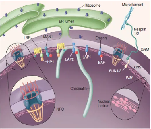
Figure 1.2: Nesprin-2 localization and its interaction partners in mammalin cells.
Nesprins-2 present at the ONM binds to SUN domain containing proteins through which it connects with the nuclear lamina. Short isoforms of Nesprin-2 have been found in the nucleoplasm. At the cytoplasmic face of the nucleus, Nesprin-2 binds to actin filaments and acts as a Linker of nucleoplasm and cytoplasm (LINC) (Stewart et al.,2007).
1.2.1 Structural composition, binding partners and role of Nesprin-2 at the nuclear envelope
Nesprin-2 at the ONM connects the nucleoplasm with the cytoplasm. To execute this function it uses its C-terminal KASH domain and N-terminal actin binding domain (ABD). Nesprin-2 giant, the largest Nesprin-2 isoform, has a molecular weight of 796 kDa, its short C-terminal isoforms are ranging from 48 to 377 kDa.
A Nesprin-2 Giant knock out (Nesprin-2 ∆ABD) mouse was generated by deleting
the calponin homology (CH1) domain 1 which together with the CH2 domain forms the functional ABD (Gimona et al., 2002; Figure 1.3). Sun proteins are highly conserved proteins present in yeast, Dictyostelium and C. elegans and human.
They are essential components of the LINC complex. The mechanisms that keep Sun proteins at the NE vary. For example, UNC84 in C. elegans and mammalian Sun-2 require Lamin binding, whereas the Sun-1 association with the NE is Lamin independent (Lee et al., 2002; Padmakumar et al., 2005; Haque et al., 2010).
For the Dictyostelium homolog a chromatin association is responsible for targeting Sun-1 to the INM (Xiong et al., 2008). Interactions with chromatin or chromatin associated proteins are also known for further NE proteins such as Lamin A/C or Emerin, which can affect gene expression (Heesen and Fornerod, 2007). For Lamin A, a direct interaction with transcription factor c-Fos was reported, whereas the lamina-associated protein 2𝛼 (LAP2𝛼) is involved in Retinoblastoma protein (Rb) activity (Dörner et al., 2006). LAP2𝛽 acts more indirectly and represses transcription through interaction with HDAC3 followed by histone deacetylation (Somech et al., 2005). Overall, the role of the NE in several of these processes may be one in which transcription factors are sequestered to the NE and by providing a surrounding, which may be inhibiting or activating gene expression (Towbin et al., 2009).
Previous data from Nesprin-2 knock out (KO) studies have shown that Nesprin-2 is an important scaffold protein in the maintenance of nuclear envelope architecture.
The loss of Nesprin-2 in dermal fibroblasts and keratinocytes showed misshapen and blebbing nuclei and an increase in the size of the nuclei causing the thickening of the epidermis and indicating its key role in the maintenance of nuclear morphology. Loss of Nesprin-2 Giant also showed a requirement for Nesprin-2 in the migration of dermal fibroblasts as the KO fibroblasts exhibited a significantly lower migration speed compared to wild type cells after 20 hours of cells scratching.
They exhibited also a defective cell polarity with respect to the Golgi complex and
microtubule organising center at the wound edge (Lüke et al., 2008). The role of Nesprin-2 Giant in other cell types of the skin tissue is not clear. Therefore we carried out experiments to analyse other cell types in the Nesprin-2 KO and test whether they are also impaired in migration. We conducted an in vivo wound healing assay and showed that Nesprin-2 is required for keratinocyte proliferation and differentiation and ultimately for healing of the wound.
Nesprin-2 Giant carries a F-actin binding site which was previously shown to be functional in vitro (Zhen et al., 2002). F-actin structures are involved in many cellular processes, including cell adhesion, migration and division. Reorganisation of the F-actin cytoskeleton is critical for different steps of cell migration, including those essential for cell protrusion, adhesion and shape change (Stricker et al., 2010). These studies have clear indication of potential role of F-actin during the wound healing process. During my PhD work, I also studied the F-actin cytoskeleton in Nesprin-2 KO fibroblast.
In general, the giant isoform of Nesprin-2 is thought to localize at the ONM
whereas the shorter isoform Nesprin-2𝛼 can enter the INM by diffusion through
the nuclear pores (Worman and Gundersen, 2006). However, there are also reports,
that Nesprin-2 Giant is present in the INM (Zhen et al., 2002; Libotte et al.,
2005). The functional relevance of Nesprin-2 Giant localization in the INM has
not been studied so far. Here I propose that Nesprin-2 Giant is associated with
chromatin thereby regulating the transcription of genes involved in the wound
healing process. I made an attempt to study the transcriptional regulation of genes
related to wound healing by Nesprin-2 Giant using the corresponding knock out
mice.
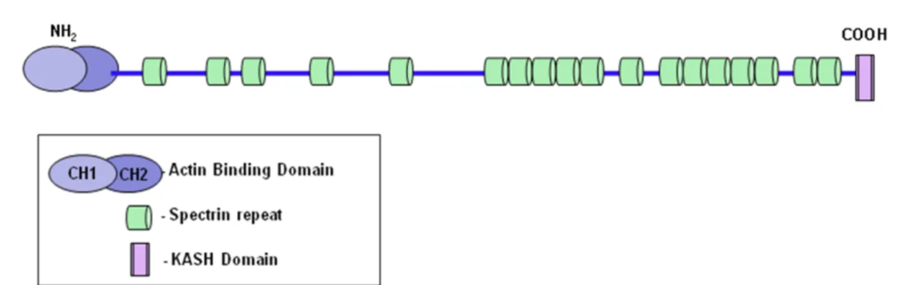
Figure 1.3: Structural features of Nesprin-2 Giant . Nesprin-2 Giant is composed of an N-terminal actin binding domain which is formed by two Calponin Homology domains, CH1 and CH2, followed by several spectrin. Not all of the 56 predicted spectrin repeats are indicated. At its C-terminal end it consists of the highly conserved KASH (Klarsicht, ANC- 1, Syne Homology) domain. This KASH domain anchors the protein at the NE (modified from Lüke et al., 2008).
1.3 Cell migration and wound healing
The process of healing is an immediate response to tissue injury, which involves different overlapping phases. During this process several cell types will become activated and act to heal the wound.
1.3.1 Phases of wound healing
The process of wound healing normally proceeds from coagulation and inflamma-
tion through fibroplasia, matrix deposition, angiogenesis, epithelialisation, colla-
gen maturation and finally wound contraction. These processes are divided into
three different but overlapping phases, namely the inflammatory phase, new tis-
sue formation phase and finally tissue remodelling phase (Schäfer and Werner,
2008).
A. Inflammatory phase
Following tissue injury, a wound must stop bleeding in order to heal and for the injured host to survive. Therefore cellular and molecular elements involved in haemostasis also signal tissue repair. Immediately after wounding or tissue dam- age the platelets activated by thrombin release insulin like growth factor 𝛼 (IGF-𝛼), transforming growth factor 𝛽 (TGF-𝛽) and platelet-derived growth factor (PDGF) which attract leukocytes, mainly macrophages and fibroblasts into the wound area.
In response to tissue damage as a defence mechanism many inflammatory cells are activated and attracted to wound area. Among the inflammatory cells neutrophils enter first followed by mast cells and monocytes, which will subsequently differ- entiate into macrophages. Macrophages play a dominant role in the synthesis of wound healing molecules as coagulation-mediated tissue repair signals fall (Franz, 2009). Neutrophils and macrophages are essential for defence against invading bacteria through their phagocytic function and through their capacity to secrete toxic mediators. Macrophages and monocyte are the source for chemo attractants which are required for the late phase of the wound healing (Figure 1.4 A).
B. New tissue formation phase
The new tissue formation phase is characterised by cellular proliferation and
migration of different cell types. This begins with migration of keratinocytes from
the epidermis at the wound edge and from injured appendages. Keratinocytes at
the leading edge alter the expression of integrin receptors to allow attachment
to new substrates and they express various proteases to allow the degradation of
connective tissue (Martin, 1997). These keratinocytes migrate forward between
fibrin and the dermis. The migration of keratinocytes is followed by their
hyperproliferation. In the later part of this phase, fibroblasts, which are attracted
from the wound edge or from the bone marrow, are stimulated by macrophages.
Figure 1.4: Phases of wound healing. In vivo wound healing occurs in three
overlapping phases - (A) Inflammatory phase (B) New tissue formation phase and (C)
Tissue remodelling phase (taken from Schäfer and Werner, 2008). (for a detailed
description please see text).
Fibroblasts also migrate, proliferate and deposit extracellular matrix and some differentiate into myofibroblasts (Schäfer and Werner, 2008). Myofibroblasts are specialized contractile fibroblasts that play a critical role in generating the contractile force responsible for wound closure and pathological contractures (Tomasek et al., 2002; Hinz et al., 2003). They are characterized by the acquisition of a contractile phenotype and the expression of 𝛼-smooth muscle actin (𝛼-SMA) (Skalli et al., 1986; Desmouliere et al., 1993), which correlates with the generation of contractile force (Hinz et al., 2001; Hinz et al., 2002). An understanding of the regulation of expression of 𝛼-SMA in myofibroblasts will be important in controlling the formation and function of myofibroblasts in wound healing and pathological contractures (Figure 1.4 B).
C. Tissue remodeling phase
During this phase wound re-epithelialization is complete and extracellular matrix
is remodeled. Formation of the new blood vessels is essential for the supply
of oxygen and nutrients. During this phase all of the processes initiated after
tissue damage wind down and cease. Most of the endothelial cells, macrophages
and myofibroblasts undergo apoptosis or exit from the wound area. Then matrix
metalloproteases that are secreted by fibroblasts, macrophages and endothelial
cells, help in strengthening the repaired tissue (Lovvorn et al., 1999). The
remodeled extracellular matrix will result in scar formation. At this phase the
wound will not have any appendages (Schäfer and Werner, 2008) (Figure 1.4 C).
1.4 Role of nuclear envelope proteins in transcrip- tion regulation and cell proliferation
The INM proteins engage in direct chromatin-independent interaction with tran- scription factors and the sequestering of transcription factors to the INM (Heessen and Fornerod, 2007). One important example which has been studied in detail with respect to sequestration of transcription factors to the NE is c-Fos by Lamin A/C. c-Fos together with c-Jun forms the activating protein 1 (AP1), which plays a role in several cellular processes including cell proliferation and differentiation.
Lamin A/C is known to regulate the process of cell proliferation by sequestering c-Fos to the NE (Ivorra et al., 2006). The process of cell proliferation was shown to be enhanced in the absence of Lamin A/C. In human fibroblasts the expression of lamina-associated polypeptide 2 𝛼 (LAP2 𝛼) upon entry and exit from G0 is tightly correlated with phosphorylation and subnuclear localization of the retinoblastoma protein (Rb) (Pekovic et al., 2007). It is not only LaminA/C but also other NE pro- teins which are participating in the regulation of transcription factor and thereby in the process of cell proliferation.
Nearly 15 INM proteins were characterized in mammalian cells so far (Dreger et al., 2003). Among them is MAN1 which is an integral INM protein consisting of a LEM domain, two transmembrane domains at its N-terminus, and a RNA recog- nition motif at the C-terminus. Several groups showed an interaction of MAN1 and Smad transcription factors. Smads are crucial regulators of transforming growth factor-𝛽 (TGF-𝛽), bone morphogenic protein (BMP) and activin signalling.
MAN1 acts as a nuclear scavenger, sequestering R-Smads that illegitimately en-
ter the nucleus (Ishimura et al., 2006). MAN1 also interacts with several other
transcriptional regulators including the transcription factor germ-cell-less (GCL),
the BCL2 associated transcription factor (BTF) and the barrier to autointegration
factor (BAF) (Mansharamani and Wilson, 2005). The INM proteins Emerin and LAP2𝛽 were also found to associate with several transcription regulators. Further- more, Lamin A/C as an important member of the nuclear lamina plays a crucial role in several cellular processes. Kandert et al., (2009) showed that LMNA mutation R545C impairs both the proliferation and differentiation capacities of myoblasts as part of the pathogenesis of AD-EDMD (Autosomal Dominant EDMD). Mutation in Lamin A/C negatively acts on the interaction between Emerin and LaminA/C, and skin fibroblasts carrying this mutation exhibited enhanced cell proliferation, collagen-dependent adhesion, larger number of filopodia and smaller cell spread size compared to control cells. Cell migration, speed and polarization were ele- vated in these cells too. The functional interaction between Emerin and Lamin A/C acts on cell spreading and proliferation through the ERK1/2 signalling pathway (Emerson et al., 2009). LaminA/C is involved in cellular processes under mechan- ical stress situation and affects cellular plasticity. Mouse embryonic fibroblasts (MEFs) deficient in Lamin A/C showed weak cytoplasmic mechanics. These MEFs also showed slower migration to cover the scratched area and the microtubule or- ganizing center (MTOC) was localized away from the scratched area. In wild type cells they are present in the direction of the scratched area showing the importance of LaminA/C in the generation of cell polarity. The distance between MTOC and the nucleus was higher in Lamin A/C deficient cells as compared to wild type cells (Lee et al., 2007).
1.5 Aim of the research
Loss of Nesprin-2 Giant resulted in a thickening of the epidermis as a consequence
of increased epithelial nuclear size; emerin localization was altered and showed in-
creased cytoplasmic staining; primary dermal knockout fibroblast and keratinocyte
nuclei were heavily misshapen displaying a striking similarity to the nuclear defor-
mations characteristic for laminopathies. In vitro wound healing was impaired in mutant fibroblasts, furthermore, fibroblasts showed a polarization defect (Lüke et al., 2008). Studies by other groups revealed that mutations in the human Nesprin- 1 or -2 genes can cause disease states that until now were considered to be a conse- quence of mutations in Lamin or in Emerin (Zhang et al., 2007). The mechanisms of disease involvement are however unclear. Similarly, a role of other NE proteins namely Lamin A/C, Emerin and SUN1 and SUN2 in cell migration and polarity was shown earlier (Lee et al., 2008). However the role of any of these proteins in- cluding Nesprin-2 during in vivo cell migration and tissue repair is not known so far. The first aim our of research is
• To reveal the role of Nesprin-2 in cell proliferation and differentiation during in vivo wound healing.
Based on its presence at the NE and its involvement in regulating nuclear shape, Nesprin-2 has the potential to be involved in regulation of gene expression.
• The next aim of our research is to study the role of Nesprin-2 in regulating the expression of genes which are involved in cell proliferation,
• To study the role of Nesprin-2 in chromatin association,
• To study the role of Nesprin-2 in regulating gene transcription using microar-
ray analysis.
2.1 Transcriptional profiling in Nesprin-2 knock- out fibroblasts
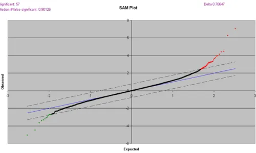
A cDNA microarray analysis was carried out to study transcriptional changes in Nesprin-2 knockout fibroblasts. For this RNA from WT and Nesprin-2 KO der- mal fibroblasts were isolated and synthesis and labelling of cDNA was done as described in Materials and Methods (Target preparation). For the microarray anal- ysis a total of 6 slides with 3 different samples each from WT and Nesprin-2 KO were used for hybridization and scanning. The normalised data were imported to SAM (Significant Analysis of Microarray), which not only identifies the differen- tially regulated genes, but also predicts the number of false positives ( Figure 2.1).
Without additional threshold SAM reported 57 genes as differentially regulated, of which 45 were up-regulated and 12 down-regulated (Table 2.1).
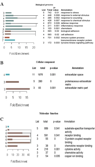
The list of differentially regulated genes was classified using the Gene Ontology
Annotation Tool (GOAT). This tool classifies the genes based on their biological pro-
cesses, the molecular functions in which they participate, and the cellular locations
in which they are active (Figure 2.2 A-C). The left panel shows the enrichment ra-
tio and hierarchy. The values in the right panel indicates information about those
GO (Gene Ontology) node. The values in the List indicate number of genes in
the target gene list which were found significantly regulated due the treatment (in
my case due to knock out of Nesprin-2). The values in the Total indicate number
Figure 2.1: Significance analysis of microarray (SAM) plot. SAM identifies statistically significant genes by carrying out gene specific t-tests. This will adjust the threshold of number of genes which are referred as significant and estimates False Discovery Rate for multiple testing.
of genes in the reference gene list means number of genes in the mouse genome sequence. The multiple-testing corrected p-value (p-value) and the GO term (An- notation). For example from Figure 2.2 A among total of 742 genes which belongs to the category (Annotation) response to stress, there were 5 genes found significantly regulated with p= 0.01.
Table 2.1: List of differentially regulated genes from microarray analysis
GENE NAME UP REGULATED GENES FOLD CHANGE
AK010675 SAA 3 1.841
X67799 Clone C1-2H(DNAfor Eg variable region-heavy chain)
1.622
L07051 Aggrecan 1.612
NM_009141 chemokine (C-X-Cmotif)ligand 5 1.293
AF087578 antisense product of high-affinity glutamate trans- porter EAAC1 and EAAC2 (ASEC1)
1.259
NM_024183 FIP 1 like 1(S.cerevisiae) 1.222
BC006914 rogdi homolog -Drosophila 1.281
GENE NAME UP REGULATED GENES FOLD CHANGE
AK004319 ELOVL family member 5,elongation of long chain fatty acid-Yeast
1.247
NM_009640 angiopoietin 1(Angpt 1) 1.358
NM_011333 chemokine (C-C motif) ligand 2 1.268
AK009960 RIKEN Cdna 1.304
AK007604 RIKEN cDNA 1810026B05 1.425
AK015050 RIKEN cDNA 4930402H24 1.349
AK003208 Impad 1 inositol monophosphatase domain contain- ing 1
1.269 BC011531 expressed sequence AI447318 and RNA binding
motif protein 26
1.64
AK014710 RIKEN cDNA 4833416J08 gene 1.241
NM_009315 TAF6 RNA polymerase II,TATA box binding pro- tein(TBP)associated factor
1.215
NM_017367 cyclin(Ccni) 1.272
AK017327 Mus musculus 6 days neonate head cDNA, RIKEN full-length enriched library
1.221
AK005760 sperm acrosome associated 4(Spaca 4) 1.273
U96635 neural precursor cell expressed,developmentally down regulated gene 4(Nedd4)
1.271
AK006699 RIKEN cDNA 1700045 I 11 gene 1.391
M25572 Mouse Ig active H-chain mRNA,V-region,subgroup IIIC,clone L10
1.26 AK008226 adult male small intestine cDNA, RIKEN full-
length enriched library SIMILAR TO 5’ NU- CLEOTIDASE, MITOCHONDRIAL homolog [Mus musculus], full insert sequence
1.171
U43512 dystroglycan(Dag1) 1.234
AK008096 adult male small intestine cDNA,
RIKEN2010004I09 product:similar to CY- TOCHROME C OXIDASE ASSEMBLY PROTEIN COX11,MITOCHONDRIAL PRECURSOR [Homo sapiens],
1.194
NM_013884 chondroitin sulfate proteoglycan 5 (Cspg5) 1.191
BC002235 tetratricopeptide repeat domain 15 1.164
GENE NAME UP REGULATED GENES FOLD CHANGE
NM_007743 collagen,type 1,alpha 2 1.26
AK014179 platelet endothelial aggregation receptor 1 1.243 NM_016892 copper chaperone for superoxide dismutase (Ccs), 1.126 NM_011330 small chemokine (C-C motif) ligand 11 (Ccl11), 1.307
AK013752 adult male hippocampus cDNA,
RIKEN2900064P18
1.24 NM_016906 Sec61 alpha 1 subunits (S.cerevisiae) 1.181
NM_007833 decorin(’Dcn) 1.229
NM_008524 lumican (Lum) 1.142
NM_011341 stromal cell derived factor 4 1.202
AK021285 RIKEN cDNA D330022H12 gene 1.233
AK008001 RIKEN cDNA 2010109N18 1.196
NM_008422 potassium voltage gated channel,shaw-related sub- family,member3 (Kcnc3)
1.118
NM_024198 glutathione peroxidase 7 (Gpx7) 1.159
AK016999 RIKEN cDNA 4933430M04 1.148
AK007533 motile sperm domain containing 1 1.26
NM_013591 mucosal vascular addressin cell adhesion molecule 1(Madcam1)
1.179 AK004919 adult male liver cDNA,RIKEN clone:1300006N24 1.373
GENE NAME DOWN REGULATED GENES FOLD CHANGE
NM_026423 RIKEN cDNA 2410018C20 0.760
M16356 major urinary protein 2(Mup2) 0.813
AK018008 Rap guanine nucleotide exchange factor 2(Rapgef2) 0.840 Z25851 immunoglobulin kappa chain variable region 0.784
AK020567 RIKEN cDNA9530022L04 0.821
AK020454 RIKEN cDNA 9430032L10 0.771
AK016502 RIKEN cDNA 4931433A01 0.813
M55561 phosphatidylinositol-linked antigen(pB7) (Cd52) 0.803 L10894 guanylate cyclase activator 2b (Guca2b) 0.809
NM_010425 forkhead box D3 0.833
AK017960 RIKEN cDNA 5830431M20 gene 0.804
NM_007561 bone morphogenin protein receptor, type II (ser- ine/threonine)( Bmpr2)
0.845
Figure 2.2: Classification of differentially regulated genes based on GOAT.
Differentially regulated genes were classified based on GOAT as (A) Biological process, based on their involvement in different processes, (B) Cellular components, based on their locations and these genes were classified based on their role in different molecular process in Molecular function (C). In A-C the left panel shows the enrichment ratio and hierarchy.
The numbers on the left of the bars are GO (Gene Ontology) node levels. The right panel
shows information about those GO nodes: number of gene in the target gene list (list),
number of genes in the reference gene list (total), the multiple-testing corrected p-value
(p-value) and the GO term (Annotation).
2.2 In vivo wound healing study in Nesprin-2 knock out mice
2.2.1 Macroscopic observation showed a delay in wound healing in Nesprin-2 KO mice as compared to wild type
In vivo wound healing is a complex process involving three main overlapping phases. When tissues get damaged there will an immediate response to the damage by flowing of blood as there will be damage of blood vessels. Immediately after the blood flow blood coagulates and at the cellular level inflammatory cells are activated and act on the wound area as a defence mechanism against bacteria.
These cells act during the initial phase of wound healing which is called the inflammatory phase followed by the new tissue formation and tissue remodeling phase. We studied the process of wound healing and analysed the different phases in detail.
At first a macroscopic analysis was carried out by taking photographs of all wounds from day 0 until day 10. Each mouse carries four circular wounds on its dorsal side.
From each mouse two wounds were considered for macroscopic observation. The
open area of the wound was measured using Image J. From the open wound area
the percentage (%) of wound closure was calculated by taking the wound area of
zero day as 100% (Figure 2.3 A). At 1day after wounding (1DAW or 1D ), Nesprin-2
KO mice showed 60% of wound closing as compared to WT wounds which showed
50% of wound closing. At 3DAW Nesprin-2 KO mice showed 68% of wound closing
while WT mice showed 62% of wound closure. The faster closing of the wound in
Nesprin-2 KO mice continued until 5DAW. At 5DAW Nesprin-2 KO wounds were
healed by nearly 71% leaving 29% of open wound area as against 67% closure
of wound area in WT mice. Although the wound healing was faster in Nesprin-
2 KO mice than in WT mice at 1, 3 and 5 DAW the difference in the recovery
rate was not significant as reflected by the t-test with p values of 0.064, 0.176 and
0.061 respectively (Figure 2.3 B). We conclude that the wound healing process is accelerated in Nesprin-2 KO mice during the inflammatory phase which normally takes place between 1 to 5 DAW as compared to WT.
However at later stages from 7 to 10DAW the process of wound healing is faster in WT compared to KO. In KO mice 86 % of the wound was healed with 14% of open wound area whereas WT wounds were healed by almost 88% with 12% of open wound area. Similarly at 10DAW, the KO wounds are delayed in closing the wounds where they showed 92% wound closure in comparison to KO wounds that closed to an extent of 95%. The fast rate of wound healing in WT mice compared to KO mice at 7 and 10DAW was statistically significant (p= 0.038 and p=0.013 respectively) (Figure 2.3 B). This indicates that, although not significant, the wound healing process is faster in Nesprin-2 KO mice as compared to WT at the inflammatory phase (1-5DAW), but this trend is reversed during later phases from 7-10DAW when the new tissue formation and tissue remodeling phase has started and thereby leading to a significantly faster recovery in WT compared to the KO.
This observation showed that Nesprin-2 absence from the mouse skin affects the in vivo wound healing process when compared to WT mice skin. The delay in the wound healing process was further confirmed and factors that might be responsible for this delay were studied.
2.2.2 The distance between the epidermal tips is altered in Nesprin- 2 KO wounds
The process of wound healing in WT and Nesprin-2 KO mice which was observed
macroscopically was confirmed by measuring the distance between the migrating
epidermal tips. The wound samples were collected at the indicated time points
(1, 3, 5, 7 and 10DAW). Among four circular wounds two wounds were used for
sectioning (paraffin and cryo sectioning) and the remaining two wounds were
stored at -80
0C for RNA and protein isolation. For further analysis the circular
Figure 2.3: Wound closure is changed in Nesprin-2 Giant knockout mice as
macroscopically analysed. (A) Four circular wounds were made on the dorsal side of
WT and Nesprin-2 KO mice. Wounds were photographed at regular time intervals-1, 3,
5, 7 and 10 days after wounding (DAW). The red dotted line indicates the wound area
left open at day 10. (B) The wound area was measured and the percentage (%) of open
wound area calculated. At each time point wounds (2 wounds / mouse) from six WT and
KO mice each were analysed. Graph showing the wound healing in WT and Nesprin-2 KO
mice.*p=0.038 (7DAW) and 0.013 (10DAW).
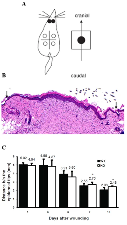
wounds were harvested together with some skin around the wound. Then the wounds were cut caudocranially (tail to head or head to tail) and fixed in paraffin blocks (Figure 2.4 A). The paraffin block, which contained skin tissue was sectioned with section size of 6µm. The sections were further used for staining with H&E (Haematoxylin and Eosin) and also with antibodies. The H&E stained sections were used for measuring the distance between the migrating epidermal tips.
The distance between the epidermal tips gives the diameter of the wound (mm)
(Figure 2.4 B black arrows). Again the wounds from all the time points were used
for measuring the distance between the epidermal tips. This measurement was
consistent with and supported the macroscopic observation. At 1DAW Nesprin-
2 KO and WT wounds showed almost the same distance between the migrating
epidermal tips (4.94 mm and 5.02 mm, respectively). This trend continued further
at 3 and 5DAW. The distance for Nesprin-2 KO and WT mice was 4.87 mm and
4.98 mm, respectively, at 3DAW and 3.60 mm and 3.91 mm, respectively, at
5DAW. Nesprin-2 KO wounds showed a reduced distance between the epidermal
tips (diameter of the wound) as was the case with macroscopic observations
suggesting hat these KO wounds showed a faster healing as compared to WT
at the inflammatory phase from 1 to 5DAW, but this difference was statistically
not significant (p= 0.62, 0.73 and 0.41 for 1, 3 and 5DAW, respectively). Then
the healing process was accelerated in WT from 7DAW onward as determined
by the decreased distance between the migrating epidermal tips in these wounds
compared to Nesprin-2 KO wounds. At 7DAW Nesprin-2 KO mice showed a value
of 2.7 mm as the distance between the epidermal tips which was significantly
different from the 2.55 mm in WT (p= 0.04). At 10DAW the wound healing process
continued to be faster in the WT mice and 2.08 mm were measured as compared
to 2.55 mm in the KO mouse (p= 0.0092) (Figure 2.4 C). This pattern of wound
healing agreed with our macroscopic observations.
Figure 2.4: The distance between the two migrating epidermal tips showed a
reduced healing in wounds from Nesprin-2 KO from 7 DAW to 10DAW compared
to WT wounds. (A) Four circular wounds were made on the dorsal side of the mice
and the entire wound harvested along with surrounding normal skin. Then this skin
is cut caudo cranially and fixed in paraffin blocks (Figure from Gerharz et al., 2006).(B)
Skin sections from WT and KO were stained with H & E and the distance between
the migrating epidermal tips (black arrows) from day 1 to day 10 was measured using
the Diskus programme (n= 3-5 sections per wound).(C) At day 7 and 10 the distance
between epidermal tips of WT and KO is significantly different (* p=0.044 and p=0.0092,
respectively; student’s t-test).
2.2.3 Inflammatory phase
Higher influx of macrophages into the wound area causes faster wound healing in Nesprin-2 KO during the early phase.
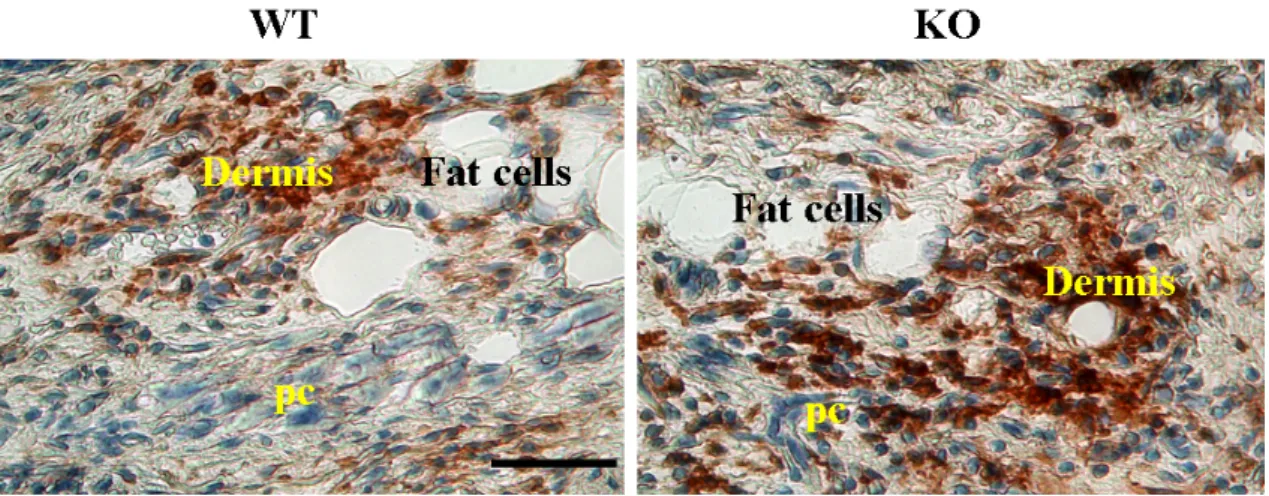
To study the earliest phase of the wound healing process, inflammatory phase wound sections from Nesprin-2 KO and WT were stained with the macrophage specific antigen F4/80 (Eming et al., 2007). The F4/80 monoclonal antibody has been used widely to identify and study macrophages in the mouse under normal and pathological conditions. The F4/80 antigen is a 160 kDa cell surface glycoprotein that is a member of the EGF 7 transmembrane (7TM) protein family of proteins which shares 68% overall amino acid identity with the human EGF module containing mucin like hormone receptor 1 (EMR1). Expression of F4/80 is heterogeneous and varies during macrophage maturation and activation (Van den Berg and Kraal, 2005). Though the inflammatory phase occurs between 1 and 3DAW some of the inflammatory cells are still present at 5DAW. Therefore the F4/80 antibody was used to stain the sections of Nesprin-2 KO and WT wounds from 1, 3 and 5DAW. The sections were subjected to immunohistochemistry with F4/80 antibody, and Hematoxylin was used as a co-stain for the nucleus. The macrophages stained with F4/80 are brown in colour while the nucleus looks blue.
The Nesprin-2 KO wounds showed a dense brown staining deposited at the wound area in the sections from all 1, 3 and 5DAW compared to WT. This indicates that in Nesprin-2 KO wounds there was an increased influx of macrophages (Figure 2.5).
This could be an indication that Nesprin-2 regulates the inflammatory cells which act during initial phase of the healing process.
Nesprin-2 might play a role in regulating the expression of some inflammatory
genes. Interestingly this is supported by the data of our microarray study where
we found among the differentially regulated genes some that are involved in the
Figure 2.5: The macrophage population is increased in the wound area in Nesprin-2KO mice. (A) Skin sections were stained for macrophages with the F4/80 antibody and photographed using the Diskus software. Nesprin-2 KO wounds showed an increased staining of F4/80 compared to WT at 1, 3 and 5 days after wounding. In the figure brown staining indicates F4/80 stained cells (shown for day 5) and blue staining from Haematoxylin indicates nuclear staining. Comparable areas in the dermis of the wounds are shown. (pc -Panniculus carnosus) Scale bar, 50 µm.
inflammatory process namely Serum Amyloid A3 (SAA3), Chemokine (C-X-C mo- tif) ligand 5, Chemokine ligand 2 or MCP1 (Monocyte chemoattract protein 1) and Small Chemokine (C-C) ligand 11 (Table 2.2). Serum amyloid (SAA) proteins are Table 2.2: List of up-regulated inflammation related genes from microarray anal- ysis.Genes involved in the inflammatory process were found up-regulated in microarray analysis.
Significantly regulated genes Fold changes
SAA3 1.84
Aggrecan 1.61
Chemokine(C-X-Cmotif) ligand5 1.3 Chemokine ligand2-MCP1 1.27 Small Chemokine(C-C) ligand11 1.31
Cyclin (Ccni) 1.27
TAF6 1.22
Collagen type1, alpha2 1.2
a family of apolipoproteins associated with high-density lipoprotein (HDL). Differ-
ent isoforms of SAA are expressed constitutively (constitutive SAAs) at different
levels or in response to inflammatory stimuli (acute phase SAAs). Three acute phase SAA isoforms have been reported in mice, called SAA1, SAA2 and SAA3.
Acute phase serum amyloid A proteins (A-SAAs) are multifunctional apolipopro- teins produced in large amounts during the acute phase of an inflammation and also during the development of chronic inflammatory diseases (Zhang et al., 2005).
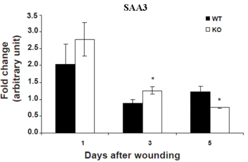
The expression of SAA3 was found upregulated in Nesprin-2 KO fibroblasts (Table 2). Next we studied the expression of the SAA3 in wound samples. The RNA from wounds of Nesprin-2 KO and WT was isolated from 1, 3 and 5DAW samples and cDNA was synthesised from these RNA samples and used for the study of expres- sion of SAA3 by q-PCR. The q-PCR results clearly showed that the expression of SAA3 was enhanced in Nesprin-2 KO wounds at 1 and 3DAW while its expression is significantly reduced after 5 days of wounding compared to WT (Figure 2.6).
Figure 2.6: The expression of the inflammation related gene SAA3 is increased in Nesprin-2 KO wounds. The expression of SAA3 was up-regulated in Nesprin-2 KO fibroblast and this was confirmed in wound samples from 1, 3 and 5DAW (* p=0.016 and 0.008 at 3 and 5DAW, respectively).
Monocytes and macrophages are a source of several chemoattractants during
the inflammatory phase which are required for the later phase of the healing
process. To check for chemoattractant production at the transcriptional level in wound samples, cDNA of Nesprin-2 KO and WT mice from 1, 3 and 5DAW was used in q-PCR experiments. We checked for MCP1 (CCL2), a monocyte specific chemoattractant protein, which is a member of the small inducible gene (SIG) family. MCP1 plays a role in the recruitment of monocytes to the sites of injury and infection. The expression of MCP1 in the wounds should correlate with the macrophages population. This was indeed the case in our analysis.
The macrophage staining was increased in the KO wounds and in parallel the expression of MCP1 was significantly up-regulated in Nesprin-2 KO mice at 1, 3 and 5DAW. The expression of MCP1 was increased in Nesprin-2 KO wounds at 1DAW (4 fold), nearly 2 fold at 3DAW and more than 1 fold at 5DAW as compared to WT . The expression of MCP1 gradually decreased in both Nesprin-2 KO and WT wounds from 1 to 5DAW as the population of macrophages also decreased from the inflammatory phase to the new tissue formation phase (Figure 2.7).
Figure 2.7: Quantitative PCR (q-PCR) data showed a significant upregulation of
the chemokine MCP1 (CCL2) in the wounds of Nesprin-2 KO. The expression of the
macrophage specific chemoattractant was found upregulated in Nesprin-2 KO wounds by
q-PCR. (*p=0.042, 0.0026 and 0.027, respectively, for 1, 3 and 5DAW).
2.2.4 New tissue formation and tissue remodelling phase
Migration of keratinocytes towards the wound area occurs during the new tissue formation phase followed by hyper proliferation of the keratinocytes. In the dermis, fibroblasts from the neighbouring dermis migrate also towards the wound area and proliferate and some of them will differentiate into myofibroblasts.
These fibroblasts deposit extracellular matrix and the tissue which is formed by these cells is called granulation tissue area. For this keratinocyte migration and proliferation and fibroblast proliferation was studied.
I - The keratinocyte migration pattern is altered in Nesprin-2 KO wounds and neo epidermis is lost in the KO wounds
Two models have been proposed to describe the epidermal migration process during wound healing: the “sliding” model and the “rolling” model (Usui et al., 2005).
Which model represents the actual migratory event best remains controversial.
The “sliding” model focuses on the newly exposed basal keratinocytes at the leading edge. Upon wounding, changes in hemidesmosomal attachments enable basal keratinocytes to retract from the basement membrane zone, degrade dermal matrix, deposit new basement membrane components such as laminin 5, and migrate laterally over the provisional wound matrix. By virtue of the strong desmosomal attachments of migratory basal cells to adjacent basal and suprabasal keratinocytes, the entire epidermis then moves as a sheet or column to eventually close the wound. Evidence in support of the “sliding” model comes from extensive in vitro studies.
For the complex stratified epidermis, the “rolling” model of epithelialization may reflect epidermal wound healing more accurately even though it is less widely accepted as a possible mechanism for the epidermal repair process. In this “rolling”
model, the primary keratinocytes participating in wound closure are suprabasal.
Suprabasal keratinocytes exposed to the wound environment upon injury are
thought to undergo cell shape change, reduce their desmosomal attachments, and tumble over basal keratinocytes that remain strongly attached to the basement membrane. In the unwounded epidermis, Keratin 14 (K14), a marker for migrating keratinocytes was shown to be localized only in basal keratinocytes and at 1DAW K14 was found in suprabasal keratinocytes along with basal keratinocytes. From 3 D onwards its expression was found throughout the epidermis or all the layers of the epidermis (Usui et al., 2005).
I studied the keratinocyte migration pattern by staining the sections from 5, 7 and 10DAW sample with a K14 specific antibody (Figure 2.8). The red staining shows the presence of K14 which stains the keratinocytes in the epidermis (Epi) and blue is nuclear staining from DAPI. The keratinocytes are migrating from the neighboring epidermis towards the wound area and migrate further between the Fibrin clot and the dermis (Derm) by forming a sharp edge (dotted line in Figure 2.8 insets) also called neo epidermis in case of WT wounds from 5DAW and 7DAW.
Whereas in Nesprin-2 KO, keratinocytes are migrating with no sharp edge which
means that they have lost the neo epidermis in both 5 and 7DAW (Figure 2.8
insets). The epidermis with neo epidermis or with a sharp edge may speed up
the migration of keratinocytes. This could be one of the reasons for faster closing
of wounds in WT than in Nesprin-2 KO mice from 7DAW onwards. At 10DAW, WT
wounds showed reduced wound area compared to Nesprin-2 KO wounds. This is
shown by drawing a line indicating the distance between the migrating epidermal
tips. The white arrow heads in the inset indicates that red staining (K14 staining)
is reducing in WT as there is DAPI staining at the basal keratinocytes. The wound
in WT at 10DAW is recovering from wound to normal skin. While in KO wounds at
10DAW, distance between the migrating tips is high as compared to WT and also
we could see the K14 staining in the all the layers of the epidermis. The wound in
KO at 10DAW is not completely recovered from the wound to normal skin.
Figure 2.8: The keratinocyte migration pattern is altered in Nesprin-2 KO
mice. Keratinocyte migration over the wound area between fibrin and the dermis was
studied with a Keratin 14 specific antibody. Red staining is from K14 which stains the
keratinocytes in the epidermis and blue staining is from DAPI, a nuclear stain. Scale
bar for 200µm. Inset showing sharp migrating tips. Line in the 10D indicates distance
between the migrating tips. The white arrow heads in the inset at 10D indicates that red
staining (K14 staining) is reducing in WT which is replaced by DAPI staining at the basal
keratinocytes. Scale bar, 50µm.
II - Proliferation of keratinocytes is reduced in Nesprin-2 KO wounds Migration of keratinocytes from the nearby epidermis towards the wound area is followed by their hyperproliferation. The expression of Ki67, a marker of cell proliferation, was restricted to basal and immediate suprabasal layers at the wound edge. In normal skin, expression of Ki67 is observed in very few nuclei along the basal cell layer, whereas during epidermal wound healing an increased expression in basal and immediate suprabasal cells is seen (Usui et al., 2005;
Patel et al., 2006). Ki67 staining had been earlier assessed in unwounded skin of Nesprin-2 KO and WT and was found to be nearly identical and restricted to the basal layer (Lüke et al., 2008).
When I carried out the analysis in skin sections of Nesprin-2 KO and WT from 5, 7 and 10DAW the expression of Ki 67 was observed both in the basal and suprabasal layer of the epidermis. However the number of proliferating keratinocytes was reduced in Nesprin-2 KO wounds (Figure 2.9 A). In order to determine the proliferation rate of keratinocytes in the wounded epidermis, I counted the number of Ki67 positive cells in the wounds from three time points, 5, 7 and 10DAW. Nesprin-2 KO wounds showed an average of 9 cells per unit area which is significantly less as compared to WT (∼30 cells per unit area) at 5DAW.
Similarly, at 7DAW the number of Ki67 positive cells was significantly reduced in
Nesprin-2 KO wounds (22 cells per unit area) as compared to WT wounds (∼41
cells per unit area) and at 10DAW Nesprin-2 KO wounds contained approximately
10 Ki67 positive cells per unit area whereas WT wounds had nearly 18 cells per
unit area (Figure 2.9 B). The reduction in keratinocyte proliferation in Nesprin-2
KO wounds may be one of the reasons for the delay in wound healing observed at
7 and 10DAW.
Figure 2.9: Keratinocyte proliferation is slowed down in Nesprin-2 KO wounds.
(A) Proliferation of keratinocytes is studied by staining the sections with the proliferation
marker Ki67. The expression of Ki67 at the basal and suprabasal layer of the epidermis
is indicated by white arrows. Scale bar, 100µm. (B) The average number of Ki67 positive
cells in Nesprin-2 KO and WT wounds per unit area was determined (*p=0.027, 0.029 and
III - Differentiation of fibroblasts into myofibroblasts in the granulation tissue area is reduced in Nesprin-2 KO wounds
During healing of an open wound, resident dermal fibroblasts proliferate from the wound margin and migrate into the provisional matrix composed of a fibrin clot.
About 1 week after wounding, the provisional matrix is replaced by newly formed connective tissue known as granulation tissue which is essentially composed of small vessels, extracellular matrix, and fibroblastic cells that become activated and differentiate into myofibroblasts. The main feature of myofibroblasts is their contractile apparatus which is similar to that of smooth muscle and in particular the expression of 𝛼-smooth muscle actin (𝛼-SMA). Myofibroblasts play a central role in closing the wound tissue through their capacity to produce a strong contractile force possibly generated within stress fibers, similar to those present in cultured fibroblasts (Skalli et al., 1986; Serini and Gabbiani, 1999). Because wound contraction takes place when de novo expressed 𝛼-SMA is incorporated in stress fibers, it has been suggested that this actin isoform plays an important role in granulation tissue contraction (Hinz et al., 2001).
Based on the fact that Nesprin-2 Giant is an actin binding protein, it might well be involved in regulating myofibroblast differentiation. Because the expression of alpha smooth muscle actin (𝛼-SMA) is considered as the most reliable marker for differentiated myofibroblasts I stained the sections from Nesprin-2 KO and WT wounds with an 𝛼-SMA specific antibody in order to study myofibroblast differentiation. The staining for 𝛼-SMA was strong in the dermis where the granulation tissue (GT) is present (Figure 2.10 A).
The visual comparison of the expression of 𝛼-SMA in the wounds of KO and
WT mice was followed by a quantification of the intensity of staining per unit
area using the Leica confocal software. Although the staining intensity of 𝛼-
SMA appeared higher in WT wounds as compared to Nesprin-2 KO wounds, the
Figure 2.10: Differentiation of fibroblasts into myofibroblasts is reduced during
the wound healing process in Nesprin-2 KO mice. (A) Differentiated myofibroblasts
were studied in wounds from 5, 7 and 10DAW. Alpha smooth muscle actin (𝛼-SMA) was
taken as a marker. Epi-epidermis and GT-granulation tissue. Scale bar, 100µm. (B)
The intensity of 𝛼-SMA staining was quantified as intensity per unit area using Leica
Confocal Software. The intensity per unit area at 7 and 10DAW differed significantly
(7DAW, p=0.04; 10DAW, p=0.04). (C) Western blot analysis of homogenates using 𝛼-SMA
antibody and 𝛽-tubulin as a loading control. Samples from 7DAW are shown.
intensity per unit area did not reveal a significant difference in the staining intensity at 5DAW between Nesprin-2 KO and WT wounds. However, there was a significantly higher intensity of 𝛼-SMA staining per unit wound area in WT at 7 and 10DAW (7DAW, p=0.04; 10DAW, p=0.04; Student’s t-test) indicating that more fibroblasts had differentiated into myofibroblasts in WT compared to Nesprin-2 KO wounds (Figure 2.10 B). The western blot analysis also showed an increased expression of 𝛼-SMA in WT as compared to KO (Figure 2.10 C). This can be further important reason for the faster wound closure in WT mice compared to KO.
III A. F-actin fibers were disturbed around the nucleus in Nesprin-2 KO fibroblasts
Induction of transforming growth factor-𝛽1 (TGF-𝛽1) in fibroblasts enhances the formation of “structural element”, bundles of actin filaments or stress fibers, vinculin-containing fibronexus adhesion complexes and fibronectin fibrils, and increases the expression of 𝛼-SMA. The formation of these structural elements is thought to serve as a prerequisite for TGF-𝛽1 induced expression of 𝛼-SMA.
They are also important for generating contractile force in myofibroblasts which is associated with wound contraction. Formation of stress fibres therefore directly correlates with the expression of 𝛼-SMA and a reduction in 𝛼-SMA is equivalent to a reduction in F-actin content (Vaughan et al., 2000).
We stained fibroblasts from Nesprin-2 KO and WT with TRITC-phalloidin for
F-actin detection we found that the F-actin network was disrupted around the
nucleus (red arrow) in many Nesprin-2 KO fibroblasts (Figure 2.11 A). To verify
this observation I determined the number of fibroblasts for Nesprin-2 KO and WT
which showed a disturbed or disrupted F-actin network around the nucleus. In
the Nesprin-2 KO ∼22% of fibroblasts had an altered F-actin staining around the
nucleus. In WT fibroblasts similar changes were seen in only ∼12 % of cells (Figure
2.11 B).
Figure 2.11: Nesprin-2 absence led to disturbed actin fibers around the nucleus in primary fibroblasts. (A) Wild type fibroblasts and Nesprin-2 knock out fibroblasts were stained for F-actin with TRITC Phalloidin. Fibroblasts with disrupted actin fibers (arrow) were counted and used for calculating the percentage. Scale bar, 50µm. The lower panel shows a single wild type and Nesprin-2 KO fibroblast at higher magnification. DAPI is used for staining the nucleus. Scale bar, 100µm. (B) Cells with defective F-actin fibers were counted and used for calculating the percentage (%). The KO fibroblasts showed a significantly higher percentage (%) of cells with a defective actin cytoskeleton than wild
−9