• Keine Ergebnisse gefunden

Reactivities of biologically active small molecules: Kinetic assessment of electrophilic enones and characterization of a photoactive phosphoantigen probe

N/A
N/A
Protected

Academic year: 2021

Aktie "Reactivities of biologically active small molecules: Kinetic assessment of electrophilic enones and characterization of a photoactive phosphoantigen probe"

Copied!
200
0
0

Wird geladen.... (Jetzt Volltext ansehen)

Volltext

(1)

Kinetic assessment of electrophilic enones and characterization of a photoactive phosphoantigen probe

Zur Erlangung des

DOKTORGRADES DER NATURWISSENSCHAFTEN (Dr. rer. nat.)

der Fakultät für Chemie und Pharmazie der Universität Regensburg

DISSERTATION

vorgelegt von Monika Enzinger aus Regensburg

Mai 2019

(2)

November 2014 bis April 2019 angefertigt.

Promotionsgesuch eingereicht am: 03.05.2019

(3)

Ich erkläre hiermit an Eides statt, dass ich die vorliegende Arbeit ohne unzulässige Hilfe Dritter und ohne Benutzung anderer als der angegebenen Hilfsmittel angefertigt habe. Die aus anderen Quellen direkt oder indirekt übernommenen Daten und Konzepte sind unter Angabe des Literaturzitats gekennzeichnet.

__________________________________

Monika Enzinger

(4)

ABBREVIATIONS VI

A ELECTROPHILES OF THE ENONE-TYPE AND THE INVESTIGATION OF THEIR

REACTIVITY AND BIOLOGICAL ACTIVITY 1

A 1 Introduction 1

A 1.1 Signal transduction pathways and their role in inflammation 1

A 1.2 Enones as electrophilic, covalently binding inflammation mediators 3 A 1.3 In-vitro cell assays to determine the toxicity and anti-inflammatory activity of compounds 6 A 1.4 General methodological access to kinetic measurements for the determination of the reactivity

of compounds 8

A 1.5 The Kinetic Thiol Assay for electrophiles and its application in the determination of the reactivity

of chalcone derivatives 10

A 1.6 α-X-Hydroxychalcones (α-X-HCs) 14

A 1.7 Synthesis of oxadiazoline enones (OXEs): a library of new electrophilic substances with an enone

unit 16

A 1.8 Synthesis of further electrophiles in the Amslinger work group 22 A 1.9 Further development of the kinetic thiol assay towards the investigation of colorless

electrophiles 25

A 1.10 Aim of the present work 29

A 2 Results and Discussion I: Synthesis, kinetic and biological evaluation of oxadiazoline enones (OXEs): a library of new anti-inflammatory electrophilic compounds 31

A 2.1 Synthesis 32

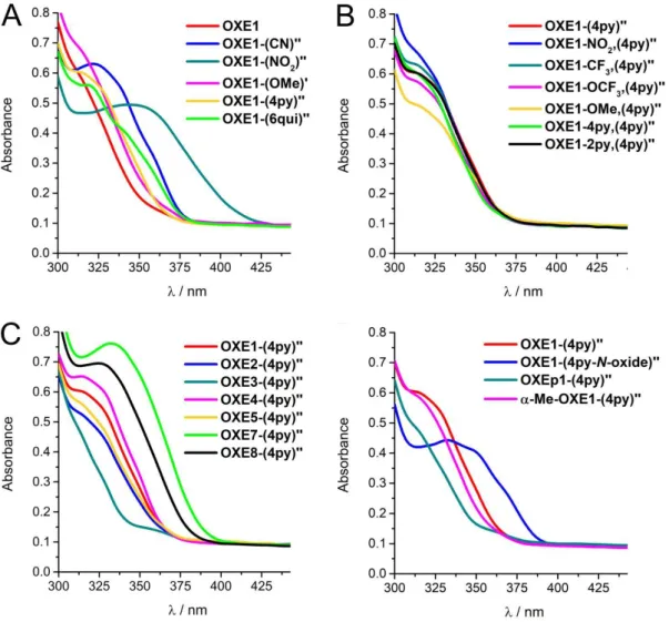
A 2.2 UV-Vis spectral properties of OXEs and OXE analogues 37

A 2.3 Kinetic evaluation of OXE and OXE analogues 40

A 2.4 Biological evaluation of the OXE and OXE analogues substance library 56 A 3 Results and Discussion II: Reactivity assessment of further electrophiles by the kinetic thiol assay

63

A 3.1 Reactivity modification of phenylacrylamide derivatives as reactive units in drug development 63 A 3.2 Juglone and juglone derivatives and their reactivity towards cysteamine 66 A 3.3 α-Nitro-hydroxychalcone (α-NO

2

-HC) as part of the α-X-hydroxychalcone (α-X-HC) substance

library 75

A 4 Results and Discussion III: Investigations for a further development of the kinetic thiol assay

towards the use of colorless enones as test substances 79

A 4.1 Syntheses 79

A 4.2 Fluorescence investigations of Michael addition reactions 85

A 5 Summary of Chapter A 88

A 6 Experimental 90

(5)

A 6.2 Syntheses of OXEs, OXE analogues and their precursors 90

A 6.3 Synthesis of NO

2

-flavanone and precursors 99

A 6.4 Synthesis of fluorescent aromatic thiol dyes 102

A 6.5 Kinetic evaluation of electrophiles 108

A 6.6 In-vitro cell assays 111

B CHARACTERIZATION OF A NEW PHOTOACTIVE PROTEIN PROBE FOR THE

LABELING OF BTN3A4 113

B 1 Introduction 113

B 1.1 The immune modulator role of HMBPP 113

B 1.2 BioBP-HMBPP as a new photoactive protein probe for the labeling of BTN3A4 115

B 1.3 Aim of this work 116

B 2 Results and Discussion 117

B 2.1 Photochemical and UV-Vis evaluation, handling of proteins and probe solutions 117

B 2.2 Stimulation of γδ T cells by BioBP-HMBPP (103) 120

B 2.3 Labeling of the B30.2 domain of the protein BTN3A1 by BioBP-HMBPP (103) in the absence and

presence of HMBPP (102) 123

B 2.4 Labeling of a BTN3A1-B30.2 charge transfer mutant 128

B 2.5 Determination of labeling efficiency by BioBP-HMBPP 129

B 3 Summary of Chapter B 130

B 4 Experimental 131

B 4.1 General information 131

B 4.2 Photochemical labeling of proteins with BioBP-HMBPP and Western Blot analysis of biotinylated

proteins 131

C LITERATURE 133

D APPENDIX 139

D 1 NMR spectra of synthesized compounds 139

D 2 Pre-kinetic UV-Vis spectra of OXEs 162

D 3 Additional figures for the kinetic evaluation of OXEs and OXE analogues 167 D 4 LC-MS data of juglone derivatives in kinetic assay buffer 177

D 5 Additional figures for protein labeling experiments 190

D 6 PhD thesis figures Fehler! Textmarke nicht definiert.

(6)

Abbreviations

15d-PGJ2 15-Deoxy-Δ12,14-prostaglandine J2

A Absorbance

α-X-TMC α-X-Substituted 2’,3,4’,4-tetramethoxychalcones α-X-HC α-X-Substituted 2’-hydroxy, 3,4’,4-trimethoxychalcones B30.2 Domain of the protein BTN3A1

BioBP-HMBPP Biotin-Benzophenone-HMBPP

BP-HMBPP (E)-4-((4-Benzoyl-benzoyl)oxy)-3-methylbut-2-en-1-yl diphosphate, Benzo-Phenone-HMBPP

BSA Bovine serum albumin

BTN3A1 Butyrophilin 3A1

CPS Counts per second

c Concentration [mol/l], [M]

CD69 Cluster of differentiation 69

CDCl 3 Deuterated chloroform

COX-2 Cyclooxygenase 2

CuI3 Cullin 3

δ Chemical shift

DCM Dichloromethane

DCY Dansyl cysteamine

DCYA Dansyl cysteamine assay

DIPEA Diisopropylethylamine

DMAPP Dimethylallyl diphosphate (formerly: pyrophosphate)

DMF Dimethylformamide

DMSO/DMSO-d 6 Dimethyl sulfoxide/deuterated dimethyl sulfoxide DOXP 1-Deoxy-D-xylulose 5-phosphate

E Electrophile

EC 50 Effective concentration

EGFR Epidermal growth factor receptor EDTA Ethylendiamintetraacetic acid

EG Ethylene glycol

EGF Epidermal growth factor

EI-MS Electron impact mass spectrometry

ESI-MS Electronspray ionization mass spectrometry

EtOAc Ethyl acetate

EtOH Ethanol

eq. Equivalents

(7)

FITR Fourier-transform infrared spectroscopy FPPS Farnesyl diphosphate synthase

GSH Glutathione

HEPES 4-(2-Hydroxyethyl)-1-piperazineethanesulfonic acid HMBPP (E)-4-Hydroxy-3-methylbut-2-enyl diphosphate

HO-1 Heme oxygenase 1

HOAc Acetic acid

HRPO Horseradish peroxidase

HTS High throughput screening

HV High vacuum

Hz Hertz

IκBα Inhibitory κB-protein α

IC 50 /IC 20 Inhibitory concentration IKK Inhibitory κB-protein kinase

IL Interleukin

iNOS Inducible NO-Synthase

IPP Isopentenyl diphosphate

IR Infrared

k Reaction rate constant

Keap1 Kelch-like ECH-associated protein 1

λ Wavelength

LC-MS Liquid chromatography Mass spectrometry

LPS Lipopolysaccharide

LR-MS Low-resolution Mass spectrometry

M Molar mass [g/mol]

[value] M Concentration [mol/l]

m/z Mass per charge number

mCPBA Metachloroperbenzoic acid

Me Methyl

MeO Methoxy

MeOH Methanol

MS Mass spectrometry

MSTI (E)-2-(4-Mercaptostyryl)-1,3,3-trimethyl-3H-indol-1-ium iodide

MTT 3-(4,5-Dimethylthiazol-2-yl)-2,5-diphenyltetrazolium bromide

NBD 7-Nitrobenzo[c][1,2,5]-oxadiazol-4-thiol

n-BuLi n-Buthyllithium

(8)

NED N-(1-Naphthyl)ethylenediamine

NF-κB Nuclear factor kappa-light-chain-enhancer of activated B cells

nm Nanometer

NMR Nuclear magnetic resonance-spectroscopy Nrf2 Nuclear factor-erythroid-2-related factor

NOS NO-synthase

OXAc Acyl-substituted oxadiazoline OXCE Oxadiazoline cyclohexane enone

OXE Oxadiazoline enone

OXEp Oxadiazoline epoxide

OXTE Oxadiazol thioester

PAg Phosphoantigen

PBMCs Peripheral blood mononuclear cells

PBS Phosphate-buffered saline

p-BZ-C-C5-OPP (E)-4-((4-Benzoyl-benzoyl)oxy)-3-methylbut-2-en-1-yl diphosphate

Ponceau S 3-Hydroxy-4-(2-sulfo-4-[4-sulfophenylazo]phenylazo)-2,7- naphthalenedisulfonic acid sodium salt

PP Diphosphate

ppm Parts per million

PVDF Polyvinylidene difluoride

R f Retention factor

RPMI medium Roswell Park Memorial Institute medium

rt Room temperature

SD Standard deviation

SDS-PAGE Sodium dodecyl sulfate polyacrylamide gel electrophoresis T cell Thymus-derived lymphocyte

TBS-T Tris-buffered saline with 0.1% Tween

TCR T cell receptor

TFA Trifluoroacetic acid

TLC Thin layer chromatography

TMC Tetramethoxychalcone

TNF Tumor necrosis factor

Tris-HCl Tris(hydroxymethyl)aminomethane UV-VIS Ultraviolet-visible-light-spectroscopy

v Reaction rate

(9)

A Electrophiles of the enone-type and the investigation of their reactivity and biological activity

A 1 Introduction

A 1.1 Signal transduction pathways and their role in inflammation

The biochemical reaction of organisms to external stimuli of all kinds is controlled by a complex signal transduction network in which biomolecules interact extra- and intracellu- larly. If a cell is activated from the outside by the binding of a signal molecule to a mem- brane receptor, this is the beginning of a multitude of possible intracellular information transmission pathways. Numerous proteins regulate gene expression and subsequent pro- duction of proteins and enzymes via signaling cascades (Figure 1).

Figure 1: Principle of intracellular signal cascades under the influence of external stimuli.

Inflammation is a well-studied biochemical process as it is the cause and/or symptom of

many diseases. Various signal transduction pathways have been identified as mechanisms

for the development of inflammation, for example the NF-κB pathway. NF-κB (nuclear fac-

tor kappa-B) is a transcription factor that, when bound to its target structures on the DNA,

triggers the biosynthesis of pro-inflammatory proteins [1] such as TNF (tumor necrosis fac-

tors), COX-2 (cyclooxygenase 2) and iNOS (inducible nitrite oxide synthase). In the cytosol,

NF-κB is present in its inactive form, bound to the inhibitor protein IκBα (inhibitory κB pro-

tein α). External stimuli activate protein kinases such as IKK (inhibitory κB protein kinase)

that phosphorylate the inhibitor protein, which leads to its ubiquitination and degradation

and activates NF-κB by release (Figure 2). [2]

(10)

Figure 2: Schematic mechanism of the NF-κB signal transduction pathway. NF-κB: nuclear factor kappa B, IKK:

IκB kinase, IκBα: kappa-B inhibitor, Ub-Ub-Ub: ubiquitination, iNOS: inducible NO synthase.

One of the pro-inflammatory enzymes produced by the influence of NF-κB is iNOS. This enzyme is one of three NOS isoforms, heme dioxygenases that cause the conversion of L- arginine to citrulline and the gaseous signaling molecule nitric oxide, NO [3] (see Scheme 1A). NO in general has multiple functions in the human body, depending on its concentra- tion and the tissue of release. For example, in the nerval system and in endothelia it is synthesized by nNOS (neuronal NOS) and eNOS (endothelial NOS), respectively, and acts as a neurotransmitter, causing for example vaso- and bronchodilatation in the latter. These functions are regulated in such way that NO is present in low concentrations and its activity is short-lasting. [4]

However, this is different in iNOS-containing cells. INOS is found in the cytosol of macro-

phages [5] in a basic amount, but its synthesis is up-regulated by external stimuli such as

bacterial lipopolysaccharides (LPS) or pro-inflammatory cytokines (e.g. TNF and interleu-

kins, ILs) in inflamed tissues, tumor tissue or by microbial infections via the NF-κB pathway

as described in Figure 2. Here, NO acts as an important mediator of inflammation and un-

specific immune response by different mechanisms of action, depending on its chemical

form, summarized in Scheme 1B. [4] NO can directly act as ligand to metal ions in cofactors,

such as iron in heme proteins, and coordination leads to increased activation or inhibition

of those enzymes. Furthermore, NO is oxidized by oxygen O 2 to NO + , the nitrosyl cation,

which is able to react with nucleophiles in amino acid residues of enzymes, transcription

factors and ion channels. In fact this leads to the nitrosylation of cysteine thiols, which

modifies the activity of these proteins. [6] Then, NO reacts also very quickly with the super-

oxide radical anion O 2 - , which leads to the formation of ONOO - , peroxonitrite. This high-

potency cellular toxin performs nitration and oxidation in biomolecules, leading to protein

de-functionalization, DNA damage and strand breaks [7] or peroxidation of lipids [8] and caus-

ing overall necrosis and apoptosis. [9]

(11)

Scheme 1: A: Biosynthesis of NO by iNOS; B: effects of NO towards biomolecules in immune response and as a trigger of inflammation.

Based on these descriptions, one can imagine that, despite the positive role of NO in the fight against infections and tumors, uncontrolled overexpression of iNOS and subsequent increased NO synthesis also damages healthy cells, leading to several diseases described as NO-induced via the NF-κB pathway. [10] [11] Asthma, arthritis and various forms of bowel dis- eases (intestinal inflammation) are amongst them. These pathological mechanisms raise the question which biochemical measures can be taken to intervene in the development of inflammation. If these biochemical connections are known, the course of associated dis- eases can be influenced.

A 1.2 Enones as electrophilic, covalently binding inflammation mediators

For pharmacologically active substances it is possible to intervene at certain points of signal transduction by covalent or non-covalent binding. If these biologically active compounds are electrophiles, covalent binding plays a particularly important role as they can react with biomolecular nucleophiles. These interactions can for example influence the activity of pro- teins in general and enzymes in particular, leading to a positive influence on many diseases.

This has been well investigated and proven for a large number of biologically active elec- trophilic natural compounds of various chemical structures and target proteins. [12]

Cysteine residues on the surface of proteins are an important target. The nucleophilic prop-

erties of their terminal thiol group (sulfhydryl group) enable them to form covalent bonds

(12)

with electrophilic compounds. Organic molecules that have a carbonyl function in the im- mediate vicinity of a carbon-carbon double bond (α,β-unsaturated carbonyl function) are such electrophiles and are called enones or Michael acceptors. Depending on their reactiv- ity, they can react on their electrophilic β-position with thiol groups in a Michael addition [13]

(Scheme 2), the organic reaction type first described by Arthur Michael. [14]

Scheme 2: Michael addition of a pharmacologically active α,β-unsaturated carbonyl compound (enone, Mi- chael acceptor) to the thiol group of a cysteine in a protein.

Enones can be divided into different substance classes due to their different structure and/or function. [12, 15] Some natural products are given as examples in Figure 3.

Figure 3: Examples of natural products with α,β-unsaturated carbonyl function.

[12, 15]

In the literature, anti-inflammatory, antimicrobial, neuroprotective and antiproliferative properties based on their Michael acceptor activity are described for some of these com- pounds. [13, 15-16] However, depending on their structure, other chemical properties of the substances, radical scavenging properties, isomerization at the double bond or oxidation and reduction can also be the reason for their biological activity. [13]

The mechanism of the anti-inflammatory effect of electrophiles and enones in particular

by reacting with surface thiols of proteins can be explained by two major signaling path-

ways. On the one hand, they can inhibit the NF-κB signal transduction pathway at multiple

(13)

points. As shown in Figure 4A, enones can react with surface thiols of IKK, IκBα or NF-κB itself. This causes protein alkylation, which inactivates them. Thus, the transcription of pro- inflammatory genes is inhibited by direct deactivation or prevention of activation of NF- κB. [2] Some natural products such as polyphenols and terpenes are NF-κB inhibitors, [17] for example curcumin (1, Figure 3) from Curcuma longa, CAPE (6) from Apis mellifera or gallo- catechins from Camellia sinensis, just to name a few. On the other hand, enone electro- philes can trigger the synthesis of anti-inflammatory enzymes by activating the Nrf2 path- way (Figure 4B). Nrf2 (nuclear factor-erythroid-2-related factor) is a transcription factor, which is inactive by the binding to a complex of the two inhibitor proteins Keap1 (Kelch- like ECH-associated protein 1) and CuI3 (cullin 3). More precisely, Keap1 and Cul3 binding leads to ubiquitination and degradation of Nrf2. If cysteines of Keap1 are oxidized to disul- fides or alkylated by electrophiles, Nrf2 is released and activated. [18] By binding to its DNA targets, Nrf2 triggers an increased synthesis of anti-inflammatory proteins, including heme oxygenase 1 (HO-1). [19] Various enones have been described to inactivate Keap1 by Michael addition on surface thiols. [20]

Figure 4: Mechanisms of the anti-inflammatory activity of α,β-unsaturated carbonyl compounds. A: Inhibition of the NF-κB pathway. E: enone, NF-κB: nuclear factor kappa B, IKK: IκB kinase, IκBα: kappa B inhibitor. B:

Mechanism of the Keap1-Nrf2 signal transduction pathway. E: enone, Keap1: Kelch-like ECH-associated pro- tein 1, CuI3: cullin 3, Nrf2: nuclear factor-erythoid-2-related factor 2, HO-1: heme oxygenase 1.

Thus, all natural substances, but also synthetically altered derivatives and completely syn-

thetically produced substances with α,β-unsaturated carbonyl functions possess the po-

tential to have an anti-inflammatory effect. However, in the search for new drugs from

nature or in synthetic drug development, the therapeutic index (or ratio) that describes the

relationship between efficiency and toxicity must always be taken into account. Electro-

philic active substances in particular are known for causing toxicity besides their pharma-

cological potency. [21] This is due to their more or less high reactivity at the electrophilic site,

with which they can also bind to non-target biomolecules in a non-specific way. As a coun-

termeasure, cells have developed detoxification systems that can mitigate or eliminate

toxic effects of highly reactive substances. Electrophiles in particular can react with the

(14)

cellular detoxification agent glutathione (GSH, 7, Scheme 3), which is intracellularly present in high concentration (1-10 mM [22] ). GSH is a tripeptide with a cysteine thiol functionality that is reactive towards electrophilic carbons of cell toxins. [23]

Scheme 3: Structure of glutathione (7) and its reaction with electrophiles (E).

In order to face the problem of the right balance between activity and toxicity, synthetic- organic drug research often uses substance libraries, which are identical in essential struc- tural elements, but whose properties can be modulated by further building blocks and/or substituents of different kinds. Since the intensity of the biological effect also depends on the reactivity of enones at the β-position of their double bond, reactivity and reaction rate play an important role in these investigations. By combining a pharmacologically active core structure with reactivity-varying substituents and building blocks, it is possible to fine- tune the reactivity and therefore also influence biological activity and selectivity. Further- more, determining the reactivity of new electrophiles can be a tool in the search for new potential pharmacological agents.

A 1.3 In-vitro cell assays to determine the toxicity and anti-inflammatory activity of compounds

Cell viability

The standard procedure performed in in-vitro assays is the following: first, the cell toxicity

of substances has to be evaluated in a cell viability assay. Toxicity is characterized by IC

values (inhibitory concentration) expressing the percentage of non-surviving cells after in-

cubation with the corresponding concentration of the test compound. Most important are

the IC 50 , which is the general value for the toxicity of the test substance, and the IC 20 , the

so-called toxicity limit. With >80% cell survival rate, the latter concentrations of test com-

pounds are generally considered as non-toxic for the specific cell line and are used in fol-

low-up activity assays. In this work, the so-called MTT cell viability assay [24] was performed

to determine the toxicity of substances. This is a quantitative, colorimetric assay in 96 well

plate format, which was performed on the murine macrophages cell line RAW264.7. After

the incubation of cells together with the test compounds at physiological conditions, MTT

(15)

( 3-(4,5-dimethylthiazol-2-yl)-2,5-diphenyl-tetrazolium bromide, 8 ) is added. Only living cells with active metabolism are able to reduce this yellow tetrazolium salt to the purple form- azan dye 9 (Scheme 4). The absorbance of 9 can be measured by UV-Vis spectroscopy at 560 nm and is a direct measure for the percentage of surviving cells.

Scheme 4: Colorimetric reaction of MTT (8) to the formazan dye 9 in the MTT cell viability assay.

Anti-inflammatory activity

After non-toxic concentrations are determined, the main activity assay can be performed.

In this work, the anti-inflammatory activity of the test substances is determined by their ability to suppress the NO synthesis caused by an iNOS up-regulation as described in chap- ter A 1.1. Murine RAW264.7 macrophages are used due to their ability to synthesize iNOS.

LPS is added to induce inflammation in the cells, which are incubated together with the test compounds. The resulting NO, quickly transformed into NO 2 - (Scheme 1), is detected in the cell culture medium by the colorimetric Griess reaction (Scheme 5).

Scheme 5: Colorimetric reaction in the iNOS assay to detect NO via diazotation and coupling to a diazo dye.

Under acidic conditions, sulfanilamide (10) reacts with NO 2 - by diazotation to the diazonium

cation 11, then couples to N-(1-naphthyl)ethylenediamine (12), which forms the pink azo

dye 13. The absorbance measured at 560 nm is inversely proportional to the inhibition of

(16)

NO production and is compared to those of cells in absence of the test compound. A high ability to inhibit NO production is thus associated with anti-inflammatory properties. [25]

A 1.4 General methodological access to kinetic measurements for the determination of the reactivity of compounds

In chemical reactions, the reaction order describes the type and number of individual mol- ecules that react with each other to form a certain number of product molecules. The re- action rate of the bimolecular reaction

𝐸𝑛𝑜𝑛𝑒 + 𝑇ℎ𝑖𝑜𝑙 ⇌ 𝑀𝑖𝑐ℎ𝑎𝑒𝑙 − 𝑎𝑑𝑑𝑢𝑐𝑡

depends linearly on the concentration of the two educts enone and thiol, which each con- tribute to the reaction with first order. Thus, the reaction follows a second order kinetics.

Since the data for these mathematical thermae are difficult to access experimentally, ki- netics research often uses the simplification of pseudo-first order kinetics. The principle is the presence of one reaction component in excess so that its concentration does not change significantly during reaction. The reaction rate still depends on the concentrations of both reaction partners, but the concentration of the excess component can be neglected mathematically. Experimental data of pseudo-first order reactions can be mathematically treated like those of first order reactions. In experiments, the time-dependent concentra- tion change of the reaction participant in the low concentration is measured by monitoring the change of analytical properties, for example pressure, conductivity or spectroscopic parameters. The parameter used for the characterization of the reactivity, the reaction rate constant k, can be determined from a first order reaction with the rate law equation ( 1 ), where v is the reaction rate and k is the reaction rate constant. If ( 1 ) is resolved, an expo- nential function ( 2 ) is obtained with c(A) being the low-concentration starting material and c(A 0 ) its initial concentration.

𝑣 = − 𝑑𝑐(𝐴)

𝑑𝑡 = 𝑘 ∗ 𝑐(𝐴) ( 1 )

𝑐(𝐴) = 𝑐(𝐴 0 ) ∗ 𝑒 −𝑘𝑡 ( 2 )

After the adjusting of the reaction equilibrium, k is obtained from the slope of ( 2 ) after linearization or by a curve fit of ( 2 ) with function ( 3 ) alternatively.

𝐴 𝑡 = 𝐴 0 − 𝑒 −𝑘

𝑜𝑏𝑠

𝑡+𝑐 ( 3 )

In the Arrhenius equation ( 4 )

(17)

𝑘 = 𝐴 ∗ 𝑒 −𝐸

𝑎

∗𝑅𝑇 ( 4 )

it becomes clear that k depends on the temperature. Therefore, in kinetic experiments, the temperature must always be taken into account and specified. Usually 25 °C is used.

There are numerous reported measurement methods to get access to the reactivity of com- pounds. A non-quantitative NMR assay was published in 2011 to identify which natural products with enone functionality are Michael acceptors towards cysteamine in DMSO-d 6

by observing the enone signals in the 1 H NMR spectrum. [26] Also, considering that thia-Mi- chael additions require polar solvents, reversibility of those reactions could be investigated with a 1:20 dilution in CDCl 3 . Another work group used LC-MS and NMR measurements to follow the reaction of various electrophiles, including enamides, towards glutathione 7 (Scheme 3) under pseudo-first order conditions. [27] Based on the time-dependent data, the kinetic rate constants k pseudo-1 were calculated and compared. A similar LC-MS method was developed to investigate the structure-activity-reactivity relationship of new covalent EGFR-2 inhibitors. [28] These methods are technically relatively complex, more or less expen- sive and not available in every work group as they require highly developed devices. How- ever, simple and fast methods that use spectroscopic measurements are also available.

For example, the amount of the unreacted thiol of the Michael reaction can be indirectly determined by adding Ellman’s reagent (14) (5,5’-dithiobis-2-nitrobenzoic acid). Since the late 1950s, this method has been used for the determination of low amounts of thiols in biochemical solutions. [29] Reagent 14 forms a mixed disulfide 15 with free or unreacted thi- ols R-SH, respectively, releasing 16 which is present as an anion in buffers with a pH > 7 (Scheme 6). The absorbance of 16 can be read out at 412 nm and is inversely proportional to c(R-SH). For example, this method was used under second order conditions to establish the reactivity-bioactivity relationship for a set of Nrf2 inhibitors [30] or to compare predicted kinetic data with experimental data of GSH (7) to Michael-type acceptors. [31]

Scheme 6: Principle of the determination of free thiols by the use of Ellmann’s reagent.

Even simpler is the direct and permanent monitoring by the absorbance decrease of the

test compounds during the reaction. This technique was used for example by Dinkova-

(18)

Kostova et al to determine k 2 rate constants under pseudo-first order conditions for the reaction of electrophiles to various simple thiols, including GSH and mercaptoethanol, in a mixture of Tris-HCl buffer pH 7.4 and acetonitrile. [32]

In this specific project, chalcone derivatives were the compounds of interest. Their reactiv- ity and anti-inflammatory activity were determined and evaluated. A new easy-to-use 96- well UV-Vis assay has been tailored to the new class of compounds by adapting and opti- mizing the solvent system and measurement conditions.

A 1.5 The Kinetic Thiol Assay for electrophiles and its application in the determination of the reactivity of chalcone derivatives

Chalcones are aromatic compounds with two aromatic units A and B, which are connected via an α,β-unsaturated carbonyl unit and belong to the natural compounds class of flavo- noids. Due to their electrophilic nature, they possess anti-inflammatory and anticancero- genic properties, among others, by alkylation and inhibition of proteins involved in the NF- κB pathway. [33] The unsubstituted chalcone diphenyl-prop-2-ene-1-one (17, Figure 5) has numerous hydroxy-, alkoxy and glycosyl derivatives, which occur in a great variety of plants, fruits and vegetables as well as spices. [33] These are also biosynthetic precursors of fla- vanones and other cyclic flavonoids. [34] One project already published concerning electro- philes of the enone-type in the working group Amslinger was the synthesis, characteriza- tion and reactivity evaluation of chalcones and their fully synthetic derivatives as depicted in Figure 5. [35] [36]

Figure 5: Basic structure chalcone 7 and synthetically varied derivatives studied in the work group of PD Dr.

Sabine Amslinger. α-X-TMCs: R

1,2,3,4

= OCH

3

; X = H, CH

3

, CF

3

, CN, NO

2

, COOH, COOEt, F, Cl, Br, I, Ph, p-H

3

CO- Ar, p-NO

2

-Ar; α-H chalcone derivatives: R

1,2,3,4

= OH, OCH

3

, OiPr; X = H. Members of the work group of PD Dr.

Sabine Amslinger contributing to this project were: Nafisah Al-Rifai (PhD thesis, 2013) and Hannelore Rücker (PhD thesis, 2014).

Substituents were introduced to the aromatic rings (hydroxy and alkoxy groups, α-H chal-

cones [35] ) and moreover the enone α-position of tetramethoxychalcone was varied with

electron-withdrawing and electron-pushing functional groups X (α-X-tetramethoxychal-

cones, α-X-TMCs) to investigate their influence on the electrophilicity. [36] In order to

(19)

estimate the reactivity of chalcones with regard to their electrophilicity at the β position of their enone system towards thiol nucleophiles, a kinetic assay using UV-Vis detection in 96 well plate format was developed. [35] In this assay, cysteamine (18, Figure 6) is used as model compound, imitating cysteine residues in biomolecules. The reaction with cysteamine in- terrupts the chalcone chromophore between 300 and 450 nm and the yellow absorbance band disappears in the spectrum. This absorbance decrease is recorded time-dependently at a suitable wavelength whereby the decreasing exponential functions ( 2 ) are obtained.

Figure 6 shows as example the absorbance spectra of α-H-tetramethoxychalcone (α-H- TMC) measured at ten-minute intervals after addition of a 60-fold excess of cysteamine as well as the kinetic absorbance decrease obtained.

Figure 6: Principle of the kinetic thiol assay for the Michael addition of cysteamine to chalcones using the example of α-H-TMC. A: Reaction equation; B: Time-dependent UV-Vis spectra with and without cysteamine at 25 °C; C: Kinetic progress of the reaction with 60-fold thiol, measurement at 360 nm.

[35]

This reaction follows a second order kinetic with the rate constant k 2 (5).

− 𝑑𝑐(𝑐ℎ𝑎𝑙𝑐𝑜𝑛𝑒)

𝑑𝑡 = 𝑘 2 ∗ 𝑐(𝑐ℎ𝑎𝑙𝑐𝑜𝑛𝑒) ∗ 𝑐(𝑐𝑦𝑠𝑡𝑒𝑎𝑚𝑖𝑛𝑒) ( 5 )

Due to the excess of cysteamine, a reaction kinetics of pseudo-first order with k obs as pseudo-first order rate constant can be assumed ( 6 ), ( 7 ).

𝑐(𝑐𝑦𝑠𝑡𝑒𝑎𝑚𝑖𝑛𝑒) ≫ 𝑐(𝑐ℎ𝑎𝑙𝑐𝑜𝑛𝑒)

→ 𝑐(𝑐𝑦𝑠𝑡𝑒𝑎𝑚𝑖𝑛𝑒) = 𝑐𝑜𝑛𝑠𝑡. = 𝑐(𝑐𝑦𝑠𝑡𝑒𝑎𝑚𝑖𝑛𝑒) 0 ( 6 )

(20)

− 𝑑𝑐(𝑐ℎ𝑎𝑙𝑐𝑜𝑛𝑒)

𝑑𝑡 = 𝑘 2 ∗ 𝑐(𝑐𝑦𝑠𝑡𝑒𝑎𝑚𝑖𝑛𝑒)

− 𝑑𝑐(𝑐ℎ𝑎𝑙𝑐𝑜𝑛𝑒)

𝑑𝑡 = 𝑘 𝑜𝑏𝑠 ∗ 𝑐(𝑐ℎ𝑎𝑙𝑐𝑜𝑛𝑒) ( 7 )

The obtained e-functions are mathematically fitted with the program Origin according to a decreasing first order exponential function, thus eliminating the need for linearization.

From these mathematical equations ( 3 ) the k obs values for all thiol concentrations can be read out. It must be considered that Michael additions are reversible, thus equilibrium re- actions and therefore the reaction must be monitored until the equilibrium is achieved.

The excess cysteamine concentration determines the associated k obs value, the reaction rate for each cysteamine concentration, which describes the net effect of the forward and backward reactions. From the linear ratio of different excess thiol concentrations to their corresponding k obs values k 2 , the desired rate constant of the reaction is obtained (8).

𝑘 𝑜𝑏𝑠 = 𝑘 2 ∗ 𝑐(𝑐𝑦𝑠𝑡𝑒𝑎𝑚𝑖𝑛𝑒) 0 → 𝑘 2 = 𝑘 𝑜𝑏𝑠

𝑐(𝑐𝑦𝑠𝑡𝑒𝑎𝑚𝑖𝑛𝑒) 0 ( 8 )

A mixture of 100 mM Tris-HCl buffer with 2 mM EDTA pH 7.4 (20 %) and ethylene glycol (80 %) was optimized as the reaction medium. The neutral pH in the buffer mimics physio- logical conditions while ethylene glycol increases the solubility of test substances and slows the reaction rate of very fast-reacting compounds to a level observable by the user. Me- dium to fast, slow and very slow reacting chalcones were easily measured in 96-well micro- titer plates via microplate photometer. Stopped-flow measurements were performed for very fast-reacting chalcones. This assay can be used to examine a large number of test sub- stances to compare their reaction rates under the same conditions. The requirement is an absorption at a wavelength > 300 nm, more precisely a sufficient chromophore, which de- creases during the reaction with cysteamine, but also complete solubility at test concen- trations of 40-80 µM. Wavelengths < 300 nm are not suitable for measurement due to the self-absorption of the assay material (buffer, 96-well plate, cover foil). In α-H-chalcones, k 2

values of 0.103 to 5.08 M -1 s -1 were obtained under these conditions. In addition, relation- ships between the substituents on both aromatic rings and the reactivity of the correspond- ing compounds could be detected. [35]

A structure-reactivity relationship was also found for the α-X-TMCs since a high influence

of the substituent X on the reaction rate was demonstrated. [36] It has been shown that the

more electron-withdrawing the substituent X is, the faster the reaction with cysteamine

takes place. The lowest reactivity is found in α-COOH-TMC, which is present as an anion

under assay conditions and thus increases the electron density to the detriment of a

(21)

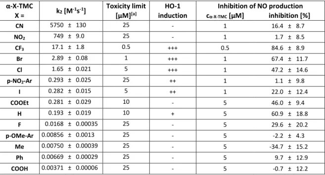
nucleophilic attack. The k 2 range for α-X-chalcones comprises a total of more than six or- ders of magnitude. Furthermore, the determined reactivity of α-X-TMCs was compared to their anti-inflammatory activity concerning HO-1 activation and NO inhibition. Structure- activity relationships were established. Kinetic and biological results are summarized in Ta- ble 1.

Reactivity of α-X-TMCs correlates with their HO-1 induction properties. The stronger the reactivity determined by the thiol assay, the stronger the HO-1 induction and thus inflam- mation inhibition, but the cell toxicity tends to increase simultaneously. The same goes for the NO-inhibition properties that follow the same order with α-CF 3 -TMC as most potent inhibitor, as it inhibits 84.6% of NO production at a very small concentration of 0.5 µM. Two obvious exceptions are the most reactive derivatives, α-CN- and α-NO 2 -TMC, in which no or only small anti-inflammatory activity and relatively low cell toxicity was detected. The electrophilic properties of these two substances are strong enough that they are most likely rapidly inactivated by the cellular detoxification agent glutathione (GSH, 7, Scheme 3).

Table 1: Results of the reactivity of α-X-TMCs determined by the kinetic thiol assay and biological data.

α-X-TMC

X = k

2

[M

-1

s

-1

] Toxicity limit [µM]

[a]

HO-1 induction

Inhibition of NO production c

α-X-TMC

[µM] inhibition [%]

CN 5750 ± 130 25 - 1 16.4 ± 8.7

NO

2

749 ± 9.0 25 - 1 1.7 ± 8.5

CF

3

17.1 ± 1.8 0.5 +++ 0.5 84.6 ± 8.9

Br 2.89 ± 0.08 1 +++ 1 67.4 ± 11.7

Cl 1.65 ± 0.021 5 +++ 1 47.2 ± 14.6

p-NO

2

-Ar 0.293 ± 0.025 25 ++ 1 1.1 ± 9.8

I 0.282 ± 0.015 5 ++ 1 22.0 ± 12.4

COOEt 0.281 ± 0.029 10 - 5 46.0 ± 9.4

H 0.193 ± 0.019 10 + 5 60.9 ± 18.8

F 0.0168 ± 0.00035 25 - 5 29.6 ± 20.2

p-OMe-Ar 0.00856 ± 0.0013 25 - 5 -2.2 ± 4.3

Me 0.00750 ± 0.00039 25 - 5 -34.7 ± 15.2

Ph 0.00669 ± 0.00029 25 - 5 9.7 ± 12.9

COOH 0.00371 ± 0.00006 25 - 5 -0.7 ± 12.2

The induction of HO-1 expression in RAW 264.7 macrophages was determined by Western Blot. +++ = strong induction; ++ = medium; + = weak induction in comparison to control; - = no induction. Kinetic and biological data were measured and/or analyzed by Nafisah Al-Rifai, Hannelore Rücker, Dita Fritsch and Sabine Am- slinger.

[35][36][37]

The excellent correlation of these data shows how well a reactivity study can predict the

biological activity of a substance class. Figure 7 visualizes this relationship by using the log-

arithmic k 2 and IC 50 values which give a linear correlation. With α-CF 3 -TMC, an optimal re-

activity/activity window was found for the α-X chalcones. However, as toxicity also in-

creases, the question arises whether optimization within the substance class could still be

(22)

achieved. It can also be promising to test potent active substances such as chalcones for further positive cell effects. In continued research, some selected compounds have been investigated in further assays concerning cytoprotectivity with very promising results for E- α-p-OMe-Ar-TMC. [38] A clear antiapoptotic effect was found at nontoxic concentrations.

Figure 7: Correlation of the inhibition of NO production with the second-order rate constants (lg(k

2

)) of the Michael addition of α-X-TMCs with an anti-inflammatory activity and cysteamine. Line indicates the linear fit.

A 1.6 α-X-Hydroxychalcones (α-X-HCs)

The investigation of chalcones was further developed towards an improvement of their reactivity and biological activity. Nafisah Al-Rifai synthesized 8 substituted, methoxylated chalcones with a 2’-OH group during her PhD thesis, called α-X-hydroxychalcones, α-X-HCs (University of Regensburg, 2014, Figure 8). [39]

Figure 8: α-X-HCs and their cysteamine addition products. Syntheses and investigations by Nafisah Al-Rifai (Dissertation, 2015, University of Regensburg).

[39]

Due to the free 2’-OH group, an enhancement of electrophilicity in comparison to α-X-

TMCs is expected, which might be beneficial also for the biological activity. Table 2 shows

the summary of the kinetic and biological investigations so far. Indeed, a two- or threefold

(23)

increased reactivity of α-F-HC and α-H-HC respectively in comparison to their TMC ana- logues was proven. [35] However, for all α-X-HCs a significant decrease or complete loss of toxicity and biological activity was found when compared to α-X-TMCs.

Table 2: Kinetic results and biological activities of α-X-HCs (unpublished results) compared to their corre- sponding α-X-TMCs.

[36]

α-X com- pound

X =

k

2

[M

-1

s

-1

] Cell viability IC

50

[µM]

Inhibition of NO production IC

50

[µM]

TMC HC TMC HC TMC HC

CN 5750

[a]

n. d. > 100 > 100 n.d. 30.8 ± 1.2

CF

3

17.1

[a]

n. d. 7.91 ± 0.98 > 100 0.120

[c]

n. a.

Br 2.89

[a]

n. d. 9.65 ± 1.04 43.1 ± 6.8 0.640

[c]

n. a.

Cl 1.65

[a]

n. d. 13.1 ± 2.4 57.0 ± 1.8 0.992

[c]

31.8 ± 7.4

p-NO

2

-Ar 0.293

[a]

n. d. > 100 > 100 n. d. 99.4 ± 11.4

H 0.193

[b]

0.717 ± 0.019 23.7 ± 5.7 37.5 ± 2.9 4.44

[c]

9.84 ± 3.32 F 0.0168

[a]

0.0347 ± 0.0029 > 100 52.2 ± 7.2 12.9

[c]

15.8 ± 3.0

Me 0.00750

[a]

n.d. > 100 > 100 n. d. 55.0 ± 21.3

[a]: Value taken from Al-Rifai et al.

[36]

; [b]: value taken from Amslinger et al.

[35]

; [c]: value taken from Rücker et al.

[37]

; n. a.: not active at non-toxic concentrations; n. d.: not determined.

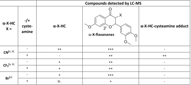
This effect is explained by the loss of the enone system due to the well-known cyclization to the corresponding flavanones (structure: α-X-flavanones, Table 3). [40] [41] The extent of this reaction depends on various factors, including the α-X-substituent. Table 3 shows UV- Vis ad LC-MS investigations of α-X-HCs under kinetic assay conditions. Indeed, flavanone formation was detected for all α-X-HCs containing an electron-withdrawing substituent X.

Due to this loss of the electrophilic position, the biomolecular mechanism of action is hin- dered or even prevented. This applies to the substances only in aqueous medium. All com- pounds were isolated as chalcones after synthesis and purification.

Table 3: Results of LC-MS studies of -X-HCs without and with 60-fold cysteamine.

Compounds detected by LC-MS

α-X-HC X =

-/+

cyste- amine

α-X-HC α-X-HC-cysteamine adduct

CN

[a, b]

- ++ +++ -

+ - ++ ++

CF

3[a, b]

- + ++ -

+ + ++ -

Br

[a]

- + +++ -

+ tr. + -

(24)

Compounds detected by LC-MS

Cl

[a]

- ++ +++ -

+ tr. + -

p-NO

2

-Ar - + +++ -

+ + +++ +

H

[a]

- > 95% tr. -

+ + - +++

F

[a]

- > 95% tr. -

+ +++ tr. ++

Me - > 95% tr. -

+ > 95% tr. tr.

Reactions were carried out in 100 mM Tris-HCl buffer pH 7.4 with 2 mM EDTA/ethylene glycol 20:80 with 40 µM of α-X-HCs and 1-2 h incubation time prior to measurement. HPLC column temperature: 40 °C. Molec- ular ratios were estimated by UV-Vis measurement and are assigned as follows: +++: 95-60%; ++: 60-30%; +:

30-5%; tr.: traces. [a]: Additional compounds (fragmentation and elimination products) were detected

[39]

; [b]:

measured directly after mixing and at 25 °C column temperature.

These results show that more aspects have to be considered when optimizing reactivity and activity, as this strategy only leads to limited success.

A 1.7 Synthesis of oxadiazoline enones (OXEs): a library of new electrophilic substances with an enone unit

The presented concept of a substance library containing a core unit, whose reactivity is influenced by variable substituents in order to optimize the biological activity, was applied in this work group to another, newly designed class of substances. The first results are taken from the doctoral thesis of Anas Rasras [42] and the Master theses of Rainer Herzog [43] and Lukas Wirth. [44]

Molecule design

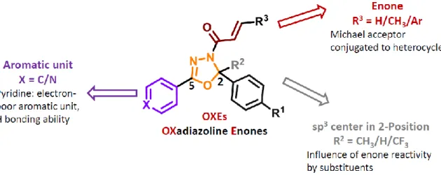
In the endeavor to create novel fine-tuned electrophiles, which are suitable model com- pounds for drug development, a new class of compounds was designed and synthesized in the work group of PD Dr. Sabine Amslinger. [42]

It was chosen to decorate a heterocyclic system with an additional enone unit, which is

able to bind covalently to pharmacological targets. Thereby it was important to use a scaf-

fold which allows for a conjugation with the enone unit to modify its reactivity. Thus, nine

classes of 3-enone-substituted 2,3-dihydro-1,3,4-oxadiazoles (oxadiazoline enones, OXEs)

were envisioned. These molecules were composed, according to Figure 9, from three vari-

ations in the 2-position, R 2 - a CH 3 , H or CF 3 group - together with three different enone

moieties in the 3-position and variations of the aromatic group attached to the 5-position

(R 1 ) of the oxadiazoline skeleton.

(25)

Figure 9: Design and structural features of the compound library of oxadiazoline enones (OXEs).

A combination of the two main structural features enone + R 3 and substituent in 2-position (R 2 ) leads to the nine different OXE classes named OXE1 to OXE9, which are summarized in Table 4. [43] [44]

Table 4: Overview and naming of nine OXE classes.

Enone + R

3

Substituent in 2-position / R

2

OXE class

acryl CH

3

OXE1

acryl H OXE2

acryl CF

3

OXE3

crotyl CH

3

OXE4

crotyl H OXE5

crotyl CF

3

OXE6

cinnamoyl CH

3

OXE7

cinnamoyl H OXE8

cinnamoyl CF

3

OXE9

The different substituents decorating the heterocyclic unit should influence the enone re-

activity by different effects. The 2-position was chosen to potentially influence the reactiv-

ity of the enone unit by a more indirect sp 3 -coupled variation in the electron density. Sub-

stituted phenyls and pyridine are the selected aromatic units in the 5-position. A stronger

modulation of electrophilicity was expected when the electron-poor 4-pyridyl-substituent

is introduced to the 5-position. Furthermore, pyridine provides a free electron pair to in-

teract with potential pharmacological targets via hydrogen bond. The largest effect was

anticipated when the 3-enone functionality itself was modified directly in the -position to

form acrylic, crotonic and cinnamic amides as the electrophilic units. A mesomeric connec-

tion between the enone carbonyl and the heterocycle is achieved via the free electron pair

of the nitrogen in the heterocyclic 3-position (Scheme 7).

(26)

Scheme 7: Mesomeric interaction of the oxadiazoline and enone units in OXEs.

There are few literature-known examples for these kinds of compounds. Prior to the work presented here, a set of six compounds having these structural features were already pub- lished, [45] however without the involvement of the pyridine unit (Figure 10). Since they carry a cinnamoyl unit in the 3-position and are 2-H-substituted, these molecules would be classified as OXE8s. They were tested for antibacterial activity on different bacterial cell lines with promising results.

Figure 10: Oxadiazoline derivatives synthesized and tested for antibacterial activity.

[45]

Synthesis of OXEs

The synthesis was done via a classical heterocyclic synthesis forming the 3,4-dihydro-1,3,4- oxadiazole ring in the last step of the sequence containing up to 4 steps (Scheme 8). [42]

Scheme 8: Synthesis of 2,5-diaryl-3,4-dihydro-1,3,4-oxadiazoles with additional 3-enone unit (OXEs).

Depending on the commercial availability of starting materials, the sequence was either

started from carboxylic acids 19 or esters 20 producing with hydrazine mono hydrate

known hydrazides 21, [46] which were transformed into the mostly known hydrazones 23 [47]

(27)

using acetophenones or benzaldehydes 22. In the last step, the heterocyclic ring was formed from hydrazones 23 in the presence of the corresponding acid chlorides 24.

A mechanism for the oxadiazoline ring formation is proposed in Scheme 9. [48] Basic condi- tions lead to the formation of anion 26 which cyclizes to 27 and is subsequently trapped by the electrophile 24.

Scheme 9: Proposed mechanism for the formation of OXEs

[48]

.

For the nomenclature, first the OXE class is named according to Table 4. Further substitu- ents are reflecting the type of aromatic unit in 5-position as well as p-substituents on the different aromatic rings R 1 and R 3 . Among the aromatic units, only the pyridine residue is termed; phenyl and aryl are not included in the name. The substituents are named in the following order:

1. Substituents on the aromatic ring in 2-position

2. Substituents on the aromatic ring in β-position of the enone unit (only in the case of cinnamoyl in OXE7s and OXE8s)

3. Substituents in 5-position (only pyridinyl or a 4-Ar substituent) and may placed in labelled brackets according to:

1. - 2. ()’

3. ()’’

Two examples given in Scheme 10 illustrate these explanations.

OXE class 2-H + cinnamoyl = OXE8 substituent on 2-Ar Cinnamoyl-Ar 5

OCH

3

NO

2

4-pyridinyl

NAME: OXE8-OMe,(NO

2

)’,(4py)’’

(28)

OXE class 2-CH

3

+ acryl = OXE1 substituent on 2-Ar Cinnamoyl-Ar 5

- - Ar-NO

2

NAME: OXE1-(NO

2

)’’

Scheme 10: Examples for the nomenclature developed for the OXE substance library.

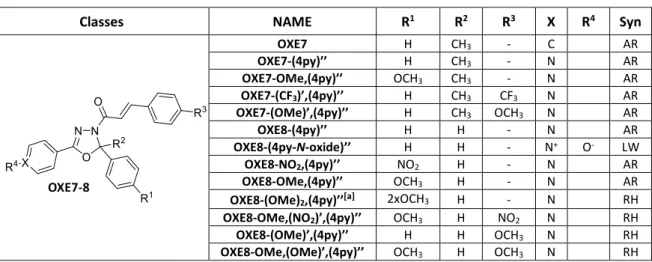
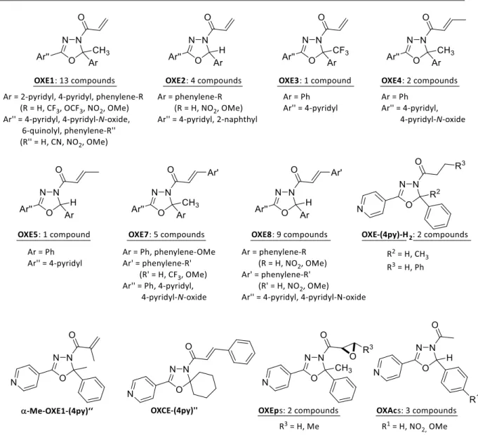
Since it is known that N-oxides of pyridines can show special binding properties, three pyr- idinyl-substituted OXE compounds were turned into their corresponding N-oxide deriva- tives [44] using the oxidation reagent mCPBA (m-chloroperbenzoic acid). [49] All synthesized OXE compounds are summarized in Table 5 and Figure 11, which reflects seven different classes of OXEs from the nine classes that were envisioned in principle. Attempts to syn- thesize representatives of OXE6 and OXE9 were not successful. Additionally, one control compound was synthesized, OXE1-(4py)’’-H 2 , which represents the dihydro derivatives lacking the enone motif. One α-substituted compound α-Me-OXE1-(4py)’’ was synthe- sized. More representatives with different structural features were included in the library:

the spiro-compound OXCE-(4py)’’ and two compounds with quinolinyl and naphthyl sub- stituents in position 5 (Figure 11).

Table 5: Overview of OXEs of the classes 1-5 and 7-8 synthesized in the Amslinger group.

Classes NAME R

1

R

2

R

3

X R

4

Syn

OXE1 H CH

3

- C H LW

OXE1-(NO

2

)’’ H CH

3

- C NO

2

RH

OXE1-(CN) ’’ H CH

3

- C CN RH

OXE1-(4py)’’ H CH

3

- N - AR

OXE1-NO

2

,(4py)’’ NO

2

CH

3

- N - AR OXE1-(4py-N-oxide)’’ H CH

3

- N

+

O

-

LW OXE1-CF

3

,(4py)’’ CF

3

CH

3

- N - AR OXE1-OCF

3

,(4py)’’ OCF

3

CH

3

- N - AR

OXE1-4py,(4py)’’ 4-py CH

3

- N - Ar

OXE1-2py,(4py)’’ 2-py CH

3

- N - Ar

OXE2-(4py)’’ H H - N - RH

OXE2-NO

2

,(4py)’’ H H - N - RH

OXE2-OMe,(4py)’’ H H - N - RH

OXE3-(4py)’’ H CF

3

- N - RH

OXE4-(4py)’’ - CH

3

- N - AR

OXE4-(4py-N-oxide)’’ - CH

3

- N - LW

OXE5-(4py)’’ - H - N

+

O

-

LW

(29)

Classes NAME R

1

R

2

R

3

X R

4

Syn

OXE7 H CH

3

- C AR

OXE7-(4py)’’ H CH

3

- N AR

OXE7-OMe,(4py)’’ OCH

3

CH

3

- N AR

OXE7-(CF

3

)’,(4py)’’ H CH

3

CF

3

N AR

OXE7-(OMe)’,(4py)’’ H CH

3

OCH

3

N AR

OXE8-(4py)’’ H H - N AR

OXE8-(4py-N-oxide)’’ H H - N

+

O

-

LW

OXE8-NO

2

,(4py)’’ NO

2

H - N AR

OXE8-OMe,(4py)’’ OCH

3

H - N AR

OXE8-(OMe)

2

,(4py)’’

[a]

2xOCH

3

H - N RH OXE8-OMe,(NO

2

)’,(4py)’’ OCH

3

H NO

2

N RH

OXE8-(OMe)’,(4py)’’ H H OCH

3

N RH

OXE8-OMe,(OMe)’,(4py)’’ OCH

3

H OCH

3

N RH Syn = Synthesized by: AR = Anas Rasras, RH = Rainer Herzog, LW = Lukas Wirth; [a]: the 3,4-dimethoxy com- pound was produced.

Figure 11: OXEs with further structural features synthesized in the Amslinger work group. Synthesized by: AR

= Anas Rasras, RH = Rainer Herzog, LW = Lukas Wirth.

Synthesis of OXE analogues: epoxides and acyl-substituted oxadiazolines

Additionally, the epoxide functionality as alternative electrophilic unit was of interest. This

functional group also occurs in natural products that are able to bind covalently to nucleo-

philic amino acid side chains in active sites of certain enzymes by undergoing epoxide open-

ing. [12, 50] Some epoxide-containing compounds have been proven to bind to thiol groups

of cysteines, e.g. in cysteine proteases, in particular. [51] Antimicrobial, anti-angiogenetic

and anti-inflammatory effects are described, [12, 52] making these compounds important in

drug discovery and development for decades. To investigate the effect of a different elec-

trophilic moiety on the OXE compounds, two oxadiazoline representatives with an addi-

tional epoxide were synthesized from their OXE1 and OXE4 analogues using n-

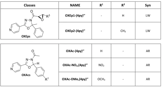
BuLi/tBuOOH, [53] whereby two compounds were produced [44] (Table 6). As further control

compounds, three representatives of OXAc-(4py)’’s known from the literature where

(30)

synthesized. [54] OXAcs stem from OXE2-(4py)’’ and carry an acyl group in 3-position instead of acryl (Table 6).

Table 6: Synthesized OXE analogues: OXEp and OXAc compounds. Syn = synthesized by: AR = Anas Rasras, LW = Lukas Wirth.

Classes NAME R

1

R

3

Syn

OXEp1-(4py)’’ - H LW

OXEp2-(4py)’’ - CH

3

LW

OXAc-(4py)’’ H - AR

OXAc-NO

2

,(4py)’’ NO

2

- AR

OXAc-OMe,(4py)’’ OCH

3

- AR

Synthesis of oxadiazol-thioesters

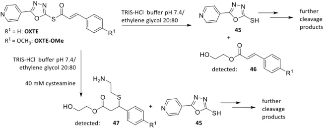
For a variation of the heterocyclic scaffold, a further structure concept differing from the OXE skeleton was designed. It was constructed from a saturated oxadiazole core unit with the enone moiety linked to the heterocycle in 2-position via a thioether and also pyridinyl in 5-position. Two compounds of this type where synthesized and named oxadiazol-thio- esters OXTE and OXTE-OMe (Table 7). [42]

Table 7: Oxadiazol compounds synthesized by Anas Rasras. Syn = synthesized by AR = Anas Rasras.

Structure NAME R

1

Syn

OXTE H AR

OXTE-OMe CH

3

AR

A 1.8 Synthesis of further electrophiles in the Amslinger work group

Phenylacrylamide derivatives as reactive units in drug development

Reactive units that differ from the enone functionality are able to react as Michael accep-

tors with nucleophilic pharmacological targets as well. Before combining those varied re-

active units to drug-like fragments, the assessment of a structure-reactivity relationship is

useful to find out if the electrophilicity of the unit is tunable by different substituents as

described before for α-X-TMCs. For that purpose, a library of simplified molecules of the

(31)

aromatic acrylamide type and of the phenylethene sulfonamide type were synthesized by Lukas Wirth. [44] Figure 12 gives an overview of all synthesized compounds.

Figure 12: N-Phenylacrylamide and phenylethenesulfonamide derivatives synthesized by Lukas Wirth.

[44]

For compounds 29 and 30 only the unsubstituted and the pyridine derivatives were syn- thesized. With 31a and 31b an unsubstituted and a OMe-decorated representative of N- phenylethenesulfonamides was obtained. But in the case of N-phenylacrylamides 32, a li- brary of 16 compounds with a great variety of electron-withdrawing and electron-pushing substituents in various positions was produced. Although OXEs can be considered as en- amide derivatives as well, the difference between acrylamides and OXEs is that the amide nitrogen is not included in a heterocycle but N-aryl substituted.

Reactivity assessment methods for electrophiles binding to thiols in a covalent fashion were previously reported also for this so-called fragment-based approach. [55] [30] The aim is to establish a structure-reactivity relationship so that the reactivity can easily be tuned by simply exchanging substituents. These reactivity fragments can subsequently be combined to one or more small, drug-like fragments to find the optimal toxicity/activity window for a certain biological effect. Since it has often been shown that reactivity and biological activity correlate, electrophilicity can be used as a modification tool in the first place.

Juglone and Juglone derivatives

Another project dealing with the synthesis, kinetic and biological evaluation of small elec-

trophilic molecules was the investigation of the natural product juglone (5-

(32)

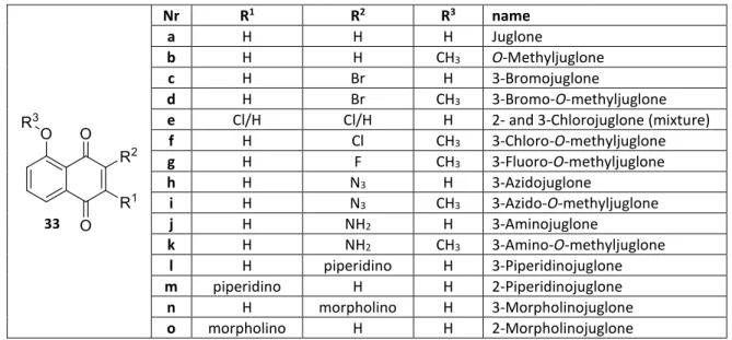
hydroxynaphthalene-1,4-dione, 33a), a naphthoquinone found in Juglans (walnut) spe- cies [56] in the course of the Master thesis of Sebastian Schlegel. [57] Table 8 summarizes the substance library of 33a and 14 of its synthesized derivatives, which are O-methylated (R 3 ) and/or substituted in position 2 or 3 (R 1 and R 2 ). Compound 33e could only be obtained as a mixture of two chloro-substituted regioisomers due to separation issues.

Table 8: Structures and names of Juglone 33a and its derivatives synthesized by Sebastian Schlegel.

[57]

Nr R

1

R

2

R

3

name

a H H H Juglone

b H H CH

3

O-Methyljuglone

c H Br H 3-Bromojuglone

d H Br CH

3

3-Bromo-O-methyljuglone

e Cl/H Cl/H H 2- and 3-Chlorojuglone (mixture)

f H Cl CH

3

3-Chloro-O-methyljuglone

g H F CH

3

3-Fluoro-O-methyljuglone

h H N

3

H 3-Azidojuglone

i H N

3

CH

3

3-Azido-O-methyljuglone

j H NH

2

H 3-Aminojuglone

k H NH

2

CH

3

3-Amino-O-methyljuglone

l H piperidino H 3-Piperidinojuglone

m piperidino H H 2-Piperidinojuglone

n H morpholino H 3-Morpholinojuglone

o morpholino H H 2-Morpholinojuglone

Biological activity such as anticancer [58] , anti-inflammatory [59] and antimicrobial [60] [61] activ- ity of 33a has been described, which is explained by the redox properties of the quinone system, including the formation of radical species, as well as Michael addition with nucle- ophiles of various pharmacological targets. [62] Therefore, not only studies of the biological activity but also the reactivity of juglone and some derivatives are published. For example, the thia-Michael addition of hydrogen sulfide [63] and GSH [64] to juglone (33a) were kinet- ically investigated. Furthermore, a study was published on the relationship between the reactivity of the addition-elimination reaction of 3-OR-substituted juglone derivatives and their anticancer activity. [65]

In the work group of PD Dr. Sabine Amslinger the compounds were tested towards their anti-inflammatory properties in the NO inhibition assay. All compounds with an amine sub- stituent in 2- or 3-position (33j-o) and bromo-substituted 33d provided anti-inflammatory activity in the µM range. 3-Amino-O-methyljuglone 33k was the most active substance with an IC 50 of 1.10 µM. All other derivatives were inactive at nontoxic concentrations. All IC 50

values in the MTT toxicity assay were determined in the range of 4.05 – 63.3 µM with a

medium of 30.0 µM; no relation between toxicity and anti-inflammatory activity was

found. [57] Biological results determined by Sebastian Schlegel and Sina Malenke are sum-

marized only for compounds with an anti-inflammatory activity in Table 9.

(33)

Table 9: Biological data of Juglone derivatives with anti-inflammatory properties. Evaluation of toxicity and NO-inhibition properties in murine macrophages RAW264.7. Cell viability (MTT) assay was performed and evaluated by Sebastian Schlegel. Anti-inflammatory (iNOS) assay was performed by Sina Malenke and evalu- ated by Sina Malenke and Monika Enzinger.

Compound Cell viability MTT

(IC

50

/ µM) ± SD

Inhibition of NO production (IC

50

/ µM) ± SD

3-Aminojuglone 33j 36.7 ± 8.1 5.95 ± 2.20

3-Amino-O-methyljuglone 33k 18.8 ± 6.9 1.10 ± 0.49

3-Piperidinojuglone 33l 63.3 ± 19.8 9.48 ± 0.66

2-Piperidinojuglone 33m 41.8 ± 7.8 4.18 ± 0.48

3-Morpholinojuglone 33n 36.4 ± 3.3 12.3 ± 8.8

2-Morpholinojuglone 33o 30.9 ± 5.0 17.0 ± 7.8

Bromo-O-methyljuglone 33d 22.5 ± 12.4 5.82 ± 1.09

A 1.9 Further development of the kinetic thiol assay towards the investigation of colorless electrophiles

Purpose and concept of the new assay

The previous chapters have dealt with enones that have an absorbance in the range of 300- 350 nm or higher. But there are some interesting electrophiles which are not suitable for the presented assay due to unsuitable absorbance properties. Figure 13 shows examples for compounds which absorbance bands do not extend as far as < 300 nm and can there- fore not be kinetically evaluated by the thiol assay.

Figure 13: Compounds that cannot be examined by the kinetic thiol assay due to insufficient absorbance properties.

Compounds 34 (cyclohexenone) and 35 (phenylvinylketone or acrylophenone) are struc- tural motifs that occur in biologically active molecules. For example, antimicrobial [66] and EGF-receptor (epidermal growth factor receptor) inhibition [67] properties have been found for 35, which are probably at least partly based on their Michael acceptor activity. Based on the kinetic properties of these two simple test substances, it would also be possible to estimate the reaction rates of substances containing these structural elements. 15d-PGJ2 (4, 15-deoxy-Δ12,14-prostaglandine J2) is the most active representative of cyclopente- none-type prostaglandins which show strong anti-inflammatory activity in vitro and in vivo.

This is based, among other mechanisms, on their Michael acceptor activity provided by the

Abbildung

Figure 2: Schematic mechanism of the NF-κB signal transduction pathway. NF-κB: nuclear factor kappa B, IKK:
Table 1: Results of the reactivity of α-X-TMCs determined by the kinetic thiol assay and biological data
Table 3: Results of LC-MS studies of -X-HCs without and with 60-fold cysteamine.
Figure 9: Design and structural features of the compound library of oxadiazoline enones (OXEs)
+7

Referenzen

ÄHNLICHE DOKUMENTE

CCDC ID Formula Formula weight Z, calculated density F000 Description and size of crystal Absorption coefficient Min/max transmission Temperature Radiation wavelength Crystal

Midven- tral scale rows of this sample appear to show some geographical variation in this series, 34-37 on specimens (n = 6) from Kaptai and 38-42 on specimens (n = 5)

This two-compartment model consisted of a fast elimination and distribution phase (T 1/2 about 20 h) followed by a slow elimination phase (renal clearance about 0.11 ml/min.) and

The analyses did not find any explicit relationships for the last non-identifiable parameter, K i6Suc6P. However, it was found that the preceding measurements were suffi- cient to

The NH 4 + ion generates medium to strong hydrogen bonds with the car- bonylic oxygen, the iminic nitrogen and the sulfonyl oxygen atoms of the acesulfamate anion.. The FTIR spectrum

The Zn(II) cation in the [Zn(bispicen)Cl(H 2 O)] + complex is in a distorted octahedral environment, coordinated to a neu- tral bispicen molecule acting as a tetradentate ligand

Ogorodnyk Inorganic Chemistry Department, Kiev University, Volodymyrska Street 64, Kiev 01033, Ukraine Reprint requests to A.. The X-ray sin- gle crystal structure and

Key words: Phosphates, Crystal Structure, Langbeinite, Magnetic