Characterization of the Anti-inflammatory Activity of Enones Based on the Evaluation of Their Heme Oxygenase-1 and Inducible NO Synthase Activity
Dissertation
zur Erlangung des Doktorgrades der Naturwissenschaften Dr. rer. nat.
an der Fakultät der Chemie und Pharmazie der Universität Regensburg
vorgelegt von Hannelore-Maria Rücker
aus
Timisoara, Rumänien
Regensburg 2014
Promotionsgesuch eingereicht am: 05.03.2014
Diese Arbeit wurde angeleitet von PD Dr. Sabine Amslinger.
Content
1 Introduction ... 1
1.1 α,β-Unsaturated carbonyl compounds and inflammation ... 1
1.2 Heme oxygenase-1 (HO-1) ... 5
1.2.1 Activity and induction of HO-1 ... 5
1.2.2 HO-1 as therapeutic target ... 6
1.2.3 Techniques for measuring HO-1 activity ... 6
1.2.4 Development of a HO-1 activity assay ... 8
1.3 The activity of inducible nitric oxide synthase (iNOS) ... 11
1.3.1 The nitrite (Griess) assay ... 12
1.4 The oxygen radical absorbance capacity-(ORAC)-fluorescein assay ... 12
1.5 Anti-inflammatory activity of a diverse group of α,β-unsaturated carbonyl compounds and polyphenols ... 14
1.6 Reactivity and biological activity of natural and synthetic chalcones ... 18
1.6.1 Reactivity of chalcones ... 18
1.6.2 Reactivity assessment of chalcones by a kinetic thiol assay ... 20
1.6.3 Biological activity of chalcones ... 20
1.7 α-X-Modified enones as a different approach in fine-tuning their Michael acceptor reactivity and biological activity ... 23
1.7.1 α-X-Modification in 2’,3,4,4’-tetramethoxychalcones (α-X-TMCHs) ... 23
1.7.2 Limno-CP and its α-X-Limno-CP derivatives ... 25
1.8 The anti-inflammatory activity of both enantiomers of arteludovicinolide A ... 26
1.9 Enzyme-triggered CO-releasing molecules (ET-CORMs) ... 27
1.10 Aim of this study ... 29
2 Materials and Methods ... 30
2.1 Materials... 30
2.1.1 Cell lines ... 30
2.1.2 Cell culture media, buffers and reagents for cell culture ... 30
2.1.3 Antibodies, proteins and enzymes ... 31
2.1.4 Kits... 31
2.1.5 Chemicals and reagents ... 31
2.1.6 Synthesis of compounds ... 33
2.1.7 Buffers and solutions ... 34
2.1.8 Equipment ... 37
2.1.9 Consumables ... 39
2.2 Methods ... 40
2.2.1 Cell culture ... 40
2.2.2 Dilution of test compounds ... 40
2.2.3 Viability assay (MTT assay) ... 41
2.2.4 Viability assay with lipopolysaccharide (MTT-LPS assay) ... 42
2.2.5 Nitrite assay (Griess assay) ... 43
2.2.6 Activity assay for heme oxygenase-1 (HO-1) ... 44
2.2.7 Western blot analysis ... 51
2.2.8 ORAC-fluorescein assay ... 53
2.2.9 Statistical analysis ... 54
3 Results and Discussion ... 55
3.1 Heme oxygenase-1 (HO-1) activity assay ... 55
3.1.1 Development and optimization of the HO-1 activity assay... 55
3.1.2 Time course of HO-1 protein expression and HO-1 activity in RAW264.7 macrophages exposed to chalcone DHDMCH ... 65
3.1.3 Inhibition of HO-1 activity in DHDMCH or LPS stimulated RAW264.7 macrophages by SnPPIX ... 68
3.1.4 Heme oxygenase-1 activity in human dendritic cells (DC) ... 69
3.2 Screening of natural products and drugs towards their HO-1 activity in RAW264.7 macrophages ... 71
3.2.1 Influence of natural products and drugs on the viability of RAW264.7 macrophages ... 71
3.2.2 Effect of natural products as well as the two drugs oltipraz and dexamethasone on
HO-1 activity in RAW264.7 macrophages ... 73
3.2.3 Effect of natural products and dexamethasone on the HO-1 protein expression in RAW264.7 macrophages ... 77
3.3 Characterization of chalcones towards their anti-inflammatory, antioxidative and cytoprotective activity... 79
3.3.1 Effect of chalcones on the viability of RAW264.7 macrophages ... 79
3.3.2 Influence of chalcones on HO-1 activity and HO-1 protein expression ... 81
3.3.3 Effect of chalcones on nitrite production ... 84
3.3.4 Antioxidant capacity of chalcones ... 86
3.3.5 Structure-activity relationship (SAR) of hydroxy- and methoxychalcones ... 88
3.4 Characterization of α-X-TMCHs towards their anti-inflammatory, antioxidative and cytoprotective activity ... 92
3.4.1 Effect of α-X-TMCHs on the viability of RAW264.7 macrophages ... 92
3.4.2 Influence of α-X-TMCHs on HO-1 activity and HO-1 protein expression ... 95
3.4.3 Effect of α-X-TMCHs on nitrite production ... 97
3.4.4 Conclusion ... 100
3.5 Characterization of α-X-Limno-CP derivatives (5-aryl-3(2H)-furanones) towards their anti-inflammatory and antioxidative activity ... 102
3.5.1 Effect of α-X-Limno-CPs on cell viability and nitrite production of RAW264.7 macrophages ... 102
3.5.2 Antioxidant capacity of α-X-Limno-CPs ... 105
3.5.3 Conclusion ... 106
3.6 Characterization of both enantiomers of arteludovicinolide A towards their anti- inflammatory activity ... 108
3.6.1 Influence on the viability and nitrite production of RAW264.7 macrophages ... 108
3.6.2 Influence of both enantiomers of arteludovicinolide A on the heme oxygenase-1 (HO-1) activity in murine macrophages RAW264.7 ... 109
3.6.3 Summary ... 110
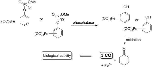
3.7 The anti-inflammatory activity of iron dienylphosphate tricarbonyl complexes as enzyme-triggered CO-releasing molecules (ET-CORMs) ... 111
3.8 Characterization of further compounds towards their cytotoxic, antioxidative and
anti-inflammatory activity ... 116
3.8.1 Cytotoxic activity of two γ-butyrolactone derivatives on the human colon cancer cell line HT-29 ... 116
3.8.2 Biological activity of different sesquiterpene lactone derivatives, γ-butyrolactones and 4-substituted cyclopentenones ... 116
3.8.3 Biological activity of chalcone-analogs ... 121
4 Summary... 122
5 References... 125
ABBREVIATIONS
2’-HOC 2’-Hydroxychalcone
AAPH 2,2'-Azobis(2-methylpropionamidine)
dihydrochloride
AP-1 Activator protein 1
APS Ammonium persulfate
ARE Antioxidant response element
AUC Area under curve
BARD Bardoxolone methyl
BR Bilirubin
BSA Bovine serum albumin
BV Biliverdin
BVR Biliverdin reductase
CDDO 2-Cyano-3,12-dioxooleana-1,9(11)-dien-28-
oic acid, bardoxolone
CH Chalcone, (1,3-diphenylprop-2-en-1-one)
CO Carbon monoxide
CORMs CO releasing molecules
COX Cyclooxygenase
DC Dendritic cells
DHDMCH 2’,4’-Dihyxdroxy-3,4-dimethoxychalcone
DMEM Dulbecco’s modified eagle’s medium
DMSO Dimethyl sulfoxide
DPPH 2,2-Diphenyl-1-picrylhydrazyl
EDTA Ethylenediaminetetraacetic acid
ELISA Enzyme-linked immunosorbent assay
eNOS Endothelial NO-synthase
ET-CORMs Enzyme-triggered CO-releasing molecules
FCS Fetal calf serum
GSH Glutathione
GST Glutathione S-transferase
HEI-OC1 cells House ear institute-organ of corti 1 cells
HO Heme oxygenase
HPLC High performance liquid chromatography
HTMCH 2’-Hydroxy-3,4’,4-trimethoxychalcone
HUVEC Human umbilical vein endothelial cells
ICAM-1 Intercellular adhesion molecule-1
IFN-γ Interferon-γ
IKK IκB kinase
IL Interleukin
iNOS Inducible NO-synthase
IP-10 Interferon γ-induced protein 10
ISL Isoliquiritigenin
IκB Inhibitor of NF-κB
Keap1 Kelch-like ECH-associated protein 1
LC-MS Liquid chromatography–mass spectrometry
LPS Lipopolysaccharide
MA Michael acceptor
MAPK Mitogen-activated protein kinase
MCE Monocyclic cyanoenone
MTT 3-[4,5-Dimethylthiazol-2-yl]-2,5-diphenyl-
tetrazoliumbromide
NADPH β-Nicotinamide adenine dinucleotide phos-
phate
NEA Non essential amino acid
NED N-(1-naphthyl)ethylenediamine
Nfr2 Nuclear factor erythroid 2 related factor 2
NF-κB Nuclear factor κB
nNOS Neuronal NO-synthase
NO Nitric oxide
NQO1 NAD(P)H:quinine oxidoreductase 1
ns Not significant
OPD ortho-Phenylenediamine dihydrochloride
ORAC Oxygen radical absorbance capacity
PBS Phosphate buffered saline
PEGF Platelet-derived growth factor
ROS Reactive oxygen species
RP-HPLC Reversed phase high performance liquid
chromatography
rpm Revolutions per minute
RPMI 1640 medium Roswell Park Memorial Institute 1640 medi-
um
RT Room temperature
RTQ-PCR Real-time quantitative polymerase chain reac-
tion
SAR Structure-activity relationship
SDS Sodium dodecyl sulfate
SERS Surface enhanced Raman scattering
TEMED N,N,N’,N’-Tetramethylethylenediamine
TGF-1β Transforming growth factor-1β
THMCH 2’,3,4’-Trihydroxy-4-methoxychalcone
TMCH 2’,3,4,4’-Tetramethoxychalcone
TNF-α Tumor necrosis factor-α
T-PBS Tween 20-phosphate buffered saline
TRIS Tris(hydroxymethyl)aminomethane
VEGF Vascular endothelial growth factor
1
1 Introduction
1.1 α,β-Unsaturated carbonyl compounds and inflammation
α,β-Unsaturated carbonyl compounds possessing a Michael acceptor functionality represent a prominent class of biologically and pharmacologically active electrophiles amongst the vast number of electrophilic compounds with numerous biological targets.1 Of great importance are thiol-regulated signal transduction pathways essential for the cellular redox homeostasis and cell protection. Especially under pathological conditions involving chronic inflammation, athero- sclerosis, diabetes, liver, lung and brain injury, kidney disease and cancer, these particular re- dox-sensitive and cytoprotective signaling cascades are promising targets of therapeutic drugs.2 Already in the late 1980’s Talalay et al. reported that the beneficial biological activity of chemoprotective agents is directly connected to their Michael acceptor activity.3 On one hand, α,β-unsaturated carbonyl compounds possess a distinct Michael acceptor reactivity by which they can react with nucleophilic sulfhydryl groups of sensor cysteins on key signaling proteins.
On the other hand the α,β-unsaturated carbonyl moiety itself as well as additional groups, i.e.
phenolic hydroxyls can react as radical scavengers or antioxidants due to their pronounced re- duction potential (Figure 1).4
Figure 1. Reactivities of α,β-unsaturated carbonyl compounds.
The electrophilic nature of the α,β-unsaturated carbonyl unit enables many natural products, which possess this functionality to act as powerful antioxidant, anti-inflammatory, neurochemo- protective and cancer chemopreventive agents. A high drug design potential emerges from the class of Michael acceptors because they can selectively address certain cellular targets in fairly complex signaling pathways, due to a distinct and moderate electrophilic behavior. Prominent α,β-unsaturated compounds, where their Michael acceptor reactivity was shown to be closely related to their biological potency, comprise phytochemicals such as the food polyphenols curcumin, butein and isoliquiritigenin (ISL), endogenous inducers like the cyclopentenone pros-
2
taglandin 15-deoxy-Δ12,14-prostaglandin J2 (15d-PGJ2
)
5 and the highly potent pentacyclic triterpenoids (CDDOs) as well as the monocyclic cyanoenone (MCE) relatives6-9 (Figure 2).Figure 2. Structures of prominent α,β-unsaturated compounds as biological active Michael acceptors. 15d- PGJ2, 15-deoxy-Δ12,14-prostaglandin J2; CDDO, bardoxolone; BARD, bardoxolone methyl; MCE, monocyclic cyanoenone.
α,β-Unsaturated carbonyl compounds can act as redox active agents by directly neutralizing reactive oxygen species (ROS) such as the superoxide radical (O2•-), hydroxyl radical (•OH) and hydrogen peroxide (H2O2) produced as a consequence of unbalanced biochemical processes in the body (e.g. in the mitochondria or under chronic inflammation) or as a result of increased exposure to xenobiotics, thus reinstalling redox homeostasis.2 A more important reactivity of the α,β-unsaturated carbonyl compounds is their ability to modulate certain sulfhydryl groups on cysteine-dependent signaling pathways. Two major signaling pathways can be targeted by α,β- unsaturated carbonyl compounds particularly leading to beneficial effects: the inflammatory signaling pathway regulated by the transcriptional factor NF-κB and the redox-sensitive and anti-inflammatory Keap1/Nrf2/ARE signaling system.
The transcriptional factor Nrf2 (NF-E2 related factor 2) is a member of the cap’n collar family of basic leucine transcription factors. Under basal conditions Nrf2 is sequestered in the cytosol as an inactive complex with Keap1 (Kelch-like ECH-associated protein 1). Electrophiles and other inducers can modify critical surface SH groups of cysteins (e.g. Cys151, Cys273 and Cys288) of Keap1 covalently or by oxidation.10 This leads to a conformational change in Keap1, thereby al- lowing Nrf2 to stabilize and accumulate in the nucleus. Here, the Nrf2-dependent gene expres- sion is triggered by binding of Nrf2 to the antioxidant response element ARE on the 5’ upstream DNA sequence. The phase II proteins encoded by the Nrf2/ARE-regulated genes are detoxifying proteins and enzymes controlling the redox status of the cell, possessing direct antioxidant activ- ity and synthesizing endogenous reducing agents such as glutathione (GSH), regulators of apop-
3
tosis, cell cycle and differentiation, heat shock proteins, regulators of immune response and in- flammation and enzymes involved in cellular metabolism11-12 (Figure 3).
Figure 3. The molecular mechanism of the redox-sensitive Keap1/Nrf2/ARE signaling pathway. Under basal conditions Nrf2 is inactivated by the chaperon protein Keap1 in the cytosol. α,β-Unsaturated carbonyl com- pounds (α,β) can modify certain SH groups of cysteines of Keap1 covalently or by oxidation. This leads to a conformational change in Keap1, which dissociates from Nrf2. The free form of Nrf2 translocates into the nucleus where it binds to the antioxidant response element (ARE) on the DNA and triggers the expression of anti-inflammatory and cytoprotective proteins.
The Keap1/Nrf2/ARE system acts as a master switch13 in the cellular redox stress response and mediates the cytoprotective signaling, especially by the induction of NAD(P)H:quinine oxido- reductase 1 (NQO1), glutathione S-transferase (GST) and heme oxygenase-1 (HO-1). Due to its sensing mechanisms activated by a wide range of electrophiles and antioxidants of natural or synthetic origin, the Keap1/Nrf2/ARE system is raised to a promising target against the devel- opment of several diseases, such as cancer, diabetes, neurodegenerative and cardiovascular dis- orders involving inflammation and oxidative stress.14-17
The nuclear factor-kappa B (NF-κB) is a major and ubiquitous transcription factor implicated in the immune and inflammatory responses through the regulation of genes encoding pro- inflammatory cytokines (IL-1β, IFN-γ, TNF-α), adhesion molecules (ICAM-1), chemokines (IP-10, IL-8), growth factors (PEGF, VEGF) and inducible enzymes such as cyclooxygenase 2 (COX-2) and inducible nitric oxide synthase (iNOS). It consists of homo- and heterodimers of the Rel pro- tein family (RelA (p65), RelB, cRel, p50 and p52) and is kept inactive in the cytosol through an association with an inhibitory protein of the IκB family (inhibitor of NF-κB). Following cell stim- ulation, NF-κB is released, accumulating within the nucleus, binding to the 5’ downstream κB promoter region on the DNA and inducing the transcription of inflammatory genes. The activa-
4
tion of NF-κB is mediated by the IκB kinase (IKK) complex, causing the phosphorylation of IκB followed by a proteasomal degradation. Electrophiles like α,β-unsaturated carbonyl compounds can inhibit the NF-κB pathway by reacting with specific SH groups of the cysteine residues of IKK, thus abolishing the NF-κB activation. Alternatively, they can directly modulate SH groups of cysteines of the NF-κB subunits, suppressing the DNA binding activity of the transcriptional fac- tor and causing a down regulation of the protein expression of inflammatory proteins and cyto- kines (Figure 4).
Figure 4. Inhibition of the inflammatory NF-κB signaling pathway by electrophiles. NF-κB is kept inactive through an association with the inhibitor IκB. The activation of NF-κB is mediated by the IκB kinase (IKK) complex, causing the phosphorylation of IκB. Subsequently, NF-κB is released and binds to the κB promoter region on the DNA inducing the transcription of inflammatory genes. α,β-Unsaturated carbonyl compounds (α,β) can react with SH groups of the cysteines of IKK, thus abolishing the NF-κB activation. Alternatively, the DNA binding of the active NF-κB can be inhibited, which leads to a suppression of the protein expression of inflammatory proteins and a reduced immune response.
The NF-κB pathway was shown to be activated by a wide range of stimuli including inflammato- ry cytokines (TNF-α), endotoxins (lipopolysaccharide, bacteria, viruses), growth factors (TGF- 1β), reactive oxygen species (ROS), therapeutic drugs (Taxol, acetylsalicylic acid), environmental hazards (heavy metals, cigarette smoke) and several chemical agents. Although NF-κB is essen- tial for normal T and B cell development in the cellular defense system and in the expression of stress response proteins (COX-2, iNOS), its dysfunction leads to various diseases such as athero- sclerosis, multiple sclerosis, Alzheimer’s disease, inflammatory bowel disease, neuropathological and renal diseases, asthma, diabetes and cancer. The inhibition of the NF-κB pathway is there- fore regarded as a potential therapeutic approach in inflammation and cancer.18-21
5
1.2 Heme oxygenase-1 (HO-1)
1.2.1 Activity and induction of HO-1
Heme oxygenase-1 (HO-1) is a redox sensitive, inducible stress protein converting heme to CO, Fe2+ and biliverdin (BV), which is further reduced to bilirubin (BR) by biliverdin reductase (BVR).
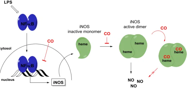
These products are particularly important for the overall chemopreventive, chemoprotective and anti-inflammatory activities of HO-1 (Scheme 1). Biliverdin and bilirubin act as radical scav- engers through their conjugated π-system that leads to a decrease of reactive oxygen species, ROS. CO is an inhibitor of proinflammatory heme-containing proteins such as inducible NO syn- thase (iNOS) or cyclooxygenase-2 (COX-2) and the released iron (II) ion can induce the anti- inflammatory protein ferritin. Finally, the breakdown of free heme itself can reduce oxidative damage, since heme acts as a promoter in the Fenton reaction.22-25
Scheme 1. Heme degradation catalyzed by heme oxygenase-1 (HO-1) and biliverdin reductase (BVR) together with the resulting cytoprotective, anti-inflammatory and antioxidative effects.
As a member of the cytoprotective phase II enzymes the transcription of HO-1 is mainly regulat- ed by the Keap1/Nrf2/ARE signaling pathway.12 Amongst other stimuli of HO-1 induction such as UV light, heavy metals, oxidative stress,26 especially electrophiles like α,β-unsaturated car- bonyl compounds can react with nucleophilic sulfhydryl groups of the Nrf2-complexing chaper- on Keap1.27-28 Thereby the Nrf2 regulated antioxidant-responsive element (ARE) is activated which leads to a transcriptional induction of HO-1 (Figure 3). Other regulatory mechanisms of HO-1 gene induction have been discussed, such as the MAPK (mitogen-activated protein kinases) signaling pathway,29 several kinases24 and transcriptional factors, such as NF-κB and AP-1 (acti- vator protein 1).30-31 However, the specific MAPK and/or other kinases involved in the HO-1 in- duction appear to vary in an inducer- and cell-specific fashion.
6 1.2.2 HO-1 as therapeutic target
The upregulation of heme oxygenase-1 has proved to be a useful tool to fight inflammation. The potent cytoprotective effects of HO-1 have been associated with therapeutic benefits in various pathological conditions such as systematic inflammation in response to infections (sepsis), asthma, oxidative lung injury (hyperoxia) and cardiovascular injury (hypertension, atheroscle- rosis) as well as organ transplantation, ischemia and reperfusion.32 Moreover, evidence that oxi- dative stress leads to chronic inflammation in HO-1 deficient mice33-34 and to an inflammatory syndrome35 in the first reported human to lack of HO-1 enzyme activity, supports the fact that the induction of HO-1 serves as an adaptive mechanism to protect against oxidative damage. In this respect, the development of anti-inflammatory, antioxidant and cytoprotective drugs based on their induction of HO-1 activity is a promising approach.
1.2.3 Techniques for measuring HO-1 activity
HO-1 activity has been measured using several techniques. HO assays that use gas chromatog- raphy of carbon monoxide have been described.36-37 However, the most common HO-1 activity assay relies on the formation of bilirubin, which is in comparison to biliverdin the more stable downstream product. Since its formation from biliverdin by BVR is a lot faster than the initial HO-1 reaction, the amount of bilirubin per time corresponds directly to the HO-1 activity, as long as there is enough active BVR present. Another enzymatically active form of heme oxygenases, namely HO-2 is constitutively expressed and present only in very low amounts in most cell types, except for brain, liver, spleen and the testis.38 Thus, the determination of bilirubin has to be con- sidered as the sum of HO-1 and HO-2 activity, which is expressed as the overall HO activity.
The amount of bilirubin can be determined by quite a range of methods such as direct spectro- photometric quantification at 468 nm39 or the difference in absorbance at 464 to 530 nm (ɛ464-530
= 40 mM-1 cm-1),40 using the specific radioactivity of 14C-bilirubin,41 quantification by HPLC,42-43 different fluorescence-based techniques,44-49 SERS Raman spectroscopy,50 LC-MS/MS quantifica- tion with 13C-labeled bilirubin as tracer51 or by ELISA with specific anti-bilirubin antibodies.52 Moreover, particularly when serum samples are analyzed, bilirubin is derivatized by diazo- tization-based methods prior to its quantification,53 oxidation by bilirubin oxidase54 or a for- mation bilirubin-zinc complexes was utilized.55
In a heme oxygenase activity assay the bilirubin is formed in situ from the active protein, which originates from stimulated cell cultures or tissues of interest. By employing microsomal frac- tions (100,000 g centrifugation precipitate) the amount of the membrane-bound proteins HO-1 and HO-2 was elevated compared to the “normal” 20,000 g supernatant in tissue fractions,42, 56-57 but a disadvantage of this method is that it cannot be scaled down to a microplate level on the centrifugation step. Variations of the HO-1 activity assay can be found throughout the literature, and many rely on the formation and the extraction/solubilization of bilirubin. Its solubility is
7
greatly enhanced in organic solvents, therefore mostly a chloroform extraction is used together with a spectrophotometric quantification58-59 or EtOH:DMSO 95:5 is added prior to an HPLC quantification.43 Based on the originally developed HO assay concept by Tenhunen,56 the typical assay components added to the 18,000 g supernatant are hemin, a NADPH-generating system60 containing NADPH, glucose-6-phosphate, glucose-6-phosphate dehydrogenase and rat liver cy- tosol prepared from a 100,000 g supernatant as a source of biliverdin reductase61-62 in buffer, pH 7.4. HO activity was measured by this method in different cell types, including porcine58, 63 or bovine aortic endothelial cells,64-66 astrocytes,67 porcine renal epithelial proximal tubule cells and rat kidney epithelial cells68 as well as murine macrophages RAW264.7.69-70
Despite these many examples, the extraction methods cannot easily be transformed into a plate assay. This makes them less attractive when many samples have to be analyzed at a time and a wider screening is targeted. One possibility to overcome this shortcoming is to ‘extract’ and ana- lyze bilirubin at the same time by using the specific monoclonal anti-bilirubin antibody 24G7.71 1.2.3.1 An-ELISA for bilirubin quantification
The group of Izumi et al. developed an enzyme-linked immunosorbent assay (ELISA), using the anti-bilirubin antibody 24G7 and a second HRP-conjugated rabbit anti-mouse antibody to de- termine unconjugated and conjugated bilirubin and also bilirubin derivatives. They could meas- ure 10-7-10-5 mol L-1 of unconjugated and conjugated bilirubin in human serum samples. The assay results gave a good correlation coefficient (CC = 0.86) compared with the HPLC results.72
Scheme 2. The bilirubin-ELISA procedure. (i) Samples containing bilirubin are incubated with an excess of specific anti-bilirubin antibody. (ii) The mixture is transferred to an immunoplate coated with a bilirubin-BSA conjugate (BR-BSA). (iii) Free, unbound anti-bilirubin antibodies are washed from the plate. (iv) A second HRP-conjugated antibody is added and allowed to bind to the anti-bilirubin antibody. (v) The HRP enzyme substrate is added to quantify the anti-bilirubin antibody bound to the immunoplate.
8
The bilirubin-ELISA is built as a non-competitive, indirect assay (Scheme 11). The sample con- taining bilirubin (BR) is incubated with an excess of the specific anti-bilirubin antibody. The free unbound anti-bilirubin antibody is captured on an immunoplate coated with the bilirubin-BSA (BR-BSA)-conjugate and detected with a second horse radish peroxidase (HRP)-conjugated anti- body. After adding the substrate solution containing ortho-phenylenediamine dihydrochloride (OPD) and H2O2, the reaction is stopped with H2SO4 and the absorbance of the yellow product can be measured at 492 nm. The intensity of the absorbance is proportional to the amount of anti-bilirubin antibody bound to the immunoplate, which is inversely proportional to the amount of bilirubin in the sample.
The anti-bilirubin monoclonal antibody 24G7 was used in immunohistochemistry to study bili- rubin IXα accumulation in atherosclerotic lesions of rabbit foam cells73 and the involvement of HO-1 activity in neuronal survival of kainate model rats.74 The ELISA method was used to meas- ure an increased bilirubin level in cerebrospinal fluid in Alzheimer’s disease,75 to assess the an- tioxidant activity of serum and urinary bilirubin oxidative metabolites76 and to examine the HO- 1 activity in oxidant-induced injury in cultured human airway epithelial cells.77
1.2.4 Development of a HO-1 activity assay
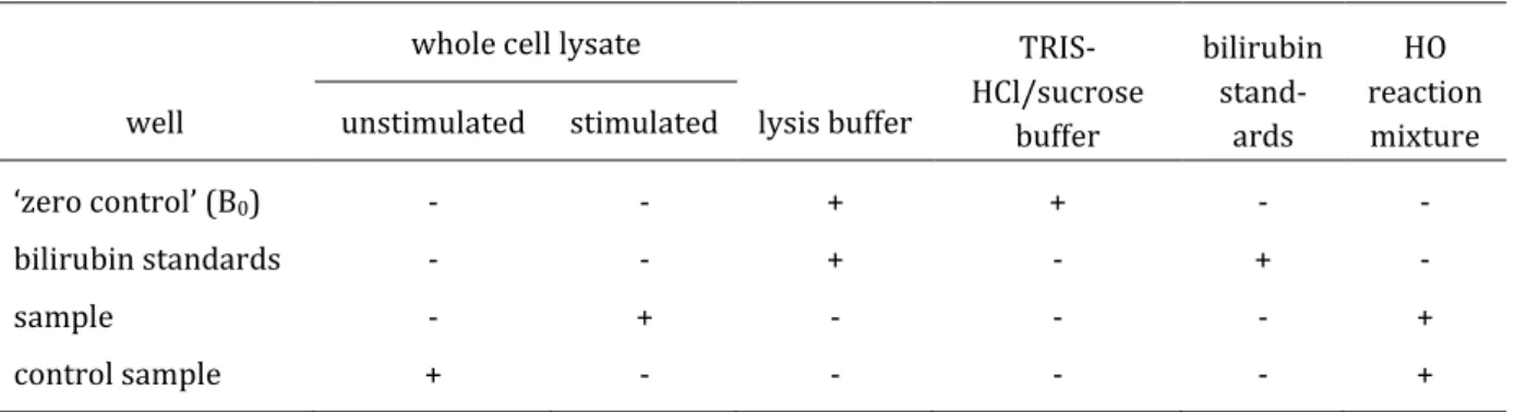
Because of the lack of a high throughput method to determine HO-1 activity to screen many po- tential new HO-1 inducers, which could be used as lead structures for drug development, a sim- ple and reliable cell line-based assay is needed. This can be achieved by developing a HO-1 activ- ity assay, which can be used in a microtiter plate setting, applicable on non-microsomal fractions of cell lysates by combining the HO enzymatic reaction with the ELISA method determining the produced bilirubin in the sample. The purpose of the HO-1 activity assay is to screen for HO-1 activity inducers in vitro in a feasible way by using a 96-well plate format for all steps of the as- say, including cell culture, cell sample preparation, HO enzymatic reaction, protein determina- tion and bilirubin quantification via ELISA. A concept of the HO-1 activity assay is shown in Scheme 3. As an in vitro model system, cells are cultured in a 96-well plate and incubated with the potential HO-1 inducers. Cell lysis is then performed in the same microtiter plate by using a mild and efficient cell lysis buffer containing a protease inhibitor cocktail and a detergent in or- der to solubilize and stabilize the target proteins from degradation. Without further centrifuga- tion step, the obtained whole cell lysate is transferred to a new microtiter plate and incubated with the HO enzyme reaction mixture consisting of hemin, biliverdin reductase (BVR) and NADPH. The final product of the HO and the BVR reaction, bilirubin, is then quantified by ELISA using the specific anti-bilirubin antibody 24G7 and a second HRP-conjugated antibody for detec- tion.
9
Scheme 3. Concept of the HO-1 activity assay to screen for new HO-1 inducers in vitro.
Finally, the total protein amount in the whole cell lysate is determined using a protein assay kit.
Bilirubin amounts in samples are calculated from a bilirubin calibration curve carried out on each plate and HO activity is expressed as pmol bilirubin h-1 mg-1 total protein. For HO-1 activity determination, the HO activity of the cells stimulated with the test compound is compared to control cells incubated only with culture medium and expressed as x-fold HO-1 activity of con- trol.
1.2.4.1 Preliminary optimizations of the HO-1 activity assay
First optimizations of the HO-1 activity assay were started in the diploma thesis (Hannelore Rücker, Universität Regensburg, October 2009)78 and were continued and further developed in the present work.
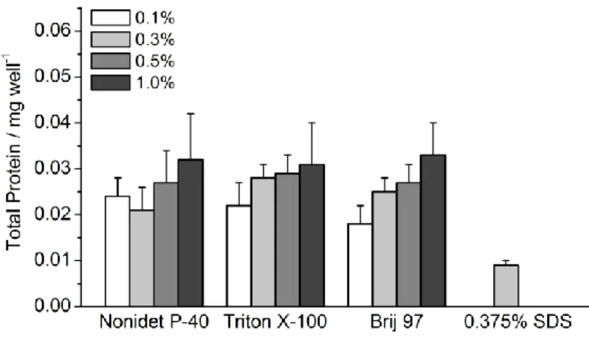
As an in vitro model system, the human colon cancer cell line HT-29 was used to develop the HO- 1 activity assay. For this purpose HT-29 cells (1 · 105 cells/well) were incubated in a 96-well plate with potential HO-1 inducers in several concentrations for a maximum of 24 h and control cells were treated with culture medium alone. The cell lysis was performed in the 96-well plate and included two steps: i) cells were incubated with a concentrated lysis buffer (40 mM TRIS- HCl, pH 7.4, 250 mM sucrose, 10 mM EDTA, 100 mM NaCl, 1% (v/v) SDS detergent, 4% protease inhibitor cocktail) on an orbital plate shaker for 15 min at 4 °C and ii) diluted with lysis buffer (without detergent and protease inhibitor) to a final detergent concentration of 0.05% in the cell lysate sample. Low concentrations of detergent in the lysis buffer (<0.1%) gave poor cell lysis
10
results. More importantly, higher concentrations of detergent (>0.05%) interfered with the pro- tein detection when using the Bradford protein assay to give false positive results.
For determining the HO activity, the whole cell lysates (10 µg protein/well) were incubated for 1 h at 37 °C with the HO reaction mixture containing 25 µM hemin, 3 mM NADPH, 100 µg guinea pig liver cytosol extract prepared by ultracentrifugation as a source of biliverdin reductase in TRIS-HCl-sucrose buffer, pH 7.4. HO assay components based on the HPLC assay method devel- oped by Ryter et al.43 The enzymatic reaction was stopped with a 1 M HCl aqueous solution and pH was then adjusted to 7.4 with 1.2 M NaOH aqueous solution. Here several stop solutions and procedures were screened and the HCl/NaOH procedure was found to be the best solution. Bili- rubin standard solutions prepared in TRIS-HCl-sucrose buffer, pH 7.4 were added after the incu- bation step to the HO reaction mixture containing only whole cell lysate and the liver cytosol extract. The bilirubin in the standards and the samples was then quantified with the ELISA method described by Izumi et al., which was slightly modified.52 Samples were incubated with the specific anti-bilirubin antibody (0.571 µg mL-1) and then the mixture was transferred on an immunoplate coated with a bilirubin-BSA conjugate (3.5 µg mL-1). Both parameters, the anti- bilirubin antibody and bilirubin-BSA conjugate concentration were optimized. Next, the bound anti-bilirubin antibody on the immunoplate was incubated with a second HRP-conjugated anti- body (0.2 µg mL-1) and then detected by adding the substrate OPD and H2O2. The reaction was terminated by a 3 M H2SO4 solution and absorbance was measured at 492 nm. Unknown biliru- bin concentration in the samples was calculated by using the linear regression curve generated from the bilirubin standards (5-50 · 10-9 M bilirubin) on each plate. Finally, the protein amount in the whole cell lysate was determined using the Bradford protein assay. HO activity was ex- pressed as pmol bilirubin h-1 mg-1 total protein.
First results with the HO-1 activity assay were promising, but insufficient stimulation of HT-29 with known HO-1 inducer compounds and a poor reproducibility of data were persistent. There- fore, further troubleshooting and optimization of the HO-1 activity assay was aimed.
Crucial for the assay is the cell lysis step which requires a more feasible one step procedure in the 96-well plate format by using a mild but sufficient detergent in the lysis buffer. Components of the lysis buffer, especially the detergent should not interfere with the protein detection or the ELISA. The use of a detergent compatible protein assay kit can avoid this problem and be also suitable for detecting small amounts of protein in the sample. Components of the HO enzymatic reaction and the reaction buffer should be suitable and sufficient to estimate the HO activity in the whole cell sample. The HO-1 activity assay should be applicable on different cell lines as in vitro model systems, as long as they are adherent. Also, the preparation of the test compound solutions for their incubation with the cells should be simplified to avoid compound precipita- tion in the cell culture medium.
11
1.3 The activity of inducible nitric oxide synthase (iNOS)
Nitric oxide (NO) is an ubiquitous key signaling molecule implicated in neurotransmission,79 vasodilatation80 and regulation of the immune system.81 NO is produced by three isoforms of NO synthase, which are: endothelial NOS (eNOS), inducible NOS (iNOS), and neuronal NOS (nNOS).
All isoforms contain heme as a prosthetic group and catalyze the reaction of L-arginine to L-citrulline using O2 and NADPH.82 The catalytic activity of the inducible NO-synthase is given in Figure 5.
Figure 5. The enzymatic activity of the inducible nitric oxide synthase (iNOS). The amino acid L-arginine is converted to L-citrulline by the active iNOS dimer with heme as the prosthetic group, using oxygen and NADPH to produce NO.
The function of the iNOS enzyme depends on its expression level, which is regulated by the proinflammatory NF-κB signaling pathway (Figure 4). iNOS, when induced in macrophages, gen- erates large amounts of NO that has cytostatic or cytotoxic effects on parasitic or tumor target cells. This is caused by the high affinity of NO to protein-bound iron, thus inhibiting the catalytic centers of iron-sulfur cluster-dependent enzymes involved in mitochondrial electron transport, DNA replication (ribonucleotide reductase) or in the citric acid cycle (acotinase). Furthermore, NO and peroxynitrites (ONOO−), yielding from the reaction with superoxide radicals (O2•-) can directly interfere with the DNA of target cells and cause strand breaks and fragmentation. High levels of NO produced by activated macrophages may not only be toxic to parasites or tumor cells, but may also harm healthy cells, contributing to the pathophysiology of inflammatory dis- eases and septic shock. In inflammation, hypertension and atherosclerosis increased reactive nitrogen species (NO, ONOO−) induce tyrosine nitration, oxidation of SH groups of cysteins and lipid peroxidation. Targeting nitrosative stress may represent a therapeutic potential in pathol- ogies like Alzheimer's disease or cancer.83-84 The activity of iNOS can be inhibited on the tran- scriptional level by inactivating the NF-κB pathway, were i.e. α,β-unsaturated carbonyl com- pounds can react with key SH groups of cysteine residues of IKK or NF-κB and suppress the pro- tein expression of iNOS and other proinflammatory enzymes as displayed in Figure 4. The pre-
12
vention of the overproduction of NO through control of regulatory pathways may assist in the treatment of NO-mediated disorders without changing the physiological levels of NO.85
1.3.1 The nitrite (Griess) assay
The potential anti-inflammatory activity of α,β-unsaturated carbonyl compounds or other com- pounds can be assessed by their ability to inhibit the iNOS activity causing a suppression in the NO production. iNOS activity can be determined by the quantification of nitrite, the more stable oxidation product of nitric oxide, using the Griess reaction in the so called nitrite assay.86 In the murine macrophage cell line RAW264.7, the NO synthesis can be induced by lipopolysaccharide (LPS), a bacterial wall constitute.87
Figure 6. The Griess reaction is used to quantify the nitrite derived from the NO production in RAW264.7 macrophages stimulated with lipopolysaccharide (LPS) in the nitrite assay. Under acidic conditions nitrite reacts with the amino group of sulfanilamide to form the diazonium cation, which couples to N-(1- naphthyl)ethylenediamine (NED) in para-position to form the azo dye.
Nitric oxide which oxidizes to nitrite and accumulates in the cell culture medium is quantified by the Griess reaction, which is a diazotization reaction followed by an azo coupling using sulfanil- amide, N-(1-naphthyl)ethylenediamine (NED) and phosphoric acid (Figure 6). Under acidic con- ditions the nitrite in the cell culture medium reacts with the amino group of sulfanilamide to form the diazonium cation, which couples to NED in para-position to form the corresponding azo dye, which absorbance can be measured at 560 nm. The NO production is quantified in LPS- stimulated macrophages in the presence or absence of a test compound. Consequently, a poten- tial inhibition of the NO production by the compound estimates its anti-inflammatory activity.
1.4 The oxygen radical absorbance capacity-(ORAC)-fluorescein assay
The radical scavenging activity of antioxidants can be determined by the cell free oxygen radical absorbance capacity-(ORAC)-fluorescein method generating peroxyl radicals from AAPH (2,2'- azobis(2-methylpropionamidine) dihydro-chloride) as a free radical initiator and using fluores- cein as a fluorescent probe.88 The protecting effect of an antioxidant, reacting as radical scaven-
13
ger can be quantified by assessing the area under the fluorescence decay curve (AUC) of the sample compared to the blank, in which no antioxidant is present (Figure 7).
Figure 7. ORAC-fluorescein assay. The fluorescence decay from the reaction of radicals generated from AAPH with fluorescein in the absence (blank) or presence of an antioxidant (sample) is measured over time. The net area under the fluorescence decay curve (net AUC) determines the antioxidant capacity of the test compound.
Figure 8. Structures of fluorescein used as fluorescence probe and Trolox, a standard antioxidant in the ORAC- fluorescein assay.
From the curves (relative fluorescence intensity versus time) the area under the fluorescence decay curve (AUC) is calculated as
Equation 1
where f0 is the initial fluorescence reading at 0 min and fi is the fluorescence reading at time i.
The net AUC corresponding to a sample is calculated by subtracting the AUC corresponding to the blank. Linear regression equations between net AUC and antioxidant concentration are de- termined for all the samples and ORAC values are compared to Trolox, a water-soluble vitamin E (α-tocopherol) derivative (Figure 8). The final ORAC values are expressed as Trolox equivalents by using the standard curve determined for each assay.
14
1.5 Anti-inflammatory activity of a diverse group of α,β-unsaturated carbonyl compounds and polyphenols
A structurally diverse group of natural products and synthetic compounds were investigated for their HO-1 induction behavior using the ELISA-based HO-1 activity assay in the model cell line RAW264.7. Known Nrf2 inducers possessing the α,β-unsaturated carbonyl moiety11 together with polyphenols which are also a prominent class of inducers of cytoprotective proteins16 were focused on for the screening. The following pharmacologically interesting compounds were ex- amined (Figure 9.): the chalcones cardamonin,89-91 flavokawain A92 and xanthohumol,93-95 the flavonoids (-)-epicatechin,96-97 kaempferol98-101 and quercetin,102-103 three cinnamic acid deriva- tives caffeic acid,104-105 chlorogenic acid104, 106-107 and CAPE,108-111 the isothiocyanate sulfo- raphane28, 112-114 and the disulfide oltipraz,115-117 curcumin118 and 3-hydroxycoumarin,27, 119 the sesquiterpene zerumbone,120-121 rosolic acid,66 dexamethasone,89, 122 a synthetic glucocorticoid, and two compounds lacking the α,β-unsaturated carbonyl moiety resveratrol123-124 and carnosol,125-126 a diterpene with a catechol unit.
The mechanism by which these compounds lead to a HO-1 induction is commonly by activating the Keap1/Nrf2/ARE pathway. Although the Nrf2 inducers are from distinct chemical classes, comprising diphenols, quinones, Michael acceptors and isothiocyanat, all have in common that they are electrophiles and can covalently modify thiol groups on Keap1 by alkylation or oxida- tion. The activation of the transcriptional factor Nrf2 that binds to the ARE and promotes the phase II protein expression, is mediated by a direct reaction with thiol groups of the suppressor protein Keap1 as shown for sulforaphane,127 zerumbone128 and xanthohumol.129 Certainly, there are also other indirect mechanisms, which lead to activation of the Nrf2/ARE pathway and thus to HO-1 induction, involving the activation of kinase pathways which phosphorylates Nrf2 and Keap1. The involvement of protein kinase in Nrf2 activation was suggested for resveratrol,130 curcumin131 and carnosol.132
A HO-1 induction on mRNA, protein or enzyme activity levels were shown for most of the biolog- ically active compounds in different cell and tissue types, summarized in Table 1. No HO-1 induc- tion tests have been described for 3-hydroxycoumarin, oltipraz, cardamonin and flavokawain A.
Since the HO-1 inductive activity is known for some compounds, the screening aimed also at the establishment and validation of the ELISA-based HO-1 activity assay as a simple and reliable screening method for HO-1 activity.
15
Figure 9. Structures of biologically active natural compounds and synthetic drugs screened in the ELISA-based HO-1 activity assay.
16
Table 1. HO-1 inducer activity of natural products and synthetic drugs.
Compound
(Dose) Mechanism of HO-1 induction Model cells or mam-
malian Ref.
Xanthohumol
2-10 µM induction of HO-1 mRNA and HO-1 protein level in normal hepatocytes (THLE-2) by activation of Nrf2;
no HO-1 induction was observed in carcinoma hepatocytes HepG2
human hepatocytes, THLE-2 and HepG2
133
CAPE 15-50 µM 5-30 µM 20 µM
increase of HO-1 protein expression and HO-1 activ- ity
increase of HO-1 protein expression and HO-1 activ- ity by binding of Nrf2 to ARE
HO-1 mRNA an protein expression induction
DI TNC1 rat astrocytes LLC-PK1 renal epithelial cells
HUVEC, human vascu- lar endothelium cells
67
68
134
Carnosol
10 µM increase of HO-1 mRNA and protein expression by binding of Nrf2 to ARE, activation of ERK, p38 MAPK and JNK
PC12 rat pheochromo- cytoma
135
(-)-Epicatechin
50 µM induction of HO-1 and Nrf2 protein expression ARPE-19 human retinal pigment epithelial cells
136
Curcumin 15-50 µM 5-30 µM 5-15 µM 200 mg kg-1 15 µM
increase of HO-1 protein expression and HO-1 activ- ity
increase of HO-1 protein expression and HO-1 activ- ity by binding of Nrf2 to ARE
increase of HO-1 mRNA, HO-1 protein expression and HO-1 activity
HO-1 expression and activity in liver, DNA binding of Nrf2-ARE in liver
increase of HO-1 mRNA expression by binding of Nrf2 to ARE, activation of PKC-δ and p38 MAPK
DI TNC1 rat astrocytes LLC-PK1 renal epithelial cells
bovine aortic endothe- lial cells
male Albino rats THP-1 human mono- cytes
67
68
65
137
131
Kaempferol
100 µM increase of HO-1 mRNA and protein expression RAW264.7 murine macrophages
138
10-100 µM increase of HO-1 mRNA expression RAW264.7 murine
macrophages
139
10 µM induction of HO-1 protein expression by activation
of Nrf2 and JNK HEI-OC1, auditory mice
cells
140
Quercetin
100 µM increase of HO-1 mRNA and protein expression RAW264.7 murine macrophages
138
17 Compound
(Dose) Mechanism of HO-1 induction Model cells or mam-
malian Ref.
30 µM increase of HO-1 protein expression microglial BV2 cells 141
100 µM induction of HO-1 activity and Nrf2 activation EtOH-treated human hepatocytes isolated from liver cancer pa- tience
142
50 µM induction of HO-1 and Nrf2 activation ARPE-19 human retinal pigment epithelial cells
136
Resveratrol
15 µM induction of HO-1 mRNA and protein expression by
activation of Nrf2/ARE and ERK PC12 rat pheochromo- cytoma
130
1-10 µM induction of HO-1 mRNA and protein expression via
NF-κB activation human aortic smooth
muscle cells
143
5-100 µM increase of HO-1 protein expression cortical neuronal mice cells
144
5-100 µM increase of HO-1 mRNA, no induction of HO-1 pro-
tein expression, reduction of HO-1 activity DI TNC1 rat astrocytes 145-
146
Rosolic acid
15 µM induction of HO-1 protein expression and HO-1
activity bovine aortic endothe-
lial cells
66
Zerumbone
1-25 µM induction of HO-1 expression and activation of
Nrf2/ARE RL34 rat liver epithelial
cells
147
10 µM induction of HO-1 mRNA and protein expression by
a Nrf2/ARE dependent pathway JB6 Cl41 mouse epi-
dermal cells and mouse skin from female hair- less mice
128
Sulforaphane
0.1 µM induction of HO-1 protein expression rat aortic smooth mus- cle cells
148
20 µM induction of HO-1 protein expression and activation
of Nrf2/ARE HepG2 human hepato-
cytes
149
Dexamethasone
5 µM induction of HO-2 mRNA, protein expression and
HO activity, no HO-1 mRNA induction observed HeLa, human cervix cancer cells
150
0.5-50 µg mL-1 suppression of HO-1 mRNA in cytokine-stimulated
cells rat astroglial cells 151
1 µg mL-1 no induction of HO-1 activity GT1-7 hypothalamic
neurons
152
1.2 mg kg-1 suppression of HO-1 protein expression and HO-1
activity in LPS stimulated macrophages alveolar macrophages of chronic bronchitis model rats
153
18 Compound
(Dose) Mechanism of HO-1 induction Model cells or mam-
malian Ref.
Caffeic acid
20 µM no HO-1 induction observed HUVEC, human vascu-
lar endothelium cells
134
150 µM no HO-1 mRNA induction bovine aortic endothe-
lial cells
154
Chlorogenic acid
150 µM no HO-1 mRNA induction bovine aortic endothe-
lial cells
154
1.6 Reactivity and biological activity of natural and synthetic chalcones
1.6.1 Reactivity of chalcones
Chalcones (1,3-diphenylprop-2-en-1-ones) are a diverse group of naturally occurring plant me- tabolites that can be regarded as open-chain flavonoids, where the two aromatic rings are bridged by an α,β-unsaturated carbonyl moiety possessing a Michael acceptor reactivity.
Chalcones can be regarded as bifunctional antioxidants: i) they possess a distinct Michael accep- tor reactivity, due to the α,β-unsaturated carbonyl moiety, allowing them to interact with reac- tive SH-groups on “sensor” proteins involved in cytoprotective and also in inflammatory signal- ing pathways, (Scheme 4, pathway A) and ii) the α,β-unsaturated carbonyl moiety itself and ad- ditional phenolic hydroxy groups on the aromatic rings can react as radical scavengers (Scheme 4, pathway B) or antioxidants due to their reduction potential (Scheme 4, pathway C).4, 155-156
Scheme 4. General structure and reactivities of chalcones.
19
Several structural and electronic characteristics of chalcones influence the reactivity of the Mi- chael acceptor moiety and thus the potential biological activity of chalcones. Aromatic OH- groups display a positive resonance effect (+M effect), due to the free electron pairs on the oxy- gen atom, which contributes to the conjugated aromatic π-electron system and further to the α,β-unsaturated carbonyl moiety of the chalcone. However, when deprotonated, hydroxylates are present in the chalcone, which is to a certain degree possible under physiological conditions (pKA (phenol) = 10). In this case the strong resonance effect is pushing electrons into the conju- gated π-electron system of the chalcone resulting in a more electron rich α,β-unsaturated car- bonyl unit, which leads to a reduced electrophilic character and therefore to a weaker Michael acceptor reactivity towards nucleophiles. If methoxy groups are present on the aromatic rings of the chalcone, the resonance effect is weaker compared to hydroxylates, due to the alkylation.
The position of the hydroxy and methoxy groups on the aromatic ring plays an important role and can influence the reactivity of chalcones in a tremendous way. As proven in several studies,27, 66, 157 the 2’-hydroxy group is essential for the Michael acceptor reactivity of chalcones, due to the intramolecular H-bond which activates the carbonyl group. Furthermore, the H-bond of the 2’-OH group contributes to a stabilized conjugation in the π-system. Generally, a replace- ment of a more electron donating OH group by a methoxy in the A- or B-ring restores the reac- tivity of the chalcone. Moreover, since the double bond of the Michael acceptor functionality can be referred to as a push-pull double bond, an exchange in the B-ring restores more reactivity compared to a similar exchange in the A-ring. Methoxy groups in 3 or 4-position on the B-ring and 4’-position on the A-ring can contribute to a stabilization of the conjugated π-system. A loss in reactivity can occur when OMe groups are present in 2 or 6-position on both rings, due to steric hindrance, which can hamper the nucleophilic attack on the β-position of the α,β- unsaturated carbonyl moiety or destabilize the conjugation of the π-system.
2’-Hydroxychalcones can readily isomerize to flavanones through an intramolecular Michael addition, leading to a loss of the functional group and thus altering the reactivity of the chalcone under biological conditions. This important effect was recently investigated by the group of Pauli under several biological conditions typical for cell-based assay systems using the chalcone isoliquiritigenin (ISL).158 Also significant for the reactivity and thus biological activity of chalcones acting as a Michael acceptors is the stability of the thiol adduct. Here, electron donat- ing groups like OMe, can decrease the acidity of the α-hydrogen, slowing down the retro-Michael reaction. A balanced activation of the Michael acceptor unit and stabilization of the resulting adduct is therefore crucial for the overall reactivity of chalcones.157
20
1.6.2 Reactivity assessment of chalcones by a kinetic thiol assay
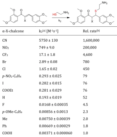
In order to predict the biological activity of electrophiles based on thiol-mediated regulation processes, a kinetic assay for the assessment of the second rate constant k2 in thia-Michael addi- tions was developed in our group. The chemical reactivity of hydroxy-alkoxychalcones was re- cently determined using the kinetic thiol assay for the thia-Michael addition reactions with the S-nucleophile cysteamine (Figure 10).159
Figure 10. Michael addition reaction of natural and synthetic chalcones with the S-nucleophile cysteamine. k2
is the second order rate constant of the addition reaction.
Reactions were carried out in 100 mM TRIS-HCl pH 7.4, 2 mM EDTA/ethylene glycol 20:80, 25 °C under pseudo-first order conditions at concentrations of 40 µM for chalcones and 12 to 500 fold cysteamine. The calculated thia-Michael addition reaction rate was displayed as k2 value in M-1 s-
1. Hydroxy- and methoxychalcones gave quite different reactivities in the Michael additions of thiols, with k2 values in the range of 5.08 - 0.193 M-1 s-1, displaying an overall good electro- philicity. The results showed that a 2’-OH group on the A-ring is essential for the reactivity of the chalcones. Not only electronic effects, but also steric effects can influence the Michael acceptor reactivity. One aspect is the conformation of the conjugated system determined by the dihedral angel between the two aromatic rings, which is in term influenced by the substituents present on the aromatic rings of the chalcone. A fairly flat chalcone (as shown by X-ray structures) dis- plays generally a higher reactivity, due to a stabilized conjugation of the π-system compared to a chalcone where the aromatic rings are twisted.
1.6.3 Biological activity of chalcones
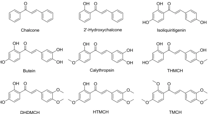
In the present study, the plant chalcones isoliquiritigenin (ISL), butein, calythropsin, 2’,3,4’- trihydroxy-4-methoxychalcone (THMCH) and 2’,4’-dihyxdroxy-3,4-dimethoxychalcone (DMDHCH) and the synthetic chalcones 2’-hydroxychalcone, chalcone, 2’-hydroxy-3,4,4’- trimethoxychalcone (HTMCH) and 2’,3,4,4’-tetramethoxychalcone (TMCH) were selected (Figure 11) to investigate their anti-inflammatory and antioxidative activity in RAW264.7 murine mac- rophages.
21
Figure 11. Structures of natural and synthetic chalcones investigated in this study towards their in vitro anti- inflammatory and antioxidative activity.
Synthetic and natural hydroxy- and methoxychalcones are of particular interest as they display a wide range of biological properties and exert diverse pharmacological activities including anti- inflammatory, antifungal, antibacterial, antiviral, antimitotic, antitumor, antituberculosis and antimalarial.160 The importance of chalcones as pharmacologically active compounds is closely connected to their Michael acceptor reactivity and antioxidant potential, by which they can ad- dress and affect multiple targets in the cell. By addition to reactive thiol groups on the surface of sensor proteins or transcriptional factors, chalcones can trigger the anti-inflammatory and cytoprotective Keap1/Nrf2/ARE signaling pathway and on the other side inhibit the pro- inflammatory pathway regulated by NF-κB.161 In case of the synthetic chalcone 3,3’,4,4’,5,5’- hexamethoxychalcone, a Nrf2-dependent HO-1 induction as well as a NF-κB down regulation was shown.162 The anti-inflammatory chalcone butein was found to be an inhibitor of IKK by direct binding on its cysteine residue 179, through which it blocks NF-κB and NF-κB-regulated gene products.163 Lee et al. reported the inhibitory effect of butein on NO production, iNOS gene expression and NF-κB activity in LPS-stimulated murine macrophages RAW264.7 cells.164 Stud- ies have also shown that butein induces HO-1 mRNA and protein expression in rat liver cells165 and that it prevents oxidative damage in human dental pulp cells by inducing HO-1 protein ex- pression and activity via a Nrf2 dependent pathway.166 The radical scavenging properties of butein was investigated towards DPPH and ABTS radicals.167 Chalcone exerts its anti- inflammatory activity by inhibiting the activation of NF-κB and inducing the HO-1 protein ex- pression, which is accompanied by an up regulated level of Nrf2 in the nucleus and an increased ARE activity in bovine aortic endothelial cells. Furthermore, the overall cytoprotective activity of
22
chalcone may be mediated through a direct modification of cysteine thiol groups on target pro- teins and regulated by the intracellular GSH level.168 The chemopreventive activity of chalcone was shown by its inhibitory effect against pulmonary and mammary carcinogenesis when given after carcinogen administration to female rats.169 2’-Hydroxychalcone was found to induce HO-1 protein expression as well as enzyme activity in RAW264.7 cells69 and bovine aortic endothelial cells.66 Additionally, 2’-hydroxychalcone promoted its anti-inflammatory activity in RAW264.7 cells by reducing iNOS expression, NO production and TNF-α release.69 Isoliquiritigenin (ISL), a plant constituent from Dalbergia odorifea (Leguminosae) and Glycyrrhiza uralensis (licorice), has been reported to posses estrogenic,170 neuroprotective,171 hepatoprotective,172 anticancer173-175 and anti-inflammatory activity.176-177 Furthermore an induction of HO-1 expression was reported in RAW264.7macrophages178 and rat hepatic stellate cells.179 ISL is a major inducer of quinine reductase and activates the ARE.180 The antioxidant and chemoprotective actions of ISL was shown in cerebral ischemia model rats, where ISL treatment protected against depletion of anti- oxidant proteins (superoxide dismutase, catalase and glutathione peroxidase) caused by free radical formation.181 The chalcone calythropsin, firstly isolated from Calythropsis aurea was found to possess weak antimitotic activity due to its cytotoxic effect.182 Furthermore, calythropsin extracted from Faramea salicifolia displayed cytotoxicity against several human cancer cell lines.183 DHDMCH and THMCH are abundant in Iryanthera polyneura (Myristicaceae) and were isolated 1979 from the trunk of the tree. It is known that Maku Indians of South Amer- ica use crushed leaves from this tree to treat infected wounds and cuts.184 The chalcone THMCH revealed a moderate cytotoxicity against several human tumor cell lines, especially against Jurkat cells and was also found to exert an impressive antiproliferative activity against the Jurkat cell cycle.185 No anti-inflammatory activity was reported for the naturally occurring calythropsin, DHDMCH and THMCH.
Chalcones possess an unique capability to address certain cysteine residues, which qualify them as a valuable tool to modulate biological activity. Several natural and synthetic hydroxy- and methoxychalcones were characterized towards their anti-inflammatory and antioxidative activi- ty, demonstrated by their induction of HO-1 activity and inhibition of NO production in RAW264.7 macrophages and also by their radical scavenging capacity in the ORAC assay. A structure-activity relationship can be assessed, due to the diverse substitution pattern of hydroxy and methoxy groups in 2’,3,4,4’-positions of the two aromatic rings, influencing not only the Michael acceptor reactivity but also the overall anti-inflammatory and antioxidative proper- ty of the chalcone. Furthermore, the chemical reactivity of the chalcones determined by the ki- netic thiol assay in thia-Michael additions159 was compared to their estimated biological activity.
23
1.7 α-X-Modified enones as a different approach in fine-tuning their Michael acceptor reactivity and biological activity
A promising approach using α,β-unsaturated compounds for the development of new potent cytoprotective, chemopreventive and anti-inflammatory drugs is to systematically modify the α-position of the α,β-unsaturated carbonyl moiety thus influencing the Michael acceptor reactiv- ity and the biological activity. Examples of such α-modifications leading to a fine-tuned biological activity are found within the class of pentacyclic triterpenoids CDDO186-187 and also among the chalcones.188
1.7.1 α-X-Modification in 2’,3,4,4’-tetramethoxychalcones (α-X-TMCHs)
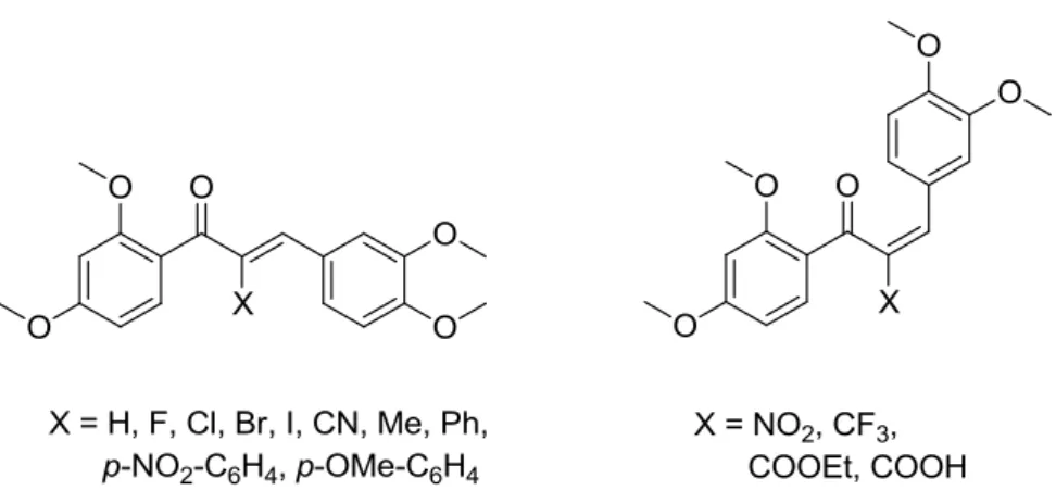
In order to manipulate the reactivity of chalcones either a change directly at the Michael system or on the aromatic rings, as seen in the chapter before, can be made. The approach of modifying the α-position of the α,β-unsaturated carbonyl system is a promising concept, because it should lead to a direct and straightforward influence on its reactivity.4 Modified chalcones in α-position and their biological studies are known for chalcones with X = halogen,188-189 aromatic,190-191 al- kyl,189, 192-193 COOEt,188 COOH,194 CN188 and alkoxy.193 But there is no fine-tuning-of-reactivity- approach, particularly on a chalcone scaffold, which investigates a clear influence of different α-X-substituents on the activity in different biological settings. The natural product-like 2’,3,4,4’- tetramethoxychalcone, TMCH was chosen as a scaffold for a diverse library of α-X-TMCHs, pos- sessing no free hydroxy groups, so that possible oxidative pathways altering the biological re- sponse can be excluded. The α-X-TMCHs used in this work were synthesized in our group by Nafisah Al-Rifai. As shown in Figure 12, thirteen distinct substituents were introduced in the α- position of the Michael system along with the α-H-TMCH, where the chalcones were obtained as two different double bond isomers. The synthetic approach of the chalcones together with their chemical reactivity and their biological evaluation were published recently.195
Figure 12. Structures of α-X-TMCHs tested for their in vitro anti-inflammatory and antioxidative activity.
For a broad range of reactivity, different electron withdrawing and electron donating substitu- ents for the α-position were chosen. The more electron withdrawing groups like CN, NO2, CF3 or