• Keine Ergebnisse gefunden

FROM INTRACELLULAR SIGNALING CASCADES TO BEHAVIOR: TOWARDS A BETTER UNDERSTANDING OF OXYTOCIN’S MOLECULAR EFFECTS

N/A
N/A
Protected

Academic year: 2021

Aktie "FROM INTRACELLULAR SIGNALING CASCADES TO BEHAVIOR: TOWARDS A BETTER UNDERSTANDING OF OXYTOCIN’S MOLECULAR EFFECTS"

Copied!
180
0
0

Wird geladen.... (Jetzt Volltext ansehen)

Volltext

(1)

FROM INTRACELLULAR SIGNALING CASCADES TO BEHAVIOR: TOWARDS A BETTER UNDERSTANDING

OF OXYTOCIN’S MOLECULAR EFFECTS

DISSERTATION ZUR ERLANGUNG DES

DOKTORGRADES DER NATURWISSENSCHAFTEN (DR. RER. NAT.) DER FAKULTÄT FÜR BIOLOGIE UND VORKLINISCHE MEDIZIN

DER UNIVERSITÄT REGENSBURG

vorgelegt von Julia Winter aus Pfarrkirchen

im Jahr 2019

(2)

Das Promotionsgesuch wurde eingereicht am: 18.10.2019

Die Arbeit wurde angeleitet von: Dr. rer. nat. Benjamin Jurek

Unterschrift:

(3)

Durchgeführt am Institut für Zoologie der Universität Regensburg

Am Lehrstuhl für Tierphysiologie und Neurobiologie

unter Anleitung von

Dr. rer. nat. Benjamin Jurek

(4)
(5)

1

Table of Contents

(6)

2

(7)

3

Table of Contents

Summary ... 9

Zusammenfassung ...13

Abbreviations ...16

Introduction ...23

1. Anxiety ...23

1.1. Animal models to study anxiety ...24

1.2. Molecular underpinnings of anxiety and anxiety disorders ...24

2. The oxytocin system ...25

2.1. Oxytocin and anxiety ...27

2.2. Oxytocin and stress ...28

2.3. Oxytocin receptor signaling ...29

2.4. Impact of cholesterol on oxytocin receptor activity ...33

3. The CRF system ...34

3.1. CRF family members ...35

3.2. Ligand-receptor interaction ...36

3.3. The CRFR2 and its role in anxiety ...38

4. Alternative splicing ...39

4.1. Alternative splicing of the CRFR2α ...41

4.2. Neural splicing regulatory networks ...43

5. MEF2 family of transcription factors...43

5.1. MEF2 signaling cascades ...44

5.2. MEF2 functions in the brain ...46

5.3. Interplay between MEF2 and oxytocin ...46

6. Neuronal morphology and its regulation ...47

6.1. Definition of neuronal morphology ...47

6.2. Neuropeptides and the regulation of neuronal morphology ...47

7. The CRISPR-Cas system: a toolbox for investigating the oxytocin system ...49

7.1. CRISPR-Cas – the immune system of bacteria ...49

7.2. Genome engineering with CRISPR-Cas9 and more ...50

8. Aim of the thesis ...53

Material and Methods ...57

1. In vivo experiments ...57

1.1. Animals and husbandry...57

(8)

4

1.2. Surgical procedures... 57

1.3. Behavioral testings ... 59

1.3.1. Elevated plus maze ... 59

1.3.2. Light-dark box ... 59

1.3.3. Open field test ... 60

1.3.4. Acute mild stress paradigm ... 60

1.4. Collection of trunk blood, brains, tissue, and CSF ... 60

1.5. Perfusion and brain slicing... 61

2. Cell culture ... 61

2.1. Cell lines and cultivation ... 61

2.2. Stimulations ... 62

2.3. Differentiation and morphological analysis ... 63

2.4. Manipulation of cholesterol content in cellular membranes ... 63

2.4.1. Cholesterol enrichment ... 63

2.4.2. Cholesterol depletion ... 64

2.4.3. Filipin staining ... 64

2.5. Plasmid-mediated oxytocin receptor overexpression ... 64

2.6. CRISPR-Cas9 ... 65

2.6.1. Otr knockout ... 65

2.6.2. Mef2a knockout... 65

2.6.3. HiBiT ... 66

2.6.4. Sequencing ... 66

2.6.5. Cell sorting ... 67

3. Molecular techniques ... 67

3.1. Protein isolation ... 67

3.2. SDS PAGE, western blot, and dot blot... 68

3.3. TransAM

®

MEF2 transcription factor activation assay ... 69

3.4 Immunohistochemistry and immunocytochemistry ... 70

3.5. RNA and DNA isolation ... 71

3.6. mRNA analysis ... 71

3.6.1. Polymerase chain reaction ... 71

3.6.2. Quantitative polymerase chain reaction ... 72

3.6.3. Polymerase chain reaction array ... 72

4. Statistical analyses ... 73

Results ... 77

1. Differential effects of acute and chronic oxytocin on anxiety-like behavior in rats ... 77

(9)

5

2. Downstream signaling of chronic and acute oxytocin ...79

2.1. Effects on MAPK and MEF2 signaling ...79

2.2. MEF2A activity induced by chronic oxytocin shifts the expression of mCRFR2α to sCRFR2α ...87

2.3. Chronic oxytocin promotes the release of sCRFR2α and leads to increased anxiety ...93

3. Effects of chronic oxytocin on neuronal morphology and signaling in vitro ... 102

3.1. Characterization of Be(2)-M17, H32, and N2a cells ... 102

3.2. Chronic oxytocin induces neurite retraction ... 103

3.3. CRISPR-mediated knockout of the oxytocin receptor impairs MAPK signaling and affects neuronal morphology ... 104

3.4. Optimizing the in vitro model for oxytocin receptor signaling with cholesterol ... 109

Discussion ... 117

1. The two faces of oxytocin in anxiety – effects of chronic vs acute oxytocin... 117

2. MAPK and MEF2 signaling induced by chronic oxytocin ... 119

3. CRFR2α alternative splicing increases anxiety induced by chronic oxytocin... 121

4. Bridging the gap between intracellular genomic effects and behavior: oxytocin- induced morphological alterations ... 127

4.1. Chronic oxytocin induces neurite retraction in vitro in various cell lines ... 127

4.2. CRISPR-Cas9-mediated gene knockout ... 128

4.3. Oxytocin receptor-mediated signaling and neuronal morphology ... 129

7. Conclusion ... 131

Perspectives ... 133

References ... 135

Acknowledgements - Danksagung ... 171

Curriculum vitae ... 175

Publications ... 176

(10)

6

(11)

7

Summary

(12)

8

(13)

9

Summary

The neuropeptide oxytocin (OT) plays a prominent role in the regulation of a variety of behavioral processes like maternal behavior, anxiety-, and stress-related behaviors as well as social behaviors. Due to the well-known link to psychiatric disorders associated with anxiety, fear, and socio-emotional dysfunctions, OT is considered a viable treatment option for these diseases (Neumann and Slattery, 2016). Despite the profound knowledge from various rodent (Blume et al., 2008; Jurek et al., 2015; Jurek et al., 2012) and human studies (MacDonald and Feifel, 2014; Meyer-Lindenberg et al., 2011) on the beneficial effects of acute or short-term OT applications, the impact of extended treatment duration on the behavioral outcomes and OT receptor (OTR) signaling cascades remains rather sparsely investigated (Neumann and Slattery, 2016).

Thus, the thesis presented here aimed to unravel the molecular mechanisms and the behavioral effects that are caused by chronic OT. Intracerebroventricular infusions of a high dose of OT (10 ng/h) via osmotic minipumps for 14 days led to increased anxiety-related behavior in male rats, whereas a lower dose (1 ng/h) had no effect. Additionally, a strong sexual dimorphism was observable. Female rats showed increased anxiety levels induced by the low dose of chronic OT and a trend in the higher dose, suggesting a higher sensitivity of females to OT.

On a molecular level, I identified a sex-specific signaling cascade mediating the observed

chronic OT-induced anxiogenic phenotype. Chronic OT stimulated mitogen-activated

protein kinase (MAPK) signaling via phosphorylation of the kinases mitogen-activated

protein kinase kinase (MEK1/2) and the extracellular signal-regulated kinase 1/2 (ERK1/2),

and recruited further downstream the transcription factor myocyte enhancer factor 2

(MEF2). In more detail, dephosphorylation of the isoform MEF2A at the amino acid residue

serine 408, a transcriptional inhibitory phosphorylation site, appeared to be the main player

in chronic OT-induced anxiogenesis in male rats. Subsequent analysis of several stress-,

anxiety-, and neuroplasticity-related MEF2-target genes revealed an involvement of the

corticotropin releasing factor receptor 2 α (CRFR2α). In comparison to the CRFR1, CRFR2

has overall anxiolytic properties, brain region- and stress-dependent anxiogenic effects

have been previously observed, though (Dedic et al., 2018; Deussing and Chen, 2018)and

references therein). I confirmed the anxiolytic properties of CRFR2 activity in the

paraventricular nucleus (PVN) of the hypothalamus of male rats in an agonist/antagonist

experiment under mild stress conditions. The close link between stress and the OT system

(14)

10

(Winter and Jurek, 2019)and references therein) was confirmed in my study, as the anxiogenic effect of chronic OT depended on a mild stressor 24 h prior to behavioral testing.

The Crfr2 mRNA undergoes several alternative splicing events, resulting in two different isoforms in rodents (CRFR2α, CRFR2β) and an additional one in humans (CRFR2γ). I found changes in the expression of the brain-specific CRFR2α in male rats after chronic OT treatment. Not only was the expression of CRFR2α altered, but also an additional alternative splicing event of Crfr2α mRNA towards a shorter soluble isoform, sCRFR2α (Chen et al., 2005; Evans and Seasholtz, 2009) could be linked to chronic OT infusions. I detected a shift from membrane-associated mCRFR2α expression towards cytoplasmic distribution and extracellular release of the soluble sCRFR2α caused by chronic OT. Manipulation of either mCRFR2α or sCRFR2α expression conferred anxiogenic properties to the splice variant sCRFR2α.

Knockdown studies in vitro linked both the expression and alternative splicing of the CRFR2α to MEF2A activity. However, in females no activation of MEF2 and downstream of CRFR2α, and alternative splicing could be detected. Signaling via alternative MAPK targets is conceivable and deserves further investigation.

Furthermore, with OT-induced changes in neuronal morphology shown in previous studies (Lestanova et al., 2016a; Meyer et al., 2018) as well as in this thesis, OT caused significant neurite retraction, and the behavioral consequences due to chronic administration of OT, treatment duration and dosage have to be considered carefully in a clinical context.

The establishment of a new OTR knockout cell line by means of CRISPR-Cas9 enables further comparative signaling as well as morphological analysis. Here, the impairment of MAPK signaling and neurite retraction could be linked to the loss of the OTR.

Overall, the presented data elucidate the differential behavioral as well as molecular

consequences of acute versus chronic OT applications in rodents. It provides a general

strategy for the analysis of neuropeptide effects both in vitro and in vivo and challenges the

beneficial effects of OT and its status as a potential treatment option for psychiatric

disorders. Treatment dosage, duration as well as the gender of the patients have to be

taken into account. However, the newly discovered connection between anxiety and the

soluble splice variant sCRFR2α might lead to new anamnesis and treatment approaches

for anxiety disorders.

(15)

11

Zusammenfassung

(16)

12

(17)

13

Zusammenfassung

Das Neuropeptid Oxytocin (OT) spielt eine zentrale Rolle bei der Regulierung verschiedener Verhaltensabläufe. Sowohl mütterliches Verhalten, Reaktionen in Stresssituation als auch Sozialverhalten werden maßgeblich von OT beeinflusst (Jurek and Neumann, 2018). Daher rückte OT ebenfalls in den Fokus der Wissenschaft als ein mögliches Medikament für die Behandlung von psychischen Erkrankungen, wie Angststörungen (Neumann and Slattery, 2016). Der zugrundeliegende Wirkmechanismus von OT ist bereits gut erforscht. Zahlreiche Studien in Mäusen, Ratten (Blume et al., 2008;

Jurek et al., 2015; Jurek et al., 2012), sowie in Menschen (MacDonald and Feifel, 2014;

Meyer-Lindenberg et al., 2011) konnten bereits positive Verhaltenseffekte von OT zeigen.

Die Aktivierung verschiedener Signalkaskaden, allen voran der mitogen-activated protein kinase (MAPK) Signalweg, konnten mit OT bereits in Zusammenhang gebracht werden.

Allerdings haben sich bis jetzt nur wenige Studien mit den Langzeiteffekten einer OT- Behandlung auseinandergesetzt. Das ist insbesondere von großer Bedeutung im Hinblick auf die potentielle Behandlung von autistischen Kindern mit OT (Neumann and Slattery, 2016).

Ziel der vorliegenden Arbeit war es, die Effekte einer chronischen OT-Behandlung sowohl auf molekularer Ebene als auch im Hinblick auf Verhaltensänderungen zu untersuchen.

Männlichen und weiblichen Ratten wurden osmotische Minipumpen implantiert, welche

über einen Zeitraum von 14 Tagen kontinuierlich zwei verschiedene Dosen (1 ng pro

Stunde und 10 ng pro Stunde) OT in den Lateralventrikel abgaben. In beiden Geschlechtern

konnte ich anschließend erhöhtes Angstverhalten in verschiedenen Verhaltenstests

feststellen. Weibchen schienen sensitiver bereits auf die niedrigere Dosis (1 ng/h) zu

reagieren, wohingegen in Männchen nur eine hohe Dosis OT (10ng pro Stunde) zu

vermehrter Angst führte. Die nachfolgende molekulare Analyse der aktivierten

Signalkaskaden zeigte auch hier geschlechtsspezifische Unterschiede. Die starke

angstfördernde Wirkung, die die hohe Dosis in Männchen hervorgerufen hat, basiert auf

einer Aktivierung des MAPK Signalwegs durch die Phosphorylierung der Kinasen MEK1/2

und nachfolgend ERK1/2. Dies wurde auch bereits in Zusammenhang mit akuter OT Gabe

gezeigt (Blume et al., 2008; Jurek et al., 2015; Jurek et al., 2012). Weiterführende

molekularbiologische Untersuchungen zeigten dann allerdings gravierende Unterschiede

zwischen einer akuten und einer chronischen Gabe von OT auf. Chronische OT-

Behandlung führte zur Aktivierung des Transkriptionsfaktors myocyte enhancer factor 2

(MEF2). Dieser verfügt über verschiedene Isoformen (MEF2A, B, C und D) und kann an

(18)

14

vielfältigen Aminosäuren phosphoryliert werden, was unterschiedliche Effekte auf die Transkription der Zielgene hat. Je nach Phosphorylierungsstelle kann die Transkription entweder aktiviert oder inhibiert werden. Insbesondere ein reduzierter Phosphorylierungsgrad von MEF2A an der Aminosäure Serin 408 scheint an der Verhaltensänderung durch hochdosiertes chronisches OT beteiligt zu sein.

Phosphorylierung an dieser Stelle führt in der Regel zur Inhibierung nachfolgender Transkription. In diesem Fall wird durch verminderte Phosphorylierung die Expression und das alternative Spleißen der corticotropin releasing factor receptor 2 α (Crfr2α) mRNA verstärkt. Je nachdem in welcher Gehirnregion dieser Rezeptor aktiviert wird und abhängig von der Stresssituation, hat er angstlösende oder angstfördernde Eigenschaften. Durch gezielte Aktivierung und Blockierung konnte ich eine angstlösende Funktion dieses Rezeptors im Paraventrikulären Nukleus (PVN) des Hypothalamus von männlichen Ratten feststellen. Dabei waren die Ratten leichten, kurzfristigen Stressbedingungen ausgesetzt.

Wie bereits erwähnt, ist auch das alternative Spleißen dieses Rezeptors von chronischem OT betroffen. Dies hat zur Folge, dass mehr von einer löslichen, kleineren Spleißvariante (sCRFR2α) produziert wird, welche nicht nur im PVN, sondern auch in der zerebrospinalen Flüssigkeit nachgewiesen werden konnte. Bis jetzt war die Funktion dieser Spleißvariante weitgehend unbekannt. Durch gezielte Manipulation auf mRNA Ebene konnte ich jedoch ihre angstfördernden Eigenschaften nachweisen. Außerdem konnte ich durch weiterführende in vitro Studien die vermehrte Expression von sCRFR2α und die veränderte Aktivität von MEF2A direkt in Zusammenhang stellen und einen Einfluss von OT auf die Morphologie von Neuronen feststellen.

Die Signalkaskade von OT über MAPK Aktivierung, reduzierte MEF2A Aktivität und anschließendes alternatives Spleißen konnte jedoch nur in männlichen Ratten nachgewiesen werden, was eine geschlechtsspezifische Regulierung nahelegt. Im Hinblick auf OT als mögliches Medikament für psychische Erkrankungen ist es unbedingt nötig, den genauen Mechanismus auch in Weibchen zu untersuchen, um optimierte Behandlungspläne für weibliche und männliche Patienten zu erstellen.

Um weitere molekularbiologische Untersuchungen der Signalkaskaden zu erleichtern, wurde eine OTR Knockout Zelllinie generiert. Erste Analysen zeigten bereits verminderte Aktivität des MAPK Signalweges, als auch Verlust der charakteristischen Änderungen der Morphologie, die Stimulationen mit OT normalerweise auslösen.

Zusammengefasst trägt die hier vorgelegte Arbeit zu einem besseren Verständnis sowohl

der molekularen Signalwege als auch der Veränderungen auf Verhaltensebene bei, welche

von chronischer OT-Behandlung hervorgerufen werden. Es zeigten sich gravierende

Unterschiede im Vergleich zu akuter OT Behandlung, die für die weitere Entwicklung von

(19)

15

OT als Medikament für psychische Erkrankungen berücksichtigt werden müssen. Die hier

aufgezeigte Herangehensweise zur Analyse von chronischen Effekten kann

verallgemeinert auch als Basis für Studien an anderen Neuropeptiden herangezogen

werden.

(20)

16

Abbreviations

acOT acute OT

ACTH adrenocorticotropic hormone

ANOVA analysis of variance

ASD autism spectrum disorder

ASO antisense oligonucleotides

ASV antisauvagine-30

AVP arginine-vasopressin

BNST bed nucleus of the stria terminalis

bp base pairs

BRET bioluminescence resonance energy transfer

BSA bovine serum albumin

CA closed arm

CaMK Ca

2+

/Calmodulin activated kinase

cAMP cyclic adenosine monophosphate

Cas CRISPR-associated

cDNA complementary DNA

cFOS cellular- Finkel-Biskis-Jinkins murine osteosarcoma virus homologue

cOT chronic OT

CREB cAMP response element-binding protein CRF corticotropin releasing factor

CRF-BP corticotropin releasing factor binding protein CRFR1/2 corticotropin releasing factor receptor 1/2

CRISPR clustered regularly interspaced short palindromic repeats

crRNA CRISPR-RNA

CRTC CREB transcriptional co-activator

CSF cerebrospinal fluid

DAG 1,2-diacylglycerol

DB dark box

dbcAMP dibutyryl-cAMP

ddH

2

O double-distilled H

2

O

DMEM/F-12 Dulbecco’s modified eagle medium F-12

(21)

17

DSB double-strand break

ECD extracellular domain

ECL extracellular loop

EDTA ethylenediaminetetraacetic acid

e.g. exempli gratia

EGFR epidermal growth factor receptor

EPM elevated plus maze

ERK1/2 extracellular signal-regulated kinase 1/2 ERK5 extracellular signal-regulated kinase 5 FACS fluorescence-activated cell sorting

FBS fetal bovine serum

GABA γ-aminobutyric acid

GC glucocorticoid

GDP guanosine diphosphate

GFP green fluorescent protein

GPCR G protein-coupled receptor

GR glucocorticoid receptor

GRK G protein-coupled receptor kinases

GTP guanosine triphosphate

HEPES 4-(2-hydroxyethyl)-1-piperazineethanesulfonic acid

HPA hypothalamic-pituitary-adrenal

i.c.v. intracerebroventricular

In/Del insertion/deletion

i.e. id est

IP3 inositol 1,4,5,-triphosphate

IZ inner zone

JNK c-Jun-N-terminal kinase

kb kilobase

KD knockdown

KO knockout

LB light box

LDB light-dark box

LDCV large-dense core vesicles

MADS MCMI, Agamous, Deficiens and Serum response factor

MAP microtubule-associated proteins

MAPK mitogen-activated protein kinase

(22)

18

mCRFR2α membrane-bound CRFR2α

MEF2 myocyte enhancer factor 2

MEK1/2 mitogen-activated protein kinase kinase MMEJ microhomology-mediated end joining

mRNA messenger RNA

MSK mitogen and stress activated kinase

MβCD methyl-ß-cyclodextrin

NHEJ non-homologous end joining

NLS nuclear localization sequence

NMD nonsense-mediated decay

NMDAR N-methyl-D-aspartate receptors

OA open arm

OFT open field test

OT oxytocin

OTR oxytocin receptor

OtrIN Otr interneurons

p phospho

p38 protein 38 kDa

PAM protospacer adjacent motif

Pax paired box

PBS phosphate-buffered saline

PCR polymerase chain reaction

PFA para-formaldehyde

PI3K phosphatidylinositol-4,5-bisphosphate 3-kinase PIP

2

phosphatidylinositol 4,5-biphosphate

PKA protein kinase A

PKC protein kinase C

PLC phospholipase C

PPT polypyrimidine tract

PTC premature termination codon

PVN paraventricular nucleus of the hypothalamus Rab4/5 Ras-related in brain 4/5

Ras rat sarcoma

RNP ribonucleoprotein

rpm revolutions per minute

RSK ribosomal protein S6 kinase

(23)

19

RT room temperature

RT-PCR reverse transcription PCR

S serine

SCP stresscopin

scr scrambled

sCRFR2α soluble CRFR2α

SEM standard error of the mean

siRNA small interfering RNA

SNARE soluble N-ethylmaleimide-sensitive factor attachment receptor

snRNP small nuclear RNP

ssDNA single-stranded DNA

TALEN transcription activator-like effector nuclease TBS tris(hydroxymethyl)aminoethane-buffered saline

TBST TBS supplemented with 0.1 % Tween-20

TGOT Thr⁴, Gly⁷-OT

Th tyrosine hydroxylase

Thr TMD

threonine

transmembrane domain/helix

TMH transmembrane helix

tracrRNA trans activating CRISPR-RNA

TRPV2 transient receptor potential vanilloid type-2 Ca

2+

TSB target site blocker

UCN urocortin

VEH vehicle

VGCC voltage-gated Ca

2+

channels

ZFN zink finger nuclease

(24)

20

(25)

21

Introduction

(26)

22

(27)

23

Introduction

1. Anxiety

“What is an emotion?” This fundamental question was already asked by William James 135 years ago (James, 1884); however, there are no scientific concepts that directly match the totality of all facets of emotions. This variety requires various models to approach the complexity and the components of emotions. From an ethological perspective, anxiety is a highly adaptive and complex response. Only appropriate conditions and correct stimuli guarantee the desired intensity and persistence of the behavior in various vertebrate species. Anxiety-related behaviors require time and effort for the successful reaction to the plethora of threats living organisms have to face. Dysregulations in intensity, persistence, or response to an incorrect stimulus are some of the sources for insufficiency in anxiety systems (Blanchard et al., 2008).

In humans, the Diagnostic and Statistical Manual of Mental Disorders (DSM V, American

Psychiatric(Association, 2013) defines 12 subtypes of anxiety disorders, namely separation

anxiety disorder, selective mutism, specific phobia, social anxiety disorder (social phobia),

panic disorder, panic attack specifier, agoraphobia, generalized anxiety disorder,

substance/medication-induced anxiety disorder, anxiety disorder due to another medical

condition, other specified anxiety disorder, and unspecified anxiety disorder. Despite the

high prevalence and burden of anxiety disorders (Wittchen et al., 2011), treatment remains

relatively elusive. Research on the molecular and neurocircuit background of anxiety

disorders has progressed substantially, nevertheless no mechanistically new treatment

options have been brought to the market within the last two decades. Standard first-line

treatments are selective serotonin-reuptake inhibitors and serotonin-norepinephrine

reuptake inhibitors. Second line and other treatments include tricyclic antidepressants, like

Clomipramine, monoamine oxidase inhibitors, benzodiazepines, and anticonvulsants

(Hoffman and Mathew, 2008; Murrough et al., 2015). Other viable candidates for the

medication of anxiety disorder that emerged are neuropeptides, like the corticotropin

releasing factor (CRF,(Baldwin et al., 1991; Dunn and File, 1987; Holsboer, 1999; Holsboer

and Ising, 2008), the neuropeptide Y (Heilig, 2004; Sorensen et al., 2004) or the

endocannabinoid system. Here, several new “druggable” targets are investigated for the

alleviation of anxiety disorders. For review see (Patel et al., 2017). The neuropeptide

oxytocin (OT) is well known for its anxiolytic, antistress, reproductive, and prosocial effect.

(28)

24

The impact on anxiety and the treatment of anxiety disorders will be discussed in more detail in the following chapters (see 2. The oxytocin system,(Neumann and Slattery, 2016).

1.1. Animal models to study anxiety

To fully understand the fundamentals of anxiety from neural circuits to molecular signaling, appropriate animal models are indispensable. In this context, rodents like mice or rats have proven to be suitable model organisms (reviewed in(Calhoon and Tye, 2015). Classical assessment of anxiety-related behavior includes approach-avoidance assays, like the elevated plus maze (EPM;(Pellow et al., 1985; Pellow and File, 1986), the open field test (OFT;(Hall and Ballachey, 1932) or the light-dark box (LDB;(Ambrogi Lorenzini et al., 1984).

These tests are based on the conflict between the innate drive of the animals to explore new contexts and the impulse to avoid bright, open and exposed areas. Other approaches to measure anxiety-related behavior are active-avoidance tests like the shock probe burying and the marble burying test. Hyponeophagia, measuring the amount and latency of food intake in a novel environment, can be addressed by the novelty-suppressed feeding test (Deacon, 2011). Concluding, social behaviors are assessed by social tests like ultrasonic vocalization and social interaction (for detailed review see(Blanchard et al., 2008).

In this study, anxiety and the effects of OT and manipulation of the CRFR2α on anxiety were assessed by EPM, LDB, and OFT. To investigate the social component of the treatments, social preference (Lukas et al., 2011) was additionally measured.

1.2. Molecular underpinnings of anxiety and anxiety disorders

To fully understand the formation of anxiety disorders, knowledge of the underlying genetic and environmental factors is crucial. In recent years, several genes involved in the regulation of anxiety were identified (Hovatta and Barlow, 2008). Although anxiety and other psychiatric disorders are most likely triggered by the combination of a large number of common and rare variants with only small effect size, several risk factors are standing out (Sokolowska and Hovatta, 2013). Just to mention few of them, variants of the glutamic acid decarboxylase 2 have been associated with panic and phobic anxiety (Lydiard, 2003;

Smoller et al., 2001), regulator of G protein signaling is related to anxiety, introversion

personality traits, and panic disorder (Okimoto et al., 2012; Sokolowska and Hovatta,

2013)and references therein), and peroxisome proliferator-activated receptor gamma

coactivator 1 alpha emerged as a novel candidate gene for anxiety disorders (Hettema et

al., 2011). Studies in mice revealed that glyoxalase 1 expression was increased in anxious

(29)

25

mouse strains (Hovatta et al., 2005). In addition to these factors, genes encoding for various neuropeptides are involved in the regulation of anxiety, like CRF (Maras and Baram, 2012), neuropeptide Y (Giesbrecht et al., 2010), neuropeptide S (Ionescu et al., 2012), arginine- vasopressin (AVP), and OT (Neumann and Landgraf, 2012). Acute treatment with OT has well studied beneficial effects on anxiety-like behavior (Blume et al., 2008; Jurek et al., 2015;

Jurek et al., 2012; Martinetz et al., 2019; Neumann et al., 2000c; Okimoto et al., 2012;

Pedersen and Boccia, 2002; Slattery and Neumann, 2010; Sobota et al., 2015; Waldherr and Neumann, 2007). However, profound knowledge on the intracellular signaling downstream of the OT receptor (OTR) induced by chronic OT treatment remains elusive and is the major part of the work presented here.

2. The oxytocin system

The neuropeptide OT and the closely related AVP were first described in 1895 as extracts of the pituitary gland with vasopressor and uterine-contracting properties. Due to these properties, OT was named after the Greek words for “quick birth” (reviewed in(Jurek and Neumann, 2018). The nonapeptide OT consists of the amino acids Cys-Tyr-Ile-Gln-Asn- Cys-Pro-Leu-Gly (Fig. 1) and is synthesized in the magno- and parvocellular neurons of the hypothalamic paraventricular nucleus (PVN), the supraoptic nucleus, and the accessory nuclei (Mohr et al., 1988; Sofroniew, 1983). As a secondary structure, OT forms a loop via a disulfide bond between the two cysteine amino acids (Busnelli et al., 2016). After storage in large-dense core vesicles (LDCV), distribution and release occur via axonal projections (Knobloch et al., 2012) as well as local dendritic release and diffusion (Gimpl and Fahrenholz, 2001; Jurek and Neumann, 2018; Landgraf and Neumann, 2004; Meyer- Lindenberg et al., 2011). Release from axon terminals is mediated by exocytosis, requiring depolymerization of F-actin and the soluble N-ethylmaleimide-sensitive factor attachment receptor (SNARE) complex to open the fusion pore. Exocytosis during axon terminal as well as dendritic release of OT is Ca

2+

dependent which originates from various sources to trigger dendritic release. Extracellular Ca

2+

can enter the cell via voltage-gated Ca

2+

channels (VGCCs), ionotropic glutamate receptors including N-methyl-D-aspartate receptors (NMDARs), or intracellular Ca

2+

levels are directly supplied from intracellular Ca

2+

stores and Ca

2+

buffering mechanisms (Ludwig et al., 2016; Ludwig et al., 2002).

Additionally, recruitment of intracellular Ca

2+

also occurs via OT-OTR binding and the

subsequent activation of transient receptor potential vanilloid type-2 Ca

2+

channels

(30)

26

(TRPV2,(van den Burg et al., 2015). Depending on the stimulus, OT either acts as a rapid neurotransmitter released from axon terminals (Dabrowska et al., 2011) or as a neuromodulator which is released from dendrites, soma, or non-terminal axonal regions (Tobin et al., 2012). In contrast to fast neurotransmission, OT can reach and bind nearby or remote receptors via diffusion through the extracellular fluid and thereby exert its effects (Neumann and Landgraf, 2012). However, recent studies support a new model of OT actions via focused axonal release (Chini et al., 2017)and references therein).

Despite broad evidence for a coordinated central and peripheral release of OT, plasma levels are only of limited suitability as a global biomarker for the activity of the central OT system, mainly due to the separation by the blood-brain barrier. However, diffusion between extracellular fluid and the ventricular cerebrospinal fluid (CSF) allows conclusions from CSF OT levels to central release (Landgraf and Neumann, 2004; Neumann and Landgraf, 2012).

Figure 1. Structure of the nonapeptide OT, consisting of a cyclic part of six amino acids that are being connected with a disulfide bridge between cysteine 1 and cysteine 6. The linear part consisting of a prolin, leucine and glycine is terminated by an amidated COOH terminus (Jurek and Neumann, 2018).

In both rodents and humans, OT plays a key role for the regulation of social behaviors

including pair bonding, sexual behavior, maternal care as well as the ability to form normal

social attachments (Bosch et al., 2005; Carter et al., 1992; Kosfeld et al., 2005; Waldherr

and Neumann, 2007; Young et al., 2001). Moreover, several studies have proven that acute

OT applications show anxiolytic and stress-protective effects in rodents and humans (Jurek

and Neumann, 2018). In this regard, the pharmacogenetic manipulation of the brain OT

system in rats and mice attenuates the response of the hypothalamic-pituitary-adrenal

(HPA) axis, reduces anxiety, and improves stress-coping (Amico et al., 2004; Neumann et

al., 2000a; Neumann and Landgraf, 2012; Windle et al., 1997). The brain regions involved

in mediating those effects are within amygdala and septal regions, the raphe nucleus, and

the hypothalamic PVN (Guzman et al., 2013; Jurek et al., 2012; Knobloch et al., 2012;

(31)

27

Neumann et al., 2000a; Yoshida et al., 2009). In summary, OT exerts a plethora of central actions, like anxiolytic and antidepressive effects, and peripheral actions, including milk ejection, uterine contractions, parturition, and lactation (Ludwig et al., 2016; Neumann and Landgraf, 2012). In humans, intranasal application of OT for pharmacological and therapeutic reasons is thought to have effects on e.g. fear, anxiety, trust, rejection, empathy, and pro-social effects have been documented as well (Veening and Olivier, 2013).

Additionally, cardiovascular effects of OT have been described, resulting in e.g. nitric oxide stimulation and therefore cardioprotective effects (Jurek and Neumann, 2018; MacDonald and Feifel, 2014).

2.1. Oxytocin and anxiety

Because of the widespread occurrence of psychosocial stress in modern societies causing

somatic diseases, depression, and anxiety-related disorders (reviewed in(Jurek and

Neumann, 2018) on the one hand, and the missing spectrum of treatment options on the

other hand, novel approaches are desperately needed, for which neuropeptides like OT are

viable candidates. As already mentioned above, OT is involved in the regulation of anxiety

and has been linked to the treatment of anxiety disorders in several studies (reviewed

in(Neumann and Slattery, 2016). Apart from the effects of the endogenous OT system on

anxiety during lactation or sexual activity (Neumann and Slattery, 2016), acute

administration of synthetic OT into various brain regions, including PVN exerts a robust

anxiolytic phenotype (Blume et al., 2008; Jurek et al., 2012). In the prefrontal cortex, OTR

interneurons (OtrIN) expressing the CRF binding protein (CRF-BP) are responsible for

sexually dimorphic anxiety-related behavior. Activation of OtrIN in male mice was anxiolytic,

whereas in females it had a prosocial effect. Li and coworkers identified the underlying sex-

specific molecular mechanism, explaining the behavioral differences between males and

females (Li et al., 2016). Not only these brain regions play a critical role in the regulation of

anxiety-related behaviors, anxiety cells in the ventral CA1 region of the hippocampus were

recently shown to be activated by anxiogenic stimuli. These cells project to the lateral

hypothalamic area and thereby control anxiety-related behavior. Learned fear remained

unaffected as anxiety cells displayed no projections to the basal amygdala (Jimenez et al.,

2018). A recent study in prairie voles illustrated the close relationship between OT and

stress, as OT infusions prior to a stressful situation inhibited stress activation of the HPA

axis by the activation and release of γ-aminobutyric acid (GABA) in the PVN (Smith et al.,

2016).

(32)

28

Chronic treatment with exogenous OT, however, exerts differential effects. The first studies on continuous central infusions of OT via osmotic minipumps revealed decreased OTR binding throughout the brain (Insel et al., 1992) as well as enhanced social interactions of male rats treated with chronic OT for 10 days and female stimulus animals (Witt et al., 1992).

With regard to anxiety-related behavior, chronic OT for 5 days at 3 different concentrations (1, 10, or 100 ng/h) had no effect on female ovariectomized rats unless a mild stressor was applied on the day of testing on the EPM; the highest dose of OT (100 ng/h) in combination with the mild stressor was then anxiolytic (Windle et al., 1997). Furthermore, chronic OT is also able to attenuate the stress response. 30 min restraint-induced release of adrenocorticotropic hormone (ACTH), corticosterone, and the increase in Crf, and cFos mRNA was reduced in female rats infused with 1 or 10 ng/h intracerebroventricularly (i.c.v.) OT for 5 days (Windle et al., 2004). In rats specifically bred for high anxiety, a high dose of chronic OT (10 ng/h over 6 days) attenuated the level of anxiety in female, but not male rats (Slattery and Neumann, 2010). Consequently, these rather neutral to beneficial effects of chronic OT seem to be highly time dependent as short-term (up to 7 days) treatment regimens seem to be less adverse. In detail, 7-day chronic OT treatment (10 ng/h) did not alter anxiety-like behavior in the OFT or EPM in male rats (Havranek et al., 2015), whereas an extended treatment duration of 14 days increased anxiety-like behavior in male mice (Peters et al., 2014). The behavioral and molecular consequences of such long-term treatment have not been assessed in male and female rats so far, and are the main subject of this thesis.

2.2. Oxytocin and stress

Stress and the OT system are very closely linked, as parvocellular OT neurons, projecting towards the brainstem, the spinal cord, or the supraoptic nucleus, belong to neuronal circuits interacting with CRF neurons in the PVN. Stress-induced CRF release and the subsequent activation of the HPA axis are likely to be accompanied by OT release.

However, the exact dynamics of stress-induced OT release are less clear (Ebner et al.,

2005; Wotjak et al., 1998). As an example, CRF-induced ACTH synthesis is potentiated by

OT (Engelmann et al., 2004; Gibbs, 1986; Gibbs et al., 1984; Lang et al., 1983; Suh et al.,

1986). Then, ACTH induces the synthesis and release of glucocorticoids (GCs) which are

able to cross the blood-brain barrier, where they bind to glucocorticoid receptors (GR) in the

PVN or hippocampus. By nuclear translocation, the GC-GR complex binds to its responsive

element in the CRF promoter region and thereby reduces CRF gene transcription

(Jeanneteau et al., 2012; Winter and Jurek, 2019). In that way, OT dampens the stress

(33)

29

response by amplification of the HPA-axis’ inherent negative feedback loop. OT also has a direct impact on CRF gene expression, in more detail it was found to delay Crf gene transcription induced by restraint stress. CREB transcriptional co-activator (CRTC3), a cofactor of the cyclic adenosine monophosphate (cAMP) response element-binding protein (CREB) is translocated to the nucleus following stressful stimuli, to stimulate Crf transcription. OT was shown to interfere with CRTC3 translocation, whereas CREB and CRTC2 activation remain unaffected by OT (Jurek et al., 2015). A second, indirect regulation of CRF neuronal activity is constituted by GABAergic inhibition that occurs under basal conditions and silences CRF neurons. The synaptic interaction of magnocellular OT neurons with parvocellular CRF neurons enhances this effect, resulting in oxytocinergic inhibition of Crf mRNA levels in the PVN (reviewed in(Winter and Jurek, 2019). This effect was shown in prairie voles, where the OT-induced anxiolysis is triggered by promoted GABAergic inhibition of CRF neuronal activity (Smith et al., 2016). The reciprocal expression of OTR-CRF and OT-CRFR in distinct neuronal populations of the PVN, and the bed nucleus of the stria terminalis (BNST) underscores the close connection between the OT- and the CRF-system (Dabrowska et al., 2011; Dabrowska et al., 2013). CRF is not the only member of the CRF family interacting with OT. For the CRF-BP a sexually dimorphic mechanism was proposed, as expression of the CRF-BP on OtrIN inhibits the activation of 2/3 pyramidal cells in the prefrontal cortex of male mice only, causing anxiolysis (Li et al., 2016). CRFR1 activation was shown to be anxiogenic in lactating and virgin female rats accompanied by the release of OT in the hypothalamic preoptic area (Klampfl et al., 2018).

For the CRFR2, differential effects could be observed depending on the brain region involved, and stress-levels experienced (Bale et al., 2002b). In addition, release of OT in the dorsolateral BNST is modulated by CRFR2 activity (Martinon and Dabrowska, 2018). In this thesis, I provide evidence for the so far unknown impact of OT on PVN CRFR2 expression and activity under mild stress conditions.

2.3. Oxytocin receptor signaling

All effects of OT described in the previous chapters are consequences of interaction of OT

with its receptor and further downstream signaling. The OTR is located on chromosome 3

with 4 exons and 3 introns with an overall length of 17 kilobases (kb), encoding for a 388-

amino-acid polypeptide (Inoue et al., 1994; Kimura et al., 1992). Two different transcripts

were identified in breast tissue (3.6 kb), and in ovary, uterine endometrium, and

myometrium (4.4 kb,(Kimura et al., 1992). Detailed splicing patterns within the brain remain

to be investigated. The precise gene structure has been described for various species

(34)

30

including humans (Inoue et al., 1994), mice (Kubota et al., 1996), and rats (Rozen et al., 1995). The receptor belongs to the G protein-coupled receptor (GPCR) family and ranks among the rhodopsin-type (class I) receptors and consists of seven transmembrane α- helices connected by 3 extracellular and 3 intracellular loops (Fig. 2,(Gimpl and Fahrenholz, 2001; Jurek and Neumann, 2018).

Figure 2. Schematic representation of the OTR structure.

The OTR is a seven-transmembrane helix (TMH1–7) receptor with three extracellular loops (2–7) and three intracellular loops (1–6, adapted from(Jurek and Neumann, 2018).

Factors that regulate OTR expression include transcription factors, estrogens, progesterone, OTR promoter methylation, micro RNAs, and, with limited evidence, ligand availability. The OTR is widely distributed in diverse brain areas (reviewed in(Jurek and Neumann, 2018).

Multiple pathways have been shown to be linked to OTR signaling. Basis of all OTR- mediated effects is classical G (guanine nucleotide-binding) protein-coupled signaling.

OTRs are coupled to 3 G proteins, namely the G

α

and G

β/γ

subunit, whose activation is

mediated by the exchange of guanosine diphosphate (GDP) to guanosine triphosphate

(GTP). A recent study identified the connection between OT concentrations and the

activation of different G protein subunits. The development of a BRET-based biosensor

enabled the determination of the EC

50

values for the different G proteins. Low

concentrations of OT (2 nM) activate G

q

proteins, whereas high ligand concentrations of 90

nM activate G

oB

as shown in Figure 4 (Busnelli and Chini, 2018; Busnelli et al., 2012).

(35)

31

Figure 3. Concentration-dependent G protein-coupling of the OTR.

Depending on ligand availability, the OTR couples to different G-proteins. Those G-proteins induce either activating (G

q

, G

oA/B

) or dampening (G

i1,2,3

) signaling cascades, with ligand concentrations ranging from 2 to 90 nM. Higher concentrations likely cross-activate the vasopressin receptor too (Busnelli and Chini, 2018).

Ligand availability but more importantly the duration of stimulation are crucial factors in GPCR-mediated signaling. Upon agonist stimulation, OTRs undergo desensitization and internalization processes, in which almost 85 % of OTR return to the cell surface within 4 h (Conti et al., 2009). The first step in this process is the uncoupling from G proteins after phosphorylation by GPCR kinases (GRK). Binding of β-arrestins mediates the removal from the plasma membrane by endocytosis. In the case of resensitization, the receptors are recycled to the cell surface, whereas degradation leads to downregulation (Conti et al., 2009; Gimpl and Fahrenholz, 2001; Grotegut et al., 2011). For the OTR, a complete internalization was observed within 30 min after agonist application. Longer exposure time up to 18 h leads to a loss of 50 % of high-affinity receptors on the cell surface. Recycling was mediated via the “short cycle” involving proteins called Ras-related in brain 4/5 (Rab4/Rab5,(Conti et al., 2009). Internalization of OTR occurs mostly via a clathrin- dependent pathway. In more detail, phosphorylation by GRK2 protein kinase promotes subsequent binding to β-arrestin and endocytosis via clathrin-coated pits (Conti et al., 2009;

Gimpl and Fahrenholz, 2001). However, agonist (Carbetocin)-binding can also induce β-

arrestin-independent internalization of the OTR (Passoni et al., 2016), which leads to

differential downstream cascade activation. Due to the structural similarities between AVP

and OT, binding of OT is not limited to the OTR but also occurs at AVP receptors (Akerlund

et al., 1999), whereby differences in signaling could be attributed to flexible receptor

interactions. Another well-known agonist to study specific receptor-mediated effects is Thr⁴,

Gly⁷-OT (TGOT,(Lowbridge et al., 1977).

(36)

32

One pathway includes two G

α

isoforms G

αq

and G

α11

which are, together with G

β/γ

, both able to activate phospholipase C-β (PLC) subunits, causing the hydrolysis of phosphatidylinositol 4,5-biphosphate (PIP

2

), which results in two products, inositol 1,4,5,-triphosphate (IP3) and 1,2-diacylglycerol (DAG). Finally, protein kinase C (PKC) is stimulated by DAG, whereas IP3 enhances Ca

2+

release from intracellular stores. Ca

2+

-dependent signaling includes, among others, downstream cascades like calmodulin-dependent protein kinases (CaMKII, CaMKIV) and Calcineurin (Busnelli and Chini, 2018; Gimpl and Fahrenholz, 2001; Jurek and Neumann, 2018)and references therein). The second pathway involves the G

i

/G

o

family, consisting of G

αi1

, G

αi2

, G

αi3

, G

αoA

, and G

αoB

. G

αi

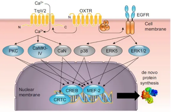
activates phosphatidylinositol-4,5- bisphosphate 3-kinase (PI3K) as well as the mitogen-activated protein kinase (MAPK) pathway via the inhibition of adenylyl cyclase and the reduction of cAMP levels (Busnelli and Chini, 2018; Busnelli et al., 2012). In more detail, MAPK activation occurs via transactivation of the epidermal growth factor (EGFR,(Blume et al., 2008), followed by recruitment of the membrane-associated proto-oncoprotein GTPase Rat sarcoma (Ras).

Ras via Ras proto-oncogene serine/threonine-protein kinase (Raf-1) triggers a chain reaction to ultimately fully activate the MAPK pathway. The three main kinases are extracellular signal-regulated kinase (ERK1/2), p38, and c-Jun NH2-terminal kinase (JNK1/2/3), the latter not being affected by OTR signaling. ERK1/2 phosphorylation occurs upon mitogen-activated protein kinase kinase (MEK1/2) activation in a cell- and tissue- specific manner, however, evidence also suggests other MEK1/2 targets, independent of the canonical ERK1/2 pathway (Jurek et al., 2012), including PPARγ, MyoD, PI3K, or LIMKinase 1 (Brandt et al., 2010; Burgermeister et al., 2007; Jo et al., 2011). Other ERK substrates include mitogen and stress activated kinases (MSK) and the ribosomal protein S6 kinase (RSK,(Jurek and Neumann, 2018).

Well-known downstream targets of MAPK signaling are the transcription factors myocyte

enhancer factor 2 (MEF2, for more details see chapter 5.) and CREB with its cofactors

CRTC2/3 (Jurek et al., 2015; Tomizawa et al., 2003). However, the pathways mentioned

here cannot be considered independently, as inhibition of OT-induced Ca

2+

influx has

deleterious effects on MAPK signaling in hypothalamic cells (van den Burg et al., 2015),

indicating an initial role of Ca

2+

for further second messenger signaling. For an overall

summary of OTR signaling, both proven as well as hypothetical see Figure 4.

(37)

33

Figure 4. Schematic summary of neuronal OTR signaling.

The 7 transmembrane GPCR OTR (shown here as OXTR) transactivates the TRPV2 cation channel and the EGFR, leading to Ca

2+

-dependent and -independent signaling. Kinases translocate from the cytoplasm to the nucleus and activate a set of transcription factors, which orchestrate transcription as a complex. Kinases and transcription factors in blue represent cascades that have been proven to be coupled to the neuronal OTR, kinases in gray are hypothetical or shown to be coupled to peripheral OTRs (adapted from(Jurek and Neumann, 2018).

In summary, OT concentration, G protein expression, OTR membrane availability, and as a last factor OTR affinity states that are addressed in the next chapter determine the quality and quantity of OTR-mediated signaling.

2.4. Impact of cholesterol on oxytocin receptor activity

OTR receptors occur in two different affinity states, the low-affinity state (K

d

> 100 nM) and

the high-affinity state (K

d

< 1-5 nM,(Gimpl et al., 1995). Transitions between those affinity

states are reversible and depend on cholesterol content in the plasma membrane (Gimpl et

al., 1995; Klein et al., 1995). In general, membranes in cellular systems are known to appear

as lipid bilayers. The “fluid mosaic model”, as originally suggested by Singer and Nicholson,

has become, with minor modifications, the standard model of membrane structure (Singer

and Nicolson, 1972). Specific areas within membranes, so-called “lipid rafts” are dynamic

microdomains that are capable of moving within the fluid bilayer. Those rafts are

(38)

34

sphingolipid- and cholesterol-enriched and contain ordered aggregations of membrane- bound signaling proteins. The whole complex represents a membrane-bound anchor, which plays an important role during signal transduction as “transduction platform” (Simons and Ikonen, 1997; Simons and Toomre, 2000). Cholesterol, as one of the most abundant components of membranes (Liscum and Underwood, 1995), is of great significance for the integrity of lipid microdomain structures like caveolae and the already mentioned lipid rafts (Incardona and Eaton, 2000; Simons and Toomre, 2000). OTR agonist binding affinity states s strongly depend on cholesterol contents in the membrane (Gimpl and Fahrenholz, 2000; Reversi et al., 2006; Wiegand and Gimpl, 2012).

On the one hand, OT inhibits cell growth and proliferation in a PI3K-dependent MAPK cascade activation, when the majority of OTRs are excluded from lipid rafts and caveolae.

On the other hand, strong PLC-independent mitogenic effects appear when OTRs are located within caveolae, induced by fusion to caveolin-2, a resident raft protein (Reversi et al., 2006; Rimoldi et al., 2003). Upon removal of cholesterol from the membrane via methyl- ß-cyclodextrin (MßCD), the OTR was found to be shifted to the low-affinity state, whereas high-affinity binding states could be regained by cholesterol enrichment via cholesterol- MßCD. Interestingly, restoring could not be achieved by other steroids, suggesting a specific and direct dependency of the OTR on cholesterol (Klein et al., 1995). Additionally, OTR stability and conformation is strongly influenced by the location in cholesterol-rich domains (Muth et al., 2011).

3. The CRF system

Stress is a major environmental factor contributing to the formation of psychiatric disorders.

The 3 physiologists Claude Bernard, Walter B. Cannon, and Hans Selye are considered as

the pioneers of stress biology. Their work was the basis for the concept of

neuroendocrinology, including the autonomic nervous system and the HPA axis as the key

players in the stress response. While the ANS represents the rapid response towards a

stressor, the HPA-axis modulates this response in duration and frequency (reviewed

in(Deussing and Chen, 2018).

(39)

35 3.1. CRF family members

The primary structure of the master regulator of the HPA axis, CRF was first identified in 1981 as a 41 amino acid peptide (Vale et al., 1981), while the evidence for an ACTH releasing mediator already existed since 1955 (Selye, 1955). CRF is synthesized in parvocellular neurons of the hypothalamus and triggers the release of ACTH into the systemic circulation (Rivier and Vale, 1983; Smith and Vale, 2006; Vale et al., 1981). Upon ACTH release, synthesis, and secretion of GCs from the zona fasciculata of the adrenal cortex are promoted. GCs are then responsible for the regulation of a variety of physiological adaptations when binding to their cognate intracellular receptors (Bamberger et al., 1996;

Munck et al., 1984; Smith and Vale, 2006). Furthermore, CRF regulates the ANS-mediated stress response and is also able to directly modulate behavioral stress responses (Deussing and Chen, 2018)and references therein). CRF and a group of 3 related proteins called urocortins (UCN), UCN1, UCN2 (also called stresscopin-related peptide), and UCN3 (also called stresscopin) elicit their function via binding to two different class B family of G-protein coupled receptor subtypes, the corticotropin releasing factor receptors 1 and 2 (CRFR1, CRFR2,(Hillhouse and Grammatopoulos, 2006; Lewis et al., 2001; Reyes et al., 2001;

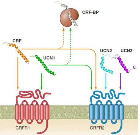
Vaughan et al., 1995). These receptors contain a large extracellular domain at the N- terminus and are able to bind large peptide ligands (Deussing and Chen, 2018). CRF and UCN1 preferentially bind to the CRFR1, while UCN2 and UCN3 bind the CRFR2 with higher affinities (Hillhouse and Grammatopoulos, 2006). CRFRs mainly signal via coupling to G

s

proteins, followed by the activation of protein kinase A (PKA) and MAPK-ERK pathways and the mobilization of Ca

2+

. Activation of ERK1/2 via PLC and for example G

is also possible. Internalization and desensitization of CRFR1 and CRFR2 is mostly β–arrestin- mediated. Additionally, receptor activity can be modulated by carboxy-terminal PDZ-domain interactions. Binding to PDZ-domains regulates the cellular location of the receptors and attributes them to larger signaling complexes (Henckens et al., 2016)and references therein). In rodents, there are two isoforms of CRFR2 (α and β) and three in humans (α, β, and γ) arising from separate promoters and 5´ exons which splice to a common set of downstream exons (Chen et al., 2005; Kostich et al., 1998). The CRF-BP regulates the activity of CRF and UCN1 by the binding between both factors. It represents a regulatory capturing function that might partially act in a CRFR2-dependent manner, however, the precise mechanism is unknown (Deussing and Chen, 2018; Eckart et al., 2001; Huising et al., 2008; Karolyi et al., 1999).

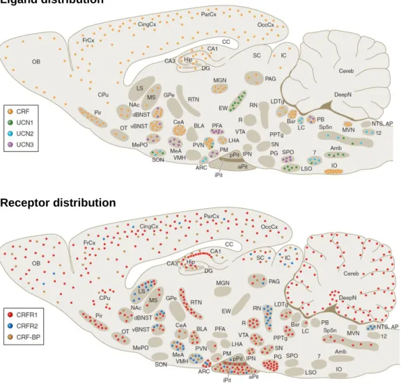
Both receptors and ligands are widely distributed throughout the brain (Fig. 5). High levels

of CRFR1 are found in the anterior pituitary, olfactory bulb, cerebral cortex, hippocampus,

and cerebellum. The mammalian brain variant CRFR2α is located in the lateral septum,

(40)

36

BNST, ventral medial hypothalamus, raphe nuclei, and choroid plexus (Chalmers et al., 1995; Lovenberg et al., 1995; Van Pett et al., 2000).

Figure 5. Distribution of CRF, UCN1-3, CRFR1, CRFR2, and CRF-BP mRNA expression in the rodent brain. Scheme taken from Deussing and Chen, 2018. Please note that in the lower scheme, PVN expression of CRFR2 seems to be absent, however, the data presented in this thesis and the according manuscript (Winter et al, in preparation) proves CRFR2 expression in the PVN.

3.2. Ligand-receptor interaction

Binding affinities of the CRFR1 and CRFR2 are different for CRF and the related UCNs (Fig. 6). These differences are mainly determined by the extracellular domain (ECD) of the receptors, but also the extracellular loops (ECL) and the transmembrane domain (TMD) contribute to the specificity of interactions. Within the N-terminus, amino acid residues 43- 50 and 76-84 play an important role in ligand binding specificity. The ECD and the TMD are linked by an amino acid segment close to the first TMD that is also essential for ligand

Ligand distribution

Receptor distribution

(41)

37

binding. Furthermore, interactions with the ECLs determine ligand binding specificity, as Arg

189

mediates binding within the first ECL for the CRFR1, whereas His

189

mediates binding to the CRFR2.

Additionally, glycosylation influences both ligand binding specificity and signal transduction for the CRFR1 as well as the CRFR2. The highly specific interaction between UCN2/UCN3 and the CRFR2 is attributed to several amino acid residues. For example, Pro

11

, Ala

35

, and Ala

39

are uniquely found in those ligands specifically binding to the CRFR2. In general, active ligands consist of 3 main parts. The N-terminal domain, important for receptor binding and activation, the central binding domain, where the CRF-BP interacts, and the C-terminus that mediates ligand affinity and specificity (Deussing and Chen, 2018)and references therein). For both CRFR1 and CRFR2, several splice variants have been identified however, only one functional splice variant of the CRFR1 is expressed in the central nervous system. Alternative splicing of the CRFR2 is further described in Chapter 4. The importance of the CRFR1 for stress-related behavior was supported by several knockout (KO) studies.

Loss of CRFR1 causes chronic GC deficiency, accompanied by reduced anxiety-related behavior and impairments in remote fear memory consolidation. Additionally, conditional KO of the CRFR1 in the forebrain lessens stress-induced dendritic remodeling and spine loss (reviewed in(Dedic et al., 2018).

Figure 6. Interactions between CRFR1, CRFR2, and the ligands CRF, UCN1, UCN2, and UCN3.

The potential role of the CRF-BP as a CRF/UCN1-capturing protein is also depicted (adapted from(Deussing

and Chen, 2018).

(42)

38 3.3. The CRFR2 and its role in anxiety

Studies on the deletion of CRFR2 yield a less clear picture compared to CRFR1. The first 3 conventional CRFR2 KO mouse models revealed normal development of the HPA axis but a hypersensitive response of the HPA axis to stress. Anxiety-like behavior was increased compared to wild-type mice. Both Ucn and Crf mRNA levels were elevated in the Edinger-Westphal nucleus and central amygdala, respectively (Bale et al., 2000). The effect of CRFR2 deletion or antagonism on baseline anxiety appears the be sex-specific, as the anxiogenic phenotype only occurred in male CRFR2 KO mice but not in females (Kishimoto et al., 2000). Despite the normal development of the HPA axis, CRFR2 is involved in the maintenance of the HPA axis drive and the recovery phase of the HPA response. CRFR2 KO mice display elevated corticosterone levels up to 90 min after stress, in contrast to an early termination of ACTH release (Coste et al., 2000). Impaired stress recovery was also seen in a triple KO mouse, lacking UCN1, UCN2, and UCN3 (Neufeld-Cohen et al., 2010).

A more recent study demonstrated that the anxious phenotype of those KOs depends on stress levels. CRFR2 KO mice display increased anxiety-like behavior 24 h after acute restraint stress. However, there were no immediate effects after acute restraint stress as well as after chronic stress in KO compared to wild-type males (Issler et al., 2014).

Furthermore, stress-levels, ligand dosage, and brain region are crucial for the impact of CRFR2 activity on anxiety (Bale et al., 2002a; Todorovic et al., 2007). Under low-stress conditions, only a relatively high dose of UCN2 in the septum increased anxiety-like behavior, whereas under high-stress conditions, also lower doses induced anxiety-like behavior (Henry et al., 2006). CRFR2-positive cells in the lateral septum exert GABAergic projections to the anterior hypothalamus. These outputs enhance stress-induced anxiety and promote persistent anxious behaviors, representing a LS circuitry that promotes stress- induced persistent anxiety (Anthony et al., 2014). Antagonism of the CRFR2 with antisauvagine-30 (ASV) also causes differential anxiety-related phenotypes (Hammack et al., 2003; Takahashi et al., 2001). The precise dosage of ASV is essential, as high concentrations might trigger non-CRFR2-mediated effects (Zorrilla et al., 2013).

As already mentioned, UCN2 (Hammack et al., 2003; Valdez et al., 2002) or UCN3 (Valdez

et al., 2003) administration and the subsequent activation of the CRFR2 has anxiolytic

effects in mice and rats. Moreover, CRFR2 activity is also thought to be related to the

serotonergic system (Hammack et al., 2003; Pernar et al., 2004). Application of UCN2 into

the dorsal raphe nucleus increased cFOS expression in a subpopulation of serotonergic

neurons. However, the precise mechanism behind the effects of UCN and CRFR2 within

the serotonergic system remains elusive (Dedic et al., 2018). The serotonergic system is

also important for stress-recovery. The CRFR2 mediates the proper functioning of serotonin

(43)

39

(5-HT

1A

) receptors, as the KO attenuates 5-HT receptor responses in the dorsal raphe nucleus (Issler et al., 2014). In addition to the consequences for anxiety-like behavior, CRFR2 activity also affects social behaviors. KO of either CRFR2 or UCN3 in mice decreases the preference for novel conspecifics and impairs their ability to cope with socially challenging situations (Shemesh et al., 2016). Members of the CRF family, like UCN3, also exhibit in combination with OT, profound effects on long-term potentiation in the rat medial amygdala (Frankiensztajn et al., 2018). Furthermore, blocking of CRFR1 signaling disrupts stress-induced hippocampal tau phosphorylation, a key factor of Alzheimer’s disease development. On the contrary, loss of CRFR2 promotes these effects (Rissman et al., 2007).

The generation of different CRFR1 and CRFR2 subforms is mediated by alternative splicing. A mechanism that is found throughout eukaryotes and provides extensive protein diversity.

4. Alternative splicing

Alternative splicing is one of many processes that regulate gene expression, and therefore leads to increased protein diversity. In eukaryotes, genes contain protein-coding sequences (exons) interrupted by non-coding segments (introns,(Berget et al., 1977; Chow et al., 1977;

Sharp, 1994, 2005). The removal of introns or alternative exons is mediated by a large

ribonucleoprotein (RNP) complex, called the spliceosome. The spliceosome mediates RNA

cleavage and ligation which are the crucial steps of splicing (Lee and Rio, 2015; Wahl et

al., 2009; Will and Luhrmann, 2011). With a number of almost 25.000 genes in the human

genome, but around 90.000 proteins being expressed, alternative splicing represents an

essential mechanism for diversity in humans, and generally, in mammals (International

Human Genome Sequencing, 2004). In fact, around 95 % of human genes undergo

alternative splicing. Originally discovered simultaneously with constitutive splicing itself

(Gilbert, 1978), there are various types of alternative splicing known today. The most

important one for my study is exon skipping, where one so-called cassette exon is either

included in the final mRNA or alternatively skipped. This mechanism is based on the

formation of the spliceosome, consisting of small nuclear RNAs and small nuclear

ribonucleoprotein particles (snRNP). The spliceosome machinery (U1, U2, U4, U5, and U6)

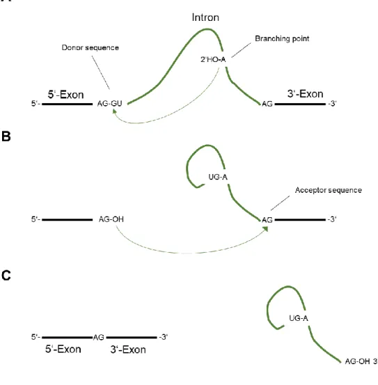
assembles on the mRNA, particularly at the 5’ splice site (GU), the 3’ splice site (AG), and

the branch point, upstream of the polypyrimidine tract (PPT). First, U1 forms a base-pairing

(44)

40

interaction with the 5’-splice site, whereas U2 base-pairs with the branch-point. Then, a tri- snRNP complex containing U4, U5, and U6 associates with the forming spliceosome, removing U1 and U4. This leads to two transesterification reactions and joins the exons, excluding the intermediate exon (Fig. 7,(La Cognata et al., 2014).

In addition to exon skipping, various other patterns of alternative splicing contribute to the

variety of mature mRNA variants. In more detail, alternative 5′ splice sites are additional

donor sites and thereby enable different exon lengths at the 3’ end. Alternative 3’ splice

sites function as additional acceptor sites changing the 5’ end of the exon. Alternative

promoters and alternative 3′ exons lead to different mature mRNA variants by altering the

first and last exons, respectively. In the last exon, 3’ untranslated regions of different lengths

can be created by alternative polyadenylation (Li et al., 2007; Vuong et al., 2016). These

mechanisms are the basis for the different transcripts of the OTR gene as well as for the

generation of CRFR1 and CRFR2 splice variants. In sheep, alterations in mRNA sequence

of the OTR can be observed and are most likely due to alternative use of polyadenylation

sites. This represents the first link between alternative splicing and the OTR (Feng et al.,

2000).

Abbildung

Figure 4. Schematic summary of neuronal OTR signaling.
Figure  5.  Distribution  of  CRF,  UCN1-3,  CRFR1,  CRFR2,  and  CRF-BP  mRNA  expression  in  the  rodent  brain
Figure 6. Interactions between CRFR1, CRFR2, and the ligands CRF, UCN1, UCN2, and UCN3
Figure 7. Basic principle of splicing in eukaryotes.
+7

Referenzen

ÄHNLICHE DOKUMENTE

To investigate the effect of DLK on the recruitment of TORC to a CRE-containing promoter, a reporter gene assay (§3.15) was performed whereby a luciferase reporter gene controlled

Inhibition of HT1, in turn, enables the protein kinases OST1 or GHR1 to activate S-type anion channel SLAC1, which initiates ion efflux from guard cells and

Brognard, J., et al., Akt/protein kinase B is constitutively active in non- small cell lung cancer cells and promotes cellular survival and resistance to chemotherapy and

(2005) LmxMPK4, a mitogen-activated protein (MAP) kinase homologue essential for promastigotes and amastigotes of Leishmania mexicana. (2009) PubChem: a public information system

Three major kinases involved in the signal transduction cascade that restarts meiosis, are Maturation Promoting Factor (MPF), Mitogen-Activated Proteinkinase (MAPK)

In den Kumuluszellen allerdings wurde die p90rsk während der Reifung, sowohl in vitro als auch in vivo, nicht aktiviert und scheint somit dort keine Rolle als Substrat

The dependence of the steroid hormone synthesis in porcine cumulus cells of the MAPK (mitogen-activated protein kinase) activation and BMP6 (bone morphogenetic protein

ROS are thought to promote atherosclerosis through a variety of mechanisms, including enhanced oxidation of lipoproteins (Steinberg 1997), activation of proinflammatory genes (Marui,