The X-linked juvenile retinoschisis protein retinoschisin is a novel regulator of mitogen-activated protein kinase signalling
and apoptosis in the retina
Karolina Pl€ ossl, Bernhard H.F. Weber, Ulrike Friedrich *
Institute of Human Genetics, University of Regensburg, Regensburg, Germany Received: August 4, 2016; Accepted: September 26, 2016
Abstract
X-linked juvenile retinoschisis (XLRS) is a hereditary retinal dystrophy in young males, caused by mutations in the
RS1gene. The function of the encoded protein, termed retinoschisin, and the molecular mechanisms underlying XLRS pathogenesis are still unresolved, although a direct interaction partner of the secreted retinoschisin, the retinal Na/K-ATPase, was recently identified. Earlier gene expression studies in retinoschi- sin-deficient (Rs1h
/Y) mice provided a first indication of pathological up-regulation of mitogen-activated protein (MAP) kinase signalling in dis- ease pathogenesis. To further investigate the role for retinoschisin in MAP kinase regulation, we exposed Y-79 cells and murine
Rs1h/Yretinae to recombinant retinoschisin and the XLRS-associated mutant RS1-C59S. Although normal retinoschisin stably bound to retinal cells, RS1- C59S exhibited a strongly reduced binding affinity. Simultaneously, exposure to normal retinoschisin significantly reduced phosphorylation of C-RAF and MAP kinases ERK1/2 in Y-79 cells and murine
Rs1h/Yretinae. Expression of MAP kinase target genes
C-FOSand
EGR1was also down-regulated in both model systems. Finally, retinoschisin treatment decreased pro-apoptotic
BAX-2transcript levels in Y-79 cells and
Rs1h/Yretinae. Upon retinoschisin treatment, these cells showed increased resistance against apoptosis, reflected by decreased caspase-3 activity (in Y-79 cells) and increased photoreceptor survival (in
Rs1h/Yretinal explants). RS1-C59S did not influence C-RAF or ERK1/2 activa- tion,
C-FOSor
EGR1expression, or apoptosis. Our data imply that retinoschisin is a novel regulator of MAP kinase signalling and exerts an anti- apoptotic effect on retinal cells. We therefore discuss that disturbances of MAP kinase signalling by retinoschisin deficiency could be an initial step in XLRS pathogenesis.
Keywords: X-linked juvenile retinoschisis retinoschisin RS1 Na/K-ATPase MAP kinase signalling apoptosis
Introduction
Pathogenic alterations affecting the
RS1gene on chromosome Xp22.1 have been shown to cause XLRS (OMIM #312700) [1], a macular degeneration disorder in young males with a prevalence of approximately 1:5000 to 1:20,000 [2]. Disorganization of retinal lay- ers and distinct abnormalities in the electroretinogram (ERG) are hall- marks of the disease. Specifically, a characteristic splitting of retinal layers, presenting as a bilateral foveal schisis, is found at an early stage of the disease and results in cystic degeneration of the central retina [3
–6]. Additionally, defects in signal transmission from pho- toreceptor to bipolar cells as visualized by ERG recordings are observed and reveal a characteristic reduction in the b-wave ampli- tude, whereas the a-wave remains almost unaffected [4, 7]. Compara- ble pathological features are also evident in XLRS mice, generated
viaa targeted disruption of the murine orthologue of
RS1, theRs1hgene [8
–10]. Due to the close resemblance of the retinal phenotype in
Rs1hknockout mice and XLRS patients, the retinoschisin-deficient mouse represents an excellent disease model widely used in experi- mental studies addressing the mechanisms of XLRS pathology but also novel treatment approaches [11
–16].
The
RS1gene is organized into six exons and encodes a 224- amino acid (aa) precursor protein [1]. It is specifically expressed in the retina by photoreceptor and bipolar cells, as well as in pinealo- cytes of the pineal gland [1, 17, 18]. During protein synthesis, a 23- aa signal sequence is cleaved to produce a 201-aa mature polypeptide which is secreted from photoreceptors and bipolar cells as a homooc- tamer held together by intermolecular disulphide bonds between aa 223 and aa 59 [19
–22]. So far, over 190 unique XLRS-associated sequence variants in
RS1have been reported (Leiden Open Variation Database, http://grenada.lumc.nl/LOVD2/eye/home.php?select_db=
RS1, accessed May 2016). Functional assessment of a subset of
*Correspondence to: Ulrike FRIEDRICH, Ph.D., E-mail: ulrike.friedrich@klinik.uni-regensburg.de
ª2016 The Authors.
doi: 10.1111/jcmm.13019
these variants demonstrated that the vast majority of mutations result in a complete loss of the functional protein [4].
Despite intensive research, the precise molecular function of reti- noschisin remains unresolved. Searching for retinoschisin interaction partners, Molday
et al.[14] identified the retina-specific Na/K-ATPase composed of the two subunits ATP1A3 (
a3) and ATP1B2 (
b2). Sub- sequently, our group confirmed the Na/K-ATPase to be required for anchoring retinoschisin to plasma membranes [23]. The Na/K-ATPase is a plasma membrane spanning ion pump, responsible for maintain- ing the cellular membrane potential by transporting Na
+and K
+ions across the plasma membrane against their electrochemical gradient [24, 25]. Despite this essential task, Na/K-ATPases also mediate inter- cellular adhesion [26
–28] and induce activation of intracellular sig- nalling pathways upon binding of glycoside hormones such as ouabain [25, 29
–34]. Members of the FXYD family, a class of Na/K- ATPase-binding proteins [35, 36], were reported to be important reg- ulators of the Na/K-ATPase, modulating its pump activity and media- tion of intercellular adhesion [37
–39]. Similar to FXYD proteins, one could consider retinoschisin to exert a role as a modulator of Na/K- ATPase activity.
A genomewide expression analysis of the
Rs1h-deficient (Rs1h/Y) murine retina first indicated an increased activation of the ERK path- way in early XLRS pathogenesis, prior to apoptotic photoreceptor degeneration [40]. The ERK pathway is one of the four major MAP kinase pathways [41, 42] known to play a crucial role in fundamental developmental and physiological processes such as apoptosis, neuro- protection, neuronal development and adhesion [41, 43
–50]. It is tempting to speculate that misregulation of MAP kinase signalling caused by retinoschisin deficiency could be an initial step in XLRS pathogenesis. However, aberrant MAP kinase activation could also be a secondary event, caused by alterations of the cellular/retinal home- ostasis in the XLRS disease process.
In this study, we examined whether retinoschisin binding to reti- nal membranes directly modulates MAP kinase signalling. Our find- ings in cultured Y-79 cells and in retinal explants of
Rs1h/Ymice demonstrate that the addition of recombinant retinoschisin, but not recombinant mutant retinoschisin, significantly down-regulates MAP kinase signalling, as well as protects against apoptosis. We conclude that retinoschisin deficiency could be a trigger for disease pathogene- sis by a defective control of MAP kinase signalling and apoptosis in the retina.
Materials and methods
Animal models
TheRs1h/Ymouse was generated as described earlier [9] and kept on a C57BL/6 background. Mice were housed under specific pathogen-free barrier conditions at the Central Animal Facility of the University of Regensburg and maintained under conditions established by the institu- tion for their use, in strict compliance with NIH guidelines. Mice were sacrificed 10 or 18 days after birth by decapitation or cervical disloca- tion after inhalation of carbon dioxide, respectively.
Cell culture
Y-79 and Weri-Rb1 (ATCC, Manassas, VA, USA) cells were cultivated in RPMI medium with 10% FCS as well as 100 U/ml penicillin/strepto- mycin. ARPE-19 cells (ATCC) were maintained in DMEM/Ham’s F12 medium containing 10% FCS and 100 U/ml penicillin/streptomycin. BV- 2 cells were grown in RPMI-1640 with 5% FCS, 100 U/ml penicillin/
streptomycin and 195 nMb-mercaptoethanol. Hek293 cells (Invitrogen, Carlsbad, CA, USA) were maintained in DMEM high glucose medium containing 10% FCS, 100 U/ml penicillin/streptomycin and 500lg/ml G418. All media and cell culture supplies were purchased from Life Technologies (Carlsbad, CA, USA). Cell lines were grown in a 37°C incu- bator with a 5% CO2environment and subcultured when they reached 90% confluency for Hek293, BV-2 and ARPE-19 or a concentration of 4–59105cells/ml for Y-79 and Weri-Rb1. Only Y-79 cells passaged less than 10 times were applied in signalling or apoptosis assays.
RNA analysis
RNA was isolated from cell lines using the Qiagen RNeasy Mini Kit (Qia- gen, Venlo, the Netherlands). RNA from murine retinae and cultured retinal explants was isolated using the PureLinkTMRNA Micro Kit (Invit- rogen), according to the manufacturers’ protocols. One microgram of total RNA was transcribed into cDNA using RevertAid M-MuLV Reverse Transcriptase (Fermentas, St Leon-Rot, Germany) and poly(dT) primers according to the manufacturer’s instructions. Semiquantitative RT-PCR was performed as described by [23], with primers given in Table S1.
Quantitative real-time RT-PCR was performed and analysed as pub- lished [51] with primers given in Table S1.
Western blot analysis
Proteins were separated after application of Laemmli buffer [52] on 12.5%
gels or gradient gels 4–20% Mini-PROTEANTGXTMPrecast Protein Gels (Bio-Rad Laboratories, Hercules, CA, USA; for analysis of retinoschisin octamers). For Western blotting, proteins were transferred to polyvinyli- dene difluoride (PVDF) membranes (Immobilon; Millipore, Schwalbach, Germany) Antibodies were used as follows: Antibodies against Myc tag, phospho-c-Raf (Ser338), c-Raf and phospho-44/42-MAPK (Erk1/2;
Thr202/Tyr204) were obtained from Cell Signaling Technologies (Danvers, MA, USA). Antibodies against ACTB and ERK1/2 were from Sigma-Aldrich (St. Louis, MO, USA). Secondary anti-rabbit or antimouse IgG horseradish peroxidase (HRP)-linked antibodies were from Calbiochem (Merck Chemi- cals GmbH, Schwalbach, Germany). Antibody dilutions were applied according to the manufacturer’s recommendations. RS1 primary antibody (diluted 1:10,000) was kindly provided by Prof. Robert Molday, University of British Columbia, Vancouver, Canada. ClarityTMWestern ECL Substrate (Bio-Rad Laboratories) and an Odyssey FC imager (LI-COR, Lincoln, NE, USA) were used to visualize Western blots. Densitometric evaluation of Western blots was carried out using ImageJ (imagej.nih.gov).
Immunolabelling of retinal cryosections
Retinal explants were washed in PBS (2.7 mM KCl, 140 mM NaCl, 10 mM phosphate, pH 7.4) once. Subsequently, they were submerged
in 4% (w/v) paraformaldehyde and incubated for 1 hr at room temper- ature. Retinae were washed in PBS twice before they were put in 30%
(w/v) sucrose overnight. Single retinae were then embedded in Richard-Allan ScientificTM Neg-50TM Frozen Section Medium (Thermo Fisher Scientific, Waltham, MA, USA) and fast frozen in liquid nitrogen.
About 10lm cryosections were cut. Immunolabelling with anti- ATP1B2 and antiretinoschisin antibodies was performed as described by [23]. Cone visualization was performed with Alexa 488-conjugated peanut agglutinin (1:250, PNA; Invitrogen). Rhodopsin staining was performed with Rho-1D4 antibody (1:1000), kindly provided by Prof.
Robert Molday, University of British Columbia, Vancouver, Canada. The sections were counterstained with 40,6-diamidino-2-phenylindol (DAPI, 1:1000; Molecular Probes, Leiden, the Netherlands). Images were taken with custom-made VisiScope CSU-X1 Confocal System (Visitron Systems, Puchheim, Germany) equipped with high-resolution sCMOS camera (PCO AG, Kehlheim, Germany).
Expression cloning
The coding sequence of non-mutant retinoschisin (NM_000330.3) was amplified from cDNA of retinal tissue using oligonucleotide primers containing aEcoRIrestriction site at the 50 end and aXhoIrestriction site at the 30 end of theRS1coding sequence (for primer sequences, see Table S1). The coding sequence of the XLRS-associated RS1 mutant RS1-C59S (NM_000330.3 (RS1):c.175T>A [p.Cys59Ser]
http://grenada.lumc.nl/LOVD2/eye/home.php?select_db=RS1) was gen- erated by site-directed mutagenesis on the retinoschisin coding sequence (primer sequences shown in Table S1). For purification, the two RS1 variants were each tagged with an N-terminal Myc tag, fol- lowing the leader sequence (after aa 23). This peptide insertion into the full-lengthRS1 coding sequence was performed by fusing the N- terminal part of both RS1 coding sequences from positions 1 to 69 (aa 1–23) to the N-terminal half of the Myc tag sequence (for primers, see Table S1). The C-terminal part of bothRS1coding sequences (aa 70 to stop codon) was fused to the C-terminal half of the Myc tag sequence (for primers, see Table S1). C- and N-terminal RS1 frag- ments were ligatedviaa BclI restriction site which was introduced into the Myc tag, and inserted into the pCDNA3.1TM expression vector (Thermo Fisher Scientific).
Expression and purification of recombinant RS1 variants
Expression constructs were transfected into Hek293 cells using calcium phosphate transfection as described by [53]. About 7 hrs after transfec- tion, the culture medium was replaced by Opti-MEM containing 100 U/ml penicillin/streptomycin (Life Technologies) and cells were cul- tured for additional 48 hrs.
Myc-tagged retinoschisin and RS1-C59S were isolated from cultiva- tion media by immunoprecipitation using PierceTM Anti-c-Myc Agarose (Thermo Fisher Scientific) according to the manufacturer’s instructions.
Concentrations of purified proteins were determined using the Bio-Rad DCTMProtein Assay Kit (Bio-Rad Laboratories).
For use as a treatment control, Hek293 cells were transfected with empty pCDNA3.1TMexpression vector, and cultivation medium of these cells was subjected to purification procedure exactly like medium from cells transfected with RS1 variants.
Purity of purified Myc-tagged RS1 proteins and control eluate was veri- fiedviasilver staining, Coomassie Blue staining and Western blot analysis using antibodies against the Myc tag and against retinoschisin (Fig. S1).
Binding of RS1 protein variants to membranes
Retinoschisin binding to adherent cell lines (BV-2, ARPE and Hek293) as well as to murine retinal membranes (P10) was assessed as described by [23], but with a prolonged incubation time of 1 hr.
Retinoschisin binding to suspension cell lines Y-79 and Weri-Rb1 was analysed by incubating 49106cells in 5 ml RS1 containing med- ium (from supernatant of stably transfected Hek293 cells [23]) for 1 hr, with subsequent washing steps as described [23].
For comparing binding affinity of retinoschisin and RS1-C59S to Y-79 cells andRs1h/Ymurine retinal membranes, 6lg of purified retinoschi- sin or RS1-C59S was added to 5 ml cultivation medium and incubated for 10, 30 and 60 min. Subsequent steps were performed as above.
For localization of bound recombinant RS1 variants onRs1h/Ymurine retinae,Rs1h/Ymurine retinal explants were incubated with 1lg RS1, RS1-C59S or control eluate, as described in signalling experiments. After 30 min. of incubation, the retinal tissue was washed with PBS once before immunolabelling of retinal cryosections was performed.
Analysis of signalling pathways in retinal explants and Y-79 cells
Y-79 cells were grown to a concentration of 4–59105cells/ml in 10 ml medium. The experiment was started by adding 1lg purified retinoschisin or RS1-C59S, or equal volume of control eluate. After 10 or 30 min. of incubation at 37°C, cells were harvested by centrifugation.
For Western blotting, cells were resuspended in 200ll of pre-cooled PBS with PhosSTOPTMphosphatase inhibitor (Sigma-Aldrich) and lysed by sonication (10 sec., 40% intensity). For RNA isolation, cells were washed once with pre-cooled PBS before they were subjected to RNA isolation.
Eyes fromRs1h/Ymice at post-natal day 10 were enucleated and retinal explants were dissected as described by [54]. Retinae were incu- bated in 800ll DMEM/Ham’s F12 containing 10% FCS, 100 U/ml peni- cillin/streptomycin, 2 mM L-glutamine and 2lg/ml insulin (Thermo Fisher Scientific). One microgram purified retinoschisin or RS1-C59S or equal volumes of control eluate were added. After 10 or 30 min. of incubation at 37°C, retinal explants were removed from medium and transferred to 200ll of pre-cooled PBS containing PhosSTOPTMphos- phatase inhibitor. For Western blot analysis, retinal explants were soni- cated for 10 sec. at 40% intensity. For RNA isolation, retinal explants were immediately transferred into lysis buffer (PureLinkTMRNA Micro Kit; Invitrogen).
Analysis of caspase-3 activity in Y-79 cells
About 29106cells/well were seeded onto poly-L-lysine-coated 24-well plates. Cells were allowed to adhere overnight before 0.1lg of purified retinoschisin or RS1-C59S, or equal volumes of control eluate were added to 1 ml medium per well. After 1 hr, the culture medium was changed to 1 ml RPMI (containing RS1 variants or control eluate as
before) and 0.2 mM H2O2to induce apoptosis or 0 mM H2O2as con- trol. After 2 hrs, the medium was replaced by 1 ml RPMI containing RS1 variants or control eluate as before. Cells were allowed to recover for 18 hrs before they were subjected to a caspase-3 activity test using the EnzChek Caspase-3 Assay Kit #2 by Thermo Fisher Scientific according to the manufacturer’s directions.
Analysis of photoreceptor degeneration in retinal explants
Eyes were enucleated from mice at post-natal day 18 and retinae were dissected as described before [55]. Five retinae each were subjected to treatment with retinoschisin, RS1-C59S and control protein. Retinal explants were transferred into pre-warmed medium (DMEM/Ham’s F12 containing 10% FCS, 100 U/ml antibiotic–antimytotic, 2 mML-glutamine and 2lg/ml insulin, all from Life Technologies) immediately after preparation, rinsed once in pre-warmed medium and then transferred onto Track Etch Membrane Filters (Whatman plc, Maidstone, UK) in 35- mm tissue culture dishes containing 3 ml of medium to which 1lg of purified retinoschisin, RS1-C59S or control eluate had been added. The retinal explants on the filters were covered in a small droplet of med- ium. Cultivation was carried out under sterile conditions in a 37°C incu- bator with a 5% CO2environment. Medium was replaced every 36 hrs and total cultivation time was 1 week. Subsequently, the retinal explants were fixed, cryopreserved and cut into 10-lm sections for subsequent histological analyses as described before. After PNA staining, cones were counted in each two different sections of the same retina, in 200- nm-wide regions to the left and the right side of the optic nerve. Rods were stained with anti-Rho-1D4 antibody. Staining signals of each two different sections of the same retina, in 200-nm-wide regions to the left and the right side of the optic nerve, were quantified using ImageJ.
Results
Increased MAP kinase signalling in murine Rs1h
/Yretinae
A study by Gehrig
et al.[40] previously indicated up-regulated MAP kinase activity in early retinal development of
Rs1h/Ymice. The authors found in the murine
Rs1h/Yretinae increased phosphoryla- tion of extracellular-signal-regulated kinases 1 and 2 (Erk1/2), as well as up-regulated expression of
Egr1(early growth response protein 1), a prominent target gene of activated MAP kinases [56].
To first verify these results, we analysed phosphorylation of Erk1/2 as well as
Egr1expression in retinae of wild-type and
Rs1h/Ymice, 7, 10 and 14 days after birth (P7, P10 and P14). Furthermore, we investi- gated phosphorylation of c-Raf, a central constituent of the ERK path- way, the activation of which precedes and is required for Erk1/2 phosphorylation [57], in P7, P10 and P14 retinae. As an additional MAP kinase target gene, we assessed expression of the FBJ murine osteosar- coma viral oncogene homologue gene (c-Fos), which is expressed in response to transient and sustained ERK signalling [56, 58
–61].
Western blot analyses showed an increase in c-Raf and Erk1/2 phosphorylation in
Rs1h/Yretinae compared with wild-type retina
(Fig. 1A). C-Raf phosphorylation levels in
Rs1h/Yretinae increased to around 150
–200%, whereas Erk1/2 phosphorylation levels rose to around 250
–300% above normal (Fig. 1B). These differences were obtained in all stages (P7, P10 and P14), and each were statistically significant (P
<0.05, except for c-Raf at P10; Fig. 1A and B) Reti- noschisin deficiency had no influence on levels of total c-Raf and Erk1/2, independent of the post-natal stages.
Expression of MAP kinase target genes
C-Fosand
Egr1was increased in
Rs1h/Yretinae, at all developmental stages;
c-Fosexpression levels in
Rs1h/Yretinae were between 150% and 250%, and
Egr1expression levels between 175% and 300% compared with wild-type retinae. Differences in expression were statistically signifi- cant between wild-type and
Rs1h/Yretinae (P
<0.05).
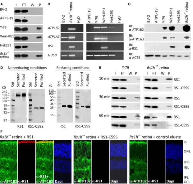
Binding of retinoschisin to different retinal cell types
Searching for an
in vitromodel system applicable for analysing the influence of retinoschisin on MAP kinase signalling, we tested differ- ent retinal cells including murine microglial cell line BV-2, human RPE-derived cell line ARPE-19, the human retinoblastoma cell lines Y- 79 and Weri-Rb1 for their capacity to bind retinoschisin (Fig. 2A). In these cells, we also investigated endogenous expression of the retinal Na/K-ATPase subunits ATP1A3 and ATP1B2, required for anchoring retinoschisin to retinal plasma membranes [23] (Fig. 2B and C).
Retinoschisin binding assays were performed on intact cells as described by [23]. Retinoschisin binding to Hek293 cells and crude membranes of
Rs1h/Yretinae served as negative and positive con- trols, respectively [23]. Efficient retinoschisin binding was found with Y-79, Weri-Rb1 and membranes of
Rs1h/Yretinae, but not with ARPE-19, BV-2 and Hek293.
Semiquantitative RT-PCR (Fig. 2B) and Western blot analyses (Fig. 2C) revealed endogenous expression of ATP1A3 and ATP1B2 only in Y-79 and Weri-Rb1. Weri-Rb1 cells also weakly expressed retinoschisin (Fig. 2B and C). In all further analyses, Y-79 cells were used to assess effects of externally added retinoschisin on intracellu- lar signalling.
Different binding affinities of normal
retinoschisin and the XLRS-associated mutant protein RS1-C59S to retinal membranes
To analyse functional properties of retinoschisin, we heterolo-
gously expressed RS1 (NM_000330.3) and the XLRS-associated
mutant protein (NM_000330.3(RS1):c.175T
>A [p.Cys59Ser], ter-
med RS1-C59S, in Hek293 cells. The mutation c.175T
>A
[p.Cys59Ser] is one of the rare XLRS variants which are not sub-
jected to co- or post-translational degradation, but instead are
translated and secreted from cells, although not as a stable octa-
mer but instead as a dimer [21, 22]. For purification, retinoschisin
(normal and mutant) was fused to an N-terminal Myc tag, which
did not influence secretion, oligomerization or binding capacities
of the resulting protein (Fig. 2D and E).
Y-79 cells and
Rs1h/Yretinal explants exposed to recombinant retinoschisin for 10, 30 and 60 min. stably bound the externally added retinoschisin, even after only 10 min. of incubation (Fig. 2E).
Notably, RS1-C59S exhibited a strongly reduced binding affinity to Y- 79 cells and
Rs1h/Yretinal explants (Fig. 2E).
Immunohistochemical stainings of murine
Rs1h/Yretinal explants after treatment with retinoschisin for 30 min. (Fig. 2F) con- firmed the binding of externally added recombinant retinoschisin.
Recombinant retinoschisin colocalized with the endogenously expressed retinal Na/K-ATPase of the murine retina at the inner seg- ments of photoreceptor cells. This is in agreement with the localiza- tion of retinoschisin in wild-type retinae [23]. In contrast to the known retinoschisin localization, no recombinant retinoschisin was detected in the plexiform layers of the retinal explants, which could possibly be explained by limited diffusion of the externally added reti- noschisin through the retinal layers. No retinoschisin staining was observed in immunohistochemical analyses of
Rs1h/Yretinal explants treated with RS1-C59S or control protein (Fig. 2F).
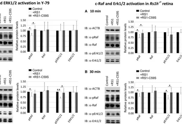
Extracellular retinoschisin modulates ERK 1/2 signalling in Y-79 cells and Rs1h
/Ymurine retinal explants
To assess the capacity of retinoschisin to directly modulate intracellular ERK1/2 signalling, we investigated an influence of extracellularly added recombinant retinoschisin (normal and
mutant) on phosphorylation of C-RAF and ERK1/2 in Y-79 cells (Fig. 3) and
Rs1h/Ymurine retinal explants (Fig. 4). As control, we applied protein purified from supernatant of mock vector- transfected cells.
In Y-79 cells, we observed a down-regulation of phosphorylated C-RAF (77.6 9.7%) after 10 min. of treatment with recombinant retinoschisin (Fig. 3A). In contrast, RS1-C59S failed to inhibit C-RAF phosphorylation (Fig. 3A). The differences in phosphorylated C-RAF levels between retinoschisin treatment and control or RS1-C59S treatment were statistically significant (P
<0.05). The effect of reti- noschisin on C-RAF phosphorylation was still evident after 30 min. of treatment with retinoschisin, where C-RAF phosphorylation was reduced to 78.4 17.0% (Fig. 3B).
In contrast to its effect on C-RAF in Y-79 cells, retinoschisin treat- ment failed to show a significant decrease in ERK1/2 phosphorylation after 10 min. of incubation (Fig. 3A). Thirty minutes of retinoschisin treatment (Fig. 3B), however, reduced ERK activation to around 69.6 5.3% in Y-79 cells (P
<0.05 compared with control protein or RS1-C59S). No alterations in total C-RAF and total ERK1/2 protein levels were detected, excluding an effect of retinoschisin on expres- sion or stability of the two proteins (Fig. 3).
MAP kinase signalling in murine
Rs1h/Yretinal explants was
similarly affected by retinoschisin treatment (Fig. 4). After
10 min. of incubation with retinoschisin, phosphorylated c-Raf
levels were decreased to 73.4 16.3% (Fig. 4A). RS1-C59S
treatment had no effect on c-Raf phosphorylation. The differences
in phosphorylated c-Raf levels were statistically highly significant
Fig. 1Influence of retinoschisin deficiency on MAP kinase signalling in the murine retina. C-Raf and Erk1/2 phosphorylation in murine wild-type and Rs1h/Yretinae, harvested at post-natal days 7, 10 and 14 (P7, P10 and P14). Retinal lysates were subjected to Western blot analyses with antibod- ies against phosphorylated c-Raf (pRaf), total c-Raf (Raf), phosphorylated Erk1 and Erk2 (pErk1/2), total Erk1 and Erk2 (Erk1/2), as well as ActB as a control (A). Densitometric quantification (B) was performed with immunoblots from three independent sample sets. Signals for pErk1/2, Erk1/2, pRaf and Raf were normalized against ActB and calibrated against signals for wild-type retinae. Data represent the mean+S.D. (C)C-FosandEgr1 expression in murine wild-type andRs1h/Yretinae harvested at post-natal days 7, 10 and 14 (P7, P10 and P14). mRNA expression ofC-Fosand Egr1was determinedviaquantitative real-time RT-PCR. Five independent sample sets were analysed. Results were normalized toHprttranscript levels and calibrated with the control. The mean+S.D. for the three (immunoblot analyses) or five (quantitative RT-PCR) independent sample sets is given. Asterisks mark statistically significant (*P<0.05) and highly significant (**P<0.01) differences.Fig. 2Binding of RS1 variants to retinal cells (A) Binding of retinoschisin to cultured retinal cell lines ARPE-19, Y-79, Weri-Rb1 and BV-2: Cells were incubated for 60 min. with retinoschisin containing supernatant (I, input) of cells stably transfected with a retinoschisin expression vector. Subse- quently, cells were centrifuged and supernatant (FT, flowthrough) was discarded. After further washing steps (last supernatant, W), cells were pel- leted (pellet, P). Fractions were subjected to Western blot analyses using an antiretinoschisin antibody. Retinoschisin binding to Hek293 cells and murineRs1h/Yretinal membranes served as negative and positive controls, respectively. (B) RT-PCR analysis ofATP1B2, ATP1A3andRS1gene expression in cell lines derived from murine microglia (BV-2), human retinal pigment epithelium (ARPE-19), human retinoblastoma (Y-79 and Weri- Rb1) and human embryonic kidney (Hek293).GUSBgene expression was assessed as control for RNA integrity. (C) Cell lysates from BV-2, ARPE- 19, Y-79, Weri-Rb1 and Hek293 were subjected to Western blot analyses using antibodies against ATP1B2, ATP1A3 and retinoschisin. Hek293+cells served as positive control. The ACTB immunoblot was performed as loading control. (D) Oligomerization of RS1 variants (non-mutant retinoschisin and RS1-C59S) before and after purification. About 48 hrs after transfection of Hek293 cells with expression constructs for N-terminally Myc-tagged RS1 variants, the cell culture medium (supernatant) was harvested and Myc-tagged proteins were purified from the supernatant. Aliquots of super- natant and purified RS1 fractions were subjected to SDS-PAGE under non-reducing and reducing conditions, followed by Western blot analyses using an antiretinoschisin antibody. (E) Binding of RS1 variants to retinal cells. Y-79 cells and murineRs1h/Yretinal explants were incubated with purified RS1 variants (I, input) for 10, 30 and 60 min. Cells were centrifuged and supernatant (FT, flowthrough) was discarded. After several wash- ing steps (last supernatant, W), cells were pelleted (pellet, P). Fractions were subjected to Western blot analyses using an antiretinoschisin antibody.
(F) Localization of recombinant RS1 variants on retinal membranes. Rs1h/Yretinal explants (P10) were incubated for 30 min. with retinoschisin, RS1-C59S or control protein, the latter purified from supernatant of empty expression vector-transfected cells. After washing and embedding, cryosections of these explants were subjected to immunohistochemical analyses using antibodies against ATP1B2 and retinoschisin. 40,6-Diamidino- 2-phenylindol (DAPI) staining shows the nuclei of the different retinal layers. IS, inner segments; ONL, outer nuclear layer; OPL, outer plexiform layer; INL, inner nuclear layer; IPL, inner plexiform layer; GCL, ganglion cell layer.
(P
<0.01) between control and retinoschisin treatment and sta- tistically significant (P
<0.05) between retinoschisin and RS1- C59S treatment. After 30 min. of incubation, the reduction in c- Raf phosphorylation by retinoschisin treatment was still observ- able (Fig. 4B): Retinoschisin decreased c-Raf phosphorylation to 81.4 14.1%, compared with the control.
Erk1/2 phosphorylation was not affected after 10 min.
(Fig. 4A), but only after 30 min. of treatment with recombinant retinoschisin (Fig. 4B). In contrast to control or RS1-C59S treat- ment, incubation with retinoschisin resulted in a clear reduction in phosphorylated Erk1/2 (68.7 18.2% compared with control, Fig. 4B). Differences in phosphorylated Erk1/2 levels were statisti- cally highly significant (P
<0.01) when compared between con- trol and retinoschisin treatment and statistically significant
(P
<0.05) between retinoschisin and RS1-C59S treatment. The different treatments caused no changes in total c-Raf and total Erk1/2 levels in the retinal explants (Fig. 4).
Extracellular retinoschisin modulates ERK1/2 target gene expression in Y-79 cells and Rs1h
/Ymurine retinal explants
Upon treatment with recombinant retinoschisin, a statistically signifi- cant down-regulation of
C-FOSmRNA expression was observed in Y- 79 cells (84.6 7.7%) and in
Rs1h/Ymurine retinal explants (68.1 18.9%) by quantitative RT-PCR when compared to control treatment (P
<0.05, Fig. 5A). In contrast, no prominent decrease in
Fig. 3Influence of retinoschisin on the ERK pathway in Y-79 cells. Reti-noschisin-dependent C-RAF and ERK1/2 phosphorylation in Y-79 cells.
Y-79 cells were treated for 10 min. (A) or 30 min. (B) with retinoschi- sin, RS1-C59S or control protein (purified from supernatant of empty expression vector-transfected cells). Subsequently, the cells were sub- jected to Western blot analyses with antibodies against phosphorylated C-RAF (pRAF), total C-RAF (RAF), phosphorylated ERK1 and ERK2 (pERK1/2), total ERK1 and ERK2 (ERK1/2), as well as ACTB as a con- trol. (AandB) Densitometric quantification was performed with immu- noblots from five independent experiments. Signals for pRAF RAF, pERK1/2 and ERK1/2 were normalized against ACTB and calibrated against the control. Data represent the mean+S.D. Asterisks mark statistically significant (*P<0.05) and highly significant (**P<0.01) differences.
Fig. 4Influence of retinoschisin on the ERK pathway in murineRs1h/Y retinal explants. Retinoschisin-dependent c-Raf and Erk1/2 phosphoryla- tion in murine Rs1h/Y retinal explants. Retinal explants harvested 10 days after birth (P10) were treated for 10 min. (A) or 30 min. (B) with retinoschisin, RS1-C59S or control protein (purified from super- natant of empty expression vector-transfected cells). Subsequently, the retinal lysates were subjected to Western blot analyses with antibodies against phosphorylated c-Raf (pRaf), total c-Raf (Raf), phosphorylated Erk1 and Erk2 (pErk1/2), total Erk1 and Erk2 (Erk1/2), as well as ActB as a control. (AandB) Densitometric quantification was performed with immunoblots from five independent experiments. Signals for pRaf Raf, pErk1/2 and Erk1/2 were normalized against ActB and calibrated against the control. Data represent the mean+S.D. Asterisks mark statistically significant differences (*P<0.05).
C-FOS
transcripts was found after treatment with RS1-C59S (98.4 7.3% for Y-79; 97.7 17.9% for retinal explants). The dif- ferences between the retinoschisin and the control or RS1-C59S treatment were statistically significant (P
<0.05; Fig. 5A).
Similarly, retinoschisin treatment caused a statistically significant decrease in
EGR1expression (Fig. 5B):
EGR1transcript levels were reduced to 82.0 8.8% in Y-79 cells and to 76.6 18.8% in
Rs1h/Ymurine retinal explants (P
<0.05) when compared to con- trol treatment. No prominent decrease in
EGR1mRNA levels was found after treatment with RS1-C59S (94.3 7.1% for Y-79 cells,
P<0.05 compared with retinoschisin treatment, and 92.4 26.2%
for retinal explants).
Retinoschisin is protective for apoptotic events
Mitogen-activated protein kinase signalling is an important mediator and regulator of several physiological processes implicated in XLRS pathogenesis [41, 43
–50, 62]. As one characteristic feature of
Rs1h/Y
mice is an early photoreceptor degeneration due to apoptotic cell death [63], we analysed the influence of retinoschisin on the expres- sion of apoptosis marker BAX in Y-79 cells and murine
Rs1h/Yreti- nal explants. Additionally, we assessed retinoschisin-dependent caspase-3 activation as a prominent marker for apoptosis induction [64
–66] in Y-79 cells, as well as the influence of recombinant reti- noschisin on cone degeneration in murine
Rs1h/Yretinal explants (Fig. 6).
Short time exposure to retinoschisin caused no change in
BAXtranscript levels in Y-79 cells (data not shown). After 20 hrs of incu- bation, however,
BAXtranscript levels were significantly decreased (77.6 14.0%) compared with control (P
<0.05) or RS1-C59S
treatment (P
<0.01, Fig. 6A). In
Rs1h/Yretinal explants, 30 min. of incubation with retinoschisin reduced
BAXtranscript levels to 85.4 9.3% (P
<0.01 compared with control and RS1-C59S-trea- ted retinae, Fig. 6A). In contrast, incubation with RS1-C59S had no significant effect on
BAXexpression in Y-79 cells (109.5 2.6%) or retinal explants (103.3 11.8%, Fig. 6A).
Caspases were shown to play a prominent role in photoreceptor cell death in the retinoschisin-deficient mouse [63]. We thus followed caspase-3 activation in Y-79 cells upon stress-induced apoptosis after treatment with 0.2 mM hydrogen peroxide [H
2O
2] [64
–66].
H
2O
2treatment caused an about 2.5-fold increase in caspase-3 activ- ity compared with unstimulated cells (Fig. 6B). Notably, in cells stim- ulated with H
2O
2, retinoschisin strongly decreased caspase-3 activation (67.6 9.9%) compared with control protein or RS1- C59S (P
<0.01). RS1-C59S led to a slight, albeit statistically not sig- nificant, reduction in caspase-3 activity (87.6 9.1%,
P=0.59) in H
2O
2-treated cells.
Finally, we addressed retinoschisin-dependent photoreceptor survival by following cone and rod degeneration in murine
Rs1h/Y
retinal explants [63]. Photoreceptor cell death in
Rs1h/Ymice
is triggered by apoptotic events initiated around 14 days after birth
(P14, [63]). We isolated
Rs1h/Yretinae 18 days after birth (P18)
and incubated them in medium containing retinoschisin, RS1-C59S
or control protein. One week of cultivation resulted in a strong
degeneration of retinal explants, shown by a markedly decreased
thickness of the central retina and a significant reduction in pho-
toreceptor cells (Fig. 6C
–F). More specifically, compared with
untreated retinae, 1 week of cultivation reduced the number of
cones to around 25% in control and RS1-C59S-treated explants
(25.6 8.5% for control and 24.8 8.9% for RS1-C59S treat-
ment (Fig. 6C and D). Notably, in explants treated with
Fig. 5Influence of retinoschisin on the expression of ERK1/2 pathway target genes. Retinoschisin-dependentC-FOS(A) andEGR1(B) expression in Y-79 cells and murine Rs1h/Yretinal explants. Cells/retinal explants were treated as described in Figures 3 and 4 for 2 hrs (Y-79 cells) and 30 min. (murine retinal explants).C-FOSandEGR1mRNA expression was determinedviaquantitative real-time RT-PCR. Five independent experi- ments were performed. Results were normalized toHPRTtranscript levels and calibrated with the control. The mean+S.D. for the five independent experiments is given. Asterisks mark statistically significant differences (*P<0.05).Fig. 6Influence of retinoschisin on apoptosis. (A) Retinoschisin-dependentBAXexpression in Y-79 cells and murineRs1h/Yretinal explants. Y-79 cells or retinal explants were treated with retinoschisin, RS1-C59S or control protein for 20 hrs or 30 min., respectively.BAXmRNA expression was determinedviaquantitative real-time RT-PCR. Five independent experiments were performed. Results were normalized toHPRTtranscript levels and calibrated with the control. The mean+S.D. for the five independent experiments is given. Asterisks mark statistically significant (*P<0.05) and highly significant (**P<0.01) differences. (B) Retinoschisin-dependent activation of caspase-3 in Y-79 cells subjected to oxidative stress. Y-79 cells, exposed to retinoschisin, RS1-C59S or control protein were treated with 0.2 mM H2O2for 2 hrs. About 18 hrs later, apoptosis was assayed by following caspase-3-specific proteolytic activity. Data represent the mean+S.D. of six independent experiments. Asterisks mark statistically highly significant differences (**P<0.01). (C–F) Retinoschisin-dependent photoreceptor degeneration in murineRs1h/Yretinal explants. Retinal explants harvested 18 days after birth (P18) were cultured for 1 week in medium containing retinoschisin, RS1-C59S or control protein (purified from supernatant of empty expression vector-transfected cells). After washing and embedding, cryosections of these explants were subjected to staining for nuclei, cones and rods. OS, outer segments; IS, inner segments; ONL, outer nuclear layer; OPL, outer plexiform layer; INL, inner nuclear layer; IPL, inner plexiform layer; GCL, ganglion cell layer. DAPI staining shows the nuclei of the different retinal layers.(C)Alexa488-conjugated pea- nut agglutinin (PNA) staining was applied to visualize cones.(D)The total number of cones per analyzed section was counted after staining with PNA.(E)Anti-Rho-1D4 antibody staining was applied to visualize rod specific Rhodopsin.(F)Rhodopsin signals per analyzed section were mea- sured using ImageJ (imagej.nih.gov). Data represent the mean + SD. Asterisks mark statistically highly significant differences (**P<
0.01).
retinoschisin, the cone number was decreased to only about 50%
(50.3 7.3% compared with untreated retinae), with a statistically highly significant difference to control and RS1-C59S-treated explants (P
<0.01). Investigations on rod degeneration revealed similar results (Fig. 6E and F). After 1 week of cultivation, rod sig- nals were reduced to around 24.8 9.3% for control treated and to 28.9 4.7% for RS1-C59S-treated explants. Treatment with retinoschisin lead to a rod signal decrease of only 59.3 7.6%
compared with untreated retinae, with statistically highly significant differences to control and RS1-C59S-treated explants (P
<0.01).
Discussion
In this study, we investigated the role of retinoschisin in the regu- lation of intracellular MAP kinase signalling. Firstly, our experi- ments confirmed strongly increased MAP kinase signalling in early retinal development of
Rs1h/Ymice. Secondly, we demonstrated that retinoschisin binding directly decreased phosphorylation of C- RAF and MAP kinases ERK1 and ERK2, as well as expression of the MAP kinase target genes
C-FOSand
EGR1in a retinal (Y-79) cell line and in murine
Rs1h/Yretinal explants. Thirdly, our data suggest a protective effect of retinoschisin against apoptotic cell death in Y-79 cells and
Rs1h/Yretinal explants. As a stringent control, the XLRS mutant RS1-C59S was deficient in binding to retinal membranes, and failed to reveal regulation on MAP kinase signalling or effects on apoptosis. Together, our results demon- strate that retinoschisin is a novel regulator of intracellular sig- nalling and protects retinal cells from apoptosis. We suggest that aberrant MAP kinase signalling due to retinoschisin deficiency could be an initial trigger in XLRS pathogenesis.
In recent years, the importance of MAP kinase signalling in retinal development and homeostasis has attracted increasing attention [67
–69]. Not surprisingly, several retinal dystrophies such as age-related macular degeneration [70
–72], diabetic retinopathy [73] or retinitis pig- mentosa [74, 75] were linked to malfunctioning MAP kinase pathways.
Aberrant MAP kinase signalling was also observed during early retinal development in the XLRS mouse model [40]. Our study verified the ear- lier observations from Gehrig
et al.[40] by showing increased activation of central constituents of the ERK pathway, c-Raf and Erk1/2 [57], in
Rs1h/Yretinae of 7-, 10- and 14-day-old mice. Furthermore, we showed up-regulation of prominent target genes of MAP kinase signalling, namely
c-Fosand
Egr1[56], indicating an early and sustained alteration in MAP kinase signalling in disease development of
Rs1h/Ymice.
To assess whether retinoschisin has the capacity to directly mod- ulate MAP kinase signalling, we investigated the effect of recombinant retinoschisin on activation of the ERK pathway in two retinal model systems; the human retinoblastoma cell line Y-79 and
Rs1h/Ymur- ine retinal explants, both capable to bind extracellularly added reti- noschisin due to an endogenous expression of the NA/K-ATPase subunits
a3 and
b2. The addition of recombinant retinoschisin had an immediate and significant influence on MAP kinase signalling in these two model systems, reflected by decreased C-RAF and ERK1/2 phos- phorylation. C-RAF activation (10 min.) occurred before ERK1/2 phosphorylation (30 min. after addition of retinoschisin), in
agreement with the established sequence of C-RAF and ERK1/2 acti- vation in the ERK signalling cascade [57, 76]. Subsequently, reti- noschisin treatment also induced down-regulation of
C-FOSand
EGR1expression in Y79 cells and
Rs1h/Ymurine retinal explants.
These results establish retinoschisin as an important regulator of the MAP kinase pathway in retinal cells.
The contribution of MAP kinase signalling to various disease processes can be explained by its key role in the regulation of complex physiological processes such as apoptosis, adhesion, pro- liferation, differentiation or development [41, 44, 49]. For instance, several studies showed a pro-apoptotic effect of ERK activation specifically connected to neuronal cells, for example in neurode- generative disease processes [77
–80]. Of note, a characteristic increase in ERK1/2 activation with an effect size similar to our results has been described for early disease stages of Alzheimer’s disease with 25% less ERK1/2 activation in temporal cortex of healthy individuals compared with patients [81], or of ocular ischaemic syndrome where 29% less ERK1 and 21% less ERK2 activation in murine retinae of control mice were found when com- pared to a mouse model of ocular ischaemic syndrome [82]. Addi- tionally, comparably small alterations in MAP kinase signalling, related to cellular survival, were found in natural killer cells of chronic fatigue syndrome [83] and in lymphocytes of patients with Alzheimer’s and Parkinson’s disease [84].
Consistently, we demonstrate a protective influence of retinoschisin against apoptosis: Transcript levels of the pro-apoptotic BAX protein [85, 86] were down-regulated in Y-79 cells and
Rs1h/Yretinae exposed to recombinant retinoschisin. Furthermore, in Y-79 cells subjected to oxida- tive stress, caspase-3 activity, a marker for the induction of apoptosis [65], was significantly decreased by retinoschisin. Similarly, apoptosis- induced cone and rod degeneration [63] in murine
Rs1h/Yretinal explants was strongly reduced in the presence of recombinant reti- noschisin. Further studies are required to verify the direct contribution of increased MAP kinase activation to photoreceptor apoptosis in XLRS pathogenesis. Nevertheless, considering the current state of knowledge on the pathological role of MAP kinase activation in neurodegeneration [77
–82], we speculate that increased MAP kinase signalling due to reti- noschisin deficiency can induce or contribute to XLRS-associated neu- rodegenerative processes in humans, and apoptotic photoreceptor degeneration in the XLRS mouse model [40].
Our investigations included studies on the functionality of the XLRS-associated retinoschisin mutant, RS1-C59S. Unlike most RS1 mutants, RS1-C59S is translated and secreted from cells, but with defective oligomerization [21, 22]. The functional consequences of this structural alteration have not been elucidated, so far. Here, we show that RS1-C59S cannot bind to retinal membranes and can thus not fulfil its function as a regulator of intracellular signalling.
The present data do not allow elucidation of how extracellular reti-
noschisin binding affects intracellular MAP kinase signalling. Previous
analysis identified the retinal Na/K-ATPase as the specific binding
partner for retinoschisin on retinal membranes [14, 23]. Several
groups reported that in addition to their function as an ion pump [87,
88], Na/K-ATPases are important regulators of intracellular MAP
kinase signalling [31, 89
–91], although the exact mechanism of signal
transduction from Na/K-ATPases to the MAP cascade is under debate
[30, 92
–96]. It would thus be conceivable that retinoschisin modu- lates the capacity of the Na/K-ATPase to regulate intracellular sig- nalling. A disruption of this retinoschisin-Na/K-ATPase signalosome complex by retinoschisin deficiency could therefore result in defective MAP kinase regulation by the Na/K-ATPase.
Taken together, we provide evidence that retinoschisin is a novel regulator of MAP kinase signalling in the retina with the capacity to protect cells against apoptotic cell death. We suggest that disturbances of intracellular MAP kinase signalling by reti- noschisin deficiency might be one of the initial steps in XLRS pathology. Thus, our data could provide a novel basis for consid- erations to therapeutic treatments for this progressive and cur- rently untreatable disease.
Acknowledgements
This work was supported in parts by a grant from the Deutsche Forschungsge- meinschaft (DFG) (FR 3377/1-1 to U.F.). We thank T.L. (Laboratory for
Experimental Immunology of the Eye, Department of Ophthalmology, Univer- sity of Cologne, Germany) for providing BV-2 cells. We thank L.P., M.R. and D.S. (Institute of Human Genetics, University of Regensburg, Germany) for excellent technical assistance.
Conflict of interest
None declared.
Supporting information
Additional Supporting Information may be found online in the supporting information tab for this article:
Figure S1
Purity of Myc-tagged RS1 proteins.
Table S1
Primers used in RNA analyses and expression cloning.
References
1. Sauer CG, Gehrig A, Warneke-Wittstock R, et al.Positional cloning of the gene associ- ated with X-linked juvenile retinoschisis.Nat Genet. 1997; 17: 164–70.
2. George ND, Yates JR, Bradshaw K,et al.
Infantile presentation of X linked retinoschi- sis.Br J Ophtalmol. 1995; 79: 653–7.
3. Kellner U, Brummer S, Foerster MH,et al.
X-linked congenital retinoschisis. Graefes Arch Clin Exp Ophthalmol. 1990; 228: 432–
7.
4. Molday RS, Kellner U, Weber BH.X-linked juvenile retinoschisis: clinical diagnosis, genetic analysis, and molecular mecha- nisms.Prog Retin Eye Res. 2012; 31: 195– 212.
5. Pimenides D, George ND, Yates JR,et al.
X-linked retinoschisis: clinical phenotype and RS1 genotype in 86 UK patients.J Med Genet. 2005; 42: e35.
6. Yu J, Ni Y, Keane PA,et al.Foveomacular schisis in juvenile X-linked retinoschisis: an optical coherence tomography study.Am J Ophthalmol. 2010; 149: 973–8 e2.
7. Renner AB, Kellner U, Fiebig B,et al.ERG variability in X-linked congenital retinoschi- sis patients with mutations in the RS1 gene and the diagnostic importance of fundus aut- ofluorescence and OCT. Doc Ophthalmol.
2008; 116: 97–109.
8. Takada Y, Vijayasarathy C, Zeng Y,et al.
Synaptic pathology in retinoschisis knockout (Rs1-/y) mouse retina and modification by rAAV-Rs1 gene delivery.Invest Ophthalmol Vis Sci. 2008; 49: 3677–86.
9. Weber BH, Schrewe H, Molday LL,et al.
Inactivation of the murine X-linked juvenile retinoschisis gene, Rs1h, suggests a role of retinoschisin in retinal cell layer organization and synaptic structure.Proc Natl Acad Sci USA. 2002; 99: 6222–7.
10. Zeng Y, Takada Y, Kjellstrom S,et al.RS-1 gene delivery to an adult Rs1h knockout mouse model restores ERG b-wave with reversal of the electronegative waveform of X-linked retinoschisis.Invest Ophthalmol Vis Sci. 2004; 45: 3279–85.
11. Bush RA, Wei LL, Sieving PA.Convergence of human genetics and animal studies: gene therapy for X-linked retinoschisis. Cold Spring Harb Perspect Med. 2015; 5:
a017368.
12. Byrne LC, Ozturk BE, Lee T, et al.Reti- noschisin gene therapy in photoreceptors, Muller glia or all retinal cells in the Rs1h-/- mouse.Gene Ther. 2014; 21: 585–92.
13. Ou J, Vijayasarathy C, Ziccardi L, et al.
Synaptic pathology and therapeutic repair in adult retinoschisis mouse by AAV-RS1 transfer.J Clin Invest. 2015; 125: 2891–903.
14. Molday LL, Wu WW, Molday RS. Reti- noschisin (RS1), the protein encoded by the X-linked retinoschisis gene, is anchored to the surface of retinal photoreceptor and bipolar cells through its interactions with a Na/K ATPase-SARM1 complex.J Biol Chem.
2007; 282: 32792–801.
15. Park TK, Wu Z, Kjellstrom S,et al.Intravit- real delivery of AAV8 retinoschisin results in cell type-specific gene expression and retinal
rescue in the Rs1-KO mouse. Gene Ther.
2009; 16: 916–26.
16. Sikkink SK, Biswas S, Parry NR,et al.X- linked retinoschisis: an update.J Med Genet.
2007; 44: 225–32.
17. Molday LL, Hicks D, Sauer CG, et al.
Expression of X-linked retinoschisis protein RS1 in photoreceptor and bipolar cells.
Invest Ophthalmol Vis Sci. 2001; 42: 816–
25.
18. Takada Y, Fariss RN, Muller M,et al.Reti- noschisin expression and localization in rodent and human pineal and consequences of mouse RS1 gene knockout. Mol Vis.
2006; 12: 1108–16.
19. Bush M, Setiaputra D, Yip CK,et al.Cog- wheel octameric structure of RS1, the dis- coidin domain containing retinal protein associated with X-linked retinoschisis.PLoS One. 2016; 11: e0147653.
20. Tolun G, Vijayasarathy C, Huang R,et al.
Paired octamer rings of retinoschisin sug- gest a junctional model for cell-cell adhesion in the retina.Proc Natl Acad Sci USA. 2016;
113: 5287–92.
21. Wu WW, Molday RS. Defective discoidin domain structure, subunit assembly, and endoplasmic reticulum processing of reti- noschisin are primary mechanisms respon- sible for X-linked retinoschisis.J Biol Chem.
2003; 278: 28139–46.
22. Wu WW, Wong JP, Kast J,et al.RS1, a dis- coidin domain-containing retinal cell adhe- sion protein associated with X-linked retinoschisis, exists as a novel disulfide-
linked octamer. J Biol Chem. 2005; 280:
10721–30.
23. Friedrich U, Stohr H, Hilfinger D,et al.The Na/K-ATPase is obligatory for membrane anchorage of retinoschisin, the protein involved in the pathogenesis of X-linked juvenile retinoschisis. Hum Mol Genet.
2011; 20: 1132–42.
24. Blanco G, Mercer RW.Isozymes of the Na- K-ATPase: heterogeneity in structure, diver- sity in function.Am J Physiol. 1998; 275:
F633–50.
25. Skou JC, Esmann M.The Na, K-ATPase.J Bioenerg Biomembr. 1992; 24: 249–61.
26. Antonicek H, Persohn E, Schachner M.Bio- chemical and functional characterization of a novel neuron-glia adhesion molecule that is involved in neuronal migration.J Cell Biol.
1987; 104: 1587–95.
27. Gloor S, Antonicek H, Sweadner KJ, et al. The adhesion molecule on glia (AMOG) is a homologue of the beta sub- unit of the Na,K-ATPase. J Cell Biol.
1990; 110: 165–74.
28. Vagin O, Dada LA, Tokhtaeva E,et al.The Na-K-ATPase alpha(1)beta(1) heterodimer as a cell adhesion molecule in epithelia.Am J Physiol Cell Physiol. 2012; 302: C1271– 81.
29. Kaplan JH. Biochemistry of Na,K-ATPase.
Annu Rev Biochem. 2002; 71: 511–35.
30. Li Z, Xie Z.The Na/K-ATPase/Src complex and cardiotonic steroid-activated protein kinase cascades.Pflugers Arch. 2009; 457:
635–44.
31. Xie Z, Askari A.Na(+)/K(+)-ATPase as a sig- nal transducer.Eur J Biochem. 2002; 269:
2434–9.
32. Xie Z, Cai T.Na+-K+–ATPase-mediated signal transduction: from protein interaction to cellu- lar function.Mol Interv. 2003; 3: 157–68.
33. Aperia AC, Akkuratov EE, Fontana JM,et al.
Na+, K+-ATPase, a new class of plasma membrane receptors. Am J Physiol Cell Physiol. 2016; 1: C491–5.
34. Schoner W, Scheiner-Bobis G.Endogenous and exogenous cardiac glycosides and their mechanisms of action. Am J Cardiovasc Drugs. 2007; 7: 173–89.
35. Geering K.FXYD proteins: new regulators of Na-K-ATPase.Am J Physiol Renal Physiol.
2006; 290: F241–50.
36. Sweadner KJ, Rael E.The FXYD gene family of small ion transport regulators or chan- nels: cDNA sequence, protein signature sequence, and expression.Genomics. 2000;
68: 41–56.
37. Jones DH, Li TY, Arystarkhova E,et al.Na, K-ATPase from mice lacking the gamma subunit (FXYD2) exhibits altered Na+affinity
and decreased thermal stability. J Biol Chem. 2005; 280: 19003–11.
38. Lubarski I, Pihakaski-Maunsbach K, Karlish SJ,et al.Interaction with the Na, K-ATPase and tissue distribution of FXYD5 (related to ion channel). J Biol Chem. 2005; 280:
37717–24.
39. Tokhtaeva E, Sun H, Deiss-Yehiely N,et al.
FXYD5 O-glycosylated ectodomain impairs adhesion by disrupting cell-cell trans-dimeri- zation of Na,K-ATPase beta1 subunits.J Cell Sci. 2016; 129: 2394–406.
40. Gehrig A, Langmann T, Horling F, et al.
Genome-wide expression profiling of the retinoschisin-deficient retina in early postna- tal mouse development.Invest Ophthalmol Vis Sci. 2007; 48: 891–900.
41. Chang L, Karin M.Mammalian MAP kinase signalling cascades.Nature. 2001; 410: 37– 40.
42. Lewis TS, Shapiro PS, Ahn NG. Signal transduction through MAP kinase cascades.
Adv Cancer Res. 1998; 74: 49–139.
43. Karmarkar SW, Bottum KM, Krager SL, et al. ERK/MAPK is essential for endoge- nous neuroprotection in SCN2.2 cells.PLoS One. 2011; 6: e23493.
44. Kolkova K, Novitskaya V, Pedersen N,et al.
Neural cell adhesion molecule-stimulated neurite outgrowth depends on activation of protein kinase C and the Ras-mitogen-acti- vated protein kinase pathway. J Neurosci.
2000; 20: 2238–46.
45. Wruck CJ, Gotz ME, Herdegen T, et al.
Kavalactones protect neural cells against amyloid beta peptide-induced neurotoxicity viaextracellular signal-regulated kinase 1/2- dependent nuclear factor erythroid 2-related factor 2 activation. Mol Pharmacol. 2008;
73: 1785–95.
46. Johnson GL, Lapadat R.Mitogen-activated protein kinase pathways mediated by ERK, JNK, and p38 protein kinases. Science.
2002; 298: 1911–2.
47. Sun Y, Liu WZ, Liu T,et al.Signaling path- way of MAPK/ERK in cell proliferation, differ- entiation, migration, senescence and apoptosis.J Recept Signal Transduct Res.
2015; 35: 600–4.
48. Xian J, Shao H, Chen X,et al.Nucleophos- min mutants promote adhesion, migration and invasion of human leukemia THP-1 cells through MMPs up-regulation via Ras/ERK MAPK signaling. Int J Biol Sci. 2016; 12:
144–55.
49. Zhang W, Liu HT.MAPK signal pathways in the regulation of cell proliferation in mam- malian cells.Cell Res. 2002; 12: 9–18.
50. Widmann C, Gibson S, Jarpe MB, et al.
Mitogen-activated protein kinase:
conservation of a three-kinase module from yeast to human. Physiol Rev. 1999; 79:
143–80.
51. Friedrich U, Myers CA, Fritsche LG,et al.
Risk- and non-risk-associated variants at the 10q26 AMD locus influence ARMS2 mRNA expression but exclude pathogenic effects due to protein deficiency.Hum Mol Genet.
2011; 20: 1387–99.
52. Laemmli UK.Cleavage of structural proteins during the assembly of the head of bacterio- phage T4.Nature. 1970; 227: 680–5.
53. Sambrook J, Russell DW. Calcium-phos- phate-mediated transfection of eukaryotic cells with plasmid DNAs.CSH Protoc. 2006;
2006: doi: 10.1101/pdb.prot3871.
54. Hsiau TH, Diaconu C, Myers CA,et al.The cis-regulatory logic of the mammalian pho- toreceptor transcriptional network. PLoS One. 2007; 2: e643.
55. Lee J, Myers CA, Williams N,et al.Quanti- tative fine-tuning of photoreceptor cis-regu- latory elements through affinity modulation of transcription factor binding sites. Gene Ther. 2010; 17: 1390–9.
56. Whitmarsh AJ.Regulation of gene transcrip- tion by mitogen-activated protein kinase sig- naling pathways. Biochim Biophys Acta.
2007; 1773: 1285–98.
57. Roskoski R Jr.ERK1/2 MAP kinases: struc- ture, function, and regulation. Pharmacol Res. 2012; 66: 105–43.
58. Hazzalin CA, Mahadevan LC. MAPK-regu- lated transcription: a continuously variable gene switch?Nat Rev Mol Cell Biol. 2002; 3:
30–40.
59. Hess J, Angel P, Schorpp-Kistner M.AP-1 subunits: quarrel and harmony among sib- lings.J Cell Sci. 2004; 117: 5965–73.
60. Murphy LO, Blenis J. MAPK signal speci- ficity: the right place at the right time.Trends Biochem Sci. 2006; 31: 268–75.
61. Whitmarsh AJ, Davis RJ.Transcription fac- tor AP-1 regulation by mitogen-activated protein kinase signal transduction pathways.
J Mol Med. 1996; 74: 589–607.
62. Wada T, Penninger JM. Mitogen-activated protein kinases in apoptosis regulation.
Oncogene. 2004; 23: 2838–49.
63. Gehrig A, Janssen A, Horling F,et al.The role of caspases in photoreceptor cell death of the retinoschisin-deficient mouse. Cyto- genet Genome Res. 2006; 115: 35–44.
64. Demelash A, Karlsson JO, Nilsson M,et al.
Selenium has a protective role in caspase-3- dependent apoptosis induced by H2O2in pri- mary cultured pig thyrocytes.Eur J Endocri- nol. 2004; 150: 841–9.
65. Li P, Nijhawan D, Budihardjo I, et al.Cyto- chrome c and dATP-dependent formation of
Apaf-1/caspase-9 complex initiates an apoptotic protease cascade.Cell. 1997; 91: 479–89.
66. Supanji, Shimomachi M, Hasan MZ,et al.
HtrA1 is induced by oxidative stress and enhances cell senescence through p38 MAPK pathway. Exp Eye Res. 2013; 112:
79–92.
67. Darling NJ, Cook SJ.The role of MAPK sig- nalling pathways in the response to endo- plasmic reticulum stress.Biochim Biophys Acta. 2014; 1843: 2150–63.
68. Donovan M, Doonan F, Cotter TG.Differen- tial roles of ERK1/2 and JNK in retinal devel- opment and degeneration. J Neurochem.
2011; 116: 33–42.
69. Mongan M, Wang J, Liu H,et al.Loss of MAP3K1 enhances proliferation and apopto- sis during retinal development. Develop- ment. 2011; 138: 4001–12.
70. Dridi S, Hirano Y, Tarallo V,et al.ERK1/2 activation is a therapeutic target in age- related macular degeneration. Proc Natl Acad Sci USA. 2012; 109: 13781–6.
71. SanGiovanni JP, Lee PH.AMD-associated genes encoding stress-activated MAPK path- way constituents are identified by interval- based enrichment analysis.PLoS One. 2013;
8: e71239.
72. Yating Q, Yuan Y, Wei Z,et al. Oxidized LDL induces apoptosis of human retinal pig- ment epithelium through activation of ERK- Bax/Bcl-2 signaling pathways.Curr Eye Res.
2015; 40: 415–22.
73. Dong N, Chang L, Wang B,et al.Retinal neuronal MCP-1 induced by AGEs stimulates TNF-alpha expression in rat microglia via p38, ERK, and NF-kappaB pathways. Mol Vis. 2014; 20: 616–28.
74. Kang MJ, Chung J, Ryoo HD. CDK5 and MEKK1 mediate pro-apoptotic signalling fol- lowing endoplasmic reticulum stress in an autosomal dominant retinitis pigmentosa model.Nat Cell Biol. 2012; 14: 409–15.
75. Sekimukai D, Honda S, Negi A.RNA inter- ference for apoptosis signal-regulating kinase-1 (ASK-1) rescues photoreceptor
death in the rd1 mouse.Mol Vis. 2009; 15:
1764–73.
76. Qi M, Elion EA.MAP kinase pathways.J Cell Sci. 2005; 118: 3569–72.
77. Cheung EC, Slack RS. Emerging role for ERK as a key regulator of neuronal apopto- sis.Sci STKE. 2004; 2004: PE45.
78. Kulich SM, Chu CT.Sustained extracellular signal-regulated kinase activation by 6- hydroxydopamine: implications for Parkin- son’s disease. J Neurochem. 2001; 77:
1058–66.
79. Lu Z, Xu S.ERK1/2 MAP kinases in cell survival and apoptosis.IUBMB Life. 2006; 58: 621–31.
80. Stanciu M, Wang Y, Kentor R,et al.Persis- tent activation of ERK contributes to gluta- mate-induced oxidative toxicity in a neuronal cell line and primary cortical neuron cul- tures.J Biol Chem. 2000; 275: 12200–6.
81. Arendt T, Holzer M, Grossmann A,et al.
Increased expression and subcellular translocation of the mitogen activated pro- tein kinase kinase and mitogen-activated protein kinase in Alzheimer’s disease.Neu- roscience. 1995; 68: 5–18.
82. Du R, Wang JL, Wang YL.Role of RhoA/
MERK1/ERK1/2/iNOS signaling in ocular ischemic syndrome.Graefes Arch Clin Exp Ophthalmol. 2016; doi: 10.1007/s00417- 016-3456-1.
83. Huth TK, Staines D, Marshall-Gradisnik S.
ERK1/2, MEK1/2 and p38 downstream sig- nalling molecules impaired in CD56 dim CD16+ and CD56 bright CD16 dim/- natu- ral killer cells in chronic fatigue syndrome/
myalgic encephalomyelitis patients.J Transl Med. 2016; doi: 10.1186/s12967-016-0859- z.
84. Wang S, Zhang C, Sheng X,et al.Peripheral expression of MAPK pathways in Alzhei- mer’s and Parkinson’s diseases.J Clin Neu- rosci. 2014; 21: 810–4.
85. Miyashita T, Krajewski S, Krajewska M, et al.Tumor suppressor p53 is a regulator of bcl-2 and bax gene expression in vitro andin vivo.Oncogene. 1994; 9: 1799–805.
86. Miyashita T, Reed JC. Tumor suppressor p53 is a direct transcriptional activator of the human bax gene.Cell. 1995; 80: 293–9.
87. Geering K.Na, K-ATPase.Curr Opin Nephrol Hypertens. 1997; 6: 434–9.
88. Therien AG, Blostein R. Mechanisms of sodium pump regulation.Am J Physiol Cell Physiol. 2000; 279: C541–66.
89. Desfrere L, Karlsson M, Hiyoshi H,et al.
Na, K-ATPase signal transduction triggers CREB activation and dendritic growth.
Proc Natl Acad Sci USA. 2009; 106:
2212–7.
90. Haas M, Wang H, Tian J, et al. Src- mediated inter-receptor cross-talk between the Na+/K+-ATPase and the epidermal growth factor receptor relays the signal from ouabain to mitogen-activated protein kinases. J Biol Chem. 2002; 277: 18694– 702.
91. Tian J, Cai T, Yuan Z,et al.Binding of Src to Na+/K+-ATPase forms a functional sig- naling complex. Mol Biol Cell. 2006; 17:
317–26.
92. Banerjee M, Duan Q, Xie Z.SH2 ligand-like effects of second cytosolic domain of Na/K- ATPase alpha1 subunit on Src kinase.PLoS One. 2015; 10: e0142119.
93. Gable ME, Abdallah SL, Najjar SM,et al.
Digitalis-induced cell signaling by the sodium pump: on the relation of Src to Na (+)/K(+)-ATPase. Biochem Biophys Res Commun. 2014; 446: 1151–4.
94. Weigand KM, Swarts HG, Fedosova NU, et al.Na, K-ATPase activity modulates Src activation: a role for ATP/ADP ratio.Biochim Biophys Acta. 2012; 1818: 1269–73.
95. Ye Q, Li Z, Tian J,et al.Identification of a potential receptor that couples ion transport to protein kinase activity.J Biol Chem. 2011;
286: 6225–32.
96. Yosef E, Katz A, Peleg Y, et al.Do Src kinase and caveolin interact directly with Na, K-ATPase?J Biol Chem. 2016; 291: 11736–
50.