Retinoschisin and the Retinal Na/K-ATPase
Towards Elucidating the Molecular Pathomechanism of X-linked Juvenile Retinoschisis
Dissertation
zur Erlangung des Doktorgrades der Biomedizinischen Wissenschaften
(Dr. rer. physiol.) der
Fakultät für Medizin der Universität Regensburg
vorgelegt von Karolina Plößl
aus Nabburg
im Jahr
2017
Dekan: Prof. Dr. Dr. Torsten E. Reichert
Betreuer: Prof. Dr. Bernhard H.F. Weber
Tag der mündlichen Prüfung:
Plössl, K., Weber, B. H. F., & Friedrich, U. (2017). The X-linked juvenile retinoschisis protein retinoschisin is a novel regulator of mitogen-activated protein kinase signalling and apoptosis in the retina. Journal of Cellular and Molecular Medicine, 21(4), 768–780. (a) Plössl, K., Royer, M., Bernklau, S., Tavraz, N. N., Friedrich, T., Wild, J., Weber, BHF,
Friedrich, U. (2017). Retinoschisin is linked to retinal Na/K-ATPase signaling and
localization. Molecular Biology of the Cell, mbc.E17-01-0064. (b)
Zusammenfassung... 1
Summary ... 3
1 Introduction ... 5
1.1 X-linked Juvenile Retinoschisis (XLRS ) – Clinical Features ... 5
1.2 Mutations in the RS1 Gene are Causative for XLRS ... 6
1.3 Rs1h Knock Out Mice – A Model System for XLRS ... 7
1.4 Prospect on Therapeutic Strategies for XLRS ... 7
1.5 The Retinoschisin Prot ein ... 8
1.6 Categoriz ation of XLRS Associated Mutations ...11
1.7 Towards Retinoschisin Function – Propos ed Interaction Partners ...12
1.7.1 Phospholipids ...12
1.7.2 Galactose ...13
1.7.3 Extracellular Matrix Proteins ...13
1.7.4 L-Type Voltage Gated Calcium Channels...13
1.7.5 The Retinal Na/K-A TPase ...14
1.8 The Na/K-A TPase ...15
1.8.1 Structure of the Na/K-A TPase ...15
1.8.2 The Na/K-A TPase as an Ionpump ...17
1.8.3 Na/K-A TPas e Mediated Signaling ...18
1.8.4 Na/K-A TPas es in Intracellular Adhesion ...20
1.9 Aim of this Study ...20
2 Material ... 21
2.1 Mouse Strains ...21
2.2 Escherichia coli (E.coli) Strains ...21
2.3 Eukaryotic Cell Lines ...21
2.4 Oligonucleotides for PCR and Sequencing Reactions ...21
2.5 Oligonucleotides and Corresponding Probes Used for qRT-P CR ...24
2.6 Plasmids and Expression Constructs ...25
2.7 Primary Antibodies ...27
2.8 Secondary Antibodies ...28
2.9 Molecular Weight Standards ...28
2.11 Kit Systems ...29
2.12 Chemicals and Ready Made Solutions ...30
2.13 Buffers and Solutions ...32
2.14 Cell Culture Media and Supplements ...34
2.15 Cons umables ...35
2.16 Instruments ...36
2.17 Software ...37
3 Methods ... 38
3.1 Cloning of Expression Constructs ...38
3.1.1 RNA Isolation...38
3.1.2 cDNA Synthesis ...38
3.1.3 PCR Amplification of Coding Sequences ...39
3.1.4 Agarose Gel Electrophoresis ...39
3.1.5 Purification of PCR Products from Agarose Gels ...40
3.1.6 Ligation into pGEM® -T ...40
3.1.7 Heat Shock Transformation of E. coli ...40
3.1.8 Plasmid DNA Miniprep...40
3.1.9 Sanger Sequencing ...41
3.1.10 Restriction Digestion...41
3.1.11 Ligation into Expression Vectors ...42
3.1.12 Colony PCR ...42
3.1.13 Plasmid DNA "Midi" Preparation ...43
3.1.14 Preparation of Glycerolstocks for Long Term Storage ...43
3.2 Generating Myc-tagged RS1 Variants ...43
3.3 Generating RS 1-C59C Expression Constructs...43
3.4 Generating Expression Constructs for A TP 1B2-A TP 1B1 Chimeras ...44
3.5 Cell Culture ...45
3.5.1 Cultivation of Hek293 Cells ...45
3.5.2 Cultivation of Y-79 Cells...46
3.5.3 Trans fection of Hek293 Cells – TransIT® -LTI Trans fection Reagent ...46
3.5.4 Trans fection of Hek293 cells – Calcium-Phosphate Method ...46
3.6 Purification of Myc-tagged Retinoschisin Variants ...47
3.7 Dialysis of Purified Retinoschisin Variants ...47
3.8 Sodiumdodecylsulfat e Polyacrylamide Gel Electrophoresis (SDS PAGE ) ...47
3.9 Western Blot ...48
3.9.1 Semi-Dry Blot...48
3.9.2 Wet Blot...49
3.10 Coomassie Staining...49
3.11 Silver Staining ...49
3.12 Bradford Assay...50
3.13 Cell Surface Biotinylation Assay ...50
3.14 Fluorescence Activated Cell Sorting (FACS) A nalysis ...50
3.15 Quantitative Real -Time P CR ...50
3.16 Animal Model ...51
3.17 Retina Preparation...51
3.18 Immunohistochemistry on Retinal Cryosections ...51
3.19 Retinoschisin Binding Assay ...52
3.19.1 Retinoschisin Binding to Hek293 Cells Transfected with Different Na/K -ATPase Subunit Combinations ...52
3.19.2 Binding of Retinoschisin Variants to Y-79 cells ...52
3.19.3 Binding of Retinoschisin Variants to Retinal Explants ...53
3.20 Investigations on the Effect of Retinoschisin on Signaling Pathways in Retinal Model Systems...53
3.21 Investigations on the E ffect of Retinoschisin on Cellular Processes in Y-79 Cells ...54
3.21.1 MTT Assay to Assess Cell Viability ...54
3.21.2 CASY TT Cell Analyzer to Assess Cell Number and Diameter ...54
3.21.3 Caspase-3 Assay to Assess Apoptosis ...55
3.22 Investigations on the E ffect of Retinoschisin on the Na/K -A TPase Activity ...55
3.22.1 Na/K-A TPas e Catalyzed ATP Hydrolysis in Murine Retinal Explants ...55
3.22.2 Na/K-A TPas e Pump Function in Xenopus leavis Oocytes ...56
3.23 Investigations on the E ffect of Retinoschisin on Na/K-A TP ase Stability ...56
4 Results... 58
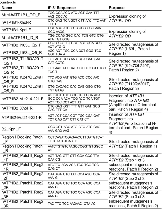
4.1 Cloning and Characteriz ation of Recombinant Myc -tagged Retinoschisin Variants ...58
4.1.1 Cloning of N-terminal Myc-tagged Retinoschisin and RS1 -C59S ...58
4.1.2 Purification of Recombinant Retinoschisin Variants ...58
4.1.3 Binding of Retinoschisin Variants to Retinal Membranes ...60
4.2 Influence of Retinoschisin on Na/K-A TP ase Stability and Turnover ...62
Affinity in Retinal Membranes...64
4.3.2 Effect of Retinoschisin on Rb+ Import into X. leavis Oocytes Mediated by Heterologously Expressed Retinal Na/K-A TPase ...66
4.4 Influence of Retinoschisin on Int racellular Signaling ...67
4.4.1 Effect of Retinoschisin on the ERK Pathway Activity in Retinal Model Systems ...68
4.4.2 Role of S RC in the Retinoschisin Induced Inhibition of ERK Singaling ...71
4.4.3 Effect of Retinoschisin on PI3K/AK T and Ca2+ Signaling ...72
4.4.4 Colocalization of the Retinoschisin-Na/K-ATPase Complex with Signaling Constituents in the Murine Retina ...74
4.5 Influence of Retinoschisin on Cellular Integrity and Homeostasis ...77
4.5.1 Influence of Retinoschisin on Proliferation and Cell Size of Y -79 Cells ...77
4.5.2 Influence of Retinoschisin on Viability of Y-79 Cells ...77
4.5.3 Retinoschisin Impairs Apoptosis in Retinal Model Systems ...78
4.6 Identification of the Retinoschisin-Interaction Site on the Na/K-A TPase ...81
4.6.1 ATP1B 2 is Responsible for Retinoschisin Binding ...81
4.6.2 Analysis von Hydrophobic Protein Surface Patches on ATP1B2 - Importance for Retinoschisin Binding ...86
5 Discussion ... 92
5.1 Pathomechanism of the Retinoschisin Mut ant RS1 -C59S ...92
5.2 Effect of Retinoschisin on the Retinal Na/K-A TPase...93
5.3 Effect of Retinoschisin on Retinal Homeostasis ...98
5.4 Identification of the Retinoschisin Binding Site at the Na/K -A TPase ...99
5.5 Implications ... 100
6 References ... 102
List of Abbreviations... 121
List of Figures ... 123
List of Tables ... 124
Acknowledgements... 125
Selbstständigkeitserklärung ... 126
Bei der X-gebundenen juvenilen Retinoschisis (engl. X-linked juvenile retinoschisis, XLRS) handelt es sich um eine degenerative Erkrankung der Netzhaut, welche auf Grund ihres X- chromosomalen Vererbungsmusters vorwiegend bei Männern auftritt (George et al., 1995).
Charakteristisch für die XLRS ist eine Aufspaltung der retinalen Schichten (Schisis) und eine defekte Signalweiterleitung von Photorezeptor- zu Bipolarzellen. Ursächlich für diese Erkrankung sind Mutationen im RS1-Gen (Sauer et al., 1997). Das vom RS1-Gen kodierte Protein, Retinoschisin, wird in der Retina spezifisch von Photorezeptoren und Bipolarzellen exprimiert und bindet an die Plasmamembranen dieser Zelltypen. Die Verankerung von Retinoschisin an der Plasmamembran wird dabei von der retinalen Na/K-ATPase vermittelt (Friedrich et al., 2011), welche bereits in einer früheren Studie als ein Interaktionspartner von Retinoschisin identifiziert wurde (Molday et al., 2007).
Der molekulare Pathomechanismus der XLRS und inwiefern die retinale Na/K-ATPase in der XLRS-Pathogenese involviert ist, ist bisher nicht bekannt. Ziel dieser Arbeit war es daher, funktionelle Konsequenzen sowie die strukturelle Basis der Retinoschisin-Na/K-ATPase Interaktion zu untersuchen.
Für die folgenden Studien wurden zunächst die kodierenden Sequenzen für Retinoschisin und eine pathogene Retinoschisin-Variante, RS1-C59S, mit einem Myc-Tag fusioniert und in einen Expressionsvektor kloniert, was die Aufreinigung rekombinanten Retinoschisins ermöglichte.
Die aufgereinigten Retinoschisin-Varianten wurden in die weiteren Versuche zur Retinoschisin-Na/K-ATPase Interaktion eingesetzt.
Das erste Projekt hatte zum Ziel, einen möglichen Einfluss von Retinoschisin auf Proteinmengen oder Membranstabilität der Na/K-ATPase in Netzhautexplantaten des RS1- Knock out (Rs1h
-/Y) Mausmodells oder in Hek293 Zellen, welche heterolog die retinale Na/K- ATPase exprimierten, aufzuklären. Rekombinantes Retinoschisin hatte in keinem der beiden Modellsysteme einen Einfluss auf Na/K-ATPase Proteinmengen, weder in Gesamtzelllysaten noch in angereicherten Membranfraktionen.
Ein weiteres Projekt befasste sich mit einem möglichen Effekt von Retinoschisin auf die aktive Ionenpumpfunktion der Na/K-ATPase. Rekombinantes Retinoschisin hatte dabei keinen Einfluss auf die ATP-Hydrolyserate oder Substrataffinitäten der Na/K-ATPase in Rs1h
-/Y- Netzhautexplantaten. Ebenso hatte es keinen Einfluss auf die Ionentransportaktivität der heterolog in Xenopus leavis Oozyten exprimierten, humanen retinalen Na/K-ATPase.
Im Rahmen des dritten Projekts wurde untersucht, ob Retinoschisin die Na/K-ATPase abhän-
gige Signaltransduktion beeinflussen kann. Die Zugabe von Retinoschisin, nicht aber von RS1-
C59S, verminderte die Aktivität von c-RAF und ERK1/2, zentraler Komponenten des MAPK
der MAPK Zielgene c-FOS und EGR1 in beiden retinalen Modellsystemen. Weitere Studien zur Retinoschisin-abhängigen Regulation von Signalkaskaden fokussierten sich auf Rs1h
-/YNetzhautexplantate. Es zeigte sich, dass die Zugabe von rekombinantem Retinoschisin, nicht aber von RS1-C59S, die Aktivität von Src, welches als initialer Signaltransmitter der Na/K- ATPase beschrieben wurde (Cui und Xie, 2017), mindert. Ebenso verminderte Retinoschisin die Aktivität des Ca
2+Signalweg-Markers Camk2. Die Aktivität des PI3K/AKT Signalwegs, repräsentiert durch Akt, wurde von Retinoschisin nicht beeinflusst. Immunhistochemische Analysen muriner, wildtypischer Netzhäute zeigten überlappende Signale des Retinoschisin- Na/K-ATPase-Komplexes mit Signalen von Gerüstproteinen und Transmittern, denen eine Beteiligung an der Na/K-ATPase-Signalweiterleitung zugesprochen wird.
Des Weiteren wurde der Einfluss von Retinoschisin auf die zelluläre Homöostase untersucht.
Die Behandlung von Y-79-Zellen mit rekombinantem Retinoschisin hatte keinen Einfluss auf deren Überlebensrate, Zellgröße oder Proliferation. Jedoch zeigte sich nach Zugabe von re- kombinantem Retinoschisin eine Minderung der apoptotischen Aktivität sowohl in Y-79-Zellen als auch in Rs1h
-/YNetzhautexplantaten. Die Expression des pro-apoptotischen Markergens BAX wurde in beiden Modellsystemen beeinflusst und nach unten reguliert. Ebenso zeigten Y-79 Zellen nach Behandlung mit Retinoschisin eine verminderte Caspase-3-Aktivität.
Netzhautexplantate der Rs1h
-/YMaus, die für eine Woche in Gegenwart von Retinoschisin ex vivo kultiviert wurden, zeigten eine deutlich reduzierte Photorezeptordegeneration.
Das letzte Projekt befasste sich mit der Identifikation der Retinoschisin-Interaktionsfläche der Na/K-ATPase. Neun verschiedene Na/K-ATPase-Isozymkombinationen (ATP1A1, ATP1A2, und ATP1A3 in Kombination mit ATP1B1, ATP1B2, oder ATP1B3) wurden hinsichtlich ihrer Fähigkeit, Retinoschisin zu binden, untersucht. Eine Retinoschisin-Bindung fand nur an Na/K- ATPasen, welche die ATP1B2-Untereinheit enthielten, statt. Die Herstellung chimärer ATP1B2-ATP1B1-Untereinheiten ermöglichte es, die Interaktionsstelle weiter auf die extrazelluläre Domäne der ATP1B2-Untereinheit einzuschränken. Mit Hilfe bioinformatischer Analysen wurden anschließend zwei mögliche Protein-Interaktionsregionen auf ATP1B2 identifiziert. Durch gerichtete Mutagenese wurden diese Regionen gezielt entfernt, in dem die entsprechenden Aminosäuresequenzen von ATP1B1 eingefügt wurden. Retinoschisin- Bindeversuche an diese chimären ATP1B2-Mutanten ergaben eine Kanditatenregion für die Retinoschisin-Interaktionsstelle in der zweiten Interaktionsregion, an Aminosäure T240.
Zusammenfassend liefert diese Arbeit neue Einblicke in initiale pathologische Prozesse der
XLRS, was die Erforschung neuer therapeutischer Strategien für diese bisher unheilbare
Krankheit vorantreiben könnte.
X-linked juvenile retinoschisis (XLRS) is a degenerative disease of the retina which primarily affects males due to an X-chromosomal recessive mode of inheritance (George et al., 1995).
Mutations in the RS1 gene were shown to be causative for XLRS (Sauer et al. 1997) which is characterized by a splitting of the retinal layers (schisis) as well as defects in signal transduction between photoreceptors and bipolar cells (Molday et al. 2012). The protein encoded by RS1, retinoschisin, is specifically expressed and secreted by photoreceptors and bipolar cells in the retina and was found to bind to plasma membranes of these cell types.
Membrane anchorage of retinoschisin was shown to be mediated by the retinal Na/K-ATPase (Friedrich et al., 2011), which had been identified as a retinoschisin interacting protein before (Molday et al., 2007).
The molecular pathomechanism of XLRS and a putative involvement of the Na/K-ATPase in XLRS pathogenesis remain elusive. Therefore, aim of this study was to investigate functional consequences and the structural basis of retinoschisin-binding to the Na/K-ATPase.
To this end, the coding sequences for retinoschisin and a pathogenic retinoschisin variant, RS1-C59S, were fused to a Myc-tag and cloned into expression vectors, enabling purification of the recombinant proteins. These purified proteins were then used in further studies addressing the interaction between retinoschisin and the Na/K-ATPase.
The first project aimed to delineate whether retinoschisin influences Na/K-ATPase levels or membrane stability in murine retinal explants of an RS1 knock out (Rs1h
-/Y) mouse model or in Hek293 cells heterologously expressing retinal Na/K-ATPase subunits ATP1A3 and ATP1B2. Recombinant retinoschisin had no effect on Na/K-ATPase protein levels in total cell lysates or enriched plasma membrane fractions from both model systems.
A second project assessed the effect of retinoschisin on the active ion pump function of the Na/K-ATPase. Retinoschisin treatment did not affect Na/K-ATPase catalysed (pump function dependent) ATP cleavage or substrate affinities in Rs1h
-/Yretinal explants. Neither did it affect ion transport activity of the heterologously expressed human retinal Na/K-ATPase in Xenopus leavis oocytes.
The third project investigated an influence of retinoschisin on Na/K-ATPase mediated
signaling. Recombinant retinoschisin, but not RS1-C59S, was shown to reduce the activity of
mitogen activated protein kinase (MAPK) signaling constituents c-RAF and ERK1/2 in Rs1h
-/Yretinal explants as well as the human retinoblastoma cell line Y-79. MAPK pathway target
genes EGR1 and c-FOS were also downregulated upon retinoschisin treatment in both model
systems. Further studies on signal regulation by retinoschisin focused on Rs1h
-/Yretinal
explants as model system. Recombinant retinoschisin, but not C59S, was found to decrease
signaling marker Camk2, but not activation of PI3K/AKT signaling marker Akt.
Immunohistochemistry in murine wildtype retinae showed overlapping signals of the retinoschisin-Na/K-ATPase complex and scaffolding proteins as well as signal transmitters involved in Na/K-ATPase mediated signal transduction.
The next project assessed the effect of retinoschisin on cellular homeostasis. Retinoschisin treatment did not influence cell viability, cell size or proliferation of Y-79 cells. However, retinoschisin but not RS1-C59S treatment lead to decreased apoptotic activity in Y-79 cells and Rs1h
-/Yretinal explants: Expression of the pro-apoptotic marker gene BAX was down regulated in both model systems. In Y-79 cells, retinoschisin reduced the activity of apoptosis marker Caspase-3. Rs1h
-/Yretinal explants, cultivated in the presence of retinoschisin for one week ex vivo, exhibited significantly decreased photoreceptor degeneration.
The last project focused on the identification of the Na/K-ATPase interaction site to retinoschisin. Retinoschisin binding to nine different Na/K-ATPase isozyme combinations (ATP1A1, ATP1A2, and ATP1A3 in combination with ATP1B1, ATP1B2, or ATP1B3) was tested and revealed that retinoschisin specifically binds to ATP1B2. Applying ATP1B2-ATP1B1 chimera, the interaction site was further narrowed down to the extracellular domain of ATP1B2.
Next, bioinformatics analyses identified two putative protein-interaction sites on the extracellular domain of ATP1B2. These two "docking patches" patches on ATP1B2 were each mutated by introducing the respective sequences of ATP1B1. Analysis of retinoschisin binding to ATP1B2 docking-patch mutants revealed a candidate region in docking patch II, in specific the amino acid (aa) T240, putatively involved in retinoschisin binding.
Taken together, this work provides novel insight into initial pathological processes of XLRS and could thus enhance the development of novel therapeutic options for this currently untreatable disease.
1 Introduction
1.1 X-linked Juvenile Retinoschisis (XLRS) – Clinical Features
X-linked juvenile retinoschisis (XLRS) is a hereditary retinal dystrophy and represents one of the most common forms of early onset macular degeneration in males (George et al., 1995).
XLRS was first described in two brothers by the Austrian ophthalmologist Josef Haas in 1898 (Haas, 1898) and later identified to be of X-linked inheritance (Pagenstecher, 1913). Due to the X-chromosomal recessive mode of transmission, mostly males are affected. Female carriers, in general, are asymptomatic, with few documented cases of subtle retinal changes (Wu et al., 1985, Rodriguez et al., 2005, Saldana et al., 2007). The worldwide estimated prevalence of XLRS ranges between 1:5000 and 1:20000 with hotspots of elevated prevalence for example in Finland, where XLRS was found to be the most common X-linked disease due to three founder mutations in the Finish population (Tantri et al., 2004). XLRS is almost fully penetrant (~ 98 %), and shows a juvenile onset in most patients (Molday et al., 2012). Affected boys often present for ophthalmologic examination at school age when reading difficulties are detected, but symptoms of XLRS were also found in patients being under one year of age, suggesting a congenital onset of the disease (George et al., 1995, Prasad et al., 2006, Renner et al., 2008, Lee et al., 2009).
The clinical manifestations of XLRS are highly variable even within families, but there are phenotypic changes characteristic for a specific diagnosis. As the name “X-linked juvenile retinoschisis” suggests, the disease is characterized by retinal ”schisis“, i.e. a splitting of retinal layers mainly affecting ganglion cell layer and inner nuclear layer, which can be visualized by spectral domain optical coherence tomography (OCT-SD) (Figure 1 A). Retinal schisis is mainly found to affect the fovea in a majority of XLRS patients, but peripheral manifestations are also seen in around 50 % of affected individuals (George et al., 1995, Molday et al., 2012).
Alongside with retinal schisis goes the development of large, fluid-filled cysts, also known as
schisis cavities. The presence of foveal cysts causes a distinctive spoke-wheel like schisis
pattern which is characteristic for XLRS and is used as a main indicator for diagnosis (Figure
1 B). Retinal schisis was shown to be more pronounced in young patients and decreases when
patients grow older (George et al., 1995, Molday et al., 2012). A further characteristic feature
of XLRS is the so called "negative" electroretinogram (ERG). Compared to healthy individuals ,
XLRS patients show a lower b-wave response, whereas the a-wave remains almost normal
(Figure 1 C). The decreased b-wave indicates a defective transmission of visual signals from
photoreceptor to bipolar cells (Khan et al., 2001). Since there have been reports of XLRS
patients showing normal ERG responses (e.g. Eksandh et al., 2005, Vincent et al., 2013) and
clinical manifestation of XLRS is highly variable with regard to disease onset, progression and
severity even in siblings, a combined approach of fundus autofluorescence imaging, OCT imaging, ERG measurement, and molecular-genetic testing is nowadays used to diagnose XLRS (Renner et al., 2008).
Secondary complications like vitreous haemorrhages or retinal detachment can occur in addition to the characteristic clinical features of XLRS. Approximately 5 % of all patients are affected by these secondary complications which in most cases develop in the first decade of life (Molday et al., 2012). Macular holes have also been reported to arise as side effects of XLRS (Brasil et al., 2011; Shukla et al., 2006).
Figure 1: Clinical features of XLRS
A) Fundus autofluorescence images of a healthy individual and a XLRS patient. A spoke -wheel like foveal schisis can be detected in the patient’s image. B) SD-OCT images of a healthy individual and a XLRS patient. A marked splitting of retinal layers is seen in the foveal area of the XLRS patient’s retina between the ganglion cell layer (GCL) and inner nuclear layer (INL) C) ERG recordings from a healthy individual and a XLRS patient. The patient’s ERG exhibits a strongly reduced b wave (Images adapted from Mo lday et al., 2012 (A, XLRS patient), Cuba and Gomez- Ulla, 2011 (A, healthy individual), Sergeev et al., 2011 (B) and Bowles et al., 2011 (C)).
1.2 Mutations in the RS1 Gene are Causative for XLRS
After localizing the XLRS-locus in close proximity to Xg blood group markers in 1984 (Wieacker
et al., 1984), it was not until 1997 that the XLRS gene was identified by a positional cloning
approach (Sauer et al., 1997). The identified gene was termed RS1 and localizes to the short,
distal arm of the X-chromosome at Xp22.2. The RS1 gene is 32.4 kb in length and is organized
in 5 introns and 6 exons. The mRNA transcript arising from RS1 is 3.1 kb long and encodes a
224 amino acid (aa) long protein named retinoschisin. RS1 expression was found to be
exclusive to photoreceptor and bipolar cells of the retina (Sauer et al., 1997, Grayson et al., 2000, Molday et al., 2001,) as well as to the pinealocytes of the pineal gland (Takada et al., 2006). Its specific expression is controlled by an upstream CpG island and two opposing CRX (Cone-Rod Homeobox) transcription factor binding regions (Langmann et al., 2008, Kraus et al., 2011). RS1 expression in murine retina is detectable already at postnatal day 1 (p1) and reaches adulthood levels at p5 to p7 which then stay constant throughout life (Weber and Kellner, 2007). Due to the X-chromosomal recessive mode of inheritance, the lack of correlation of mutation type and disease phenotype, and various biochemical studies on mutant retinoschisin, it is thought that XLRS arises as a result of a functional loss of the retinoschisin protein (e.g. Wang et al., 2002, Wu and Molday, 2003, Vijayasarathy et al., 2010;
reviewed by Molday; 2007).
1.3 Rs1h Knock Out Mice – A Model System for XLRS
Identification of the RS1 gene has not only facilitated XLRS diagnostics but also provided the possibility to generate a mouse model for XLRS. Murine retinoschisin is termed Rs1h and shares 96% sequence identity with its human homologue. Rs1h knock out mouse lines were generated and characterized independently by several groups (Weber et al., 2002, Zeng et al., 2004, Jablonski et al., 2005). The phenotype resulting from Rs1h
-/Yknock out models reflects the human condition of XLRS in the independent mouse lines: Retinal structures are disorganized and a marked retinal splitting is observable in the inner nuclear layer. Also, the characteristic negative ERG is comparable to the human condition (Molday et al., 2012).
Together, these features render the retinoschisin-deficient mice excellent model systems for studies on XLRS pathomechanisms as well as treatment options.
1.4 Prospect on Therapeutic Strategies for XLRS
To date, there is no cure for XLRS. Prescription of low-vision aids is still the most common treatment for XLRS patients and the only drug which is under testing for XLRS treatment is 2
% dorzolamide, a carbonic anhydrase inhibitor (CAI). CAIs such as dorzolamide and
acetozolomide have been used to successfully treat macular oedema, where inhibition of
carbonic anhydrase in the retinal pigment epithelium leads to acidification of the retina and
elevated fluid extrusion. This, in turn, results in a lowered intraocular pressure (Wolfensberger,
1999). Because of this known beneficial effects on macular oedema, CAIs were also
suggested to reduce schisis cavities in XLRS. Studies have analysed the effects of CAI
treatment in 8 or 9 XLRS patients and observed positive effects on foveal thickness, cystic
spaces and/or visual acuity in 7 or 5 of these, respectively (Apushkin and Fishman 2006,
Verbakel et al., 2016). Successful CAI treatment of XLRS patients was also shown to be
irrespective of the class of disease-associated mutation (Walia et al., 2009). However, there are also studies that have reported no improvement in visual acuity after CAI treatment, but only a decrease in retinal splitting (Genead et al., 2010, Khandhadia et al., 2011). For a subset of XLRS patients, CAI treatment had no effect at all (Apushkin and Fishman 2006, Genead et al., 2010, Khandhadia et al., 2011, Verbakel et al., 2016). A clinical evaluation on the use of 2
% dorzolamide or 1 % brinzolamide, another CAI, in 66 XLRS patients for 18 months has been completed in October 2016 (https://clinicaltrials.gov/ct2/show/NCT02331173, accessed August 31
st2017), but final data on the outcome of this study are not yet available. Due to the few and partially conflicting studies on XLRS patients treated with CAIs, this type of medication is not routinely used in XLRS treatment.
RS1 gene replacement using AAV (adeno-associated virus) vectors in the Rs1h
-/ymouse model has successfully reduced XLRS phenotypes and opened up gene replacement therapy as a novel way to cure XLRS (Zeng et al., 2004, Janssen et al., 2008, Park et al., 2009, Ou et al., 2015, Ye et al., 2015, Bush et al., 2016a). Pre-clinical evaluations had already confirmed that the vectors are well tolerated in both mice and Cynomolgus macaques: vector DNA was only detectable in eyes and there were no antibodies against the exogenously expressed retinoschisin (Ye et al., 2015a, Ye et al., 2015b). Currently there is a Phase I clinical trial ongoing on gene replacement therapy for XLRS using AAV based vectors (https://clinicaltrials.gov/ct2/show/NCT02317887, accessed August 2nd 2017). Solid Lipid Nanoparticles (SLNs) have also been used to deliver RS1 to the eyes of Rs1h
-/Ymice resulting in reduced photoreceptor loss and schisis formation (Apaolaza et al., 2015, Apaolaza et al., 2016). SLNs may thus provide an alternative approach to viral gene replacement therapies.
For all three XLRS therapeutic approaches, however, further studies and especially long-term follow up of patients will be needed to evaluate their value for XLRS treatment.
1.5 The Retinoschisin Protein
Due to the very limited therapeutic options for XLRS, several research groups aim to dissect the basic pathomechanisms caused by retinoschisin deficiency, and thus the function of retinoschisin in the retina. Retinoschisin is a 24 kDa protein and highly conserved among species. It exhibits four distinct domains: an N-terminal signal peptide (23 aa), a discoidin domain (157 aa), an RS1 domain (39 aa), and a short C-terminal fragment of 5 aa (Sauer et al., 1997, Molday, 2007) (Figure 2 A ).
The signal peptide is responsible for retinoschisin secretion and is cleaved off during this
process. Two cleavage sites, between aa 21-22 and aa 23-24 have been identified within the
signal peptide, resulting in two mature retinoschisin isoforms which differ by two aa in length
in mouse (Vijayasarathy et al., 2006). Next follows the RS1 domain, which has been given its
name due to the lack of sequence homology with any other known protein (Molday et al., 2012).
The subsequent discoidin domain, however, is highly conserved and found in many other proteins. After the discoidin domain, the last element to follow in the retinoschisin protein is the short C-terminal segment.
The discoidin domain makes up about 75 % of the mature retinoschisin protein and is thought to be the major functional unit of retinoschisin (Wu et al., 2003, Molday, 2007). A discoidin domain was first identified in the discoidin I protein of Dictyostelium discoideum (Poole et al., 1981) and later found as a recurrent motif in a huge variety of eukaryotic and prokaryotic proteins (Baumgartner et al., 1998, reviewed by Kiedzierska et al., 2007). Discoidin domain containing proteins are involved in a plethora of cellular processes such as cellular adhesion, fertilization, migration, signaling and development (reviewed by Kiedzierska et al., 2007).
Discoidin domains are organized in a core domain which is highly conserved, and so called spike or loop regions which, in contrast, are highly variable (reviewed by Kiedzierska et al., 2007). The core domain of discoidin domains consists of a five-stranded antiparallel ß-sheet which is tightly packed up against a three-stranded antiparallel ß-sheet forming a distorted barrel like structure. From this core barrel two or more of the highly variable spike regions are protruding. The spike regions form a groove which serves as the recognition site for discoidin domain’s interaction partners. Since the spike regions are highly variable, discoidin domains can bind to a variety of ligands including lipids, carbohydrates and proteins which goes along with the diverse functions they have been attributed (reviewed by Kiedzierska et al., 2007).
Even though the 3-D structure of the retinoschisin discoidin domain has not been determined yet, the availability of high resolution structures of many other discoidin domains allowed for modelling of the retinoschisin discoidin domain. In case of the retinoschisin discoidin domain, three spikes protrude from one end of a core hydrophobic structure of eight ß-strands (Wu and Molday, 2003).
Retinoschisin is secreted from photoreceptors and bipolar cells as a homo-octameric complex (Figure 2 B). Oligomerization was shown to take place before the protein is secreted from the endoplasmatic reticulum (ER) (Molday et al., 2001, Wu and Molday, 2003, Wu et al., 2005).
Intensive studies on known pathogenic retinoschisin variants determined the cystein residues
responsible for retinoschisin oligomerisation: While two retinoschisin monomers are linked
together by a disulfide bond between their cystein residues at aa position 40 (C40),
octamerization is achieved by disulfide bonds between cystein at position 59 (C59) in the RS1
domain and cystein at position 223 (C223) in the C-terminal segment of two retinoschisin
monomers (Wu and Molday, 2003, Wu et al., 2005). Recently, technical advances enabled the
determination of the structure of the retinoschisin octamer. Using single particle electron
microscopy, Bush and colleagues were the first to present retinoschisin to be arranged in a
cog-wheel like ring structure, with the RS1 domains facing the inside of the ring and the
discoidin domains and especially their spikes facing outwards (Bush et al., 2016). Using cryoelectron microscopy, Tolun and colleagues refined the structure and Ramsay and colleagues analysed the influence of XLRS associated mutants on the octamer struc ture (Tolun et al., 2016, Ramsay et al., 2016). Tolun and colleagues also provided evidence that two retinoschisin octamers assemble in a back-to-back fashion, forming a hexadecamer (Figure 2 C).
Figure 2: Schematic presentation of retinoschisin domains and 3D structure of hexadecameric complexes A) Schematic representation of the retinoschisin domains, arrows depict cystein residues which are essential for retinoschisin oligomerization. B) Top view onto a retinoschisin double-octamer organized into a cog-wheel like structure. C) Side view onto two retinoschisin octamers organized into a hexadecamer in a back -to-back fashion.
Subfigures B) and C) were created using the 3D structure of retinoschisin provided by Ramsay et al., 2016 as a template with the EXPASY swissmodell tool available at https://swissmodel.expasy.org/repository/uniprot//O15537.
Immunolabelling experiments detected retinoschisin on the inner segments of both rod and
cone photoreceptors, as well as in plexiform layers with most intensive staining found in
photoreceptor inner segments (Figure 3) (Grayson et al., 2000, Molday et al., 2001, Reid et
al., 2003, Takada et al., 2004).
1.6 Categorization of XLRS Associated Mutations
To date, 197 pathogenic variants of the RS1 gene have been reported (https://databases.lovd.nl/shared/genes/RS1, accessed August 3rd 2017). In 2012, a review by Molday and colleagues categorized the then known 191 XLRS-associated mutations as follows: 13 % were deletions, 3 % duplications, 1.5 % insertions, 1.5 % insertions/deletions and the majority of 80 % were single nucleotide polymorphisms (SNPs) which may result in pre-mature stop-codons, aa changes or alterations of splice sites. Around 40 % of these mutations are expected to be true null alleles, not allowing translation (Molday et al., 2012).
The rest is represented by missense mutations categorized into three different classes with regard to their position in the retinoschisin protein and thus their effects on retinoschisin protein folding, secretion and stability/octamerization.
The first class of missense mutations is represented by mutations in the discoidin domain which seems to be a hotspot for missense mutations and harbours 85 % of all RS1 missense mutations. The discoidin domain missense mutations biochemically characterized so far in general led to defects in retinoschisin secretion caused by ER retention (Wang et al., 2002, Wu et al., 2003, Iannaccone et al., 2006, Wang et al., 2006, Walia et al., 2009, Gleghorn et al., 2010). As shown by Gleghorn and colleagues, misfolded retinoschisin does not induce the unfolded protein response (UPR), which suggests that it is rather the null phenotype that causes disease than cellular stress induced by UPR (Gleghorn et al., 2010).
Missense mutations in the signal peptide of retinoschisin represent the second class of distinctive RS1 mutations. These mutations (e.g. RS1-L12H (NM_000330.3(RS1):c.35T>A [p.Leu12His] and RS1-L13P (NM_000330.3(RS1):c.38T>C [p.Leu13Pro]) were shown to prevent translocation of the nascent protein into the ER, resulting in complete absence of mature retinoschisin and thus a null phenotype (Wang et al., 2002, Vijayasarathy et al., 2010).
The third class of RS1 mutations leads to protein variants which are still secreted, but exhibit abnormal oligomerization. As shown by Wu and colleagues the cystein residues C59 and C223 are essential for assembly of retinoschisin octamers and mutations at both of these residues
Figure 3: Immunolabeling of a murine retinal cryosection showing retinoschisin localization
Left: DIC/DAPI (differential interference contrast/ 4′,6-Diamidin-2-phenylindol) visualisation of nuclei in retinal layers . Right: Immunohistochemistry (IHC) with an anti-retinsochsin antibody OS/IS:
outer/inner segments; OPL/IPL outer/ inner plexiform layers; ONL/INL: outer/inner nuclear layer; GCL: ganglion cell layer.
(Molday et al., 2012)
have been reported in XLRS patients (Wu et al., 2005). Pathogenicity of these mutations led to the suggestion that octamer formation is necessary for correct retinoschisin function.
There are few known mutations which have also been characterized biochemically, and do not fit into any of the mutation categories described above. The retinoschisin variants RS1-R141H (NM_000330.3(RS1):c. 422G>A [p.Arg141His]), RS1-R141G (NM_000330.3 (RS1):c.421>T/G [p.Arg141Gly]), RS1-H207Q (NM_000330.3(RS1):c. 621C>G [p.His207Gln]), and RS1-R209H (NM_000330.3(RS1):c.626G>A [p.Arg209His]) have been shown to be secreted as octameric complexes. Ramsay and colleagues analysed the structural changes introduced into the retinoschisin octamer by mutations R141H and H207Q.
Whereas H207Q was reported to destabilize both the retinoschisin monomer and octamer, R141H did not affect overall protein stability. It was suggested that the pathomechanism underlying the R141H mutation potentially arises from defective interaction of the mutant protein with its interaction partners (Ramsay et al., 2016).
1.7 Towards Retinoschisin Function – Proposed Interaction Partners
In spite of the clinical features being well defined and the disease causing gene being known for 20 years, the pathomechanism of XLRS still remains elusive. Efforts have been made to identify the molecular mechanisms underlying retinoschisin function and determine why retinoschisin deficiency has such tremendous consequences for retinal integrity. Retinoschisin has been proposed to interact with galactose, phosphatidylserine, extracellular matrix proteins like laminin, L-type voltage gated ion channels, as well as the retinal Na/K-ATPase (Fraternali et al., 2003, Steiner-Champliaud et al., 2006, Vijayasarathy et a., 2007, Molday et al., 2007, Dyka et al., 2008, Shi et al., 2009, Kotova et al., 2010, Friedrich et al., 2011, Shi et al., 2017), each interaction partner providing room for speculations towards retinoschisin function.
1.7.1 Phospholipids
One of the molecules retinoschisin was proposed to interact with is phos phatidylserine.
Phosphatidylserine is an acidic membrane phospholipid which is found in the plasma
membrane. A possible interaction of retinoschisin and phosphatidylserine was assumed
because other discoidin domain containing proteins were also shown to interact with
phosphatidylserine (Ortel et al., 1992, Zwaal et al., 1998). An initial study addressing the
interaction between retinoschisin and phosphatidylserine was conducted by Fraternali and
colleagues in 2003. Bioinformatical evaluation of XLRS associated mutations leading to aa
exchanges at Y89, V90, W92, Y93, F108, and I144 revealed that these aas may form a
hydrophobic and exposed region on retinoschisin similar to sites on Factor V and Factor VIII
which mediate interactions with phospholipid bilayers (Fraternali et al., 2003). Later studies
showed retinoschisin to co-purify with phosphatidylserine (Vijayasarathy et al., 2007) and atomic force microscopy analyses by the same group detected retinoschisin binding to phosphatidylserine containing planar lipid bilayers (Kotova et al., 2010). However, other groups were not able to verify an interaction between retinoschisin and phosphatidylserine (Molday et al., 2007, Friedrich et al., 2011). In addition to the contradictory experimental data obtained by different in vitro systems, the physiological relevance of an interaction between retinoschisin and phosphatidylserine is also questionable. In a native cellular system phosphatidylserine mostly localizes to the inner leaflet of plasma membranes whereas retinoschisin binds to the outer plasma membrane, hence the proposed interaction might not be feasible in a native system (Molday et al., 2012).
1.7.2 Galactose
Discoidin proteins from Dictyostelium discoideum were shown to function as lectins and exhibit a high affinity for galactose residues to promote cell aggregation (Poole et al., 1981, Valencia et al., 1989). This finding encouraged Dyka and colleagues to analyse whether retinoschisin is capable of binding to immobilized carbohydrates. They found efficient retinoschisin binding to agarose-coupled galactose and lactose, whereas no retinoschisin binding was observed to agarose-coupled N-acetylgalactosamine, N-acetylglucosamine, mannose or heparin (Dyka et al., 2008). To date, there are no further studies addressing the physiological relevance of the interaction between retinoschisin and galactose. Nevertheless, its high affinity to galactose has been used to purify recombinant retinoschisin (e.g. Bush et al., 2016).
1.7.3 Extracellular Matrix Proteins
In an affinity chromatography based approach to identify retinoschisin binding proteins , the extracellular matrix component β2-laminin and αB crystalline were found to bind to immobilized retinoschisin (Steiner-Champliaud et al., 2006). Interaction with extracellular matrix proteins would strengthen the notion that retinoschisin is involved in cell-cell contact formation, but no further investigations with regard to a physiological relevance of these interactions have been conducted so far.
1.7.4 L-Type Voltage Gated Calcium Channels
In 2009, Shi and colleagues performed co-immunoprecipitations and Yeast-2-Hybrid
experiments which identified L-Type Voltage Gated Calcium Channels (L-VGCCs) as novel
retinoschisin interaction partners (Shi et al., 2009). L-VGCCs are heteromeric complexes of
four different subunits which span the plasma membrane and function as Ca
2+specific
channels (De Waard et al., 1996). Mutations in the gene encoding the Cav1.4 α1 subunit of L-
VGCCs (CACNA1F) were shown to cause X-linked stationary night blindness. Cacna1f knock out in mice resulted in a negative ERG, highlighting the importance of L-VGCCs in photoreceptor bipolar synapse integrity (Mansergh et al., 2005). As a negative ERG is also a clinical hallmark of XLRS, an involvement of L-VGCCs in XLRS pathogenesis, possibly due to misregulation in a retinoschisin deficient retina, might be conclusive. A more recent study by Ou and colleagues also showed that retinoschisin is needed to maintain the synaptic localization of L-VGCC in retina (Ou et al., 2015). The latest publication by Shi and colleagues described successful co-immunoprecipitation of retinoschisin and Cav1.3 and Cav1.4 from porcine retinae. Furthermore, retinoschisin was shown to increase L-VGCC currents in transfected Hek293 cells. The same study also presented immunohistochemical analyses indicating that Cav1.4 expression is reduced in retina of Rs1h
-/Ymice, but does not provide quantification (Shi et al., 2017). This finding is contradictory to results from Ou and colleagues, who did not observe any differences in Cav1.4 expression (quantification given) when comparing Rs1h
-/Yand wildtype murine retinae. In addition, Cav1.4 α1 subunits are only expressed and located at release sites of mammalian photoreceptors in the outer plexiform layer, additionally exhibiting a punctate immunostaining in the mouse inner plexiform layer (Boycott et al., 2000, Ball and Greg, 2002, Berntson et al., 2003, Baumann et al., 2004).
Retinoschisin signals in contrast are most abundant in the photoreceptor inner segments.
Finally, immunohistochemical analyses reveal poor colocalization of Cav1.4 and retinoschisin in the plexiform layers. (Shi et al., 2017). Further analysis will be needed to gain deeper insight into possible effects of retinoschisin on L-VGCC regulation.
1.7.5 The Retinal Na/K-ATPase
Another proposed protein interaction partner of retinoschisin is the retinal Na/K-ATPase consisting of Na/K-ATPase subunits α3 and ß2. This complex was first identified by co- immunoprecipitation of human (and bovine) retina by Molday and colleagues in 2008 (Molday et al., 2008). This study also showed a perfect colocalization of retinoschisin and the retinal Na/K-ATPase in retinal cryosections (Molday et al., 2008). Later studies at the Institute of Human Genetics at the University of Regensburg confirmed the importance of the Na/K- ATPase for membrane anchorage of retinoschisin (Friedrich et al., 2011). Heterologous expression of ATP1A3 and ATP1B2 in Hek293 cells was sufficient to enable retinoschisin binding to Hek293 membranes. Western blot and immunohistochemical analyses demonstrated that retinoschisin no longer binds to retinal membranes in Atp1b2 deficient mice.
In return, immunostaining of the Na/K-ATPase subunits α3 and ß2 in Rs1h
-/Yretinae revealed
retinal Na/K-ATPase localization different from that in wildtype mice, suggesting an effect of
retinoschisin on Na/K-ATPase localization and/or stabilization in the membrane (Friedrich et
al., 2011).
1.8 The Na/K-ATPase
1.8.1 Structure of the Na/K-ATPase
The Na/K-ATPase is a plasma membrane spanning ion pump which is ubiquitously expressed.
The minimal functional unit of this enzyme is a heterodimeric complex composed of an α and a ß subunit (Figure 4).
The α subunit is the catalytic subunit of the complex, responsible for ion transport as well as ATP cleavage (Skou et al., 1957). It is also responsible for signal transmission upon binding of cardiac glycosides (reviewed in Cui and Xie, 2017). The α subunit is an integral membrane protein of 110-130 kDa which has 10 transmembrane domains, 5 short extracellular loops, and 4 cytosolic domains. Specific functions have been attributed to the first and second intracellular loops and three domains have been named accordingly: the first loop comprises the nucleotide (ATP) binding domain (N domain), while the second loop contains the phosphorylation domain (P domain) and the actuator domain (A domain) which is responsible for occlusion of cargo ions in the transmembrane binding sites and for dephosphorylation of the P-domain (Kühlbrandt, 2004) (Figure 4).
Four different isoforms of the Na/K-ATPase α subunit have been identified and were shown to be expressed in a tissue specific manner. The α1 isoform is omnipresent in all tissues and the predominant isoform in epithelial and heart tissue. The α2 isoform is mainly expressed in heart and skeletal muscle, as well as in astrocytes and glial cells of the brain. The α3 isoform is most strongly expressed in neuronal tissue and α4 was exclusively detected in testis (summarized in Kaplan 2002, Cui and Xie, 2017, Clausen et al., 2017). The α1, α2, and α3 isoforms share about 87 % aa sequence identity while α4 is about 78 % identical to the other three α isoforms (Shamraj and Lingrel, 1994). Even though there are sequence differences, the overall three dimensional structure of all four α isozymes seems to be identical and mainly the extracellular surface of the protein differs between the individual isoforms (Clausen et al., 2017). However,
Figure 4: Schematic overview of the Na/K-ATPase structure.
The α subunit is shown in medium purple, numbers 1-10 represent the 10 transmembrane domains; The actuator (A) domain is shown in orange, the phosphorylation (P) domain in blue and the nucleotide binding (N) domain in green. The ß subunit is depicted in light purple, N- linked glycosylation of the extracellular domain is indicated by white hexagons; shown in dark purple is an auxiliary FXYD subunit (modified after Horisberger 2004).
there are functional differences in the catalytic activity of the isoforms, affecting substrate affinities, voltage dependency and pump activity (Crambert et al., 2000, Blanco, 2005).
Within the retina, different α isoforms were attributed to different retinal cell types. In rodent retina, α1 was found in Müller cells, horizontal cells and to a small extent in photoreceptors, α2 only in some Müller cells, and α3 in photoreceptors and other neuronal cells (Wetzel et al., 1999, Schneider and Kraig, 1990).
The ß subunit does not have any catalytic properties, but is responsible for correct assembly, membrane integration and stability of the α-ß heterodimeric Na/K-ATPase complex (Ackermann and Geering, 1990, Geering 2001, Reinhard et al., 2013). Further, the ß subunit was also shown to modulate activity of the α subunit (Jaisser 1994, Horisberger and Rossier 1992; Eakle et al. 1994; Blanco et al. 1995) and is also involved in mediating intracellular adhesion (e.g. Antonicek et al., 1987, Shoshani et al., 2005, Vagin et al., 2012, Tokhtaeva et al., 2016).
The ß subunit consists of a short cytosolic N-terminal part, a single transmembrane domain and a large extracellular domain (reviewed in Kaplan 2002, Reinhard et al., 2013) (Figure 4).
Three different isoforms of the ß subunit have been identified, which in contrast to the α isoforms, show only little aa sequence identity (35-47 % in humans) (Reinhard et al., 2013). A common feature of all three ß isoforms is post-translational modification by N-linked glycosylation: ß1 has three glycosylation sites, while ß2 has eight and ß3 two glycosylation sites, respectively (Miller and Farley, 1988, Malik et al., 1996, Tokhtaeva et al., 2010).
Similar to the α subunits, the different ß subunits also show a tissue specific expression pattern.
Expression of ß1 has been detected in brain, heart, and kidney, ß2 subunits are expressed predominantly in brain, pineal gland and retina and ß3 expression has been detected in lung, liver and testis (Shyjan and Levenson, 1989, Shyjan et al., 1990, Arystarkhova and Sweadner, 1997). In rodent retina, ß1 is expressed in photoreceptors, horizontal, amacrine and ganglion cells, ß2 is expressed in bipolar cells, photoreceptors and Müller cells, while ß3 is restricted to photoreceptors (Wetzel et al., 1999, Schneider and Kraig, 1990).
In several tissues, members of the FXYD proteins build accessory subunits of the Na/K-
ATPase. FXYD proteins are type I membrane proteins, which have a single membrane
spanning domain (Swaender and Rael, 2000). The FXYD protein family consists of seven
members, five of which are associated with the Na/K-ATPase complex. FXYD1
(phospholemman), FXYD2 (γ-subunit of Na-K-ATPase), FXYD3 (Mat-8), FXYD4 (CHIF), and
FXYD7 were shown to interact with the Na/K-ATPase via their transmembrane domain and
are capable of regulating its function (reviewed by Geering, 2006). FXYD family members
exhibit a distinct, tissue specific expression, but, until now, there is no data on FXYD protein
expression in the retina.
1.8.2 The Na/K-ATPase as an Ionpump
The most crucial function of the Na/K-ATPase is the maintenance of a steep gradient of Na
+and K
+ions across the cellular plasma membrane. This gradient is essential for many processes such as neuronal excitability, cellular uptake of ions, nutrients, or neurotransmitters, but also for regulation of cell volume and intracellular pH (reviewed by Reinhard et al., 2013).
At the expense of one molecule of ATP, the Na/K-ATPase transports three Na
+ions out of and two K
+ions into the cell. For a single cell, this process may consume up to 80 % of the energy stored in ATP molecules. At baseline activity, Na/K-ATPases go through an average of 60-80 phosphorylation-dephosphorylation cycles per minute (reviewed by Orlov et al., 2017). The Na/K-ATPase is a member of the so called P-Type ATPase family of proteins, which all share the presence of a phosphorylated enzyme intermediate during their reaction cycle (Pedersen and Carafoli, 1987). The reaction cycle of the Na/K-ATPase confers two major steps and enzyme conformations, E1 and E2, and is described by the Albers -Post scheme (Figure 5).
The E1 state is also referred to as the Na
+-bound state and the E2 as K
+-bound state. The change from one state to the other is accompanied by many intermediate states and conformational changes.
The transition of the Na/K-ATPase through its reaction cycle can be prevented by the application of a variety of inhibitors. The most commonly used and best studied Na/K-ATPase inhibitors are so called cardiotonic steroids (CTS). Low levels of endogenously produced CTS digoxin, ouabain and marinobufagenin have been detected in mammalian tissue and biological fluids (Schoner and Schreiner-Bobis, 2005, Aperia et al., 2016). For decades, the main medical use of CTS has been for treatment of congestive heart failure. Most frequently used CTS are the plant-derived digoxin, ouabain and the vertebrate-derived bufalin and marinobufagenin
Figure 5: Schematic picture of the Albers-Post reaction cycle of the Na/K- ATPase
Per reaction cycle the Na/K-ATPase transports three Na+ ions (yellow circles) out of the cell and two K+ ions (purple circles) inside. During this reaction cycle one molecule of ATP is hydrolysed and
the Na/K-ATPase becomes
phosphorylated. E1: Na+-bound conformation, E2: K+-bound conformation, E1P/E2P: phosphate-bound states of E1/E2.
The Na/K-ATPase specific class of cardiotonic steroid inhibitors (CTS, orange triangle) binds to the Na/K-ATPase in its E2P conformation and puts the reaction cycle to a halt at this stage.
(Figure adapted from Orlov et al., 2017)
(Schoner and Schreiner-Bobis, 2007). CTS bind to the Na/K-ATPase from the extracellular side and enter the putative ion-exchange pathway with highest affinities for the E2P state (Laursen et al., 2015). By putting the pump to a halt in this position, no more Na
+ions are transported out of the cell and intracellular Na
+levels rise, which leads to an inhibition of the Na
+/Ca
2+exchanger (NCX). In patients suffering from congestive heart failure, CTS treatment via NCX inhibition leads to raised cytoplasmic Ca
2+levels, which allows for enhanced contractility of the diseased cardiac muscle (reviewed in Kaplan 2002, Clausen et al., 2017).
1.8.3 Na/K-ATPase Mediated Signaling
There is growing evidence that the Na/K-ATPase also functions as a receptor for signal transduction via CTS as hormones (summarized in Aperia et al., 2016, Cui and Xie, 2017).
First findings were made in the 1970s, when several studies showed effects of low (sub-pump- inhibitory) nM concentrations of ouabain on gene expression, differentiation and proliferation (Cuff and Lichtman, 1975, Kaplan, 1978, summarized in Cui and Xie, 2017). Later studies from many laboratories then showed that CTS treatment could stimulate different intracellular signaling pathways in various cell types and by that also affect cellular processes like proliferation, development, or apoptosis (e.g. Huang et al., 1997, Kometiani et al., 1998, Xie et al., 1999, Haas et al., 2000, Haas et al., 2002, Dmitrieva and Doris, 2003, Golden and Matrin, 2006, Ramirez-Ortega et al., 2006, Kulikov et al., 2007, Wang et al. 2015). Until today, there is experimental evidence that CTS can trigger the activation of the mitogen activated protein kinase (MAPK) signaling pathway, the phosphoinositide 3-kinase/ Protein kinase B (PI3K/AKT) pathway as well as Ca
2+dependent signaling cascades (reviewed in Aperia et al., 2016, Cui and Xie, 2017, Orlov et al., 2017).
The extracellular signal-regulated (ERK) pathway, one of the four MAPK pathways (Lewis et al., 1998, Chang and Karin, 2001), was the first Na/K-ATPase activated signaling pathway to be discovered by Kometiani and colleagues (Kometiani et al., 1998). Upon binding of CTS, various cell types show a strong activation of intracellular ERK signaling. The non-receptor tyrosine kinase SRC is widely accepted to be involved in activation of MAPK signaling through the Na/K-ATPase. However, the exact mechanism of how SRC is implicated in this pathway is discussed controversially (Haas et al., 2000, Haas et al., 2002, Tian et al., 2005, Weigand et al., 2012, Gable et al., 2014, Yosef et al., 2016). Despite the discrepancies on the mechanism of SRC activation by the Na/K-ATPase, SRC is thought to act as the initial, Na/K-ATPase associated signal transmitter which gets activated upon CTS binding to the Na/K-ATPase.
Activated SRC stimulates the endothelial growth factor receptor (EGFR), which then signals
on towards the MAPK cascade through RAS and RAF (Figure 6).
The PI3K/AKT pathway has also been shown to be activated by CTS-bound Na/K-ATPases, in a manner independent of SRC kinase activation (Liu et al., 2007). It was described to be induced by a physical interaction of the p85 subunit of phosphatidyl inositol 3 kinase (PI3K) with the Na/K-ATPase α subunit, which then causes AKT activation (Liu et al., 2007, Wu et al., 2013) (Figure 6).
The third signaling cascade activated upon CTS binding to the Na/K-ATPase is Ca
2+dependent signaling. Two different mechanisms were suggested to be responsible for Ca
2+pathway activation via the Na/K-ATPase. Several studies described a direct interaction of the Na/K- ATPase α subunit with the inositol trisphosphate receptor (IP3R). Upon CTS binding to the Na/K-ATPase structural alterations of the Na/K-ATPase α subunit are suggested to act on the IP3R and induce Ca
2+oscillations from the ER, leading to variable Ca
2+dependent cellular responses (Aizman et al., 2001, Miakawa-Naito et al., 2003, Yuan et al., 2005) (Figure 6). An involvement of Phospholipase C (PLC) in the Na/K-ATPase-IP3R interaction is under discussion (Yuan et al., 2005). Other studies report on an involvement of the NCX in the induction of Ca
2+signaling. Activation of the NCX by CTS was originally thought to arise from elevation of intracellular Na
+upon CTS dependent inhibition of the Na/K-ATPase (Langer 1972, Blaustein et al., 1998). However, more recent studies provide evidence of a direct interaction between NCX and the Na/K-ATPase, leading to the formation of specific Ca
2+signaling microdomains (Song et al., 2006, reviewed in Tian and Xie, 2008).
Figure 6: Schematic overview of Na/K-ATPase associated signaling cascades
CTS-bound Na/K-ATPases are implicated in the induction of three different signaling pathways. The MAPK pathway was described to be activated via SRC and the EGFR, the AKT pathway via PI3K, and Ca2+ dependent signaling can be activated via the IP3R, which can modify Ca2+ oscillations from the endoplasmatic/sarcoplasmatic reticulum (ER/SR) or may also be induced by the NCX (Figure modified after a figure kindly provided by Dr. Ulrike Friedrich, Institute of Human Genetics, University of Regensburg).
It is important to notice that most studies mentioned above were conducted with α1 subunit containing Na/K-ATPases. Until now, there is very little data on signaling properties of the other three α subunits. The α2 subunit was shown to be able to restore ion pumping but not α1 signaling function in a α1 knock out cell line (Xie et al., 2015), which rises the question whether α2 is capable of functioning as a signal transmitter. In contrast, the α3 subunit was shown to activate the MAPK cascade upon CTS treatment, but notably, in a mechanism independent of SRC activation (Madan et al., 2017). CTS treatment was also demonstrated to activate ERK signaling in an α4 expressing murine spermatogenic cell line, but no data on the mechanism were provided in this study (Upmanyu et al., 2016).
1.8.4 Na/K-ATPases in Intracellular Adhesion
In addition to its ion transport and signaling functions, the Na/K-ATPase further acts as a mediator of intracellular adhesion. Transdimerization of the ß1 subunit was shown to tether two cells together (Shoshani et al. 2005, Cereijido et al., 2012, Vagin et al., 2012, Tokhtaeva et al. 2016). The ß2 subunits mediates heterotypic adhesions between astrocytes and neighbouring neurons (Antonicek et al., 1987, Antonicek and Schachner, 1988, Gloor et al., 1990, Vagin et al., 2012). The mechanism underlying intracellular adhesion mediated by the ß2 subunit has not been resolved, yet. Transdimerization of two ß2 subunits was not consistent with experimental data and other factors or proteins are believed to be involved in the ß2 mediated intercellular interaction.
1.9 Aim of this Study
Despite substantial research efforts in the last 20 years, the consequences of retinoschisin deficiency and thus the initial pathomechanisms of XLRS are still not fully understood.
Nevertheless, it has been shown that the Na/K-ATPase is an essential interaction partner of retinoschisin in retinal membranes (Molday 2007, Friedrich 2011). The present study focused on functional consequences and the molecular interface of this interaction.
A first project investigated an effect of retinoschisin on Na/K-ATPase levels in retinal model systems. In addition, an effect of retinoschisin on two functions of the Na/K-ATPase, namely ion transport and intracellular signal transduction, was assessed. Furthermore, the influence of retinoschisin on several cellular processes (cell volume, proliferation, viability, and apoptosis) associated with XLRS was investigated. Finally, it was sought to identify the interaction site between the Na/K-ATPase and retinoschisin.
Results of these investigations are suited to provide insight into the basic molecular
pathomechanisms of XLRS, and should help to obtain a deeper understanding to initiate
innovative targeted therapeutic approaches for this currently untreatable disease.
2 Material
2.1 Mouse Strains
Table 1: Mouse strains used
Mouse Strains Source
Rs1
tm1Web(Rs1h
-/y) Bernhard HF Weber, Institute of Human Genetics,
University of Regensburg, Germany
C75BL/6 The Jackson Laboratory, Bar Harbor, Maine, USA
2.2 Escherichia coli (E.coli) Strains
Table 2: E. coli strain used
Strain Source
E. coli strain DH5α Life Technologies, Carlsbad, CA, USA
2.3 Eukaryotic Cell Lines
Table 3: Names and tissues of origin of cell lines used
Cell Line Organism Tissue of Origin Source
Hek293 Homo sapiens Embryonic kidney ATCC;LGC Standards GmbH, Wesel, Germany
Hek293
+RS1 Homo sapiens
Embryonic kidney, cell line stably transfected with an RS1 expression vector
Institute of Human Genetics, University of Regensburg, Germany
Y-79 Homo sapiens Retinoblastoma ATCC;LGC Standards GmbH, Wesel, Germany
2.4 Oligonucleotides for PCR and Sequencing Reactions
Table 4: Names, sequences and purposes of oligonucleotides used for cloning of retinoschisin (RS1) and Na/K-ATPase expression constructs
Name 5‘-3‘ Sequence Purpose
RS1-HindII-F
AAG CTT TTC ATG TCA CGC AAG ATA GAA GGC TTT TTGExpression cloning of RS1 RS1-XhoI-R
CTCGACGAGGCATCACTCGGCACACTTGCTRS1-EcoRI-F
GAA TTC ATG TCA CGC AAG ATA GAA GGC TTT TTGCloning of Myc-tagged RS1 variants
RS1-n-termMyc-R
TGATCAATTTCTGCTCCGATAATC CCAATGTGGCTTCATAGCCAAAGA GA
RS1-ntermMyc-F
TGATCAGTGAGG AAG ATCTGTCTA CCGAGGATGAAGGCGAGGACC CCT
RS1-C59S-mut-F
CCAAATGCCCCTCCTTGGACAGTATACCAGSite directed mutagenesis of RS1-C59S-mut-R
GGC ATT CTG GTA TAC TGT CCARS1
AGG AGG TGG
hATP1A2_NotI-F
GCG GCC GCA TGG GCC GTG GGG
CTG GCC GTG AG