Endothelin signalling in the retina
DISSERTATION
ZUR ERLANGUNG DES DOKTORGRADES DER NATURWISSENSCHAFTEN (DR. RER. NAT.)
DER FAKULTÄT FÜR BIOLOGIE UND VORKLINISCHE MEDIZIN DER UNIVERSITÄT REGENSBURG
Durchgeführt am Lehrstuhl für Humananatomie und Embryologie der Universität Regensburg
vorgelegt von Sabrina I. Schmitt
aus München im Jahr 2019
Das Promotionsgesuch wurde eingereicht am:
25.04.2019
Die Arbeit wurde angeleitet von:
Prof. Dr. Dr. Barbara Braunger
______________________________________
Sabrina I. Schmitt
Abstract
The three endothelin isoforms (Endothelin 1, 2, 3), signal through two G-protein coupled receptors: Endothelin receptor type a (Eta) and Endothelin receptor type a (Etb). In the eye, the role of Endothelin signalling is discussed controversially.
Thus, the main aim of the current thesis was to investigate the role of endothelin signalling in the eye and in particular whether Etb mediates neuroprotective effects for photoreceptor survival following experimentally induced photoreceptor degeneration.
Moreover, the underlying molecular mechanisms mediating the potential neuroprotection were examined in detail.
First, the localization of Etb in the eye was analysed and its cell-type specific localization was identified using immunohistochemical co-staining. Furthermore, its quantitative expression in the retina was determined.
Next, we examined an activation of the entire Endothelin signalling in the retinae of wildtype mice in response to different ocular traumata (puncture of the eye, PBS- injections, light-induced damage).
Then, conditional knockout mice with a deletion of Etb in the entire eye (EtbΔeye) and a second mouse line with a cell type specific deletion of Etb in retinal neurons and Müller cells (EtbΔOC) were generated using the Cre-loxP-System. Additionally, photoreceptor cell lines (661WΔEtb) with a stable deletion of Etb were created via CRISPR/Cas9.
The in vivo ablation of Etb in Müller cells and retinal neurons showed no obvious alterations of retinal morphology and vasculature. However, EtbΔOC mice showed a higher vulnerability of photoreceptors after light-induced photoreceptor degeneration concomitant with a significantly thinner outer nuclear layer (ONL). In accordance, our in vitro data obtained a significantly higher apoptosis rate in Etb-deficient photoreceptor cells after serum-deprivation compared to controls. Quite intriguingly, the expression levels of neuroprotective molecules leukaemia inhibitory factor (Lif), and fibroblast growth factor 2 (Fgf2) were significantly elevated in light-exposed EtbΔOC mice compared to light-exposed control littermates. Still we detected a significantly upregulated mRNA expression of Caspase 8 indicating that the increased apoptosis of photoreceptors was mediated via extrinsic death-receptor mediated apoptosis in EtbΔOC mice. Furthermore, we identified an impaired activation (phosphorylation) of the neuroprotective Protein Kinase B (Akt) signalling pathway.
Taken together, our data identified Etb mediated signalling as the essential and even more potent neuroprotective event for photoreceptor survival compared to the neuroprotective factors fibroblast growth factor 2 (Fgf2) and leukaemia inhibitory factor (Lif). Downstream of Etb its neuroprotective signalling is mediated through activation of Akt signalling.
In summary, our data identified Etb mediated signalling for the first time as the essential pathway for photoreceptor survival. Thus, targeting Endothelin signalling might be a promising approach for the development of new therapeutic strategies in the context of retinal degenerations.
Zusammenfassung
Das vasoaktive Peptid Endothelin wird als drei Isoformen (Endothelin 1, 2, 3) exprimiert, die an zwei G-Protein gekoppelte Rezeptoren Endothelin Rezeptor Typ a (Eta) und Endothelin Rezeptor Typ b (Etb) binden können. Die Rolle von des Endothelin- Signalwegs im Auge ist noch weitgehendst unbekannt und wird kontrovers diskutiert.
Deshalb war es das Hauptziel dieser Arbeit die Rolle des Endothelin-Signalwegs im Auge zu untersuchen und besonders einem potenziellen neuroprotektiven Effekt von Etb für das Überleben von Photorezeptoren nach Licht-induzierter Schädigung nachzugehen. Außerdem sollten die zugrundeliegenden molekularen Mechanismen aufgeschlüsselt werden.
Zunächst wurde die Lokalisation des Etb im Auge untersucht und durch immunhistochemische Doppelfärbungen dessen zelltypspezifische Lokalisation bestimmt. Außerdem wurde die retinale Expressionstärke von Etb im Vergleich zu Eta ermittelt. Des Weiteren sollte die Expression des gesamten Endothelin-Signalwegs in wildtypischen Retinae im Falle von okulärem Trauma, hervorgerufen durch alleinige Perforation des Auges, PBS-Injektion oder Lichtschaden, untersucht werden. In allen drei Schadensmodellen kam es zu einer Aktivierung des gesamten Endothelin- Signalwegs.
Anschließend generierten wir mittels Cre-loxP-System eine Mauslinie mit einer Deletion von Etb im gesamten Auge (EtbΔeye) und eine weitere Mauslinie mit einer Etb Deletion in Müllerzellen und retinalen Neuronen (EtbΔOC). Darüber hinaus etablierten wir Photorezeptorzelllinien mit einer stabilen Deletion von Etb mittels des CRISPR/Cas9- System.
Die Deletion von Etb in retinalen Neuronen und Müllerzellen führte zu keinen offensichtlichen Veränderungen der retinalen Morphologie und Gefäßstruktur. Allerdings war die Vulnerabilität der Photorezeptoren bei EtbΔOC Mäusen nach Licht-induzierten Photorezeptor Degeneration signifikant höher, einhergehend mit einer signifikant dünner äußeren Körnerschicht 14 Tage nach Lichtschaden. Auch die Etb-defizienten Photorezeptor-Zelllinien wiesen eine signifikant höhere Apoptoserate nach Zellstress, verursacht durch Serum-Entzug, auf. Weiterführende molekulare Analysen zeigten, eine signifikante Erhöhung der neuroprotektiven Faktoren leucaemia inhibitory factor (lif) und fibroblast growth factor (fgf2) bei lichtgeschädigten EtbΔOC Mäusen im Vergleich zu lichtgeschädigten Kontrolltieren. Überraschenderweise detektierten wir dennoch eine signifikante Erhöhung von Caspase 8 bei lichtgeschädigten EtbΔOC Mäusen im Vergleich
zu lichtgeschädigten Kontrolltieren, was den Rückschluss nahelegt, dass die beobachtete verstärkte Degeneration der Photorezeptoren durch den extrinsischen, d.h.
durch Todesrezeptoren vermittelten Signalweg ablief. Weiterführende Analysen identifizierten eine verminderte Aktivierung (Phosphorylierung) des neuroprotektiven Protein Kinase B (Akt) Signalwegs als den Signalweg worüber Etb sein neuroprotektives Signal vermittelt.
Zusammenfassend zeigen die erhobenen Daten erstmals, dass das durch den Etb vermittelte Signal essenziell und weitaus potenter für das Überleben von Photorezeptoren ist als die Wirkung der neuroprotektiven Faktoren fibroblast growth factor 2 (Fgf2) und leukaemia inhibitory factor. Downstream von Etb wird die Neuroprotektion über den Akt-Signalweg vermittelt.
Zukünftig kann dieser durch Etb vermittelte Signalweg einen vielversprechenden Ansatz für die Entwicklung von neuen Therapieansätzen zur Behandlung retinaler Degenerationen wie Retinopathia pigmentosa oder altersbedingte Makuladegeneration darstellen.
Table of content
Abstract ... IV Zusammenfassung ... VI Table of content ... VIII
1 Introduction ... 1
1.1 The retina ... 1
1.1.1 Retinal neurons ... 2
1.1.2 Retinal glia cells: Müller cells, astrocytes and microglia cells ... 3
1.1.3 Retinal vasculature ... 4
1.2 Retinal degeneration in the context of photoreceptor cell death ... 6
1.2.1 Retinopathia pigmentosa ... 6
1.1.1 Light-induced damage paradigm as mouse model for photoreceptor degeneration ... 7
1.1.2 Neuroprotection in the retina following light-induced damage paradigm ... 7
1.2 The Endothelin system ... 8
1.2.1 Distribution of Endothelin signalling in the eye ... 9
1.2.2 Diverse roles of Endothelin signalling in the ocular system ... 10
2 Aim of study ... 11
3 Results ... 14
3.1 Expression of Endothelin receptor b in the eye ... 14
3.2 Role of Endothelin signalling in the damaged retina ... 19
3.3 Etb-deficiency in vitro and in vivo ... 21
3.3.1 Characterization of EtbΔeye mice ... 21
3.3.2 Characterization of EtbΔOC mice ... 34
3.3.3 Characterisation of 661WΔEtb ... 45
3.4 Impact of an Etb-deficiency in vivo and in vitro under physiological conditions.. 46
3.4.1 Relative mRNA expression levels of Endothelin signalling ... 47
3.4.2 Relative mRNA expression levels of transforming growth factor β (Tgf-β) signalling ... 48
3.4.3 Relative mRNA expression levels of pro-apoptotic and anti-apoptotic factors 50
3.4.4 Relative mRNA expression levels of neuroprotective factors ... 53
3.5 Neuroprotective role of Etb in vivo and in vitro under pathological conditions ... 55
3.5.1 In vivo: Neuroprotective role of Etb on photoreceptors in the light-damaged retina ... 55
3.5.2 In vitro: Neuroprotective role of Etb on cultured photoreceptor cells following serum-deprivation ... 63
3.6 Proteomics analysis: proteasomal dysregulation in photoreceptor cells following Etb-deficiency (in vivo) ... 71
4 Discussion ... 73
4.1 Lens phenotype: Endothelin receptor b is essential for cellular homeostasis and Collagen IV synthesis in the lens ... 73
4.2 The regulation of the endothelin receptors ... 75
4.3 Etb deficiency and its effects on the retinal and choroidal vasculature ... 76
4.4 Etb is essential for the neuroprotection of photoreceptors after light induced- damage... 78
5 Future directions ... 83
6 Material and methods ... 84
6.1 In vivo experiments ... 84
6.1.1 Housing conditions ... 84
6.1.2 Animal models and Cre/loxP-System ... 84
6.1.3 Light damage ... 85
6.1.4 Puncture of the eye and intravitreal injections ... 86
6.1.5 Preparation of ocular tissue ... 86
6.1.6 Cardial perfusion ... 86
6.1.7 Preparation of retinal flatmounts ... 86
6.2 In vitro experiments ... 87
6.2.1 Cell line and culture conditions ... 87
6.2.2 Passaging of cells ... 87
6.2.3 Serum-deprivation for 24 h ... 87
6.2.4 CRISPR/Cas9-System ... 88
6.3 List of material ... 94
6.3.1 Reagents ... 94
6.3.2 Commercial kits and enzymes ... 95
6.3.3 Oligonucleotides for genotyping ... 96
6.3.4 Oligonucleotides for quantitative RT-PCR ... 96
6.3.5 Antibodies ... 99
6.3.6 Solutions and buffers ... 100
6.3.7 Technical equipment ... 102
6.3.8 Consumable supplies ... 103
6.4 Molecular biology ... 105
6.4.1 DNA analysis ... 105
6.4.2 RNA analysis ... 110
6.4.3 Protein analysis ... 113
6.4.4 Proteomic analysis (Proteomics) ... 117
6.5 Histology ... 117
6.5.1 Morphometric analysis of semi-thin sections ... 117
6.5.2 Embedding and preparing of frozen sections ... 118
6.5.3 Embedding and preparing of paraffin sections ... 118
6.5.4 Immunohistochemistry... 118
6.5.5 Antibodies for immunochemistry ... 119
6.5.6 Terminal deoxynucleotidyl transferase dUTP nick end labeling (TUNEL) .. 119
6.5.7 In situ hybridization Base Scope® ... 120
6.5.8 Microscopy ... 122
6.5.9 Statistical analysis ... 122 References ... XII Abbreviations ... XXII Figure legend ... XXVII Table legend ... XXXI Danksagung ... XXXIII
Ehrenwörtliche Erklärung ... XXXV
1 Introduction
1.1 The retina
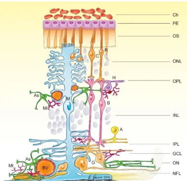
The retina, a light-sensitive layer, which lines the inner surface of the posterior part of the eye and can be described as visible part of the central nervous system (CNS) (Kaufman et al., 2003). The retina is organized in several different layers that can be clearly defined histologically (Figure 1).
Figure 1 Schematic structure of the mammalian retina. Detailed schema is shown with the layers beginning from the outermost side (PE=
pigment epithelium) to the innermost side (NFL= nerve fiber layer).
Different cell types are situated in the retina. Ch=Choroid, PE=pigment epithelium, OS=outer segments of photoreceptors, ONL=outer nuclear layer, OPL=outer plexiform layer, INL=inner plexiform layer, IPL=inner plexiform layer, GCL=ganglion cell layer, ON=optic nerve, NFL=nerve fibre layer, R=rod, C=cone, H=horizontal cell, G=ganglion cell M=Müller cell, B=bipolar cell, A=amacrine cell, As=astrocyte, Mi=microglia, BV=blood vessel (Vecino et al., 2016)
The single layered retinal pigment epithelium (RPE or PE) is part of the blood retina barrier and provides nutrition and waste removal to photoreceptors, the light sensitive cells of the retina (Strauss, 2005). The outer limiting membrane is rather a narrow zone comprising a series of heterotypic adherens junctions between Müller cells themselves and between Müller cells and photoreceptors (Williams et al., 1990). The photoreceptors’
cell bodies and processes constitute the outer nuclear layer (ONL), whereas their synapses form together with the synapses of bipolar and horizontal cells the outer plexiform layer (OPL). The cell bodies of horizontal, bipolar and amacrine cells and the nuclei of the Müller cells are located in the inner nuclear layer (INL), followed by the synapses between bipolar, amacrine and ganglion cells resulting in the inner plexiform layer (IPL) (Purves and Williams, 2001). The perikarya of the ganglion cells are located together with “displaced” amacrine cells in the ganglion cell layer (GCL), their axons build the nerve fiber layer (NFL) converging to form the optic nerve. At last, the Müller cell
endfeet form the inner limiting membrane, that separates the retina from the vitreous body (Purves and Williams, 2001; Kaufman et al., 2003)
1.1.1 Retinal neurons
The neural retina comprises an interacting network of five different specialized sensory neurons (Sachsenweger and Klauss, 2014): Ganglion cells, amacrine cells, bipolar cells, horizontal cells and photoreceptors. Photoreceptors are light-sensitive cells and capable of visual phototransduction (Kefalov, 2012), the initial step of vertebral vision. The visual phototransduction is the conversion of incident light energy into neural signals that can be transmitted to the brain by the optic nerve (Mannu, 2014).
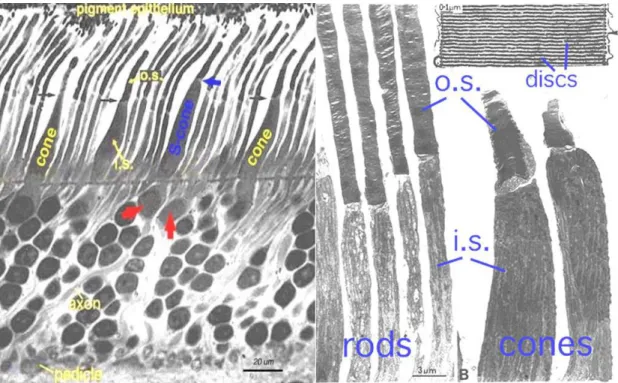
There are two different types of classic photoreceptors: the highly light-sensitive rods (100-125 million) for scotopic (night) vision, located mainly in the peripheral retina and the less sensitive cones (6-7 million) for achromatic (colour) vision located mainly at the fovea centralis in humans (Figure 2). Cones are divided in three types, each with different visual pigments, namely: S-cones, M-cones and L-cones. Each cone is therefore sensitive to visible wavelengths of light that correspond to short-wavelength (S-cones), medium-wavelength (M-cones) and long-wavelength (L-cones) (Mustafi et al., 2009).
Figure 2 Left. Semithin section of a human retina illustrating rod and cones, differentiated in short wavelength cones (S-cone, blue) and long wavelength cones (red arrows) (Kolb et al., 1995). Right.
Electronic microscopy of rod and cones of a monkey retina showing the inner and outer segments of rod and cones and a high magnification of the membrane discs (Anderson and Fisher, 1976). I.S.=inner segment, O.S.=outer segment
Both photoreceptors show the same basic structure: An outer segment (O.S.) for light- absorption by containing photopigment-filled disks (rods) or membrane enfoldings (cones), an inner segment (I.S.) full of ATP-providing mitochondria and endoplasmatic reticulum, the cell body (located in the outer nuclear layer) with the nucleus and other cell’s organelles and the synaptic terminal which is located in the outer plexiform layer (OPL). It releases neurotransmitter (e.g glutamate) to amacrine cells, interneurons which are responsible for lateral interaction in the retina, or to horizontal cells and bipolar cells which transmit the signal onwards to the ganglion cells, the neurons that transfer all visual information processed in the retina to the brain (Masland, 2012).
1.1.2 Retinal glia cells: Müller cells, astrocytes and microglia cells
The retinal glia cells consist of Müller cells, astrocytes and microglia cells (Vecino et al., 2016).
Müller cells radially span the retina with their processes from the outer to the inner limiting membrane and with their cells bodies residing in the inner nuclear layer (INL). They also infold all retinal neurons which enables a multifunctional interaction between Müller cells and neurons such as a homeostatic and metabolic support of retinal neurons (Bringmann et al., 2006; Reichenbach and Bringmann, 2013). A further important function of Müller cells is the release of neuroprotective (e.g. Fibroblast Growth Factor 2 (Fgf2), angiogenic factors (e.g. vascular endothelial growth factor (Vegf) and the recycling of neurotransmitters (Pierce et al., 1995; Bringmann et al., 2006). In case of retinal injury or disease, Müller cells become reactive and undergo a cytoskeleton remodelling called reactive gliosis which is characterized by a rounded shape and enlarged endfeet (García and Vecino, 2003) as well as an upregulation of the intermediate filament protein glial fibrillary acidic protein (Gfap) (Lupien et al., 2004).
In the healthy retina, Gfap is regularly expressed by astrocytes, a second type of glial cells in the central nervous system (Hol and Pekny, 2015). In the retina, their processes are almost confined to the nerve fiber layer (NFL) and ganglion cell layer (RGC) (Büssow, 1980; Hol and Pekny, 2015). As a main producer of the angiogenic factor Vegf (West et al., 2005), astrocytes are of significance in retinal vascularization (O'Sullivan et al., 2017).They migrate ahead of the vessels to promote the formation of superficial or deep vasculature (Stone et al., 1995; van der Wijk et al., 2018). Together with endothelial cells lining retinal microvessels and pericytes, they also support the integrity of the inner blood retinal barrier (BRB), which is constituted through the tight junctions of the endothelial cells (Klaassen et al., 2013).
The third glial subpopulation, the microglia cells, are the phagocytic immune cells of the CNS including the retina. Under physiological conditions, microglia cells remain in the
plexiform layers in a resting state and surveil their neuronal microenvironment to cleanse metabolic products and tissue debris (Langmann, 2007). In case of retinal injury, microglia react rapidly by transitioning to an activated status and undergoing gliosis.
Therefore, they switch their appearance through dynamic remodeling of their cytoskeleton and they withdraw their filopodic processes to acquire an oval, amoeboid shape. (Wang and Wong, 2014).
1.1.3 Retinal vasculature
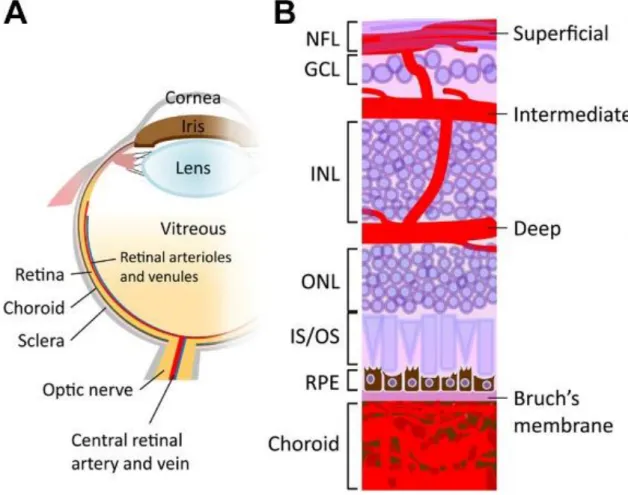
The eye obtains nutrients and oxygen from two different vascular systems: the retinal vessels within the inner parts of the retina and the uveal vessels within the iris, ciliary body and choroid (Luo et al., 2015). The choroid mostly persists of a fenestrated capillary network (choriocapillaris) and supplies the outer retinal layers and photoreceptors (Nickla and Wallman, 2009).
The inner ocular blood supply is predominantly provided by the ophthalmic artery, which is the first branch of the internal carotid artery. The branches of the ophthalmic artery extend form three intra-retinal vascular beds (plexus). In the murine retina, the plexus formation starts with the superficial plexus arising from the optic nerve head by a radial outgrowth to the periphery and lies on the RGC (Stahl et al., 2010). The superficial capillaries continue sprout vertically to first form the deep plexus in the outer plexiform layer and then the intermediate plexus located in the inner plexiform layer (Anand-Apte and Hollyfield, 2010; Stahl et al., 2010). Also in the human eye, the plexus formation also starts with the generation of the superficial plexus, but is then – in contrast to the murine retina- followed by the intermediate plexus and at least deep plexus (Figure 3).
Figure 3 Schematic illustration of the ocular vasculature. A. Drawing showing a sagittal section of an entire eye B. Detailed illustration of a sagittal section of the retina including retinal and choroidal vasculature.
Three capillary plexus of the retina are embedded among retinal neurons: the superficial plexus along the ganglion cell (GCL) and nerve fibre layer (NFL), the intermediate and deep plexus along each side of the inner nuclear layer (INL). The choroidal vessels are located beneath the RPE and Bruch's membrane and supply oxygen and nutrients to the outer portion of the retina. GCL=ganglion cell layer; INL=inner nuclear layer; IS/OS=inner segment/outer segment of photoreceptor; NFL=nerve fibre layer; ONL=outer nuclear layer; RPE=retinal pigment epithelium (Liu et al., 2017).
In human embryos, the development of vessels is dependent on oxygen-supply and bases on two different mechanisms: vasculogenesis and angiogenesis (Patan, 2004).
Whereas vasculogenesis describes the de novo formation of primary vessels out of common progenitor cells (Anand-Apte and Hollyfield, 2010; Lutty and McLeod, 2018), angiogenesis characterises the maturation or rather sprouting of vessels from already existing vessels (Risau, 1997; Hughes et al., 2000). The retinal vessels differentiation out of the avascular, immature retina starts at 14-16 weeks of gestation and is nearly complete vascularized by term birth (Penn, 2008). In mice, the developmental process of the retinal vasculature is angiogenesis only and vessel growth begins at the optic nerve and sprouts peripherally. In contrast to humans, mice still possess immature vasculature with hyaloid vessels postnatal and (Gyllensten Lars J. and Hellstöm, 1954) the three vascular plexus will develop during the first three weeks postnatal (Stahl et al., 2010)).
1.2 Retinal degeneration in the context of photoreceptor cell death
The vision loss in hereditary retina degenerations is generated by a progressive dysfunction and apoptosis of photoreceptors (Wright et al., 2010). As in the healthy retina only a small number of photoreceptors undergo apoptosis, our workgroup uses the light- induced damage model to mimic photoreceptor degeneration that occur in retinal degenerations like Retinopathia Pigmentosa.
1.2.1 Retinopathia pigmentosa
Retinopathia pigmentosa (RP) is a clinically and genetically heterogeneous group of hereditary disorders, which are characterized by a progressive loss of photoreceptors and pigment epithelial function (Pagon, 1988). Its genetic heterogeneity is reflected in the different inheritance patterns (autosomal-dominant, autosomal-recessive or X- linked) and mapping of mutations in over 45 causative gene loci in the typical RP, with the majority of them expressed in either the photoreceptors or the retinal pigment epithelium (Dryja and Li, 1995; Hamel, 2006). With a prevalence of 1:4000 worldwide (Hamel, 2006), RP represents one of the most common causes of blindness or severe low-vision at the age from 20 to 60 years (Parmeggiani, 2011).
The typical RP is described as rod-cone dystrophy, which implies a primary degeneration of rod photoreceptors and a following degeneration of cones in later stages (Ferrari et al., 2011; Hamel, 2006). This order of photoreceptor degeneration correlates with the typically slow progress (over three to five decades) after its usual first manifestation from age 20 to 30 years (Dryja and Li, 1995), with initial symptoms of night blindness the loss of the mid-peripheral visual field, due to the fact that rod photoreceptors are almost entirely responsible for night vision and mainly located in the retinal periphery (Shintani et al., 2009). Further visual loss progresses to the central visual field resulting in tunnel vision and eventual blindness, which can also be accompanied by further clinical
Figure 4 Fundus of patient with normal fundus (left) and patients with retinitis pigmentosa at mid stage (left) and end stage (right) show bone spicule-shaped pigment deposits and vascular atrophy (Hamel, 2006).
hallmarks like photopsia and diminished or absent electroretinogram (Ferrari et al., 2011) and an abnormal fundus with bone-spicule deposits (Figure 4), formed by migration of pigment-containing cells to perivascular sites in the inner retina (Li et al., 1995).
At current status, there is no cure of RP available. The only treatment options aim to slow down the progression of disease. Besides a vitamin and nutritional supplementation therapy such as vitamin A and omega-3 fatty acid, several emerging technologies like retinal cell transplantation, retinal prosthesis devices and gene therapy may provide additional promising therapeutic options (Hamel, 2006).
1.1.1 Light-induced damage paradigm as mouse model for photoreceptor degeneration
In rodents, the exposure of eyes to bright, white light leads to the death of photoreceptors, which is typically confined to rod photoreceptors (Organisciak and Vaughan, 2010). This susceptibility to light was used for establishing an experimental animal model for the induction of photoreceptor degeneration, the light-induced damage paradigm (Grimm and Remé, 2013; Wenzel et al., 2005). Hereby, the exposure to excessive light leads to photoreceptor cell death via apoptosis, which is followed by morphological and functional retinal alterations, quite similar to photoreceptor cell death in retinal hereditary dystrophies and age-related macular degenerations (Marc et al., 2003). With the help of the light-induced damage model, a large number of photoreceptors undergo apoptosis at the same time, which simplifies the analysis of molecular mechanisms in photoreceptor apoptosis and neuroprotective signalling pathways in the retina. According to the length of light exposure, the light damage can be induced in an acute or chronic way. There are also different types of light qualities used such as energy-rich blue light or cool, white light with a similar emission spectrum like daylight (Grimm and Remé, 2013; Wenzel et al., 2005). In the current thesis, we used the acute protocol with cool white light to induce photoreceptor degeneration and to examine whether Endothelin receptor b has neuroprotective effects on photoreceptor cells and which underlying mechanisms might mediate this effect.
1.1.2 Neuroprotection in the retina following light-induced damage paradigm
Light-induced photoreceptor degeneration leads to the activation of Müller cells, which suggests the existence of a signalling system that informs Müller cells about the health status of photoreceptors and monitors the release of neuron-derived signalling molecules (Rattner and Nathans, 2005). Microarray analysis showed that transcripts
coding for Endothelin 2 (Et-2) were highly upregulated following light induced damage (Rattner and Nathans, 2005). Its localization in the ONL was determined by using in situ hybridization, which indicates that photoreceptors react to excessive light exposure by the release of Et-2 (Rattner and Nathans, 2005). A simultaneous upregulation of Endothelin receptor b (Etb) in Müller cells indicated that the release of Et-2 leads to an activation of Müller cells via binding to Etb as response to light-induced damage (Rattner and Nathans, 2005).
Moreover, Müller cells react directly to photoreceptor cell stress by an upregulation of the neuroprotective factor leukemia inhibitory factor (Lif) (Joly et al., 2008; Gao and Hollyfield, 1996; Joly et al., 2007), which is essential for Müller cell activation and photoreceptors’ survival (Joly et al., 2008; Gao and Hollyfield, 1996; Joly et al., 2007) Furthermore, Lif was also shown to interact with Et-2, as the expression of Et-2 reduced to 2% of wild-type levels in Lif deficient mouse retinae, as well with fibroblast growth factor 2 (Fgf2), as intravitreal injections of recombinant Lif lead to an induction of Et-2 and Fgf2 in wildtype and remarkably also in Lif-deficient mice, (Joly et al., 2008; Gao and Hollyfield, 1996; Joly et al., 2007). Fgf2 is considered to promote survival on photoreceptor cells in a paracrine manner in different models of photoreceptor degeneration (Faktorovich et al., 1990; Joly et al., 2007; Gao and Hollyfield, 1996). It is likely, that photoreceptor damage leads to the activation of Et-2 in photoreceptors, which mediates expression of Lif and Fgf-2 expression in Müller cells. As photoreceptor-derived Et-2 itself leads to an activation of Müller cells and in turn the expression of Lif, a positive feedback loop is very likely. Thus, the activation of Et-2 and its mediation of Lif and Fgf- 2 expression is essential for retinal neuroprotection in response to photoreceptor degeneration.
1.2 The Endothelin system
In 1985, Hickey et al. first discovered a vasoconstricting factor obtained from the culture media of bovine aortic endothelial cells (Hickey et al., 1985). Three years later, Yanagisawa et al. identified the structure of this endothelin-derived vasoconstrictor and termed it endothelin (Yanagisawa et al., 1988). It is a 21-amino acid peptide linked with a free amino terminus and C-terminal carboxylic acid. This C-terminal helical tail seems to be crucial for receptor interaction, as well as two intramolecular disulfide bonds between Cys residues. Endothelin is expressed in three isoforms Endothelin 1, 2 and 3 (Et-1, Et-2, Et-3), which differ by the amount of amino acids (Davenport et al., 2016).
All three isoforms are first encoded as large precursor peptides (Prepro-Et-1/-2/-3) which are then abbreviated by 17 amino acids into proPeptides (proEt-1/2/3) by a signal peptidase. A second cleavage by furin enzymes removes 35 and 122 amino acids and
results in the immediate precursor of Endothelin (BigEt-1/2/3) (Xu et al., 1994). Its activation to mature Endothelin (Et-1/2/3) occurs via cleavage by membrane-bound metalloproteinases, the endothelin converting enzymes (ECE-1, ECE-2). Amongst the endothelin isoforms, Et-1 is the most extensively studied isoform and one of the most known potent vasoconstrictors (Yanagisawa et al., 1988).
Endothelins activate two main class I G-protein coupled receptor subtypes: endothelin receptor a (Eta) and b (Etb). ET-1 and ET-2 bind with equal affinity to the receptors, whereas Et-3 has a lower binding affinity for Eta (Sakurai et al., 1990; Arai et al., 1990;
Barton and Yanagisawa, 2008; Kedzierski and Yanagisawa, 2001). Eta is predominantly expressed by vascular smooth muscle cells, whereas Etb is expressed by both vascular smooth muscle and endothelial cells (Guan et al., 2015). Given physiological conditions, the two receptors are considered to possess synergistic but also opposite actions with Eta and Etb promoting vasoconstriction by the release of calcium by the sarcoplasmic reticulum resulting in increased smooth muscle contraction and vasoconstriction (Schneider et al., 2007), whereas Etb on endothelial cells contributes additionally to vasodilation (Filep et al., 1991; Schneider et al., 2007; Hirata et al., 1993). In addition, Etb acts as clearing receptor of circulating Et-1 (Fukuroda et al., 1994a). Endothelin signaling is involved in a variety of physiological functions, above all the regulation of vasomotricity, blood pressure and vascular homeostasis (Rautureau et al., 2015).
1.2.1 Distribution of Endothelin signalling in the eye
In the eye, the expression of Et-1 is well described in tissues like lens epithelium, optic nerve , ciliary body, trabecular meshwork as well as vascular endothelial cells in the choroid and retina (Chakravarthy et al., 1994; Wollensak et al., 2002), whereas Et-2 is expressed by photoreceptors (Bramall et al., 2013; Alrashdi et al., 2018). The expression of Et-3 is barely known. It has been shown that Et-1 modulates pericyte contractility to regulate retinal blood flow (Kawamura et al., 2002, 2002) and is involved in the regulation of intraocular pressure and aqueous humor dynamics (Taniguchi et al., 1994) Eta and Etb are expressed on perivascular cells of retinal and choroidal blood vessels to promote vasoconstriction (Choritz et al., 2005; Torbidoni et al., 2005; MacCumber and D'Anna, 1994). Etb is additionally expressed on vascular endothelial cells to promote vasodilation (Clozel et al., 1992; Schneider et al., 2007). Furthermore, Etb is expressed on photoreceptors and Müller cells (Braunger et al., 2013a; Rattner and Nathans, 2005).
1.2.2 Diverse roles of Endothelin signalling in the ocular system
The role of the endothelin signalling in the eye is discussed controversially. Recently, our group and others could demonstrate its contribution to the protection of retinal neurons (Braunger et al., 2013a; Rattner and Nathans, 2005; Joly et al., 2007), with Et-2 being discussed to act as a general stress factor following photoreceptor damage and to mediate neuroprotection on photoreceptors in the light-induced damage model, presumably via Etb (Rattner and Nathans, 2005; Braunger et al., 2013a).However, there are conflicting reports concerning the protective properties of endothelin signalling, as it is dysregulated in several ocular diseases (Vingolo et al., 2010; Sorrentino et al., 2018;
Good and Kahook, 2010). In particular, Et-1 has been suspected as contributing factor for the development of glaucoma as Et-1 levels were elevated in the aqueous humor (Noske et al., 1997; Tezel et al., 1997) and in the blood plasma of glaucoma patients (Li et al., 2016). The precise underlying mechanisms are still not fully understood, but it is discussed that Et-1 could promote ganglion cell death and contribute to optic nerve neuropathy through mechanisms of vascular dysregulation and (Krishnamoorthy et al., 2008; Lau et al., 2006). Furthermore, Et-2 is discussed to contribute to a breakdown of the blood retinal barrier with increased vascular leakage, vascular endothelial growth factor expression and infiltrating macrophages (Alrashdi et al., 2018). Thus, we aimed to examine the role of endothelin signaling in the retina in the current thesis.
2 Aim of study
This thesis focuses on Endothelin receptor b (Etb) mediated signalling and its potential neuroprotective function for the survival of photoreceptors. To this end, animals with an inducible, tamoxifen-dependent deletion of Etb in the entire retina (EtbΔeye) and animals with a conditional deletion of Etb in retinal neurons and Müller cells (EtbΔOC) will be generated using the Cre/loxP system. Additionally, an Etb-deficient murine photoreceptor cell line (661WΔEtb) will be created via CRISPR/Cas9-System. A successful Etb-deletion in EtbΔeye and EtbΔOC mice and 661WΔEtb cells will be validated via molecular analysis (quantitative realtime RT-PCR and western blot analyses) and in situ hybridization (BaseScope®). To address this we will:
• We will characterize the localization of Etb in the eye
For a general characterization of the localization of Etb in the eye, we will first use wildtype albino (CD1) mice to examine the distribution of Etb in the murine eye by performing in situ hybridization (BaseScope®) and co-immunohistochemical staining for different retinal cell types (astrocytes: glial fibrillary acidic protein, Müller cells: glutamine synthetase, vasculature: Collagen IV).
• We will investigate the expression levels of the endothelin signalling family following ocular trauma
To this end, we will investigate the mRNA expression levels of the Endothelin signalling family following different damages of the retina or the eye, respectively (puncture of the eye, PBS-injection, light-induced photoreceptor degeneration).
• We will characterize the impact of an Etb deletion on the morphology of the eye and the molecular expression levels of the endothelin signalling family, the Tgf-β signalling family, pro-and anti-apoptotic factors and neuroprotective factors in the retina.
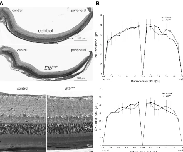
We will furthermore characterize the effect of the Etb-deficiency on the retinal architecture in EtbΔeye and EtbΔOC mice using semi-thin sections. To obtain statistically meaningful data, we will perform morphometric analyses and measure the thickness of the INL and ONL to generate Spider diagrams. We will analyse the morphology and a
potential leakiness of the retinal vessels in EtbΔeye and EtbΔOC mice by high molecular FITC-dextran perfusion, and immunohistochemical staining for their basal lamina (collagen IV) and for pericytes (neuron glia antigen 2 (Ng-2)). We will complete these analyses by studying the mRNA expression levels of the endothelial cell maker cluster of differentiation 31 (Cd31) and the perivascular cell markers alpha smooth muscle actin (α-Sma), neuron glia antigen 2 (Ng-2) and platelet-derived growth factor receptor beta (Pdgfrb). Moreover, we will analyse whether the Etb deletion might result in retinal hypoxia using hypoxia-inducible factor 1 alpha (Hif1α) western blot analyses. We will furthermore use specific antibodies (glial fibrillary acidic protein (Gfap) and ionized calcium-binding adapter molecule 1 (Iba1)) for immunohistochemical staining to analyse whether the deletion of Etb in EtbΔeye and EtbΔOC mice might influence the reactivity of Müller cells or microglia cells. To study whether the deletion of Etb will affect the mRNA expression levels of the other members of the Endothelin signalling pathway we will analyse their expression levels (Endothelin receptor type a (Eta), Endothelin 1/2/3 (Et- 1/-2-3). In addition, we will analyse the mRNA expression levels of the transforming growth factor beta signalling pathway (Tgf-β receptor 1 (Tgfbr1), Tgf-β receptor 2 (Tgfbr2), Tgf-β 1, Tgf-β 2, Tgf-β 3), pro-apoptotic factors (Caspase 8, Caspase 9, BH3- only BCL-2-interacting mediator of cell death (Bim), Bcl2 associated X (Bax), Bcl2 associated death promotor (Bad)) and the anti-apoptotic factor B-Cell Lymphoma 2 (Bcl2) as well neuroprotective factors (leukaemia inhibitory factor (Lif), fibroblast growth factor 2 (Fgf2)) .
Proteomic analyses of 661WΔEtb cells and controls will give unbiased and hypothesis- free information about the impact of an Etb deletion in photoreceptor cells.
• We will investigate a potential neuroprotective role of Etb mediated signalling for the survival of photoreceptors.
Therefore, we will use the light damage paradigm to induce photoreceptor apoptosis in EtbΔOC mice and controls. We will additionally cause cell stress by serum-deprivation in 661WΔEtb cells and controls to induce their apoptosis. TUNEL labelling will be used to detect apoptotic cells and we will quantify the TUNEL labelled apoptotic cells to clarify, whether the apoptotic rate might be altered in Etb-deficient mice and 661WΔEtb cells compared to their respective controls. Furthermore, we will use semi-thin sections to investigate the retinal morphology and perform morphometric analyses to compare the thickness of the ONL between light-exposed EtbΔOC mice and light-exposed controls. To learn more about the underlying molecular mechanisms, the transcript levels of the
members of the Endothelin signalling family (Eta, Et-1/-2/-3), transforming growth factor beta signalling pathway (Tgf-βr1, Tgf-βr2, Tgf-β 1, Tgf-β 2, Tgf-β 3), pro-apoptotic factors (Caspase 8, Caspase 9, Bim, Bad, Bax) and the anti-apoptotic factor Bcl2 as well neuroprotective factors (Lif, Fgf2) will be examined by quantitative realtime RT-PCR.
Western blot analyses for the cell survival factor Protein Kinase B (Akt) and its phosphorylated (p) form pAkt, a main component of the PI3K-Akt pathway, will be performed in light-exposed EtbΔOC mice and controls to clarify whether Etb might use this signalling pathway to mediate its neuroprotective effect.
3 Results
3.1 Expression of Endothelin receptor b in the eye
Initially, we used 6-week old albino (CD1) wildtype mice to study the expression and localization of Etb in the eye.
We first performed quantitative real-time RT-PCR of retinal lysates to detect the relative mRNA expression levels of Endothelin receptor a (Eta) and Endothelin receptor b (Etb) in the retina. Both receptors were detectable in the retina of wildtype mice (Figure 5).
Quite intriguingly, Etb was identified as predominant receptor in the retina as the relative mRNA expression level of Etb (24.34 ± 3.34) was significantly (p=0.02) higher than Eta mRNA expression (9.72 ± 3.01).
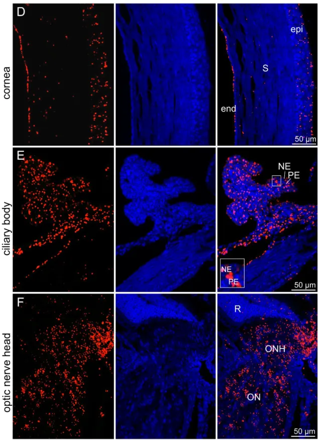
Next, we performed in situ hybridization BaseScope® (Chapter 6.5.7); which was friendly provided by Prof. Dr. Charlotte Wagner (University Regensburg, Germany) on sagittal sections of the entire eye to detect the mRNA signal of Etb and further localize Etb in the entire eye via conventional fluorescent microscopy (Figure 6 A-F). In the retina (A), the Etb signal was confined to the retinal ganglion cell layer (GCL), inner nuclear layer (INL), outer nuclear layer (ONL) and retinal pigment epithelium (RPE).There was also a distinct Etb mRNA signal detectable in the lens (B), strictly defined to the lens epithelium and lens fibres, whereas Etb signal was not visible in the lens capsule. In the iris (C), the Etb mRNA signal was only traceable in the stroma, whereas the iris epithelium had no signal for Etb. Furthermore, there were Etb mRNA signals in the entire cornea (D) detectable.
The corneal epithelium as well endothelium showed intense Etb signals, whereas the stroma had less positive Etb mRNA signals. The entire ciliary body (E) showed an intense signal for Etb, in its pigmented as well as its non-pigmented epithelium.
Furthermore, intense Etb mRNA signals were detected in the entire area of the optic nerve head and the optic nerve (F).
Figure 5 Relative mRNA expression of Eta and Etb in the retina.
Both receptors were detectable, but Etb was identified as predominant receptor with a significantly higher expression than Eta. The mean value of the housekeeping genes Gapdh and Gnb2l was used for normalization. Data are means ± SEM. Eta n = 6; Etb n = 4;
p* = 0.02, student’s t-test.
Figure 6 In situ hybridization BaseScope® of a 6-week old wildtype mouse.Etb mRNA signal (red); Nuclei were Dapi stained (blue). (A) Retina. There was an Etb mRNA signal in several layers of the retina detectable. GCL=ganglion cell layer, INL=inner nuclear layer, ONL=outer nuclear layer, RPE=retinal pigment epithelium (B) Lens. Etb was located in the lens epithelium as well as lens fibres, but not in the capsule. epi= epithelium, fib= lens fibres, cap=capsule (C) Iris. Only the iridal stroma depicted a positive Etb mRNA signal. epi= epithelium, S= stroma (D) Cornea. Etb was detectable in the entire cornea. epi=
epithelium, S= stroma, end= endothelium (E) Ciliary body. Etb was detectable in the entire ciliary body. PE=
pigmented epithelium, NE= non-pigmented epithelium (F) Optic nerve head. Etb was detectable in the entire area of the optic nerve head. R=retina, ONH=optic nerve head, ON=optic nerve
For a cell-specific Etb localization in the retina, the in situ hybridization BaseScope®- labelled sagittal sections were co-stained against Glial fibrillary acidic protein (Gfap) (Figure 7 A), a marker for astrocytes and reactive Müller cells and glutamine synthetase (Gs) (Figure 7 B), a Müller cell specific marker. Again, a positive Etb mRNA signal was detected in the GCL, INL and ONL, indicating a localization on neurons and photoreceptor cells. Immunohistochemical staining against Gfap (Figure 7 A) revealed a characteristic staining on top of the GCL, the typical localization of astrocytes. Here, no Etb mRNA signal was detectable, showing that astrocytes did not express Etb in the retina. The immunohistochemical staining against Gs (Figure 7 B) displayed the regular pattern of Gs Müller glia staining in the inner plexiform layer as well a distinct staining along the elongated glia processes stretching through the entire retina from the inner limiting membrane to the outer limiting membrane. Here, the Etb mRNA signal partly overlapped with GS within the inner plexiform layer and additionally in the region of the GCL, where the Müller cell endfeet are located, indicating an Etb expression in Müller cells, especially in their end feet.
A
B
Figure 7 (A) In situ hybridization BaseScope® (red) and immunohistochemical staining against Gfap (green) of a 6 week-old wildtype mouse.Immunohistochemical staining against Gfap revealed a characteristic staining on top of the ganglion cell layer (GCL), the typical localization of astrocytes. Here, no Etb signal was detectable (detailed magnification, arrows) indicating that astrocytes do not express Etb. (B) In situ hybridization BaseScope® (red) and immunohistochemical staining against Gs (green) of a 6 week-old wildtype mouse. Gs staining displayed the regular pattern of Gs Müller glia staining with a distinct staining along the elongated glia processes stretching through the entire retina from the inner limiting membrane to the outer limiting membrane. Etb mRNA signal partly overlapped within the inner plexiform layer (detailed magnification, arrows) and additionally the GCL, indicating an Etb expression in Müller cells. GCL =ganglion cell layer, INL = inner nuclear layer, ONL = outer nuclear layer, RPE = retinal pigment epithelium. Nuclei were Dapi stained (blue). In situ hybridization BaseScope® was performed in cooperation with Dr. Thomas Neder (University Regensburg)
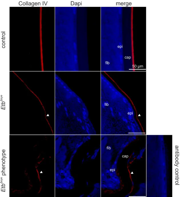
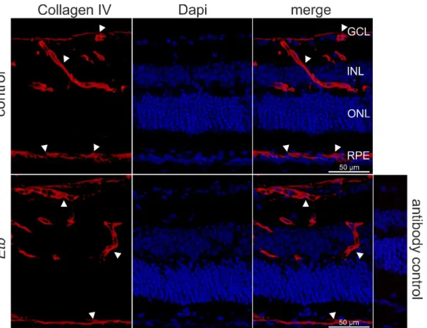
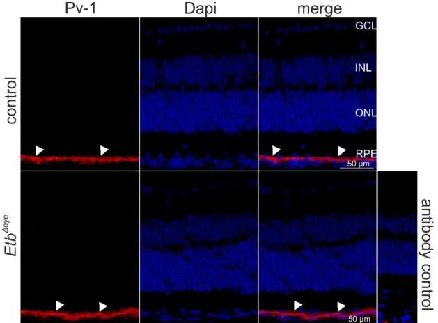
Next, we performed immunohistochemical staining against Collagen IV, an abundant protein of the basal lamina of blood vessels, that encloses endothelial cells located on the inner (luminal) side of the basal lamina, and perivascular cells still enclosed by the Collagen IV positive basal lamina but located at the outside of the vascular lumen. Thus, Collagen IV is a reliable marker to label the retinal vasculature and to distinguish between endothelial and perivascular cells, based on their inner (endothelial cells) or outer (perivascular cells) luminal position. E.g. positive Collagen IV staining was detected in retinal vessels located in the GCL showing a distinct Etb mRNA expression on the luminal side of the retinal vessels, indicating that Etb is expressed in retinal endothelial cells. Furthermore, we detected Etb positive signals at the outer luminal side of the vessels indicating the expression of Etb in perivascular cells.
Figure 8 In situ hybridization BaseScope® (red) and immunohistochemical staining against Collagen IV (green) of a 6 week-old wildtype mouse.Collagen IV positive staining (green) was detected in retinal vessels located in the GCL showing a distinct Etb mRNA expression on the luminal side of the retinal vessels, indicating that Etb is expressed in retinal endothelial cells (arrowheads). Furthermore, we detected Etb positive signals at the outer luminal side of the vessels indicating the expression of Etb in perivascular cells (arrows). GCL =ganglion cell layer, INL = inner nuclear layer, ONL = outer nuclear layer, RPE = retinal pigment epithelium. Nuclei were Dapi stained (blue).
3.2 Role of Endothelin signalling in the damaged retina
Our workgroup and other already showed that Et-2 is upregulated in experimentally induced photoreceptor degeneration (Braunger et al., 2013a; Rattner and Nathans, 2005; Joly et al., 2007). We aimed to characterize the role of the entire Endothelin signalling pathway in the damaged retina and therefore performed three different experimental setups leading to induce a retinal damage: Puncture of the eye (wildtype puncture, Figure 9 A) intravitreal injections of PBS (wildtype PBS,Figure 9 B) and light- induced damage (wildtype light, Figure 10)
Quite intriguingly, the sole puncture of the bulbus (wildtype puncture) led to significantly elevated mRNA expression levels of Eta (6.40 ± 1.53, p**=0.005) and Etb (8.34 ± 1.01, p***=0.00005) as well as Et-1 (6.89 ± 1.05, p***=0.0001) and Et-2 (5.66 ± 0.93, p *=0.04) compared to untreated controls (Eta: 1.00 ± 0.21; Etb: 1.00 ± 0.28; Et-1: 1.00 ± 0.33; Et- 2: 1.00 ± 3.57). The relative mRNA expression of Et3 remained unaffected (control: 1.00
± 0.02; wildtype PBS: 1.33 ± 0.32, p=0.7). Mice that received additionally an intravitreal injection of 3 μl PBS (wildtype PBS) showed a comparably increased relative mRNA
Figure 9 Quantitative real-time RT-PCR of retinal lysates of 6 to 8-week old wildtype mice (A) relative mRNA expression levels of Eta, Etb, Et-1 and Et2 were significantly elevated after the solely puncture of the eye into the vitreous (wildtype puncture) compared to controls. (B) The intravitreal injection with PBS (wildtype PBS) resulted in significantly elevated mRNA expression levels of Eta, Etb, Et1 and Et2 compared to controls. Expression was normalized to Gnb2l. Data are means ± SEM. Wildtype puncture: Eta (n≥ 3;
p**=0.005), Etb (n≥ 3; p***=0.00005), Et1 (n≥ 4; p***=0.0001), Et2 (n≥ 2; p*=0.04), Et3 (n≥ 3; p=0.7 (not significant); wildtype PBS: Eta (n≥ 3; p=0.09 (not significant)), Etb (n≥ 3; p*=0.02), Et1 (n≥ 4; p**=0.003), Et2 (n≥ 2; p*=0.05), Et3 (n≥ 3); p=0.6 (not significant). Student’s t test. [AU]= arbitrary unit; Figures taken from Schmitt et al. New insights into endothelin signalling and its diverse roles in the retina, in press.
Experiment performed by Sabrina Schmitt
A B
expression of Eta (7.34 ± 4.25, p=0.09) and a significant increase of Etb (8.99 ± 3.12, p*=0.02), Et-1 (3.85 ± 0.73, p**=0.003) and Et-2 (4.52 ± 0.95, p*=0.05) compared to untreated controls (Eta: 1.00 ± 0.21; Etb: 1.00 ± 0.28; Et1: 1.00 ± 0.33; Et2: 1.00 ± 3.57).
Again, the relative mRNA expression level of Et-3 (control: 1.00 ± 0.02, wildtype PBS:
1.33 ± 0.32, p=0.6) remained unaffected.
Quite similarly, the light exposed mice (Figure 10) showed significantly elevated mRNA expression levels of Eta (10.15 ± 2.69, p**=0.004), Etb (7.10 ± 3.31, p*=0.05), Et-1 (4.77
± 0.56, p***=0.0004) and predominantly, an up to 36-fold increase of Et-2 (36.37 ± 2.32, p**=0.002) compared to controls (Eta: 1.00 ± 0.22; Etb: 1.00 ± 0.30; Et-1: 1.00 ± 0.37;
Et-2: 1.00 ± 3.60). However, Et-3 (control: 1.00 ± 0.01, wildtype light: 1.2 ± 0.40, p=0.7) expression levels were not significantly altered following experimentally induced photoreceptor degeneration. Taken together, our results show, that endothelin signaling is upregulated following various retinal damages, indicating that the endothelin signaling pathway may act neuroprotective in the retina.
Figure 10 Quantitative real-time RT-PCR of 6 to 8-week old albino mice after light- induced damage.Relative mRNA expression levels of Eta, Etb, Et1 and Et2 are significantly upregulated after light- induced damage (wildtype light) compared to controls that were not exposed to light.
Expression was normalized to Gnb2l. Data are means ± SEM. Eta (n≥ 3; p**=0.004), Etb (n≥ 4; p*=0.05), Et1 (n≥ 4;
p***=0.0004), Et2 (n≥ 3; p**=0.002), Et3 (n≥ 4; p=0.7. Student’s t test. [AU]=
arbitrary unit. Figures taken from Schmitt et al. New insights into endothelin signalling and its diverse roles in the retina, in press.
Experiment performed by Sabrina Schmitt
3.3 Etb-deficiency in vitro and in vivo
To analyse the potential neuroprotective effect of Etb on photoreceptor cells in detail, we generated two mouse strains, one with a conditional deletion of Etb in the entire retina (EtbΔeye mice) and a second mouse strain with a cell type specific deletion in Müller cells and retinal neurons (EtbΔOC mice) via the CreloxP-System. Furthermore, we generated a stable Etb deletion in a photoreceptor cell line (661WΔEtb) via the CRISPR/Cas9-System to further study its physiological and molecular function.
3.3.1 Characterization of EtbΔeye mice
For simplicity, Etbflox/flox mice lacking the Cre recombinase are referred to as controls and Etbflox/flox mice expressing Cre recombinase are referred to as EtbΔeye or conditional knockout mice.
3.3.1.1 Successful deletion of Endothelin receptor b in the entire retina
To activate the CAG-CreERTM-Recombinase and thereby induce the conditional deletion of Etb in the entire retina via the CreloxP-System (Chapter 6.1.2), all mice were injected intraperitoneally with tamoxifen [20mg/ml] at the age of 4 weeks twice a day for five days.
The mice were sacrificed at the age of 6 weeks for further analyses.
The relative mRNA expression levels of Etb in retinal tissue of EtbΔeye mice and controls were compared via quantitative real-time RT-PCR analysis. The mean value of the housekeeping genes Gapdh and Gnb2l was used for normalization. A significant (p = 0.001) decrease of 87% of the relative Etb mRNA expression level in the EtbΔeye mice (0.13 ± 0.30) compared to controls (1.00 ± 0.16) confirmed a successful deletion of Etb in the entire retina (Figure 11 A).
Figure 11 (A) Relative mRNA expression of Etb in EtbΔeye and controls showed a successful deletion of Etb in the entire retina following tamoxifen treatment. Mice with a conditional deletion of Etb expressed significantly lower amounts of Etb mRNA in the retina compared to controls. The mean value of the housekeeping genes Gapdh and Gnb2l were used for normalization. Data are means ± SEM. control n = 4; EtbΔeye n = 4; p*** = 0.001, student’s t-test. (B) Western blot analysis of retinal lysates and densitometry of Etb in EtbΔeye and controls following tamoxifen treatment. Significant decrease of Etb expression in the entire retina after tamoxifen treatment in EtbΔeye mice compared to controls. Gapdh was used as housekeeping protein. Data are means ± SEM. control n = 6, EtbΔeye n = 4; p* = 0.03, student’s t-test;
Experiments were performed by Anna Huber and Sabrina Schmitt
The densitometric analysis of the relative protein expression level of Etb (54 kDa) showed a significantly (p* = 0.03) decreased expression of Etb in EtbΔeye mice (0.55 ± 0.12) compared to controls (1.00 ± 0.12) again confirming the successful deletion of Etb in the entire retina (Figure 11). Gapdh (37 kDa) was used as housekeeping gene.
To further verify a successful deletion of Etb we performed in situ hybridization BaseScope® on sagittal sections, to detect mRNA signal of Etb and to precisely localize potentially remaining Etb in retina (Figure 12). In control mice, Etb was distributed throughout the entire retina (Figure 12 A). The detailed magnification (Figure 12 B) showed Etb mRNA signals as described before (Figure 6). In contrast, there was no Etb mRNA signal detectable in EtbΔeye mice validating a successful deletion of Etb in the retina of conditional knockout mice.