Molecular analysis of the neuroprotective role of Norrin for photoreceptors in the
mammalian retina
DISSERTATION
Zur Erlangung des Doktorgrades der Naturwissenschaften (Dr. rer. nat.) der
Naturwissenschaftlichen Fakultät III Biologie und vorklinische Medizin der
Universität Regensburg
vorgelegt von Barbara Maria Braunger
aus Biberach an der Riß
Regensburg, 2013
Das Promotionsgesuch wurde eingereicht am:
8.10.2013
Die Arbeit wurde angeleitet von:
Prof. E.R. Tamm
Unterschrift:
The human being, creature of eyes, needs the image.
Leonardo da Vinci (1452 – 1519)
Meinen Eltern
Table of Contents
1 Introduction: ... 1
1.1 Retina... 1
1.1.1 Retinal neurons and glia cells ... 1
1.1.1.1 Retinal ganglion cells ... 1
1.1.1.2 The inner nuclear layer (INL): ... 2
1.1.1.3 Neurons of the INL: amacrine cells, bipolar cells and horizontal cells ... 2
1.1.1.4 Retinal glia cells: astrocytes, Müller glia cells and microglia ... 3
1.1.1.5 Cells of the outer nuclear layer (ONL): photoreceptors ... 4
1.1.1.6 The retinal pigment epithelium ... 6
1.1.2 Phototransduction and visual cycle ... 7
1.1.2.1 Phototransduction ... 7
1.1.2.2 Visual cycle ... 8
1.1.3 Electrophysiology: Electroretinography (ERG) ... 9
1.1.4 Retinal vessels in development and adulthood ... 10
1.2 Norrin ... 12
1.2.1 Norrie disease: symptoms and genetics ... 12
1.2.2 The protein Norrin ... 12
1.2.3 The biological functions of Norrin ... 13
1.2.3.1 Angiogenic properties of Norrin ... 13
1.2.3.2 Neuroprotective properties of Norrin ... 14
1.2.3.3 Norrin in reproduction and development ... 15
1.2.4 Norrin signaling and the Wnt/β-catenin pathway ... 16
1.3 Animal models to induce cell specific stress ... 19
1.3.1 The retinopathia of prematurity (ROP) model ... 19
1.3.2 A damage model for photoreceptor degeneration: light damage ... 19
1.3.3 Neuroprotective signaling pathways in the context of photoreceptor degeneration .. 19
1.4 Aim of the current work ... 22
2 Results ... 25
2.1 Characterization of Rpe65-Norrin-2 mice ... 25
2.1.1 Southern Blot: successful genomic insertion of the transgene construct ... 25
2.1.2 Northern blot analysis: effective transcription of the construct ... 26
2.1.3 Western blot analysis: effective protein translation ... 27
2.1.4 Immunohistochemistry: retinal localization of the transgenic Norrin protein ... 27
2.1.5 Rpe65-Norrin-2 mice and Wnt/β-catenin activation ... 28
2.2 Phenotype analysis of Rpe65-Norrin-2 animals ... 31
2.2.1 Retinal morphology ... 31
2.2.2 Ultrastructure of Rpe65-Norrin-2 animals ... 32
2.2.3 Vascular phenotype ... 33
2.3 The retinopathia of prematurity (ROP) model ... 35
2.4 Light damage experiments ... 40
2.4.1 Morphometric analysis ... 40
2.4.2 In vivo SLO and OCT imaging before and after light damage ... 43
2.4.3 Functional analysis: electroretinography (ERG) analysis ... 45
2.4.4 Analysis of the visual cycle ... 47
2.4.5 Light exposure and apoptosis ... 49
2.4.6 Wnt-/β-catenin signaling and Norrin mediated neuroprotection ... 51
2.5 Neuroprotective factors ... 54
2.5.1 Transgenic Norrin overexpression and neuroprotective factors ... 54
2.5.2 Correlation between Norrin induced Wnt/β-catenin activation, EDN2 upregulation and light induced apoptosis ... 58
2.5.3 Brain-derived neurotrophic factor and the protective effects of Norrin ... 61
3 Discussion ... 67
3.1 Norrin in the context of neuroprotection ... 67
3.1.1 Characterization and morphology of Rpe65-Norrin-2 mice ... 67
3.1.2 Norrin and light exposure ... 68
3.1.3 Norrin, Wnt/β-catenin and Edn2 signaling ... 69
3.1.4 Norrin neuroprotection and BDNF after light exposure ... 70
3.2 Norrin in the context of angiogenesis ... 73
3.2.1 Vascular phenotype of Rpe65-Norrin-2 animals ... 73
3.2.2 Norrin in the ROP model ... 74
3.3 Future directions ... 75
4 Summary – Zusammenfassung ... 77
4.1 Summary... 77
4.2 Zusammenfassung ... 78
5 Material and methods ... 79
5.1 Methods ... 79
5.1.1 Animal models. ... 79
5.1.1.1 Rpe65-Norrin-2 mice ... 79
5.1.1.2 βB1-Crystallin Norrin mice ... 79
5.1.1.3 Wnt-Reportermice: TOPGAL mice ... 79
5.1.2 DNA analysis ... 80
5.1.2.1 DNA isolation ... 80
5.1.2.2 Genotyping: Polymerase chain reaction (PCR) ... 80
5.1.2.2.1 Genotyping for the transgenes: Norrin and LacZ ... 80
5.1.2.2.2 Genotyping for the leucine variant of Rpe65 ... 81
5.1.2.3 Agarose gel electrophoresis ... 82
5.1.2.4 Southern blot analysis ... 82
5.1.2.5 Southern and northern blot probe ... 83
5.1.3 RNA analysis ... 83
5.1.3.1 RNA isolation ... 83
5.1.3.2 Complementary DNA (cDNA) synthesis ... 84
5.1.3.3 Quantitative realtime RT-PCR ... 85
5.1.3.4 Primer for real-time RT-PCR ... 85
5.1.3.5 Northern Blot analysis ... 86
5.1.4 Protein analysis... 87
5.1.4.1 Protein isolation ... 87
5.1.4.2 Western blot analysis ... 87
5.1.4.3 Antibodies and blocking solutions for Western blot analysis ... 88
5.1.4.4 Apoptosis: cell death detection ELISA ... 88
5.1.5 Histology ... 89
5.1.5.1 Phenotype analysis ... 89
5.1.5.2 Immunohistochemistry ... 89
5.1.5.3 Antibodies for immunohistochemistry... 90
5.1.5.4 Apoptosis: TUNEL analysis ... 91
5.1.6 Animal experiments ... 92
5.1.6.1 Oxygen-induced retinal damage: The retinopathia praematurorum (ROP) model .. 92
5.1.6.2 Light damage ... 92
5.1.6.3 Light damage and morphometric read out: the spider diagram ... 93
5.1.6.4 Intravitreal injections ... 94
5.1.6.5 Fundus imaging and angiography ... 94
5.1.6.6 Retinal whole mounts... 94
5.1.6.7 Functional analysis: electroretinography (ERG) ... 95
5.1.6.8 Rhodopsin recovery in darkness... 95
5.1.7 Statistical analysis ... 96
5.2 Material ... 97
5.2.1 Chemicals and reagents ... 97
5.2.1.1 Enzymes and Taq polymerase ... 98
5.2.1.2 Kits ... 99
5.2.2 Consumable supplies and equipment ... 99
5.2.2.1 Consumable supplies ... 99
5.2.2.2 Equipment ... 99
5.2.3 Gels, buffers, dilutions ... 101
5.2.3.1 Buffers and dilutions ... 101
5.2.3.1.1 Histology ... 101
5.2.3.1.2 Protein analysis ... 101
5.2.3.1.3 DNA analysis ... 103
5.2.3.1.4 RNA analysis ... 103
5.2.3.1.5 Animal experiments ... 104
5.2.3.2 Polyacrylamide gel electrophoresis ... 104
6 References ... 105
7 Abbreviations ... 115
8 Figure and table legend ... 119
8.1 Figure Legend ... 119
8.2 Table Legend ... 121
9 Danksagung ... 123
10 Erklärung ... 125
Introduction
1
1 Introduction:
1.1 Retina
The vertebrate retina consists of several neuronal layers interconnected by synapses. From the vitreous side, the first neuronal layer is the ganglion cell layer (GCL) with the soma of retinal ganglion cells and aberrant amacrine cells. The axons of the retinal ganglion cells (RGC) form the optic nerve.
The cells of the RGC layer are connected by synapses with the neurons of the inner nuclear layer (INL). Within the INL, there are several different neuronal populations: the amacrine, bipolar and horizontal cells. Additionally the cell somas of the Müller glia cells are located in the INL. The INL neurons are again interconnected by synapses with the light sensitive photoreceptor cells, their cell soma are located in the outer nuclear layer (ONL). Figure 1 shows a schematic of the retina and a corresponding semithin section of the retinal morphology.
Figure 1: Morphology of the retina
Schematic and corresponding semithin section of retinal morphology. Abbreviations: NFL: nerve fiber layer, GCL: ganglion cell layer, IPL: inner plexiform layer, INL: inner nuclear layer, OPL: outer plexiform layer, ONL:
outer nuclear layer, RPE: retinal pigment epithelium. Slightly modified after (1), semithin section (right hand side) by Barbara Braunger.
1.1.1 Retinal neurons and glia cells
1.1.1.1 Retinal ganglion cells
The retinal ganglion cells are located in the innermost neuronal layer of the retina. The axons of the retinal ganglion cells form the nerve fiber layer (stratum opticum) which is situated on top of the retinal ganglion cell soma, next to the side of the vitreous. These axons conjoin in the central retina as optic nerve head and form thereafter the optic nerve, which extend after the optic chiasm as optic
Introduction
2
tract in the optic center of the brain (2). There are different subpopulations of retinal ganglion cells (3) that use different neurotransmitter gamma-aminobutyric acid (GABA), glycine or glutamate (4).
The retinal ganglion cells have different functions, apart from sending photoreceptor derived signal into the optic center of the brain. For example, photosensitive retinal ganglion cell are involved in the pupillary reaction and circadian rhythm (5,6).
1.1.1.2 The inner nuclear layer (INL):
The inner nuclear layer (INL) is composed of various different cell types. One has to distinguish between neuronal cells like amacrine, bipolar and horizontal cells and Müller glia cells (see 1.1.1.4.).
1.1.1.3 Neurons of the INL: amacrine cells, bipolar cells and horizontal cells
Amacrine cells are located in the retinal ganglion cell layer, where they are called aberrant amacrine cells and in the inner aspect of the inner nuclear layer (2). There are various subtypes (> 30 different subtypes) of amacrine cells (7,8). Amacrine cells have large dendrites that extend laterally in the inner plexiform layer. They modulate ganglion cell function by utilizing inhibitory neurotransmitters like GABA or glycine, but also the excitatory neurotransmitter acetylcholine (9). Additionally, amacrine cells with extensive ramified dendrites are thought to support the function of horizontal cells by sending inhibitory feedback to bipolar cells and ganglion cells.
Bipolar cells are the major neuronal cell population in the inner nuclear layer (2). They are interneurons, transporting the signals from the photoreceptor cells to the ganglion cells. One distinguishes between cone and rod bipolar cells and the way they mediate the electric excitation as on- and off-bipolar cells (10). In the mouse retina, there is one class of rod driven bipolar cell and 11 subtypes of cone driven bipolar cells (11). Bipolar cells use the neurotransmitters glutamate and to a small extend glycine (4). After light induced hyperpolarization of photoreceptor cells, the on-bipolar cells depolarize resulting in an excitation of the subsequent retinal ganglion cell. In contrast off-bipolar cells hyperpolarize after photoreceptor hyperpolarization leading to an inhibition of the consecutive retinal ganglion cell.
Horizontal cells are situated at the outer aspect of the inner nuclear layer with large ramified dendrites in the outer plexiform layer (2). Depending on the species, there are up to 3 different subtypes of horizontal cells which release the inhibitory neurotransmitter GABA (12). Although the detailed mode of how horizontal cells mediate their effects is still under discussion (10,13), in the end horizontal cells sum up the light responses across a wide retinal region and subtract it from the local signal. This results in an improved contrast vision and allows seeing properly under dim as well as under bright light conditions (14).
Introduction
3
1.1.1.4 Retinal glia cells: astrocytes, Müller glia cells and microglia
The retinal astrocytes are found in the optic nerve fiber layer of the retina (15). Together with the Müller glia end feet they form the inner limiting membrane (16). Astrocytes play an important role during vasculogenesis for the proper guiding of the vessels towards the periphery of the retina. Later in development, they are part of the inner blood retinal barrier, which is composed of astrocytes, endothelial cells and pericytes (17). They also secrete the neuroprotective and angiogenic factor vascular endothelial growth factor (VEGF). This factor expressed from astrocytes turned out to be more important for the maintenance of vessel stability than the guiding of the vessels during vasculogenesis (18).
Müller glia cells span the retina vertically (19). Their cell bodies are located in the inner nuclear layer (2), of which they comprise 16% of all the cells present in the INL (15). Müller glia end feet give rise to the outer and inner limiting membrane (16). The outer limiting membrane lies between the outer nuclear layer and the inner segments of the photoreceptors, whereas the inner limiting membrane lies on top of the nerve fiber layer. During angiogenesis, Müller glia cells build up a gradient of the angiogenic factor vascular endothelia growth factor (VEGF) that guides endothelial cells towards their designated localization. Additionally, Müller glia cells are of major importance for the maintenance of the retina in general. They express growth factors, neurotransmitter transporters and antioxidant agents to prevent retinal neurons from excitotoxic damage (16,19). The various Müller cell functions are shown as a schematic in Figure 2. The ability of Müller cells to secrete neuroprotective factors is of major importance for this project.
Figure 2: Schematic of the different Müller cell functions
Role of Müller glia cells in potassium (K+) and water homeostasis (I), neurotransmitter clearing (II), protection from oxygen free radicals (III) and the secretion of neuroprotective factors (IV). Picture taken from (20).
Introduction
4
Microglia cells are the third glial population in the retina. Microglia cells represent the mononuclear phagocyte population of the nervous system (21). In the retina, they are located in the inner and outer plexiform layer and have important functions in immune surveillance and neuronal homeostasis (22–25).
1.1.1.5 Cells of the outer nuclear layer (ONL): photoreceptors
There are two types of photoreceptor cells: rod and cones. Rod photoreceptor cells are specialized for vision in dim light and provide black and white vision, whereas the cones operate during bright light, like daylight, and provide color vision (10).
In the dark, the photoreceptor cells permanently release the neurotransmitter glutamate (4), which is thought to be the neurotransmitter of the vertical neurons in the retina (photoreceptor cells, bipolar cells and retinal ganglion cells). When light hits the rhodopsin molecule in the photoreceptor cell this results in a closure of ion channels (Na+ and Ca2+) which in turn results in a hyperpolarization of the photoreceptor cell and subsequently a diminished release of its neurotransmitter glutamate.
(The detailed process of phototransduction is described in 1.1.2.1).
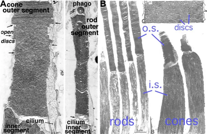
Regarding the photoreceptor`s morphology one speaks of an inner and an outer segment, a cell body, which is situated in the outer nuclear layer (ONL) containing the nucleus of the photoreceptor cell, and the synaptic terminal, which is situated in the outer plexiform layer (OPL) where neurotransmission to downstream neurons occurs (2). The inner segment of the photoreceptors contains cell organelles like mitochondria, golgi apparatus, endoplasmatic reticulum and ribosomes (26). A cilium connects the inner with the outer segment of the photoreceptor. Figure 3 shows the ultrastructure of a rod and cone photoreceptor and a membranous disc stack. Photoreceptor cells have an outer segment composed of membranous discs (rod) or membrane enfolding (cones) that contain the photopigment (26).
Introduction
5 Figure 3 Ultrastructure of photoreceptors
A. Electron microscopy of rod and cones photoreceptors of a squirrel retina with the connecting cilium between the inner and the outer segments and an example of disc shedding in the retinal pigment epithelium.
B. Electron microscopy of rod and cone photoreceptors of a monkey retina with enlargement of the outer segment discs. o.s.: outer segment, i.s.: inner segment. Slightly modified after (27).
The opsin part is a membrane receptor that is assembled in the photoreceptor cell organelles of the inner segment and transported through the connecting cilium to their final destination in the membrane of the outer segment. In rod outer segments the final destination is in intracellular membrane disc stacks. These stacks are without contact to the plasma membrane (see Figure 3 and Figure 4). In cones, the disks are in contact with the plasma membrane (28,29), which is called 'membrane enfolding' (30).
Figure 4: Schematic of rod outer segment disc
A. The rod outer segment is filled entirely with discs of folded double membranes. These membranes do not contact the plasma membrane of the photoreceptor cell. The light sensitive visual pigment is embedded in the intracellular disc membrane. Picture taken from (29).
B. Schematic that illustrates the continuous distance between the plasma membrane and the intracellular situated membranous discs stacks. Picture taken from (28).
Introduction
6
The complete renewal of a photoreceptor disc occurs about every 10 days. Newly formed discs are located at the base of the outer segment and within 10 days the discs displace progressively towards the retinal pigment epithelium. When they reach the RPE they are phagozytosed (26). This process is called shedding. To facilitate this process, the outer segments lay folded in the microvilli of the retinal pigment epithelium (1). The retinal pigment epithelium plays an essential role in removing and recycling the photoreceptor discs.
To be precise, there is a third, but rare type of photoreceptor cell: the light sensitive ganglion cells that contain melanopsin as the photopigment. These cells are important for the circadian physiology, they are involved in the pupillary reaction to light by reflecting signals into brain regions like the olivary pretectal nucleus and they respond to blue spectral light (~ 480nm) independent of rod and cones (31).
The arrangement of the photoreceptor cells lying at the very end of the light entering the retina seems to be counteractive. But apart from its formation during retinal development, there may also be functional reasons in the context of regeneration, as supported by the special relationship that exists between the outer segments of the photoreceptor and the retinal pigment epithelium..
1.1.1.6 The retinal pigment epithelium
The retinal pigment epithelium has many different and very important functions for the maintenance of the eye (outlined in Figure 5). Among these are the secretion of growth factors (vascular endothelial growth factor (VEGF), pigment epithelium derived growth factor (PEDF), the epithelial transport to uphold the ion homeostasis for glia cells, the phagocytosis of sorted photoreceptor disks (shedding) and its ability of light absorption (32).
Figure 5: The retinal pigment epithelium and its functions.
Picture taken from (32). For details see main text.
Introduction
7
One of its functions which is of major importance for the current project, lies in the biochemical machinery for the regeneration of the photopigment molecules, after they have been exposed to light. This process is called visual cycle.
1.1.2 Phototransduction and visual cycle
1.1.2.1 Phototransduction
Vision starts with the conversion of light in an electric signal in the photoreceptor cells. This process is called phototransduction (Figure 6). After light (photon) absorption in the photopigment of the photoreceptor cell, a signaling cascade is initiated that changes the membrane potential of the receptor and leads in the end to a change of the amount of the neurotransmitter glutamate released by the photoreceptor synapses onto the cells in the inner nuclear layer (4).
Figure 6: Phototransduction
Photon absorption (hν) activates the rhodopsin molecule (R*) in the disk membrane. The activated rhodopsin initiates the exchange of the G-protein (transducin, G) bound GDP to a GTP. The activated α-subunit (G*) of the G-protein binds to phosphodiesterase (PDE) which results in its activation (PDE**) and hydrolysis of cGMP to GMP. This leads to closure of the cation ion channels (Na+, Ca2+) and the photoreceptor cell membrane hyperpolarizes. GDP= guanosine diphosphate; GTP= guanosine triphosphate; cGMP/cG= cyclic guanosine monophosphate; GMP= guanosine monophosphate. Figure taken from (34).
The signaling cascade is mediated via G-proteins. The photopigment opsin itself is a membrane- bound G-coupled receptor. The chromophore is 11-cis retinal, which is covalently bound to the opsin part. The opsin molecule is generated in the inner segment of the photoreceptor whereas the chromophore 11-cis retinal is delivered via interphotoreceptor matrix carrier molecules (retinal binding proteins, IRBP) from the retinal pigment epithelium (32). When a light photon hits the G- coupled receptor, the 11-cis retinal changes its conformation into all-trans retinal – a phenomenon called photo isomerization. Subsequently the G- coupled receptor changes its conformation, resulting in its activation. Then the light activated photopigment activates the GTP-binding protein transducin. Transducin stimulates cyclic guanosine monophosphate (cGMP) phosphodiesterase (PDE). The PDE enzyme hydrolyzes cGMP to 5`GMP. Subsequently the cGMP gated ion (Na+ and Ca2+)
Introduction
8
channels in the cell surface membrane close. This results in the hyperpolarization of the photoreceptor cell and modulation of the neurotransmitter release at the synaptic terminal (33).
1.1.2.2 Visual cycle
The visual cycle describes the regeneration process of all-trans retinal. As mentioned above (in 1.1.2.1) when 11-cis retinal is photo isomerized, it changes its conformation into all-trans retinal.
During the visual cycle, a “used” all trans-retinal is recovered back into the active 11-cis-retinal using several enzymatic recovery steps. This process takes place in the outer segments of the photoreceptors and the retinal pigment epithelium (see Figure 7). Between these structures and in the interphotoreceptor matrix, the retinal is transported using carrier proteins.
Figure 7: Visual cycle
Schematic showing the visual cycle with its light induced conformation change of 11-cis retinal into all-trans retinal and the following regeneration process in the interphotoreceptor matrix and retinal pigment epithelium, respectively. Figure taken from (28).
The 11-all-trans retinal is released from the opsin protein, reduced to all-trans retinol and esterified by lecithin retinol acyltransferase (LRAT), converted to 11-cis retinol by the protein RPE65. Finally, 11-cis retinol is oxidized to 11-cis retinal and moved back to the rod outer segment, where it is reconjugated to an opsin. A new functional visual pigment (opsin) is formed (28).
In the context of light damage experiments (see 1.3.2) the proper function of the visual cycle is essential. Animal experiments with RPE65 knockout (RPE65-/-) mice and animals that did not express opsin in photoreceptors (Rho-/- mice) showed that these mice both are not sensitive to intense light exposure (35). RPE65-/- and Rho-/- animals did not develop any kind of light induced damage. These
Introduction
9
results highlight the importance of rhodopsin in the context of light damage and its main impact on retinal degeneration, indicating that the G-coupled receptor is the receptor mediating the damaging effects of light (35). Additionally, the publication of Grimm and co-workers showed the importance of the RPE65 protein (visual cycle, see 1.1.2.2 and Figure 7) in the context of light induced photoreceptor damage. RPE65 deficiency protected against early signs of light mediated damage (vesiculation of photoreceptor rod outer segments), as well as against late effects (photoreceptor apoptosis). The authors pointed out the importance of “excessive” adsorption of photons by rhodopsin that induces a death cascade leading to apoptosis (35).
Another observation highlights the importance of the visual cycle and the RPE65 gene in the context of light susceptibility. A point mutation at position 450 in the RPE65 gen has been shown to be of major important for the susceptibility of the animals to light damage. This mutation results in an amino acid exchange from methionine to leucine (36–38). Animals carrying the leucine variant of the Rpe65 protein are about 120-fold more sensitive to intense light than methionine carrying animals. In the current thesis, experimental animals were tested for the mutation and leucine carriers only were used for the experiments.
1.1.3 Electrophysiology: Electroretinography (ERG)
The electroretinography (ERG) method is used to in vivo evaluate the activity of the retinal neurons.
In a standard ERG one distinguishes between a- and b-wave amplitude (see Figure 8). The negative a- wave amplitude provides information about photoreceptor function and reflects their hyperpolarization upon activation, while the b-wave amplitude is indicative for the function of the neurons of the inner retina (post synaptic second and third order neurons). Using different light stimuli one can selectively activate either the rod or the cone pathway. In the current project, we used the ERG methods to analyze the neuronal function in the retina of our animals before and after light exposure.
Figure 8: ERG response and parameters that are measured for diagnosis
The a-wave indicates the activation of the photoreceptor cells, whereas the b-wave is the response of post- synaptic second and third order neurons. The implicit time is the time between the light stimulus to the peak.
Experiment performed by Barbara Braunger in cooperation with Prof. H. Jägle.
Introduction
10
1.1.4 Retinal vessels in development and adulthood
In the adult retina 3 different vascular layers can be distinguished. These layers are called plexus and named according to their localization in the retina: superior/superficial, intermediate and inferior/deep (39). The superior plexus lies on top of the retinal ganglion cell layer next to the vitreous; the intermediate vascular layer is situated in the inner plexiform layer next to the inner aspect of the inner nuclear layer. The inferior vascular plexus is localized in the outer plexiform layer next to the outer aspect of the inner nuclear layer (see Figure 9) (2,39).
Figure 9: FITC- coupled Dextran perfusion showing intra-retinal vessel localization
Retinal vessels of a 3 weeks old 129SV wild-type mouse. The vessels are perfused with high molecular FITC- Dextran (green) that stays in the vessel lumen due to its high molecular weight, which does not allow a passage through the blood retinal barrier. Cell nuclei are stained with DAPI (blue).The white arrows indicate the correct position of the superior, intermediate and inferior vascular plexus. GCL: ganglion cell layer, INL: inner nuclear layer, ONL: outer nuclear layer.
In contrast to humans, retinal vessel development in the murine retina starts postnatally. This process in the mouse retina is thought to be restricted to angiogenesis (humans: vasculogenesis and angiogenesis) (40). During murine angiogenesis vessel sprouting starts at the optic nerve head (see Figure 10).The immature vessels sprout from the optic nerve head into the periphery following the guidance of astrocytes on the very top of the retina. At postnatal day (P) 8 the vessels reach the periphery (Figure 10). Then, in the further process of angiogenesis, the vessels start to grow into the retina as early as P9. They form the deep vascular plexus in the outer plexiform layer. The intermediate vascular plexus begins to develop starting at P12 (39). In contrast, in the human retina, the plexus are formed in the following order: superior, intermediate and inferior plexus).
Introduction
11 Figure 10: Retinal vessel development – superior vascular plexus
Superior vascular plexus development of C57Bl6 mice retinae. The endothelial cells are labeled using isolectin B4-Alexa 496 (red). At postnatal day 1 (P1N), the vessels are only present at the optic nerve head. From there, they extend radial, reaching the periphery at P8N. Figure taken from (39).
Introduction
12
1.2 Norrin
1.2.1 Norrie disease: symptoms and genetics
The Norrie syndrome is a congenital X linked recessive disease. The major symptoms of this disease are ophthalmological findings such as atrophic irides, corneal clouding, cataract and retinal dysplasia with early vascular proliferation (pseudoglioma) followed by bulbar atrophy. Additionally, the affected patients show progressive sensorineural hearing loss, psychotic features and one third of the patients demonstrate mental retardation (41–47). The localization of the Norrie disease gene on the proximal short arm of the X chromosome (Xpll.3) was assigned using deletion and linkage analysis (48–52). The gene is located 60 kilobases (kb) proximal of the monoamine oxidase B (MAOB) gene and its 5`end is oriented towards the centromere. The Norrie gene is evolutionary-conserved (53).
Interestingly, two other ophthalmologic diseases are either genetically linked to the same region (familial exudative vitreoretinopathy, FEVR) (54–59) or linked to the receptor which binds Norrin, the frizzled 4 receptor (autosomal-dominant familial exudative vitreoretinopathy) (60). The X-linked form of familial exudative vitreoretinopathy (FEVR) is caused by missense mutations in Norrin (54–59), while the autosomal-dominant familial exudative vitreoretinopathy is caused by mutant frizzled-4 and is characterized by incomplete vascularisation of the peripheral retina (61).
1.2.2 The protein Norrin
The Norrin gene contains three exons that are transcribed into an mRNA of 2.1 kb (52). The murine Norrin gene encodes for a secreted protein of 131 amino acids with a molecular weight of about 17 kDa (62). Based on multiple sequence alignments of Norrin with TGF and the resulting similarities one assumes Norrin has a cysteine rich domain. In silico analyses of its tertiary protein structure predict a cysteine knot motif (63,64).
Norrin, or Norrin disease pseudoglioma (Ndp), is expressed in retinal neurons (outer nuclear layer (ONL), inner nuclear layer (INL), ganglion cell layer (GCL), Müller glia cells, the choroid, the fetal brain (cerebellar granular layer, hippocampus, olfactory bulb and cortex), the stria vascularis of the cochlea, decidua, uterus and human placenta. It is expressed in fetal, as well as in adult stages.
(51,52,67–71). Recent data coming from a Norrin knock-in mouse that carries the coding sequence of human placental alkaline phosphatase (AP) as a reporter gene inserted at the Ndp locus (Ndp(AP)) further elucidated the expression pattern of Norrin during mouse embryonic development (72).
These mice showed distinct AP-staining, which indicated Norrin expression, in the central nervous system (CNS) starting from E10.5. Postnatally, the expression in the CNS is restricted to astrocytes in the forebrain and midbrain, the Bergmann glia in the cerebellum and in the retina, the Müller glia cells. In the cochlea, the Ndp(AP) expression is associated with two compactly vascularized regions
Introduction
13
the first is the stria vascularis and the second a capillary plexus between the Corti organ and the spiral ganglion (72).
Figure 11: Structure of the Norrin gene and protein
(A) The genomic structure of the Norrin gene. (B) The protein sequence of Norrin. The 3 braces indicate putative evolutionarily conserved disulfide bonds. They are expected to form the cysteine knot motif. (C) Predicted 3D structure of the human Norrin protein. The yellow stick models show the cysteine residues of the cysteine-rich domain. This domain is predicted to adopt a cysteine knot motif. Figure taken from (65,66).
1.2.3 The biological functions of Norrin
1.2.3.1 Angiogenic properties of Norrin
Norrin deficient (Ndpy/-) mice show a prominent retinal phenotype. It is mainly characterized by the complete absence of the intermediate and inferior vascular plexus and the formation of vascular membranes at the vitreal surface of the retina (73). The number of blood vessels in the interface of the ganglion cell layer and the nerve fiber layer was significantly increased in Ndpy/- mice. In contrast, a drastic decrease was observed in the vessel number of the inner and outer plexiform layers in 9 day old Ndpy/- mice (74). Vessels were adjacent to the inner limiting membrane, which they penetrated occasionally and the hyaloid vessels were still present. Electron microscopy revealed fenestrations of
Introduction
14
the vessels (74). Taken together, these observations in Ndpy/- mice demonstrated angiogenic malformations in the absence of Norrin, comparable to those observed in humans with Norrie disease (75). This indicates that Norrin is essential for vascular development of the inner retinal layers, especially for the development of inferior vascular plexus, and for the atrophy of hyaloid vessels (74,76). Vascular changes are also described for the auditory system, where the stria vascularis of Ndpy/- mice is the most affected structure (69). Taken together, this implies the important role for Norrin in basic vascular biology.
To further investigate the angiogenic properties of Norrin, Ndpy/- mice were crossed with transgenic Norrin animals (Nor-29). Nor-29 mice overexpress Norrin lens-specifically under the control of the
B1-crystallin promoter (73). The ectopic Norrin expression in the lens restored retinal structure and function in Ndpy/- mice. Additionally, double transgenic animals (Nor-29/Ndpy/-) also developed both retinal capillary beds. The capillaries were covered by pericytes and surrounded by a complete basal lamina and they showed typical ultrastructural characteristics of retinal capillaries. In summary, the ectopic transgenic expression of lens-derived Norrin completely restored normal vascular development of the retina, compared to Ndpy/- mice (73).
1.2.3.2 Neuroprotective properties of Norrin
Ndpy/- mice showed disorganized retinal ganglion cells which migrate into the inner plexiform layer.
Quantification of the number of retinal ganglion cells in Ndpy/- mice compared to wild-type demonstrated a decreased number of retinal ganglion cells in knockout animals (74). In contrast, the cell number (retinal ganglion cells and during retinal development: neurons in the marginal and ventricular zone) in the transgenic Norrin overexpressing Nor-29 animals was significantly increased.
Furthermore, double transgenic animals (Nor-29/Ndpy/-) showed a rescue of retinal ganglion cells loss in cell number (73).
Ndpy/- mice showed sporadic variations the inner and outer nuclear layer cell morphology. Those regions that were severely affected exhibited a degeneration of the outer plexiform layer and the outer segments of the photoreceptors. Furthermore, in these regions the retinal pigment epithelium (RPE) was hyperpigmented (77). Loss of the anti-apoptotic functions of Bcl-2 interacting death suppressor (Bis) and Fas apoptosis inhibitory molecule (Faim) in Ndpy/- mice may contribute to the degeneration of photoreceptor cells during disease progression (78).
Taken together, these findings support the idea of a neuroprotective activity of Norrin. Cell culture experiments with RCG-5 cells, treated with only staurosporine (SS) resulted in cell death, whereas RCG-5 cells treated with SS and Norrin showed less apoptosis (79). In another approach, the possible neuroprotective role of Norrin was investigated using an animal model, in which an exitotoxic (using
Introduction
15
N-methyl-D-aspartic acid (NMDA)) retinal ganglion cell death was induced. To this end, intravitreal Norrin injections in N-methyl-D-aspartic acid (NMDA) treated animals provided the evidence for a protective role of Norrin for retinal ganglion cells (RGC) survival (80). The double treated animals (NMDA+Norrin) demonstrated a minor loss of axons in the optic nerve and the area of glial scarring appeared to be considerable smaller, compared to eyes treated with NMDA alone. TUNEL labeling confirmed the significant inhibition of the RGC apoptosis after NMDA+Norrin injections (80). Norrin injections into NMDA damaged eyes resulted in Müller glia cell activation and enhanced Endothelin-2 (EDN2)/leukemia inhibitory factor (LIF) signaling. Experiments with an inhibition of the Wnt/β-catenin pathway using Dickkopf-1 indicated that the neuroprotective effect of Norrin is most probably mediated by the Wnt/-catenin pathway (80). In summary, this study showed that Norrin is part of an endogenous protective system in the retina which is important for RGC survival during and after injury (80). The endogenous protective system appears to act largely independent from the effects of Norrin on vascular development and repair (71,80).
1.2.3.3 Norrin in reproduction and development
The expression of Norrin in murine deciduae was confirmed using RNA in situ hybridization (70).
Transcripts of the NDP gene could be detected in decidua and uteri of wild-type mice and in human placentae, indicating an additional role for Norrin in reproduction (70). The distinct reduction of decidualization in female Ndp-/- mice resulted in an impaired female fertility. Compared to wild-type mice, the deciduae were also smaller in female Ndp-/- mice and did not reach as deep into the endometrium (70). In addition, fibrocytes of the deeper endometrial zone did not show transition to large roundish decidua cells and they were separated from each other by wide intercellular spaces (70). In summary, Norrin affects reproduction in two possible ways: on the one hand it impairs the correct vascularization of the deciduae resulting indirectly in a decreased female fertility. On the other hand, Norrin deficiency resulted in the malformation of endometrial fibrocytes, which implicates a more direct role of Norrin in decidualization.
During retinal development, Norrin stimulates the proliferation of neurons. This has been shown in Norrin overexpressing animals (Nor-29 mice) that exhibited a significant higher number of bromo-desoxyuridine (BrdU) positive cells in the retina at E14.5. During DNA synthesis, BrdU incorporates in the DNA of the replicating cell and is therefore used for the detection of cell proliferation in living tissue. The increased number of BRDU positive cells in Nor-29 mice indicated a growth stimulatory effect of Norrin on retinal neurons. The increased proliferation led to a thicker retina and an increased cell number of retinal ganglion cells and other neurons at P2. However, Norrin most probably does not influence the rate of apoptosis directly, since no significant difference
Introduction
16
was found in the number of apoptotic cells (TUNEL, see 5.1.5.4) at P7 between Nor-29 and wild-type littermates (73).
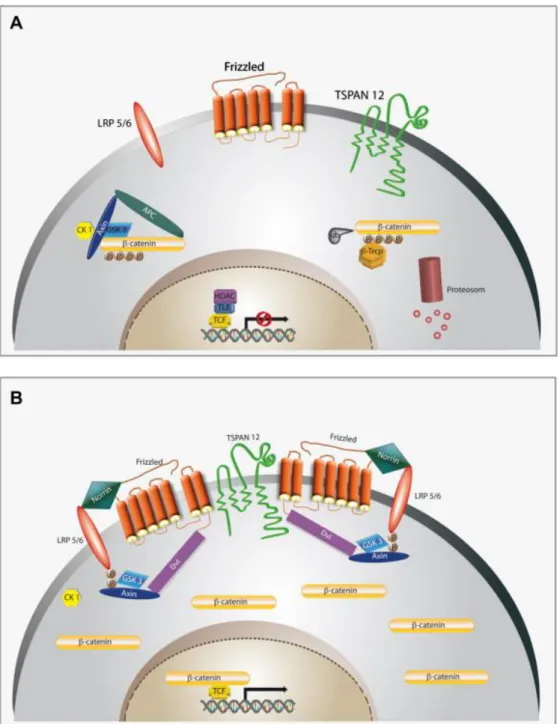
1.2.4 Norrin signaling and the Wnt/β-catenin pathway
In the absence of a Wnt (Wingless-related integration site) ligand (inactive Wnt pathway, see Figure 12, left hand side), β-catenin forms a complex composed of APC (Adenomatous polyposis coli), GSK- 3β (Glycogen synthase kinase-3β), CK1 (Casein kinase 1, alpha 1) and Axin in the cytosol (81,82). In this situation β-catenin is phosphorylated, ubiquitinylated through the E3-ubiquitin-ligase β-Trcp (β- transducin repeat containing) and degradated by the proteasome (81). In summary, an inactive Wnt pathway results in a constant degradation of the intracellular β-catenin levels.
Figure 12: Schematic of the canonical Wnt pathway.
Left: in the absence of Wnt ligand, a complex of Axin, APC, GSK3-β, CK1 and β-catenin is located in the cytosol.
β-catenin is phosphorylated by CK1 and GSK3-β and degraded by the proteosomal machinery mediated by β-Trcp.
Right: Wnt signaling through the Fz receptor and LRP5/6 co-receptor complex induces the phosphorylation of LRP6 and this leads to a translocation of the protein complex containing Axin from the cytosol to the plasma membrane. Dsh is also recruited to the membrane and binds to Fz. This results in a stabilization of β-catenin, which subsequently translocates into the nucleus. There it complexes with Lef/Tcf family members to mediate the transcription of target genes. APC= Adenomatous polyposis coli). GSK-3β= Glycogen synthase kinase-3β.
CK1= Casein kinase 1, alpha 1. β-Trcp= β-transducin repeat containing. Dsh= dishelved. Figure taken from (83).
Wnt pathway activation (see Figure 12, right hand side) leads to a phosphorylation of Lrp5/6, which in turn results in a translocation of the some proteins from the β-catenin complex from the cytosol to the plasma membrane. Axin binds to phosphorylated Lrp5/6 and Dsh is recruited to binds to the Fz receptor (81). This complex formed of Fz/Lrp5/6 induces the stabilization of β-catenin in the cytosol either through sequestration and/or degradation of Axin. β-catenin translocates into the nucleous
Introduction
17
where it forms a complex with Lef/Tcf family members resulting in the transcription of Wnt- signaling target genes (81,82).
Despite the canonical Wnt/β-catenin pathway, which results in a β-catenin accumulation in the cell, there are two other possible Wnt-pathway activations that do not result in an intracellular β-catenin accumulation: the non-canonical planar cell polarity pathway and the non-canonical Wnt/Ca2+
pathway (83).
Norrin shows specific and strong affinity binding to the frizzled 4 receptor (Fz4) (84). In consequence Norrin and fz4 mutations in both, human and mice (see 1.2.3.1 and 1.2.3.2) result in very similar morphological phenotypes (60,84,85). After Norrin binds to the Fz4 receptor, the canonical Wnt/- catenin pathway is activated as shown in Figure 13, B (84).
Dickkopf-1 (DKK1) binds with high affinity to Lrp5 and 6 and is used in the current thesis to inhibit the Norrin mediated Wnt/β-catenin pathway activation (86,87).
A recent study showed that the transmembrane protein Tspan12 (Tetraspanin 12) is able to enhance specific the activity of Norrin/β-catenin signaling and not Wnt/ β-catenin signaling (88). Tspan 12 is expressed in the retinal vasculature, but not in other neuronal cell types. The authors discuss a Tspan 12 mediated enhanced clustering of Fz4 receptor complexes (receptor multimerization) thus also enhancing Norrin/β-catenin activity. Consistently, the phenotype of Tspan 12 knockout mice is comparable to those of the Ndpy/- (see 1.2.3.1), Fz4-/- and Lrp5-/- knockout (88).
Norrin is in addition able to bind to the Lgr4 receptor (Leucine-rich repeat containing G protein-coupled receptor 4) and stimulates the canonical Wnt signaling though Lgr4 binding and its activation (89). However, binding studies using AP (alkaline phosphatase) coupled Norrin showed a higher Norrin binding affinity to Fz4 than to Lgr4 (89). In a study using xenopus as a model, Norrin interacted with the TGFβ (transforming growth factor β) signaling ligands BMP4 (bone morphogenic protein 4) and Nodal1 resulting in an inhibition of the TGFβ signaling pathway (90). Similar results were obtained in mammal cell culture assays, where Norrin acted as an antagonist of BMP2 and 4, resulting in a BMP signaling pathway inhibition (affecting intracellular Smad1/5/8) (89). Taken together, Norrin is considered as a multifunctional ligand to three receptor/binding proteins that are of major importance for Wnt and BMP signaling pathways.
Introduction
18
Figure 13: Schematic of Norrin binding to frizzled 4/LRP5/6 receptors
A. Inactive: β-catenin is part of a complex composed of APC (Adenomatous polyposis coli), GSK-3β (Glycogen synthase kinase-3β), CK1 (Casein kinase 1, alpha 1) and Axin in the cytosol. β-catenin is phosphorylated, ubiquitinylated processed to proteasomal degradation.
B. Active: Wnt pathway activation leads to a phosphorylation of Lrp5/6, which allows a translocation of Axin to the phosphorylated Lrp5/6. Dsh is recruited to bind to the Fz receptor. β-catenin is stabilized in the cytosol and translocates in the nucleous. There β-catenin and Lef/Tcf family members induce the transcription of Wnt- signaling target genes.
APC= Adenomatous polyposis coli). GSK-3β= Glycogen synthase kinase-3β. CK1= Casein kinase 1, alpha 1. β- Trcp= β-transducin repeat containing. Dsh= dishelved. Figure taken from (66)
Introduction
19
1.3 Animal models to induce cell specific stress
1.3.1 The retinopathia of prematurity (ROP) model
The disease retinopathia of prematurity (ROP) in humans is characterized through 2 phases: a time period of vaso-obliteration followed by retinal neovascularization (91,92). The ROP animal model reflects the pathogenesis of the human disease. This animal model is used to investigate vascular conditions in which vascular development becomes unbalanced (93). In the ROP model, mice are exposed to high levels of oxygen (75%) for a designated duration (detailed methodical explanation:
see 5.1.6.1). The high levels of oxygen induce a vaso-obliteration and growth arrest of the developing retinal vessels (93). Relocalization of the experimental animals back to normal room air leads to a relative hypoxia in the tissue (due to the former growth arrest) and this results in uncontrolled vessel growths (neovascularizations: due to the expression of vascular endothelial growth factor (VEGF) and insulin growth factor (IGF)) (91,93). In the current thesis, we used this model to investigate the influence of Norrin on vessel growth and retinal vascularization.
1.3.2 A damage model for photoreceptor degeneration: light damage
Exposure of rat or mouse eyes to bright, visible light leads to death of their rod photoreceptors (94).
This vulnerability to light has been used to establish the experimental model of light-induced damage. Photoreceptors exposed to excessive amounts of light die through apoptosis, a scenario that is quite similar to their apoptotic death seen in inherited retinal dystrophies or in age-related macular degeneration (AMD) (95,96). Animal models for inherited retinal dystrophies or AMD are available and commonly show photoreceptor apoptosis over a relative long period of time, with only a few neurons entering apoptotic cell death each day. In contrast, photoreceptor death induced by excessive light leads to a large number of photoreceptors dying at the same time. Accordingly, studies on the molecular mechanisms of photoreceptor apoptosis and/or on neuroprotective signaling pathways preventing apoptotic death are considerably easier. Light damage can be induced in an acute or chronic way, by exposing animals to light for a short or longer period of time. In addition, different qualities of light can be used, such as fluorescent white light with an emission spectrum similar to that of daylight, or energy-rich blue light (94,97,98). In the current thesis, light exposure was used to investigate whether Norrin has neuroprotective effects on photoreceptor cells and which molecular mechanism may mediate this effect.
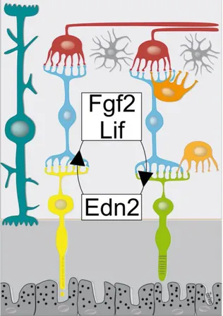
1.3.3 Neuroprotective signaling pathways in the context of photoreceptor degeneration In the context of light damage and neuroprotection several neuroprotective factors are discussed.
Microarray analysis revealed a relatively small number of genes activated after light exposure (99).
Among those transcripts endothelin 2 (Edn2) was highly upregulated (99). Its localization in the ONL was determined using in situ hybridization (99). In addition, the endothelin receptor type B (Ednrb)
Introduction
20
was upregulated in Müller glia cells indicating that photoreceptor derived Edn2 signals to Müller glia cells to probably increase their sensitivity to Edn2 as part of the retinal tissue response to the light damage (99).
It was also shown that photoreceptor cell death in VPP mice (a model for inhereditary photoreceptor degeneration) strongly induced the expression of leukemia inhibitory factor (LIF) in Muller glia cells (100). Lif-/- animals revealed accelerated photoreceptor degeneration in the VPP mouse model without activating the Müller glia cells (100). Similar to the situation in light damaged retinae, Edn2 was significantly upregulated in VPP retinae as well (99,100). Intravitreal injections of recombinant LIF in turn induced molecular factors like Edn2, signal transducer and activator of transcription (Stat3), fibroblast growth factor 2 (Fgf2) and glial fibrillary acidic protein (Gfap) in wild-type, VPP and interesstingly in Lif–/- mice, too. These findings strongly indicate that the upregulation of those factors are mediated through LIF. The fact that Edn2 levels in Lif-/- animals were reduced to the level of 2%
compared to wild-types and that VPP;Lif-/- double mutants did not reveal an upregulation of Edn2 (although VPP animals did) further indicates that Edn2 expression is dependent on LIF.
Figure 14: The response of photoreceptors to injury: EDN2, LIF and FGF2
Light or genetically induced photoreceptor cell stress results in the upregulation of LIF. Strikingly, Lif induction leads to an upregulation of EDN2 in photoreceptor cells and FGF2 in Müller glia cell. Since EDN2 itself further stimulates the expression of LIF, a positive feed forward loop is very likely.
Introduction
21
FGF2 is upregulated in different models of photoreceptor degeneration and it is speculated that endogenous FGF2 may function in a parakrine way, secreted from the Müller glia cells, as a protective factor activated in response to photoreceptor cell stress (101,102). When Fgf2 and Müller glia cell activation marker Gfap were studied in Lif-/- and VPP;Lif-/- mice respectively, again, the Fgf2 and Gfap levels in Lif-/- animals were reduced to a level of 38% (Fgf2) and 11% (Gfap), respectively.
VPP;Lif-/- double mutants did not reveal an upregulation of Fgf2 or Gfap (although VPP animals did) (100). This further indicates that the expression of FGF2 and GFAP is dependent on LIF as well.
Taken together LIF/EDN2/FGF2 signaling pathways (see Figure 14) seem to play an essential and early role in the retinal response to photoreceptor injury.
Introduction
22
1.4 Aim of the current work
The main focus of this thesis is to investigate the neuroprotective role of Norrin on photoreceptor survival and maintenance. To this end, we will use a transgenic mouse line that overexpresses Norrin specifically in the retinal pigment epithelium (RPE).
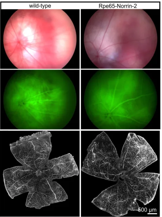
First, we will carefully characterize the overexpression of Norrin in the transgenic animals using various molecular biology techniques. Then we will analyze the effect of Norrin on photoreceptor survival in untreated animals. To this end, we will characterize the morphology of the animals at different timepoints, followed by morphometric analysis. For morphometry, we will measure the thickness of the photoreceptor cell layer (outer nuclear layer, ONL) at 20 measure points throughout the entire retina and blot these measure points in a so called spider diagram. We will evaluate the different measure points in the spider diagram using Student's t-test. This will give us a good indication, if Norrin has proliferative effects on the outer nuclear layer (ONL). Since Norrin has strong angiogenic properties, the retinal vasculature will be examined by dextran perfusion, in vivo fluorescence angiography and immunohistochemical staining (Isolectin B4, plasmalema associated protein (PLVAP)).
To investigate a possible neuroprotective function of Norrin for photoreceptors, we will consequently establish a light induced damage model for photoreceptor apoptosis. Using the established model, we will treat transgenic animals and littermate controls. TUNEL labeling for apoptotic cells will show, if the transgenic overexpression of Norrin might have a protective effect on photoreceptor survival (less apoptosis). These results will be confirmed with an ELISA, which detects free nucleosomes as a sign for apoptosis. Additionally, we will focus on the morphology and use Spider diagrams and Student's t-test to compare the thickness of the ONL of light exposed animals.
To learn more about the underlying molecular mechanisms, transcript levels of different neuroprotective factors (leukaemia inhibitory factor (Lif), fibroblast growth factor 2 (Fgf-2), endothelin-2 (Edn-2), brain derived neurotrophic factor (Bdnf), lens epithelium derived growth factor (Ledgf), ciliary neurotrophic factor (Cntf), pigment epithelium derived growth factor (Pedf)) and as a marker for Müller glia cell activation: glial fibrillary acidic protein (GFAP) will be determined before and after light exposure. Western blot analysis and immunohistochemical staining will be performed to confirm realtime-PCR data on the protein level. In vivo examinations like, e.g. ERG-, OCT- and SLO- analysis will be performed in cooperation with Prof. Seeliger (Tübingen) to complete the dataset with a functional aspect.
Finally we will investigate in detail the possible mechanism by which Norrin mediates its neuroprotective effects. Norrin can activate the canonical Wnt/β-catenin pathway (103). Hence, Western blot analysis for β-catenin will be performed. In addition, crossbreeding of transgenic Norrin
Introduction
23
animals with TopGal mice (Wnt signaling reporter mice, see 5.1.1.3) will reveal if the Wnt pathway is more active in the transgenic animals. Wnt signaling is neuroprotective (80,104,105). Consequently, if transgenic animals show neuroprotective effects following light damage, we will investigate, if this effect might be mediated through the classical Wnt/β-catenin pathway. To this end, we will inhibit Wnt signaling using vitreal Dkk-1 injections. We will inject Dkk-1 into the vitreous of the left eye; the right eye will be injected with PBS only, followed by light damage experiments. Spider diagrams of PBS and Dkk-1 injected transgenic and wild-type control animals will be constructed and statistical analysis will be performed. The expression levels of possible target genes of Norrin and Wnt/β-catenin signaling will be investigated after injection of Dkk-1 in untreated and light exposed animals. Possible downstream signaling pathways of the target genes will be analyzed using Western Blot analysis and immunohistochemical staining.
Introduction
24
Results
25
2 Results
2.1 Characterization of Rpe65-Norrin-2 mice
2.1.1 Southern Blot: successful genomic insertion of the transgene construct
To analyze if the transgene construct was successfully integrated into the genome of Rpe65-Norrin mice, southern blot analysis was used. To this end, genomic DNA was cut with the restriction enzyme EcoRV, transferred on a agarose gel on which the DNA fragment were separated by their size.
Subsequently, the DNA was blotted onto a positively charged nitrocellulose membrane. After membrane hybridization with the digoxygenin (DIG) labeled RNA-probe, a RNA-DNA hybrid was formed, which was detected using the DIG easy system with alkaline phosphatase as chemiluminescence.
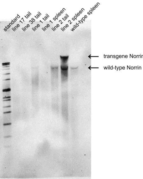
Figure 15: Southern blot analysis of the different Rpe65-Norrin founder mouse lines
Southern blot analysis showed a distinct signal for wild-type Norrin DNA. Line 25 showed a second band positive for the Norrin probe indicating a successful insertion of the transgene construct into the genomic DNA.
Results
26
Rpe65-Norrin-line 2 animals showed a distinct second signal in addition to the wild-type Norrin signal, which was detectable in control animals (wild-type) and Rpe65-Norrin-line 1 animals (second transgene mouseline) (Figure 15). Mentioning the fact that the lanes are obviously not equally loaded, one might have the impression of a very weak additional second signal above the wild-type signal in the Rpe65-Norrin-line 1 tail DNA (Figure 15, lane 4).
2.1.2 Northern blot analysis: effective transcription of the construct
Northern blot analysis was used to further verify if the successfully integrated Norrin transgene construct is being transcribed in the eye. To this end carefully isolated RNA samples from the posterior bulb (sample containing the retina, RPE, choroid and sclera) were pretreated as described in 5.1.3.5, loaded on an agarose gel and separated by size. The RNA in the gel was transferred on a positively charged nitrocellulose membrane, hybridized with the same probe as used for Southern blot analysis and the RNA-RNA Hybrid was detected using DIG easy system with alkaline phosphatase as chemiluminescent reagent. Rpe65-Norrin-2 animals showed a signal on the same height as the positive control (βB1-Norrin). The signal in Rpe65-Norrin-2 retinae appeared weaker compared to controls, a phenomenon which is not surprising at all, because it is well known, that the promoter (βB1-crystallin) in these positive controls is far more effective than the RPE65 promoter used in our mouse line in this project (Figure 16).
Figure 16: Northern Blot analysis of Rpe65-Norrin-2
Rpe65-Norrin-2 animals show a distinct signal at the appropriate size. On the right: coomassie blue staining of the membrane to show equally loaded lanes, especially for the wild-type, since there was no signal visibly in the northern blot. 28 S and 18 S indicate the large and small ribosomal RNAs. Experiment published in (106), performed by Barbara Braunger.
Results
27
2.1.3 Western blot analysis: effective protein translation
To further confirm, whether the RNA was successfully translated into Norrin protein, which in the end would stand for a successful overexpression of Norrin protein in the retina, Western blot analysis was performed. After statistical evaluation of the Western blot densitometry, Rpe65-Norrin-2 animals had indeed a significant, about 2-fold increase of Norrin protein in the retina (Figure 17).
Figure 17: Western blot for Norrin in Rpe65-Norrin-2 mice
Left: Western blot analysis for Norrin protein in retinal lysates of Rpe65-Norrin-2 mice compared to wild-type littermates.
Right: statistical evaluation (students t-test) of the densitometric analysis.
Rpe65-Norrin-2 animals showed a significant increase of Norrin in the retina compared to their wild-type littermates. Mean and SEM are shown; n= 8; *p<0.05. Experiment published in (107), performed by Barbara Braunger.
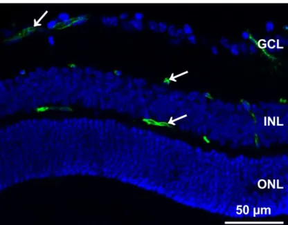
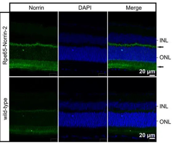
2.1.4 Immunohistochemistry: retinal localization of the transgenic Norrin protein To investigate in which region or cellular layer of the retina the significant overexpressed Norrin was localized, immunohistochemical staining was performed (Figure 18). In adult control animals, specific immunostaining for Norrin was weak and barely detectable in the outer retina. In contrast Rpe65-Norrin-2 animals showed an intense staining for Norrin in the inner and outer segments of the photoreceptors, the ONL and the outer plexiform layer. This strongly indicated that Norrin was secreted from the retinal pigment epithelium into the retina.
Results
28
Figure 18: Immunohistochemical staining for Norrin in the retina
Rpe65-Norrin-2 showed an intense staining for Norrin (green) in the outer and inner segments of the photoreceptors, the ONL and the outer plexiform layer (arrows). Cell nuclei were stained with DAPI (blue). INL=
inner nuclear layer; ONL= outer nuclear layer. Experiment published in (107), performed by Barbara Braunger.
2.1.5 Rpe65-Norrin-2 mice and Wnt/β-catenin activation
Norrin activates the canonical Wnt/β-catenin pathway by binding to the Frizzled-4 receptor (see Figure 13) (60,85,103). To investigate whether the Wnt/β-catenin pathway was activated in the transgenic Rpe65-Norrin-2 animals, western blot analysis of β-catenin levels was performed.
Densitometric and statistic evaluation showed that Rpe65-Norrin-2 animals had a significant increase in β-catenin protein in the retina compared to control wild-type littermates (Figure 19).
Figure 19: Western blot analysis for β-catenin in Rpe65-Norrin-2 animals
Rpe65-Norrin-2 animals had a significant increase for β-catenin protein in the retina. Mean and SEM are shown; n≥7; *p>0.05. Experiment published in (107), performed by Barbara Braunger.
Results
29
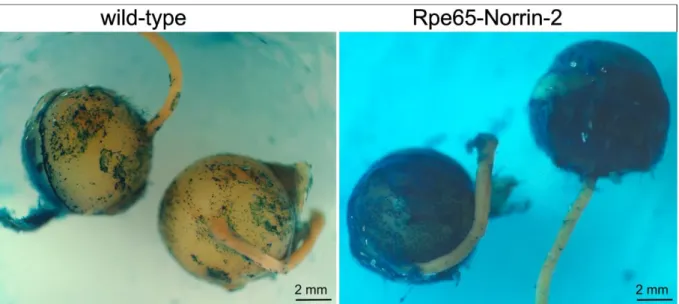
To investigate if the increase in retinal β-catenin in the retina correlated with an activation of the canonical Wnt/β-catenin signaling pathway, Wnt reporter mice (TOPGAL) were crossed to the Rpe65-Norrin-2 line and β-galaktosidase staining was performed.
Figure 20: Wnt pathway reporter mice (TOPGAL): macroscopic picture of β-galactosidase stained eye.
Macroscopic pictures of the eyes of Lac-Z stained TOP-Gal;wild-type and TOP-Gal; Rpe65-Norrin-2 animals. The sample of Rpe65-Norrin-2 mice showed a strong β-galactosidase reactivity compared to wild-types.
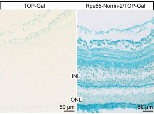
In control animals only a weak positive signal was detected in the innermost layers of the retina, in contrast double transgenic Rpe65-Norrin-2 X TOP-Gal animals showed an intense β-galaktosidase signal (Figure 20) throughout the retina (Figure 21), which indicates an active Wnt/β-catenin pathway. Taken together, these experiments strongly pointed out that the canonical Wnt/β-catenin pathway was activated in the retina of transgene Rpe65-Norrin-2 animals.
Results
30
Figure 21: Wnt pathway reporter mice (TOPGAL): β-galactosidase staining
Rpe65-Norrin animals showed an intense β-galactosidase staining, which indicates an active Wnt/β-catenin pathway. INL= inner nuclear layer; ONL= outer nuclear layer. Experiment published in (107), performed by Barbara Braunger.
Results
31
2.2 Phenotype analysis of Rpe65-Norrin-2 animals
2.2.1 Retinal morphology
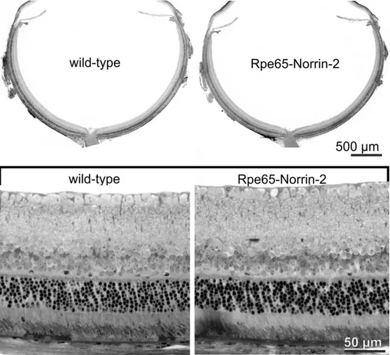
Conventional light microscopy was used to analyze the morphology of Rpe65-Norrin-2 animals and wild-type littermates at different time points by using Richardson stained semithin sections. Although we observed a strong overexpression of Norrin from the retinal pigment epithelium into the retina, the phenotype of the transgenic animals showed no morphological differences. The sizes of wild-type and transgenic eyes were similar, the anterior segment of the eye was regular formed, and the retinae showed a normal architecture with all layers present from the optic nerve to the periphery.
The retinal pigment epithelium (RPE) looked normal with no visible alterations (Figure 22).
Figure 22: Semithin sections of adult Rpe65-Norrin-2 and control wild-type littermates eyes
Semithin (1 μm thick) horizontal sections of eyes of a 6 week-old Rpe65-Norrin-2 mouse and its wild-type littermate. The eyes were of similar sizes and the architecture of the retina was normal with all layers present from the optic nerve to the periphery. Lower panel shows higher magnification of the central retina Rpe65-Norrin-2 animals. There was no obvious morphological change compared to control wild-type littermates. Experiment published in (107), performed by Barbara Braunger.
Because the thickness of the outer nuclear layer is the major read out data point for morphometric analysis of light damage experiments, prior to the actual light damage experiments the thickness of the outer nuclear layer throughout the entire retina was measured. The ONL thickness did not differ