of microglial reactivity in the retina
Inaugural Dissertation zur
Erlangung des Doktorgrades Dr. nat. med.
der Medizinischen Fakultät und
der Mathematisch-Naturwissenschaftlichen Fakultät der Universität zu Köln
vorgelegt von Alexander Aslanidis
aus Regensburg
Köln, 2016
Berichterstatter/Berichterstatterin: Prof. Dr. Sabine Eming Prof. Dr. Peter Kloppenburg
Tag der letzten mündlichen Prüfung: 16. November 2015
For my beloved parents
1
I Table of contents
I Table of contents... 1
II Summary ... 3
III Zusammenfassung ... 5
1. Introduction ... 7
1.1 Structure and function of the mammalian retina ... 7
1.2 Retinal genetics ... 9
1.2.1 Inherited retinal degenerations ... 9
1.2.2 Regulation of photoreceptor development ... 10
1.2.3 Genome-wide identification of CRX target genes ... 12
1.2.4 Sterile alpha motif containing proteins and SAMD7 ... 13
1.3 Microglia ... 14
1.3.1 Microglia in the CNS and retina ... 14
1.3.2 Functions of microglia ... 14
1.3.3 Microglia in retinal degeneration ... 16
1.3.4 Microglial (reactivity) markers ... 19
1.3.5 Microglia as a therapeutic target ... 21
1.3.5.1 Natural compounds ... 22
1.3.5.2 Whey acidic proteins (WAPs) and AMWAP ... 23
1.3.5.3 Translocator protein (18 kDa) (TSPO) and its ligands . 24 1.4 Aims of the thesis ... 26
2. Results ... 28
2.1 Sterile alpha motif containing 7 (SAMD7) is a novel CRX-regulated transcriptional repressor in the retina ... 28
2.2 Activated microglia/macrophage whey acidic protein (AMWAP) inhibits NFκB signaling and induces a neuroprotective phenotype in microglia . 30 2.3 Translocator protein (18 kDa) (TSPO) is expressed in reactive retinal microglia and modulates microglial inflammation and phagocytosis ... 32
3. Discussion ... 35
3.1 Results from the three presented studies in summary ... 35
3.2 SAMD7, a novel photoreceptor gene repressor ... 35
3.2.1 Photoreceptor-specific SAMD7 gene regulation ... 36
3.2.2 SAMD7, a gene regulator without DNA binding sequence... 36
3.2.3 Potential role of SAMD7 in retinal health and disease ... 37
3.3 AMWAP, a counter-regulator of microglial reactivity ... 38
3.3.1 Obtaining recombinant AMWAP for therapy ... 38
2
3.3.2 AMWAP, an inhibitor of microglial NFκB signaling... 38
3.3.3 AMWAP induces a neuroprotective microglial phenotype ... 39
3.3.4 AMWAP, current state and future perspectives ... 40
3.4 TSPO, emerging roles in the CNS and retina ... 41
3.4.1 TSPO, a novel marker for reactive retinal microglia ... 41
3.4.2 Functions of TSPO and its ligands in reactive microglia ... 42
3.4.3 TSPO, current state and future perspectives ... 44
3.5 Concluding remarks ... 45
Reference list ... 46
Acknowledgements ... 61
Erklärung ... 62
Publications ... 63
3
II Summary
Photoreceptors are highly specialized cells required for phototransduction within the retina and thus crucial for visual perception. Inherited retinal diseases are mainly caused by mutations in photoreceptor-specific genes, the majority of which is regulated by the key transcription factor cone rod homeobox (CRX). Using genome- wide chromatin immunoprecipitation data (CRX ChIP-seq), we have identified a novel sterile alpha motif (SAM) domain containing protein, SAMD7, as a CRX target.
SAMD7 is expressed in the mouse retina and pineal gland and localizes to the cytoplasm and nucleus of photoreceptor cells. SAMD7 expression is regulated by CRX, which binds to two specific regions in the promoter and first intron (enhancer) of the SAMD7 gene. Consequently, CRX knock-down leads to a significant decrease of SAMD7 enhancer activity and protein levels in the retina. Functionally, SAMD7 acts as a transcriptional repressor of CRX-mediated photoreceptor gene expression, indicating that mutations in or dysregulation of SAMD7 could lead to disturbed photoreceptor homeostasis and ultimately retinal degeneration.
Microglial cells are the resident macrophages of the central nervous system (CNS), including the retina, and play pivotal roles in innate immune responses and regulation of homeostasis in the healthy and degenerating CNS. Reactive microgliosis is a common hallmark of neurodegenerative diseases and chronic pro-inflammatory microglial reactivity contributes to disease progression. We have previously identified activated microglia/macrophage whey acidic protein (AMWAP) as a biomarker for microglial reactivity and counter-regulator of pro-inflammatory response. AMWAP is actively secreted from lipopolysaccharide (LPS)-activated microglia and recombinant AMWAP is taken up by microglial cells in a paracrine fashion and effectively reduces TLR2- and TLR4-mediated pro-inflammatory gene expression. AMWAP exerts its anti-inflammatory function through blockade of NFκB activation, as it inhibits proteolysis of the NFκB pathway mediators IRAK-1 and IκBα without preventing IκBα phosphorylation and ubiquitination or affecting overall 20S proteasome activity.
Functionally, AMWAP reduces pro-inflammatory microglial nitric oxide (NO) secretion and neurotoxicity on photoreceptor cells in vitro. Further, AMWAP promotes filopodia formation of microglia and increases the phagocytic recognition and uptake of apoptotic photoreceptor debris, common features of homeostatic regulatory microglia. We therefore hypothesize that anti-inflammatory whey acidic proteins
4 (WAPs) could have therapeutic potential in neurodegenerative diseases of the brain and retina.
The translocator protein (18 kDa) (TSPO) is a mitochondrial protein expressed in reactive glial cells and a biomarker for gliosis in the brain but has not been investigated in a retinal context so far. Various TSPO ligands have been shown to reduce neuroinflammation in neurodegenerative mouse models. We could show strong upregulation of TSPO transcript and protein levels in reactive microglial cells in vitro, microglia of the retinoschisin-deficient retinal degeneration mouse model as well as TSPO expression in microglia of the human retina. TSPO mRNA expression is high in the developing mouse retina and declines to low levels in the adult tissue.
The synthetic TSPO ligand XBD173 effectively suppresses pro-inflammatory microglial gene expression, migration, proliferation, NO secretion and neurotoxicity on photoreceptors in vitro. Further, XBD173 treatment promotes filopodia formation and increases the phagocytic recognition and uptake of latex beads and apoptotic photoreceptor debris by murine and human microglial cells in vitro. Finally, XBD173 effectively reduces the number of amoeboid alerted microglia in organotypic murine retinal explant cultures stimulated with LPS. In conclusion, we have identified TSPO as a novel marker for microglial reactivity in the retina and a potential therapeutic target to reduce chronic neuroinflammation during retinal degeneration.
In summary, our studies on the novel photoreceptor gene regulator SAMD7 and the microglial reactivity markers AMWAP and TSPO provide insights into potential disease mechanisms of retinal degeneration and suggest future strategies of identifying and therapeutically modulating pro-inflammatory microglial reactivity in degenerative diseases of the CNS and retina.
5
III Zusammenfassung
Photorezeptorzellen sind hoch spezialisierte Zellen innerhalb der Netzhaut, welche die Phototransduktion vermitteln und daher essentiell für die Aufnahme und Verarbeitung visueller Sinnesreize sind. Erbliche Netzhautdystrophien werden vornehmlich durch Mutationen in Photorezeptor-spezifischen Genen verursacht, von welchen die Mehrzahl durch den Schlüsseltranskriptionsfaktor cone rod homeobox (CRX) reguliert wird. Mit Hilfe eines genomweiten Chromatin Immunpräzipitations- Datensatzes (CRX ChIP-seq), konnten wir ein neuartiges, sterile alpha motif (SAM) Domänen beinhaltendes Protein, SAMD7, als CRX Zielgen identifizieren. SAMD7 wird in der Mausnetzhaut und Zirbeldrüse exprimiert und lokalisiert im Zytoplasma und Nukleus von Photorezeptorzellen. Die SAMD7 Expression wird durch CRX reguliert, welches an zwei spezifischen Regionen im Promotor und ersten Intron (Enhancer) im SAMD7 Gen bindet. Somit führt CRX knock-down zu einer signifikanten Abnahme der regulatorischen Aktivität des SAMD7 Enhancers und der SAMD7 Proteinlevel in der Netzhaut. Funktionell agiert SAMD7 als Transkriptionsrepressor der CRX-vermittelten Photorezeptor-Genexpression, was darauf hindeutet, dass Mutationen in oder Dysregulation von SAMD7 zu gestörter Photorezeptor-Homöostase und letztlich zu Netzhautdegeneration führen könnten.
Mikrogliazellen sind die gewebsansässigen Makrophagen des zentralen Nerven- systems (ZNS), inklusive der Netzhaut, und spielen eine wichtige Rolle in der Immunantwort und Regulation der Homöostase im gesunden sowie degenerierenden ZNS. Reaktive Mikrogliose ist ein typisches Merkmal neurodegenerativer Erkrankungen und chronische proinflammatorische Mikrogliaaktivierung beschleunigt den Krankheitsverlauf. Wir konnten in Vorarbeiten das activated microglia/
macrophage whey acidic protein (AMWAP) als Biomarker für reaktive Mikroglia und Gegenspieler der proinflammatorischen Mikrogliaantwort identifizieren. AMWAP wird aktiv von Lipopolysaccharid (LPS)-aktivierten Mikroglia sezerniert und rekombinantes AMWAP wird in parakriner Weise von Mikroglia aufgenommen in welchen es effektiv die TLR2- und TLR4-vermittelte proinflammatorische Genexpression reduziert.
AMWAP erzielt diese antiinflammatorische Wirkung durch Blockade der NFκB Aktivierung, indem es die Proteolyse der NFκB Signalwegsmediatoren IRAK-1 und IκBα verhindert ohne die IκBα Phosphorylierung, Ubiquitinierung oder die allgemeine 20S Proteasomaktivität zu beeinflussen. Funktionell reduziert AMWAP die proinflam-
6 matorische mikrogliale Stickstoffoxidsekretion und Neurotoxizität gegenüber Photo- rezeptorzellen in vitro. Des Weiteren induziert AMWAP die Bildung mikroglialer Filopodien und erhöht die phagozytische Aufnahmefähigkeit von apoptotischem Photorezeptordebris, typische Charakteristika homöostatischer und regulatorischer Mikroglia. Wir schließen aus diesen Ergebnissen, dass antiinflammatorische whey acidic proteins (WAPs) therapeutisches Potential in neurodegenerativen Erkran- kungen des Gehirns und der Netzhaut haben könnten.
Das Translokatorprotein (18 kDa) (TSPO) ist ein mitochondriales Protein, welches in reaktiven Gliazellen exprimiert ist, als Gliosemarker im Gehirn fungiert und bisher noch nicht im retinalen Kontext untersucht wurde. Verschiedene TSPO Liganden reduzieren Neuroinflammation in neurodegenerativen Mausmodellen. Wir konnten eine starke Hochregulation der TSPO Transkript- und Proteinlevels in reaktiven Mikrogliazellen in vitro, Mikroglia des Retinoschisin-defizienten Mausmodells sowie TSPO Expression in Mikroglia der humanen Netzhaut nachweisen. TSPO ist auf mRNA Ebene stark in der sich entwickelnden Mausnetzhaut exprimiert und sinkt auf ein niedrigeres Niveau im adulten Gewebe. Der synthetische TSPO Ligand XBD173 verringert effektiv die proinflammatorische mikrogliale Genexpression, Migration, Proliferation, Stickstoffoxid-Sekretion und Neurotoxizität gegenüber Photorezeptor- zellen in vitro. Außerdem induziert XBD173 die Bildung mikroglialer Filopodien und erhöht die phagozytische Aufnahmefähigkeit von Latexkügelchen und apoptotischem Photorezeptordebris durch murine und humane Mikrogliazellen in vitro. Weiterhin reduziert XBD173-Behandlung effektiv die Zahl amöboider, alarmierter Mikroglia- zellen in LPS-stimulierten organtypischen Netzhautexplantatkulturen. Schluss- folgernd konnten wir TSPO als neuartigen Marker der Mikrogliaaktivierung in der Netzhaut und potentielle therapeutische Zielstruktur zur Reduktion chronischer Neuroinflammation während Netzhautdegeneration identifizieren.
Zusammenfassend ermöglichen unsere Untersuchungen zum neuartigen Photorezeptor-Genregulator SAMD7 und den mikroglialen Reaktivitätsmarkern AMWAP und TSPO einen Einblick in mögliche Pathomechanismen retinaler Degeneration und offenbaren Möglichkeiten zur Identifizierung und therapeutischen Modulation pro-inflammatorischer Mikrogliaaktivität in degenerativen Erkrankungen des ZNS und der Netzhaut.
7
1. Introduction
1.1 Structure and function of the mammalian retina
Visual perception represents one of our most important senses and enables us to experience our environment in a remarkable fashion and detail. The complex mechanisms underlying the transformation of light into visual stimuli have fascinated humans for centuries. Electromagnetic rays between 400-750 nm wavelengths are recognizable to humans as visible light, and the tissue crucial for this process is the retina with its different cellular layers. In opposition to the peripheral sensory organs, the retina is a direct component of the central nervous system (CNS), as it developmentally represents an evagination of the diencephalon.
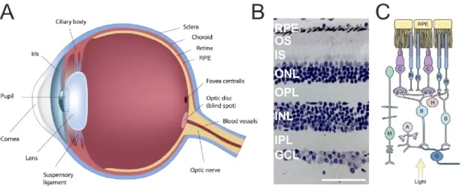
Figure 1: Gross anatomy of the eyeball and detailed cross-section of the retina and its different cell types.
(A) The retina lines the posterior segment of the eyeball and lies adjacent on top of the retinal pigment epithelium (RPE). (B) Histological cross-section of the retina depicting its different cell layers. GCL, ganglion cell layer; IPL, inner plexiform layer; INL, inner nuclear layer; OPL, outer plexiform layer; ONL, outer nuclear layer; IS, photoreceptor inner segment; OS, photoreceptor outer segment; RPE, retinal pigment epithelium. (C) Schematic overview of the different cell types within the retinal layers. G, ganglion cell; A, amacrine cell; B, bipolar cell; H, horizontal cell; M, Mueller cell; R, rod photoreceptor; C, cone photoreceptor; RPE, retinal pigment epithelium.
(Eyeball modified from http://www.garetina.com/about-the-eye, retinal cross section modified from http://www.oculist.net/downaton502/prof/ebook/duanes/pages/v8/v8c013.html, schematic section adapted from Sung and Chuang, 2010).
Within the eye, the retina lies directly adjacent on top of the retinal pigment epithelium (RPE) (Fig. 1 A), which has important housekeeping functions like vitamin A metabolism, heat regulation and constant shedding of photoreceptor outer segments containing the visual pigment (opsins) through phagocytosis. Further, the
8 RPE functions as outer blood retinal barrier, ensuring that only molecules below a certain size translocate from the choroidea into the neuroretina. The choroidea serves as a key structure for supporting blood vessels, and the sclera, as the outermost layer of the eye, provides stability and is attached to the ocular muscles. In spite of being only ~200 µm thick, the retina consists of three distinct cellular layers, harboring many different cell classes (Fig. 1 B, C) (Klinke et al., 2003). The innermost layer consists of ganglion cells (GCs), whose axons converge to form the optic nerve and transmit visual signals to the brain. Adjacent lays the inner nuclear layer (INL), consisting of amacrine, bipolar and horizontal cells, preprocessing visual signals and increasing contrast. After passing through these layers, light reaches the outer nuclear layer (ONL), which harbors the cell bodies of photoreceptors. There are two distinct types of photoreceptors: cones and rods. The human retina harbors around 130 million photoreceptors, of which 95% represent rods (Sung and Chuang, 2010).
Cones are responsible for high resolution color vision and the perception of objects in bright light conditions (photopic vision), whereas rods are responsible for perception in dim light conditions and night vision (scotopic vision) (Baylor et al., 1979, Klinke et al., 2003).
Absorption of photons by opsins in the photoreceptor outer segment (OS) leads to isomerization of 11-cis retinal to all-trans retinal and triggers the phototransduction cascade including hyperpolarization of photoreceptor cell membranes. The signal is then transmitted to the neurons of the INL and finally reaches the ganglion cells, which possess long axons that unite at the so-called “blind spot”, which is devoid of photoreceptors and where the optic nerve exits the eyeball. Roughly 3.5 mm distant lies the fovea centralis (in the center of the macula), which is comprised solely of cones and represents the area enabling sharp central vision within the retina. In the fovea, each cone cell is connected to a single bipolar and ganglion cell, enabling particularly focused vision in this area compared to the peripheral retina. Mueller cells represent one out of three glial cell moieties in the retina (Mueller glia, astroglia and microglia). They serve as support cells for retinal neurons and span throughout all retinal layers. In recent years, an additional function of Mueller cells as optic fibers facilitating the transmission of light through the retina towards the photoreceptors has been described (Reichenbach and Bringmann, 2013).
9 The retina is devoid of pain receptors, therefore neuroretinal diseases remain largely pain-free and impairment of visual perception should be monitored and examined thoroughly. One of the main causes of impaired vision in inherited retinal degenerations is the irreversible loss of photoreceptors due to retinal damage or mutations in photoreceptor-specific genes leading to apoptosis (Rattner et al., 1999).
The murine retina is to a large extent structurally similar to the human retina, there are however certain distinctions to be considered. Around 75% of the murine retina account for photoreceptors, of which 97% are rods. Mice almost exclusively rely on rod-mediated scotopic vision and, in contrast to humans (red, green and blue), are only equipped with two types of cones for either short or middle-to-long wavelengths of light (Morrow et al., 1998). Most importantly, the murine retina does not harbor a macula or fovea.
1.2 Retinal genetics
1.2.1 Inherited retinal degenerations
Since the retina is comprised of a complex network of highly specialized cells, it is highly susceptible to genetic defects leading to hereditary retinal dystrophies, a major cause of legal blindness worldwide. To date, the Retnet database (https://sph.uth.edu/retnet/ as of July 28th 2015) lists a total of 278 genes and loci linked with retinal degenerations. 64 of these genes are causative for the most frequent monogenic group of disorders, retinitis pigmentosa (RP). On the other hand, the leading cause of legal blindness in the elderly of industrialized nations today is age-related macular degeneration (AMD), a complex genetic disease. In contrast to RP, a multitude of genetic risk variants as well as environmental risk factors contribute to the development of AMD and ultimately loss of vision (Jager et al., 2008, Chen et al., 2010).
Various animal models have been generated over the last decades, mimicking hereditary retinal degenerative diseases like retinitis pigmentosa (Rivas and Vecino, 2009). X-linked juvenile retinoschisis is another retinal dystrophy affecting patients as early as in their adolescence. Retinoschisin-deficient (Rs1h-/Y) mice show an early splitting (schisis) of the inner retinal layers followed by photoreceptor degeneration
10 (Weber et al., 2002). Although a plethora of different cellular malfunctions can be the underlying cause for monogenic or complex inherited retinal degeneration, photoreceptor apoptosis represents a common hallmark of all retinal dystrophies during disease progression (Portera-Cailliau et al., 1994, Sancho-Pelluz et al., 2008).
Within the large group of causative genes, defects in retina-specific genes and master transcription factors are frequently associated with inherited retinal dystrophies (Rattner et al., 1999, Tran and Chen, 2014). Photoreceptors are highly specialized cells with a multitude of intracellular components and functions and therefore highly prone to various genetic malfunctions, ultimately leading to photoreceptor degeneration (Bramall et al., 2010). High transcript levels of a gene in photoreceptors and a dysfunction of the corresponding protein are tightly associated with retinal degeneration, indicating that knowledge about abundantly expressed photoreceptor-specific genes, transcription factors and their regulation may help to identify novel retinal disease genes (Blackshaw et al., 2001, Wagner et al., 2013).
1.2.2 Regulation of photoreceptor development
Photoreceptors have a bridging function between our environment and central nervous system as they transform light into visual signals within the retina. The development of retinal progenitor cells into differentiated, fully functional photo- receptors is orchestrated by many regulatory events like expression of transcription factors, their target genes and cis-regulatory elements that mediate their interaction (Hsiau et al., 2007).
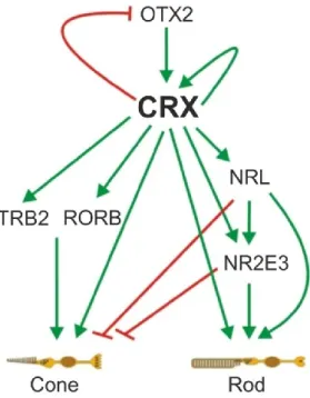
During early retinal development, expression of the transcription factor OTX2 (orthodenticle homeobox 2) in retinal progenitor cells directs them towards a photoreceptor cell fate (Nishida et al., 2003). 12.5 days into the mouse embryonal stage (E12.5) OTX2 induces expression of another photoreceptor-specific key transcription factor, CRX (cone rod homeobox), in the developing rods and cones (Furukawa et al., 1997). From then on, CRX is highly expressed in developing and mature photoreceptors indicating its important role in photoreceptor function and homeostasis. Additionally, CRX interacts with a variety of other transcription factors to influence photoreceptor development and function (Fig. 2). CRX orchestrates this network of transcription factors, which regulates whether a progenitor develops into a rod or cone photoreceptor. TRB2 (thyroid hormone receptor β2) and RORB (retinoid-
11 related orphan receptor β) are crucial factors towards a cone cell fate (Yanagi et al., 2002, Srinivas et al., 2006), whereas NRL (neural retina leucine zipper) and NR2E3 (nuclear receptor subfamily 2, group E, member 3) are important mediators of a rod cell fate and predominantly expressed in rods (Mears et al., 2001, Milam et al., 2002).
Figure 2: The transcriptional network of photoreceptor development. The key transcription factor CRX is induced by OTX2, represses OTX2 and regulates its own expression. CRX further activates a variety of transcription factors important for either a cone or rod photoreceptor cell fate. Activatory (green) and repressory (red) influences are depicted as arrows. (Modified from Hsiau et al., 2007).
This is in line with the observation that NRL- and NR2E3-mutant mice exhibit a significant transformation of rods into cones or cone-like photoreceptors (Mears et al., 2001, Corbo and Cepko, 2005). CRX interacts synergistically with NRL in mature photoreceptors to regulate the expression of rod-specific genes like rhodopsin (Mitton et al., 2000). Among the above described, a multitude of additional transcription factors play subordinate roles in photoreceptor development (Hsiau et al., 2007). It is however fair to say that the beforementioned factors, majorly orchestrated by CRX, play the central part (Hennig et al., 2008).
Severe retinal diseases like cone-rod dystrophy or Leber’s congenital amaurosis are caused by mutations in the human CRX gene (Freund et al., 1997, Freund et al., 1998, Sohocki et al., 1998), while NRL mutations have been associated with retinitis pigmentosa (Bessant et al., 1999). CRX overexpression leads to a disproportionally
12 high number of rods and CRX-mutant mice develop photoreceptors lacking outer segments and are therefore devoid of visual function (Furukawa et al., 1999).
1.2.3 Genome-wide identification of CRX target genes
In a genome-wide approach to identify potential novel photoreceptor-specific genes including potential disease genes using CRX chromatin immunoprecipitation coupled with massively parallel sequencing (CRX ChIP-seq), we could previously identify 5,595 CRX-bound regions (CBRs) in the regulatory regions of murine photoreceptor genes (Corbo et al., 2010). These CBRs show high phylogenetic conservation, indicating their functional importance in photoreceptor gene regulation (Visel et al., 2007). After mapping those target genes to their human orthologues, a total of 724 CRX-regulated genes, containing a large number of confirmed retinal disease genes, could be identified. Employing this dataset, two novel retinitis pigmentosa genes, FAM161A and MAK could already be identified (Langmann et al., 2010, Ozgul et al., 2011) and a recently generated mouse model showing severe FAM161A-associated retinal ciliopathy confirmed its role in photoreceptor degeneration (Karlstetter et al., 2014b). Moreover, the gene causative for cone dystrophy with supernormal rod response (CDSRR), KCNV2, was also identified to be highly CRX-regulated which we could previously confirm employing electroporation reporter assays in living mouse retinas (Aslanidis et al., 2014).
Interestingly, among the most prominently CRX-bound genes not only known disease genes, but also genes that have not been investigated in a retinal context so far, were identified. One prominent example of such a gene was sterile alpha motif containing 7 (SAMD7). Out of all identified CRX target genes, SAMD7 shows the highest CRX binding affinity towards its two single CBRs in the promoter region and the first intron, respectively. CRX ChIP-seq of NRL-/- retinas still revealed SAMD7 to be highly CRX-bound, indicating that SAMD7 is expressed in both rods and cones, since NRL-deficient mice are largely cone-enriched. Additionally, a recent genome- wide NRL ChIP-seq study confirmed binding of NRL at the above mentioned SAMD7 cis-regulatory elements (Hao et al., 2012). Further, another ChIP-on-Chip study has shown that the promoter region and first intron of SAMD7 are actively bound by RNA- polymerase II and are thus active transcription start sites in the adult retina (Tummala et al., 2010). These findings on SAMD7 as a novel heavily CRX- and NRL-regulated
13 gene in photoreceptors prompted us to take a closer look into its expression, regulation and function in the retina.
1.2.4 Sterile alpha motif containing proteins and SAMD7
Sterile alpha motif (SAM) domains are ~70 amino acid long protein-protein interaction domains present in a variety of proteins of different function (Qiao and Bowie, 2005).
Their initial identification and role in yeast and drosophila fertility together with their proposed α-helical structure coined their nomenclature (Ponting, 1995). Around 1,000 proteins containing SAM domains are found in eukaryotes and some bacteria.
However, no common functional theme exists for SAM domains (Kim and Bowie, 2003). These proteins can self-assemble via their SAM domain to build functional complexes as well as bind to many other proteins lacking a SAM domain (Kim and Bowie, 2003). SAM domain proteins can therefore fulfil a multitude of functions like acting as kinases (Tu et al., 1997), regulatory enzymes (Harada et al., 2008), scaffolding proteins (Baron et al., 2006), RNA-binding proteins (Aviv et al., 2003) and transcriptional regulators (Slupsky et al., 1998).
Little is known about the role of SAM domain proteins in a retinal context. The SAM domain containing ETS transcription factor Yan acts as a negative regulator of photoreceptor development in drosophila (Lai and Rubin, 1992) and recently the first SAM domain protein exclusively expressed in mouse photoreceptors and the pineal gland, Mr-s alias SAMD11 was identified. SAMD11 is CRX-regulated and functions as a repressor of CRX-mediated photoreceptor gene expression (Inoue et al., 2006).
Interestingly, in situ hybridization experiments describe high SAMD7 transcript levels in the developing and adult mouse retina (Blackshaw et al., 2001). The emerging role of SAM domain proteins in retinal photoreceptors, together with the identification of SAMD7 as a highly CRX-regulated gene via CRX ChIP-seq therefore make SAMD7 a promising gene with potentially important function in photoreceptor development and homeostasis. Indeed, we could recently show that SAMD7 acts as a CRX- regulated repressor of CRX-mediated photoreceptor gene expression in the retina (Hlawatsch et al., 2013).
14
1.3 Microglia
1.3.1 Microglia in the CNS and retina
Microglial cells are the resident macrophages of the CNS, including the retina, and play pivotal roles in innate immune responses and regulation of homeostasis in the healthy and degenerating CNS (Hanisch and Kettenmann, 2007, Streit, 2002).
Initially observed by Rio-Hortega as distinct cells with long cellular processes within the brain parenchyma, he hypothesized that microglia are mobile, macrophage-like cells playing a role in inflammatory nerve tissue (Rio-Hortega, 1939). This hypothesis was later confirmed by showing that microglia are indeed bone marrow-derived immune-competent cells of the CNS (Hickey and Kimura, 1988). It is now an established concept that microglia play important roles in the maintenance of the healthy and degenerating CNS in various neurodegenerative diseases (Hanisch and Kettenmann, 2007, Heneka et al., 2014). In contrast to macrophages, which are derived from postnatal hematopoietic progenitors, microglia originate from myeloid precursor cells in the yolk sac during early embryogenesis and populate their respective resident tissues in the CNS before formation of the blood-brain and blood- retina barrier (Ginhoux et al., 2010, Ginhoux and Jung, 2014). Under healthy homeostatic conditions, microglia exhibit a ramified morphology with small somata and long cellular protrusions, actively scanning their microenvironment for changes or injuries in the neuronal tissue (Raivich, 2005). These protrusions are highly motile to ensure complete monitoring of the brain parenchyma every few hours (Nimmerjahn et al., 2005). In the healthy retina, microglia reside in the inner and outer plexiform layers with their small cell bodies, branching their long protrusions into the nuclear layers actively surveying the retinal microenvironment (Hume et al., 1983, Langmann, 2007, Karlstetter et al., 2015).
1.3.2 Functions of microglia
Microglia carry out a multitude of physiological functions in the developing and adult CNS such as regulating apoptosis of specific subpopulations of developing neurons, synaptic pruning, modeling and regulation of synaptic plasticity (Marin-Teva et al., 2004, Coull et al., 2005, Casano and Peri, 2015, Salter and Beggs, 2014). In contrast to other neuroglia, microglial cells keep their distance to one another with each single
15 cell surveying its individual territory. It is therefore likely that microglia signal through auto- and paracrine mechanisms. Neuron-microglia crosstalk takes place through various ligands, receptors, neurotransmitters and neurotrophins (Biber et al., 2007, Pocock and Kettenmann, 2007, Vecino et al., 2015). As an example, two well characterized ligands, CD200 (cluster of differentiation 200) and CX3CL1 (CX3C chemokine ligand 1, alias fractalkine) find their corresponding receptors expressed on microglia (Hoek et al., 2000, Cardona et al., 2006). Signaling between those ligands and receptors leads to an immunosuppression of microglia, preventing them from becoming reactive as well as maintaining their homeostatic regulatory state. In addition, these ligands affect microglial migration and ramification movements, contributing to the effective monitoring function of microglia (Cardona et al., 2006).
Another example is the exposition of glycocalyx components on the surface of healthy neurons, which microglia recognize via specific Siglec (sialic acid-binding immunoglobulin like lectins) receptors (Linnartz-Gerlach et al., 2014). Upon ligand binding, Siglecs signal through an immunoreceptor tyrosine-based inhibition motif (ITIM) to keep the microglial cell in a regulatory homeostatic state (Wang and Neumann, 2010, Claude et al., 2013). However, in early neuronal damage, loss of these glycocalyx components leads to rapid complement-mediated pro-inflammatory activation of microglia through an immunoreceptor tyrosine-based activation motif (ITAM).
Figure 3: Phenotypic plasticity of microglia in the healthy and diseased CNS. Under homeostatic conditions (left), microglia exhibit small somata with long, branched protrusions to actively scan their microenvironment and carry out tissue maintenance tasks. During degenerative processes in the CNS, microglia are early sensors of perturbed tissue homeostasis and undergo a gradual morphological transition (middle) towards amoeboid pro- inflammatory reactive phagocytes (right). (microglia drawings modified from Karperien et al. (2013)).
16 Upon recognition of disturbed or insulted tissue integrity, microglia undergo morphological changes, retracting their cellular protrusions and adopting an amoeboid reactive phenotype (Fig. 3) (Hanisch and Kettenmann, 2007). These microglial activation cues include damage-associated molecular patterns (DAMPs) released from neurons, which are recognized by microglia and lead to toll-like receptor (TLR)-mediated pro-inflammatory microglial reactivity (Gao et al., 2011).
Upon activation, these amoeboid microglia actively migrate towards the lesion site where they proliferate, phagocytose apoptotic cells and secrete pro-inflammatory cytokines and chemokines like IL6 (interleukin 6), TNFα (tumor necrosis factor α) or CCL2 (chemokine (C-C motif) ligand 2) (Banati et al., 1993). Further, they actively secrete reactive oxygen species (ROS) and nitric oxide (NO) (Minghetti and Levi, 1998, Agostinho et al., 2010). Microglia represent the first line of damage response, as microglial reactivity occurs very early in response to tissue damage, often preceding physiological reactions of any other cell type, like apoptosis (Gehrmann et al., 1995). Only minutes after injury, microglia polarize their processes towards the lesion site and initiate the inflammatory response (Nimmerjahn et al., 2005). Once the pro-inflammatory stimulus has resolved, microglia will return to their regulatory state. However, in the case of prolonged insult or neurodegenerative disease, these mechanisms may become dysregulated, leading to chronic neuroinflammation which can have neurotoxic and detrimental effects including microglial phagocytosis of still viable neurons (Block et al., 2007, Brown and Neher, 2014, Zhao et al., 2015). The contribution of this reactive microgliosis is nowadays recognized to play a role in various neurodegenerative diseases including Alzheimer’s disease, Parkinson’s disease, multiple sclerosis, inherited retinal degenerations and several other retinal diseases (Perry et al., 2010, Orr et al., 2002, Raivich and Banati, 2004, Langmann, 2007, Karlstetter et al., 2015).
1.3.3 Microglia in retinal degeneration
As the retina is part of the CNS, microglia also exert their physiological and immunoregulatory functions in this tissue. Regardless of the underlying genetic defect or source of retinal injury, pro-inflammatory microglial reactivity is an early hallmark and active contributor in retinal degeneration (Thanos, 1991, Langmann, 2007, Karlstetter et al., 2015). At early stages of disease progression in various
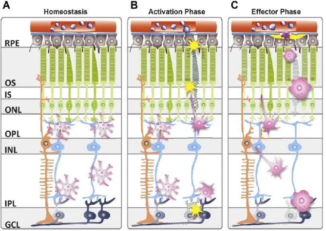
17 retinal degeneration models, reactive retinal microglia retract their cellular protrusions and migrate from the plexiform layers to the areas of tissue damage, e.g. the outer nuclear layer (ONL) containing photoreceptors, where they act as pro-inflammatory phagocytes as mentioned above (Fig. 4) (Zeiss and Johnson, 2004, Combadiere et al., 2007, Cuenca et al., 2014).
Figure 4: Schematic overview of microglial distribution and behavior in retinal health and disease. (A) In the healthy retina, microglia reside in the plexiform layers, actively scanning their microenvironment with their long protrusions (simplified depiction), phagocytosing insoluble metabolites and cellular debris. (B) Upon sensing tissue damage, microglia get alarmed, retract their protrusions and migrate into the sites of lesion, where they become full-blown pro-inflammatory phagocytes (C), phagocytosing apoptotic cells, secreting cytokines, chemokines and neurotoxic radicals. GCL, ganglion cell layer; IPL, inner plexiform layer; INL, inner nuclear layer;
OPL, outer plexiform layer; ONL, outer nuclear layer; IS, inner segment; OS, outer segment; RPE, retinal pigment epithelium. (Karlstetter et al., 2015).
In the PDE6B (phosphodiesterase 6b)-deficient rd1 and rd10 mouse models, which exhibit strong photoreceptor degeneration, a significant number of reactive microglial cells is found in the outer nuclear layer secreting high amounts of the pro- inflammatory mediators TNFα, CCL2 and CCL5 (Zeng et al., 2005, Yoshida et al., 2013). Further, a recent study has shown that phagocytosis of living photoreceptors by reactive microglia contributes to disease progression in the rd10 mouse model
18 (Zhao et al., 2015). In the murine model of X-linked juvenile retinoschisis (Weber et al., 2002), an early transformation of microglia into reactive phagocytes, even before retinal neurons undergo apoptosis, is observed (Gehrig et al., 2007, Ebert et al., 2009). These microglial cells show elevated pro-inflammatory transcript expression days before neuronal cell death is observable. We recently described a novel model of FAM161A-associated photoreceptor ciliopathy, which also exhibits strong microgliosis accompanied by pro-inflammatory microglial gene expression in the ONL during disease progression which seems to partially resolve after most photoreceptors are lost in late disease stages (Karlstetter et al., 2014b). These findings indicate that microglial reactivity is a key event in the onset and progression of retinal degeneration.
There is now ample evidence that inflammation and microglia also play important roles in the onset and disease progression of AMD (Newman et al., 2012, Penfold et al., 2001). In accordance, in post mortem retinas from human AMD and RP patients, the presence of reactive microglia bearing phagocytosed apoptotic photoreceptor debris was confirmed in the ONL (Gupta et al., 2003). Since mice harbor no macula and no archetypical murine AMD model exists to this date, most AMD models focus on mimicking single pathological features of the disease like deposition of drusen, RPE damage or choroidal neovascularization leading to photoreceptor degeneration.
In a model of geographic atrophy, which accumulates drusen-like deposits in the RPE, reactive phagocytes including microglia accumulate in the outer retina by a CCR2 (C-C chemokine receptor type 2)-dependent recruiting mechanism (Hollyfield et al., 2010, Cruz-Guilloty et al., 2013). Another strategy of mimicking photo-oxidative damage and retinal degeneration present in AMD is irradiation with focused blue or intense white light. Blue light-damaged retinas show a high focal accumulation of blood-derived macrophages and reactive retinal microglia, and transcriptomic analyses confirm the strong inflammatory response of these cells as early as a few hours after light damage (Joly et al., 2009, Ebert et al., 2012). Similar observations have been made for microglia in the white light damage model which shows early CCL2-mediated chemotaxis of reactive microglia to the outer nuclear layer (Santos et al., 2010, Suzuki et al., 2012). In recent years, animal models mimicking the wet form of AMD including choroidal neovascularization (CNV), employing local rupture of the Bruch’s membrane through laser coagulation have proven to be successful (Lambert et al., 2013). In this model, reactive microglia are recruited to the lesion site within
19 four hours and peak numbers of reactive phagocytes are found in the subretinal space two days after laser damage, long before CNV can be observed (Eter et al., 2008, Liu et al., 2013).
These findings indicate the key role of microglia and inflammatory processes during early pathogenesis and disease progression of monogenic inherited retinal degenerations as well as complex retinal diseases like AMD.
1.3.4 Microglial (reactivity) markers
With microglia playing a key role in the onset and progression of neurodegenerative diseases of the CNS and retina, there is a high interest in discovering microglia- specific markers to allow for their identification and discrimination between regulatory and reactive microglia. Lacking microglia-specific candidates, for decades classical macrophage markers like F4/80, CD68 and CD11b were employed to identify microglia in the brain and retina (Hume et al., 1983, Xu et al., 2007). In recent years, however, IBA1 (ionized calcium-binding adapter molecule 1) has emerged as the most reliable and widely used target structure for immunohistochemical detection of microglia in the CNS and retina (Ito et al., 1998, Ohsawa et al., 2000).
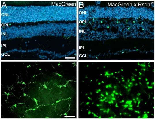
Transgenic mice harboring GFP (green fluorescent protein)-positive macrophages and microglia are also widely used to simplify recognition of microglia in the immune- privileged CNS, which is devoid of macrophages under healthy conditions. The CX3CR1-GFP knock-in mouse is a widely used example, where all myeloid cells express GFP under control of the cell type-specific CX3CR1 promoter on one of their CX3CR1 gene copies (Jung et al., 2000). This model has enabled in vivo and live- imaging of microglial distribution, dynamics and behavior in the healthy and degenerating retina (Eter et al., 2008, Lee et al., 2008, Liang et al., 2009). Another microglial reporter animal model is the MacGreen mouse. In comparison to the CX3CR1-GFP knock-in mouse, this model has the advantage of not replacing a functional locus and therefore homozygous mice with potentially stronger GFP expression can be used without altering microglial behavior (Sasmono et al., 2003).
GFP is expressed under the control of the macrophage-specific c-fms (colony stimulating factor 1 receptor) promoter and GFP signals in the healthy and
20 degenerating retina overlap nicely with classical immunohistochemical markers IBA1 and F4/80 (Fig. 5) (Ebert et al., 2009).
Figure 5: Microglial reactivity in the retinoschisin-deficient (Rs1h-/Y) mouse retina. (A) In the healthy MacGreen GFP reporter mouse, microglia reside in the plexiform layers, exhibit small somata with long cellular protrusions (top) and populate the retina in an evenly distributed fashion as shown in retinal flat mount microscopy (bottom). (B) In the dystrophic retina, microglia adopt an amoeboid phenotype, lose their even distribution and migrate into the site of retinal damage, in this model an early splitting (schisis) of the inner nuclear layer, where they act as pro-inflammatory phagocytes. Scale bar, 50 µm. (Flat mount pictures modified from Ebert et. al, 2009).
Reactive microglia express and secrete a plethora of molecules including classical pro-inflammatory mediators like IL1, IL6 and TNFα (Banati et al., 1993, Perry et al., 2010). This response is largely triggered by pro-inflammatory cytokines like IFNγ (interferon γ), TNFs or pathogen-associated molecular patterns (PAMPs) like bacterial lipopolysaccharide (LPS) and leads to a classical activation of microglia towards an aggressive M1-like macrophage phenotype (Laskin, 2009). However, alternative activation of microglia through IL10 or TGFβ (transforming growth factor β) towards an M2-like phenotype is associated with resolution of inflammation, reduced pro-inflammatory gene expression and increased expression of neurotrophic factors, a phenotype more favorable during neurodegenerative diseases (Duffield, 2003, Cherry et al., 2014). Knowledge about microglial reactivity markers and their function is therefore crucial for understanding their individual role in degenerative diseases of the CNS and retina.
21 Employing genome-wide expression profiling of microglia from the Rs1h-/Y mouse retina, we could recently identify activated microglia/macrophage whey acidic protein (AMWAP, alias WFDC17, Gm11428) as a novel marker for microglial reactivity, counter-regulating their inflammatory response and thereby serving as a feedback- loop mechanism to help attenuate pro-inflammatory microgliosis (Gehrig et al., 2007, Karlstetter et al., 2010, Aslanidis et al., 2015). We could also show induction of AMWAP gene expression in the FAM161A-deficient retinal ciliopathy mouse model, congruent with the course and severity of observed microgliosis (Karlstetter et al., 2014b). In addition, in a recent study we also observed elevated AMWAP transcript levels in the retina of the acid sphingomyelinase (aSMase)-deficient mouse model of impaired retinal function and reactive microgliosis (Dannhausen et al., 2015).
Interestingly, transcriptomic profiling using microarray analysis of isolated microglia previously also reported a strong upregulation of the back then uncharacterized AMWAP transcript (Gm11428) in reactive microglia in a mouse model of severe neuronal impairment (Huyghe et al., 2006).
Further, we and others could show that reactive retinal microglia from Rs1h-/Y and aSMase-deficient mice as well as in vitro exhibit strong upregulation of the mitochondrial translocator protein (18 kDa) (TSPO) (Karlstetter et al., 2014a, Dannhausen et al., 2015, Wang et al., 2014b). These are the first reports of TSPO as a microglia reactivity marker and potential therapeutic target in the retina, while TSPO has already been implicated to play a role in neuroinflammation of the brain and CNS (Girard et al., 2012, Chen and Guilarte, 2008).
These novel microgliosis markers now serve as assessment tools to evaluate microglial reactivity in the retina and are subject of further investigation to elucidate their function and employ them as potential mediators of anti-inflammatory immunotherapy.
1.3.5 Microglia as a therapeutic target
Given the important and often detrimental role of chronic pro-inflammatory microglial reactivity in degenerative diseases of the CNS and retina, there is a growing interest in modulating microglia towards a regulatory, homeostatic phenotype in a therapeutic fashion (Schuetz and Thanos, 2004, Karlstetter et al., 2015). Various therapeutic
22 strategies have been investigated so far, including the use of natural compounds, endogenous microglial factors as well as synthetic immunoregulatory pharma- ceuticals.
1.3.5.1 Natural compounds
Curcumin, a major constituent of turmeric, has been used in Ayurvedic medicine for centuries and has broad anti-inflammatory properties (Ammon and Wahl, 1991).
Curcumin treatment effectively reduces microglial NO secretion, pro-inflammatory cytokine expression and migration in vitro (Karlstetter et al., 2011). It further attenuates microglial reactivity via blockade of NFκB signaling and is neuroprotective in mouse models of intracerebral hemorrhage and traumatic brain injury (Yang et al., 2014, Zhu et al., 2014a). It was also shown to be protective in various retinal degeneration models, inhibiting microglial neurotoxicity and attenuating photoreceptor degeneration (Mirza et al., 2013, Gupta et al., 2011, Vasireddy et al., 2011).
Luteolin, another natural compound, is a flavonoid abundant in parsley, green pepper and celery. Luteolin acts anti-inflammatory through inhibition of pro-inflammatory NFκB and AP-1 signaling as well as acting as a radical scavenger (Jang et al., 2008, Chen et al., 2007). We could previously show that Luteolin triggers global changes in the microglial transcriptome inducing an anti-inflammatory, anti-oxidative and neuroprotective phenotype in vitro (Dirscherl et al., 2010). Luteolin further blocks pro- inflammatory NFκB activation and microglial neurotoxicity in vitro (Zhu et al., 2014b).
Finally, Luteolin reduces the neurotoxicity of microglia cells on primary hippocampal neurons and enhances spatial working memory in aged mice by reducing microglia- mediated inflammation in the hippocampus (Zhu et al., 2011, Jang et al., 2010).
Docosahexaenoic acid (DHA) is a polyunsaturated fatty acid enriched in fish oil and is highly abundant in the retina as a precursor of neuroprotectin D1 (NPD1), which promotes the survival of RPE and photoreceptor cells (Bazan et al., 2010). DHA levels were shown to be decreased in various retinal degenerations and DHA is therefore regarded as a potential compound for beneficial dietary supplementation (Anderson et al., 2002, Schaefer et al., 1995). DHA reduces microglial reactivity through inhibition of NFκB signaling and confers neuroprotection in animal models of
23 spinal cord compression and experimental stroke (Wang et al., 2015, Lim et al., 2013, Eady et al., 2014). In the eye, DHA protects from microglia-mediated retinal degeneration in mouse models of X-linked juvenile retinoschisis and neuronal ceroid lipofuscinosis (NCL) (Ebert et al., 2009, Mirza et al., 2013).
1.3.5.2 Whey acidic proteins (WAPs) and AMWAP
Whey acidic proteins (WAPs) are a group of secreted proteins carrying one or more WAP four-disulfide core (WFDC) domains, which comprise eight cysteines forming four disulfide bonds in a specific manner (Simpson and Nicholas, 2002). They are highly enriched at mucosal surfaces like the lung, oral cavity or colon and also expressed by neutrophils and macrophages. They fulfil a role as “alarmins”, since they are upregulated and actively secreted during tissue damage and inflammation (Bingle and Vyakarnam, 2008). They have a broad range of functions, including anti- microbial, anti-protease and anti-inflammatory potential in the respective tissues (Moreau et al., 2008). The best characterized WAPs are secretory leukocyte protease inhibitor (SLPI) and elafin (Scott et al., 2011). Both these WAPs serve as markers in inflammatory diseases of the lung and colon and have been shown to be beneficial for the resolution of inflammation (Tsoumakidou et al., 2010, Eriksson et al., 2008, Small et al., 2015, Marino et al., 2011, Reardon et al., 2011). Functionally, they can inhibit pro-inflammatory NFκB and AP-1 signaling in innate immune cells, restoring a regulatory protective phenotype (Greene et al., 2004, Butler et al., 2006, Taggart et al., 2005).
Over the last years, an emerging role of WAPs in the onset and resolution of neuroinflammation in the CNS has been proposed (Hannila, 2014). SLPI is upregulated in various CNS pathologies, including animal models of multiple sclerosis and stroke, as well as in amyotrophic lateral sclerosis (ALS) patients (Mueller et al., 2008, Wang et al., 2003, Lilo et al., 2013). In addition, SLPI was shown to be immunomodulatory and neuroprotective in models of optic nerve and spinal cord injury, multiple sclerosis and stroke (Hannila et al., 2013, Ghasemlou et al., 2010, Mueller et al., 2008, Wang et al., 2003). We have previously identified the novel activated microglia/macrophage whey acidic protein (AMWAP) as a highly upregulated molecule in microglia from Rs1h-/Y, the FAM161A- and aSMase-deficient retina as well as reactive microglia and macrophages in vitro (Gehrig et al., 2007,
24 Karlstetter et al., 2010, Karlstetter et al., 2014b, Dannhausen et al., 2015). AMWAP is a 76 aa polypeptide containing a single WAP domain and a N-terminal signaling peptide for cellular export. AMWAP overexpression in cultured microglia reduces their pro-inflammatory gene expression, induces expression of alternative activation markers and reduces microglial migration, whereas RNAi-mediated AMWAP knock- down has adverse effects (Karlstetter et al., 2010). In addition, we could recently show that AMWAP is actively secreted from reactive microglia in vitro to counter- balance pro-inflammatory microglial activation through inhibition of NFκB signaling in a paracrine fashion (Aslanidis et al., 2015).
1.3.5.3 Translocator protein (18 kDa) (TSPO) and its ligands
The translocator protein (18 kDa) (TSPO), formerly known as peripheral benzodiazepine receptor (PBR), is a transmembrane protein located in the outer mitochondrial membrane, highly expressed in steroidogenic cells of the adrenal gland, gonad and brain (Papadopoulos et al., 2006, Rupprecht et al., 2010). Within the CNS, TSPO is expressed predominantly in glial cells and to a lesser extent in certain neurons, e.g. of the olfactory bulb or dorsal root ganglia sensory neurons (Casellas et al., 2002, Zhang et al., 2007, Karchewski et al., 2004, Maeda et al., 2007). One of the main functions of TSPO is the cholesterol transport from the outer to the inner mitochondrial membrane, which is a crucial step in steroid hormone and neurosteroid synthesis (Papadopoulos et al., 2006). Additionally, TSPO plays a role in mitochondrial respiration and permeability, apoptosis and cell proliferation (Casellas et al., 2002, Maeda et al., 2007, Karchewski et al., 2004).
Over the last years, many studies have surfaced observing an upregulation of TSPO expression during CNS pathologies (Chen and Guilarte, 2008, Rupprecht et al., 2010). For example, TSPO expression was shown to be induced in Alzheimer’s disease, Huntington’s disease, Parkinson’s disease as well as in psychiatric disorders like anxiety or post-traumatic stress disorder (Yasuno et al., 2008, Politis et al., 2012, Nakamura et al., 2002, Da Pozzo et al., 2012). In a retinal context, we and others could recently show microglial upregulation of TSPO in the Rs1h-/Y mouse model of retinal degeneration, the aSMase-deficient retina as well as in an in vivo model of LPS-induced retinal microglia activation (Karlstetter et al., 2014a, Dannhausen et al., 2015, Wang et al., 2014b). The selective upregulation of TSPO in
25 microglia and astrocytes during CNS pathologies makes it a suitable biomarker to monitor the location, course and severity of neuroinflammatory processes. Therefore, TSPO serves as a target structure for neuroimaging procedures and specific TSPO ligands are employed as diagnostic tools e.g. for positron emission tomography (PET) (Chen and Guilarte, 2008).
Since TSPO is selectively upregulated in pathologies of the CNS, in recent years growing interest in the use of specific TSPO ligands as a strategy to attenuate neuroinflammation has emerged (Rupprecht et al., 2010, Nothdurfter et al., 2012).
TSPO has a number of endogenous ligands like cholesterol, porphyrins or endozepines (Li and Papadopoulos, 1998, Gavish et al., 1992, Costa and Guidotti, 1991). Especially endozepines have been found to be highly produced during CNS inflammation, indicating that paracrine mechanisms of modulating glial reactivity through TSPO may exist. As an example, it was recently shown that Mueller cells and astrocytes express and secrete diazepam binding inhibitor (DBI) in the inflamed retina, which effectively reduces pro-inflammatory microglial cytokine expression and ROS secretion (Wang et al., 2014b).
These mechanisms raised the interest in developing highly specific synthetic TSPO ligands as therapeutic tools to attenuate neuroinflammation. The isoquinoline carboxamide derivative PK11195, used as a radioligand in PET studies and the benzodiazepine diazepam as a classic anxiolytic drug are prime examples for synthetic TSPO ligands (Venneti et al., 2013, Gavish et al., 1992). It was implicated that TSPO ligands can act therapeutically through induction of anti-inflammatory cytokine and neurotrophic factor expression in microglia and protective effects have already been shown for ALS, neuropathic pain, brain inflammation, depression and anxiety disorders (Bordet et al., 2007, Wei et al., 2013, Papadopoulos and Lecanu, 2009, Rupprecht et al., 2010). Further, TSPO ligands have been shown to reduce pro-inflammatory microglia reactivity and the production of pro-inflammatory cytokines in animal models of hippocampal and striatal brain damage (Veiga et al., 2005, Ryu et al., 2005). Novel synthetic ligands include etifoxine (Stresam®) and XBD173 (Emapunil®). Etifoxine reduces neuroinflammation and is protective and regenerative in a mouse model of multiple sclerosis (Daugherty et al., 2013).
XBD173 is a phenylpurine, which binds TSPO at a nanomolar affinity (Kita et al., 2004). XBD173 was shown to possess potent anxiolytic functions in rodents and
26 humans, without exerting negative diazepam-like side effects like sedation or habit formation (Rupprecht et al., 2009). In addition, we could recently show that XBD173 is a potent anti-inflammatory mediator and modulates microglial reactivity, neurotoxicity and phagocytosis towards a regulatory phenotype (Karlstetter et al., 2014a).
The exact mechanisms, through which TSPO ligands modulate microglial reactivity and exert their neuroprotective effects, are still largely unknown. Possible mechanisms include the modulation of the mitochondrial transmembrane potential, leading to reduced ROS production and preventing neuronal damage (Carayon et al., 1996, Chelli et al., 2004). Another suggestion is a reduction of apoptosis-related caspase activation by attenuating mitochondrial cytochrome C release (Strohmeier et al., 2002, Veiga et al., 2005). However, since TSPO ligands increase neurosteroid synthesis in the CNS, which exert immunomodulatory and neuroprotective functions, it is likely that this could be a key mechanism of their protective action (Schule et al., 2011). It remains to be elucidated how specific TSPO ligands fulfill their therapeutic function in detail, and it is likely that a combination of the described effects contributes to their effectiveness.
1.4 Aims of the thesis
In a comprehensive approach, we focused on the understanding of photoreceptor homeostasis and disease mechanisms, the identification and characterization of novel microgliosis markers as well as potential future therapeutics in neuro- degenerative diseases with an emphasis on microglia-directed anti-inflammatory therapy in the brain and retina.
Thus, one aim of this work was to investigate the novel CRX-regulated gene SAMD7 in the retina. Elucidating its spatiotemporal expression pattern, the exact regulatory fine-tuning of SAMD7 by the photoreceptor-specific transcription factor CRX and the putative function of SAMD7 as a repressor of CRX-mediated photoreceptor gene expression, we aimed to gain understanding into the fine-tuning of photoreceptor homeostasis and potential disease mechanisms of inherited retinal degenerations.
Another goal was to elucidate the paracrine anti-inflammatory potential of the novel activated microglia/macrophage whey acidic protein (AMWAP) in microglial cells.
27 Using recombinant AMWAP, we sought to shed light on AMWAPs effects on microglial NFκB signaling and classical physiological functions of microglia in vitro, with the aim of establishing whey acidic proteins as potential future therapeutics in neurodegenerative diseases of the brain and retina.
Finally, we were interested in the role of the translocator protein (18 kDa) (TSPO) in the context of neuroinflammation in the retina. Investigating its potential as a novel biomarker for pro-inflammatory microglia activation in the degenerating retina and employing the specific TSPO ligand XBD173 to modulate microglial reactivity in vitro and ex vivo, we aimed to establish TSPO as a potential target structure for microglia- directed anti-inflammatory therapy in the brain and retina.
28
2. Results
2.1 Sterile alpha motif containing 7 (SAMD7) is a novel CRX-regulated transcriptional repressor in the retina
Julia Hlawatsch*, Marcus Karlstetter*, Alexander Aslanidis*, Anika Lückoff, Yana Walczak, Michael Plank, Julia Böck, and Thomas Langmann
*these authors contributed equally
Inherited retinal diseases are mainly caused by mutations in genes that are highly expressed in photoreceptors of the retina. The majority of these genes is under the control of the transcription factor Cone rod homeobox (CRX), which acts as a master transcription factor in photoreceptors. Using a genome-wide chromatin immuno- precipitation dataset that highlights all potential in vivo targets of CRX, we have identified a novel sterile alpha motif (SAM) domain containing protein, SAMD7.
mRNA Expression of SAMD7 was confined to the late postnatal and adult mouse retina as well as the pineal gland. Using immunohistochemistry and Western blot, we could detect SAMD7 protein in the outer nuclear layer of adult mouse retina. Ectopic over-expression in HEK293 cells demonstrated that SAMD7 resides in the cytoplasm as well as the nucleus. In vitro electroporation of fluorescent reporters into living mouse retinal cultures revealed that transcription of the SAMD7 gene depends on evolutionary conserved CRX motifs located in the first intron enhancer. Moreover, CRX knock-down with shRNA strongly reduced SAMD7 reporter activity and endogenous SAMD7 protein, indicating that CRX is required for retinal expression of SAMD7. Finally, using co-transfections in luciferase reporter assays we found that SAMD7 interferes with CRX-dependent transcription. SAMD7 suppressed luciferase activity from a reporter plasmid with five CRX consensus repeats in a dose dependent manner and reduced CRX-mediated transactivation of regulatory sequences in the retinoschisin gene and the SAMD7 gene itself. Taken together, we have identified a novel retinal SAM domain protein, SAMD7, which could act as a transcriptional repressor involved in fine-tuning of CRX-regulated gene expression.
29 Own contribution to publication I:
I carried out the characterization of the novel SAMD7 gene by analyzing its tissue- specific mRNA expression (Fig. 2 B) and its expression in the pineal gland (Fig. 2 C).
I further elucidated the retina-specific localization of SAMD7 protein to the photoreceptor layer using immunohistochemistry (Fig. 3 A). Further, I established, designed, performed and analyzed ex vivo electroporation knock-down experiments in murine retinal explants combined with immunohistochemistry to show the CRX- dependent regulation and expression of SAMD7 (Fig. 5 A,B). Finally, I demonstrated the important transcriptional repression function of SAMD7 using Luciferase reporter assays in HEK293 cells, after optimizing the protocol (Fig. 6 A-D). I co-supervised Bachelor student Michael Plank, who also contributed experimentally to this project.
All images depicting my experimental data were created by me. I presented posters and oral presentations as first author on this project at both national and international symposia during my PhD.
Contribution of co-authors to publication I:
Dr. Julia Hlawatsch was a medical student, who participated in the early stages of the project and helped to establish anti-SAMD7 immunohistochemistry. Dr. Marcus Karlstetter carried out bioinformatic analysis (Fig. 1 A-D) and characterization of the SAMD7 protein, helped with dissection of pineal glands and carried out Flag-SAMD7 transfections and immunocytochemistry in HEK293 cells (Fig. 3 E-P). Anika Lückoff cloned the Flag-SAMD7 construct, carried out transfection of HEK293 cells and Western blot analysis (Fig. 3 B-D). Yana Walczak carried out SAMD7 qRT-PCR of postnatal murine retinas (Fig. 2 D). Michael Plank and Julia Böck performed ex vivo electroporation of SAMD7 reporter constructs into living mouse retinas under my co- supervision (Fig. 4 C-E). Prof. Thomas Langmann, as principal investigator, supervised the study and wrote the manuscript.
30
2.2 Activated microglia/macrophage whey acidic protein (AMWAP) inhibits NFκB signaling and induces a neuroprotective phenotype in microglia
Alexander Aslanidis, Marcus Karlstetter, Rebecca Scholz, Sascha Fauser, Harald Neumann, Cora Fried, Markus Pietsch* and Thomas Langmann*
*these authors contributed equally
Background: Microglia reactivity is a hallmark of neurodegenerative diseases. We have previously identified activated microglia/macrophage whey acidic protein (AMWAP) as a counter-regulator of pro-inflammatory response. Here, we studied its mechanisms of action with a focus on toll-like receptor (TLR) and nuclear factor κB (NFκB) signaling.
Methods: Recombinant AMWAP was produced in Escherichia coli and HEK293 EBNA cells and purified by affinity chromatography. AMWAP uptake was identified by fluorescent labeling, and pro-inflammatory microglia markers were measured by qRT-PCR after stimulation with TLR ligands. NFκB pathway proteins were assessed by immunocytochemistry, Western blot, and immunoprecipitation. A 20S proteasome activity assay was used to investigate the anti-peptidase activity of AMWAP.
Microglial neurotoxicity was estimated by nitrite measurement and quantification of caspase 3/7 levels in 661W photoreceptors cultured in the presence of microglia- conditioned medium. Microglial proliferation was investigated using flow cytometry, and their phagocytosis was monitored by the uptake of 661W photoreceptor debris.
Results: AMWAP was secreted from lipopolysaccharide (LPS)-activated microglia and recombinant AMWAP reduced gene transcription of IL6, iNOS, CCL2, CASP11, and TNFα in BV-2 microglia treated with LPS as TLR4 ligand. This effect was replicated with murine embryonic stem cell-derived microglia (ESdM) and primary brain microglia. AMWAP also diminished pro-inflammatory markers in microglia activated with the TLR2 ligand zymosan but had no effects on IL6, iNOS, and CCL2 transcription in cells treated with CpG oligodeoxynucleotides as TLR9 ligand.
Microglial uptake of AMWAP effectively inhibited TLR4-dependent NFκB activation by preventing IRAK-1 and IκBα proteolysis. No inhibition of IκBα phosphorylation or ubiquitination and no influence on overall 20S proteasome activity were observed.
Functionally, both microglial nitric oxide (NO) secretion and 661W photoreceptor