cGMP-dependent kinase I in renal fibrosis
Elisabeth Schinner
1, Veronika Wetzl
1,2, Andrea Schramm
1, Frieder Kees
1, Peter Sandner
3, Johannes-Peter Stasch
3, Franz Hofmann
4and Jens Schlossmann
11 Department of Pharmacology and Toxicology, University of Regensburg, Germany 2 Novartis Pharma GmbH, Nuremberg, Germany
3 Bayer Pharma AG, Wuppertal, Germany
4 Institute of Pharmacology and Toxicology, Technical University of Munich, Germany
Keywords
cGMP-dependent protein kinase I; cyclic guanosine monophosphate; renal fibrosis;
soluble guanylate cyclase stimulation Correspondence
J. Schlossmann, Lehrstuhl f € ur
Pharmakologie und Toxikologie, Institut f € ur Pharmazie, Universit€ at Regensburg, Universit € atsstr. 31, 93040 Regensburg, Germany
Fax: +49 941 943 4772 Tel: +49 941 943 4771
E-mail: jens.schlossmann@chemie.uni- regensburg.de
(Received 8 November 2016, revised 16 January 2017, accepted 23 January 2017) doi:10.1002/2211-5463.12202
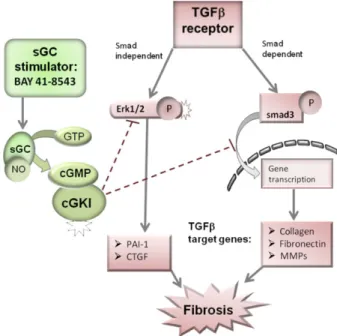
Agents that enhance production of nitric oxide (NO) and cyclic guanosine monophosphate (cGMP) ameliorate the progression of renal fibrosis. How- ever, the molecular mechanism of this process is not fully understood. We hypothesize that the antifibrotic effects of cGMP and cGMP-dependent kinase I (cGKI) are mediated via regulation of the TGF b signalling path- way, both via ERK and the Smad-dependent route. Kidney fibrosis was induced by unilateral ureter obstruction (UUO) in wild-type and cGKI- deficient (cGKI-KO) mice. The cGMP/cGKI signalling pathway was acti- vated by application of the soluble guanylate cyclase (sGC) stimulator BAY 41-8543 (BAY), beginning 1 day after UUO. After 7 days, the antifi- brotic effects of BAY were analysed by measuring mRNA and protein expression of characteristic fibrotic biomarkers. The effects of cGMP/
TGF b on cultured fibroblasts were also analysed in vitro. BAY application influenced the activity of the extracellular matrix (ECM)-degrading matrix metalloproteases (MMP2 and MMP9) and their inhibitor tissue inhibitors of metalloproteinase-1, the secretion of cytokines (e.g. IL-6) and the expression pattern of ECM proteins (e.g. collagen, fibronectin) and profi- brotic mediators (e.g. connective tissue growth factors and plasminogen- activator inhibitor-1). Activation of the cGMP/cGKI signalling pathway showed protective effects against fibrosis which were mediated by inhibi- tion of P-Erk1/2 and translocation of P-smad3. The elucidation of these signalling mechanisms might support the development of new therapeutic options regarding cGMP/cGKI-mediated antifibrotic actions.
Fibrosis is characterized by excessive expression of extracellular matrix (ECM). Fibrogenic factors promote the fibrotic process such as transforming growth factors (TGF b ), plasminogen-activator inhibitor-1 (PAI-1) or connective tissue growth factors (CTGF) [1]. TGF b is
involved in the differentiation of fibroblasts to myofi- broblasts, which are characterized by the expression of a -smooth muscle actin ( a SMA). Myofibroblasts synthe- size ECM proteins including collagen and fibronectin, and they secrete cytokines, for example, IL-6.
Abbreviations
cGKI, cGMP-dependent protein kinase I; cGKI-KO, cGKI-knockout; cGMP, cyclic guanosine monophosphate; Co-IP, coimmunoprecipitation;
Col1a1, collagen1a1; CTGF, connective tissue growth factor; ECM, extracellular matrix; ERK1/2, extracellular-signal regulated kinase; GTP, guanosine triphosphate; MMPs, matrix metalloproteinases; NO, nitric oxide; PAI-1, plasminogen-activator inhibitor-1; sGC, soluble guanylyl cyclase; TGF β , transforming growth factor β ; TIMP, tissue inhibitors of metalloproteinases; UUO, unilateral ureter obstruction; wt, wild-type;
α SMA, α -smooth muscle actin.
In addition, synthesis and degradation of ECM pro- teins are determined by metalloproteinases (MMPs) and tissue inhibitors of metalloproteinases (TIMPs).
Their expression pattern is regulated by MAPK/Erk kinase, which promotes the progression of fibrosis [2,3].
We evaluated the effects of the soluble guanylate cyclase (sGC) stimulator BAY 41-8543 (BAY) on the fibrotic kidney. Under physiological conditions, sGC can be activated by nitric oxide (NO). Activated sGC synthesizes the second messenger cyclic guanosine monophosphate (cGMP) which then stimulates cGMP-dependent protein kinases (cGK) [4]. We have previously reported that cGMP suppresses renal fibro- sis in particular via cGKI a , an isoform of cGK.
cGKIa is expressed in fibroblasts and myofibroblasts, which are excessively produced after unilateral ureter obstruction (UUO) [5]. Protective effects of sGC stim- ulation on renal fibrosis in rats have already been shown [6,7]. Thereby, BAY reduced apoptosis and macrophage infiltration after relief of UUO [7], and Sharkovska et al. [6] reported that sGC stimulation improved creatinine clearance in hypertensive renin- transgenic rats. However, the molecular mechanism by which cGMP via cGKI affects the development of kid- ney fibrosis has not yet been fully elucidated. There- fore, we analysed the impact of BAY on fibrosis in a mouse model of UUO using cGKI-knockout (cGKI-
KO)-mice. The present study investigates the functional role of sGC stimulation in the fibrotic process, the sig- nalling pathway as well as the underlying mechanisms involved.
Results
Effect of BAY and function of cGKI on the mRNA expression of different fibrotic biomarkers
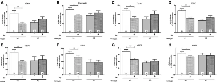
As marker for fibrosis induction, we examined the mRNA levels of a SMA, fibronectin, collagen1a1 (Col1a1), CTGF, TIMP-1, PAI-1, MMP2 and MMP9 (Fig. 1). One week after UUO surgery, the mRNA levels were elevated in comparison to the contralateral control kidney. Especially, aSMA (Fig. 1A), Col1a1 (Fig. 1C), TIMP-1 (Fig. 1E), PAI-1 (Fig. 1F) and MMP2 (Fig. 1G) were strongly increased by UUO. In contrast, the mRNA expression of fibronectin (Fig. 1B) and CTGF (Fig. 1D) were only moderately upregulated, and MMP9 (Fig. 1H) was nearly unchanged.
To examine the role of NO/cGMP signalling in renal fibrosis, we injected the sGC stimulator BAY. A significant raise of cGMP in kidney tissues of BAY- treated mice in comparison to untreated mice indicat- ing a BAY-induced stimulation of sGC was measured (Fig. S1). BAY treatment decreased the mRNA
Fig. 1. Effect of BAY in wt- and cGKI-KO-kidneys on the mRNA expression levels of (A) a SMA, (B) fibronectin, (C) Col1a1, (D) CTGF, (E)
TIMP-1, (F) PAI-1, (G) MMP2 and (H) MMP9. In wt-mice, BAY caused a significant decrease in the mRNA expression of (A) a SMA, (B)
fibronectin, (C) Col1a1, (D) CTGF, (E) TIMP-1, (G) MMP2 and (H) MMP9 with the exception of (F) PAI-1. In cGKI-KO-mice sGC stimulation
showed no significant decrease in the mRNA levels (A – H). The results are shown as the x-fold change in mRNA expression in the fibrotic
kidney relating to the opposite healthy kidneys whose mRNA expression was set to one. In each mouse strain the untreated mice were
compared with BAY-treated mice. Significant differences between two groups are indicated with asterisks ( *P < 0.05, **P < 0.01). The
columns show the number of animals which were used. The right columns illustrate the data of GKI-KO-mice and patterned columns the
data of BAY-treated mice.
expression of all investigated biomarkers of fibrosis with the exception of PAI-1 (Fig. 1F). The expression of PAI-1 was reduced but the difference did not reach significance. To explore whether cGKI is involved in the impact on the fibrotic process, we analysed cGKI- KO-mice. As previously reported, untreated cGKI- KO-mice showed less mRNA expression than untreated wild-type (wt) mice [5]. However, the mRNA expression of cGKI-KO-mice was not influ- enced by BAY application (Fig. 1).
Effect of BAY and role of cGKI on the protein expression of: aSMA, fibronectin, Col1a1 and total collagen
After UUO, the interstitial accumulation of protein expression of aSMA, fibronectin and Col1a1 was increased in wt- and cGKI-KO kidneys as demon- strated by immunofluorescence analysis (Fig. 2A – C).
The quantitative analysis revealed that BAY signifi- cantly reduced the protein expression of a SMA, fibro- nectin and Col1a1 in wt-, but not in cGKI-KO- kidneys (Fig. 2A – C). The same pattern was present when we used the Sirius red/fast green staining for total collagen. In wt-, but not in cGKI-KO-kidneys, sGC stimulation by BAY significantly downregulated the level of total collagen (Fig. 2D).
Effect of BAY and function of cGKI on the activity or protein expression of TGFb target genes As expected, UUO increased the protein expression of the TGFb target gene CTGF in comparison to the healthy kidney (Fig. 3A). The quantitative analysis, which compared only fibrotic kidneys, confirmed that the protein expression of CTGF was significantly diminished by BAY in fibrotic wt-kidneys. However, treatment of cGKI-KO-mice did not result in a reduc- tion of CTGF (Fig. 3A). Figure 3B demonstrates that PAI-1-expression was not significantly influenced by BAY. The protein expression of TIMP-1 was not changed by UUO in comparison to the contralateral healthy kidney (Fig. 3C). Intriguingly, TIMP-1 was significantly higher in BAY treated than in untreated
fibrotic wt-mice. In cGKI-KO-mice, we detected no increase in TIMP-1 expression following BAY admin- istration (Fig. 3C). The latent and active forms of MMP2 and the latent forms of MMP9 were elevated, but the active forms of MMP9 were reduced by UUO (data not shown). In agreement with the increase in TIMP-1, which is an inhibitor of MMPs, the latent and active forms of MMP2 (Fig. 4A,B) and the latent forms of MMP9 (Fig. 4A,C) were significantly dimin- ished by BAY. This was again only observed in wt-, but not in cGKI-KO-kidneys.
Effect of cGMP/cGKI on the TGFb signalling pathway
At first we analysed the influence of cGMP/cGKI on the TGFb/smad signalling pathway. Isolated fibroblasts of wt- (left side of Fig. 5A) and cGKI-KO-kidneys (right side of Fig. 5A) were pretreated with cGMP or vehicle followed by exposure to TGF b or vehicle (Fig. 5A). We quantified the intranuclear and extranu- clear fluorescence intensity of P-smad3 respectively. Fig- ure 5B shows that TGF b treatment significantly enhanced nuclear fluorescence intensity of P-smad3 but pretreatment with cGMP significantly limits nuclear translocation of P-smad3 in fibroblasts of wt-kidneys in the presence of TGF b . cGMP alone had no effects (data not shown). Intriguingly in fibroblasts of cGKI-KO-kid- neys pretreatment with cGMP did not change the translocation of P-smad3 (Fig. 5C). In contrast to P- smad3, P-smad2 was not influenced by preincubation with cGMP (data not shown). Isolated fibroblasts expressed sGC but during culturing the expression of sGC was downregulated (data not shown). Therefore, we stimulated the cells only with cGMP and not with the sGC stimulator BAY. Furthermore, we quantified the total cellular fluorescence intensity of P-smad3 which was significantly increased by TGF b treatment but interestingly not significantly changed by cGMP pretreatment (Fig. S2). In pulmonary artery smooth muscle cells activation of cGMP/PKG limited TGF b - induced nuclear translocation of smad3 by sequestering smad3 with cytosolic b 2-tubulin [8]. Therefore, we per- formed a coimmunoprecipitation (Co-IP) of stimulated
Fig. 2. Effect of BAY in wt- and cGKI-KO-kidneys on the protein levels of (A) a SMA, (B) fibronectin, (C) Col1a1 and (D) total Collagen.
Metamorph offline was used for the quantification of fluorescence-intensity of (A) a SMA, (B) fibronectin and (C) Col1a1.
Immunofluorescence staining of (A) a SMA (Alexa488, shown in red), (B) fibronectin (Alexa647, shown in red) and (C) Col1a1 (Alexa647,
shown in red) in healthy, UUO-untreated and UUO-BAY-treated kidneys of wt- and cGKI-KO-mice. Total collagen levels in the kidneys were
measured by Sirius red/fast green staining (D). The protein expression of (A) a SMA, (B) fibronectin, (C) Col1a1 and (D) total Collagen was
significantly diminished in wt-mice, but not in cGKI-KO-mice by BAY. Thereby, the increase in protein by UUO was related to the healthy
kidney. In each mouse strain the untreated mice were compared with BAY-treated mice. Significant differences between two groups are
indicated with asterisks ( *P < 0.05, **P < 0.01). The columns show the number of animals which were used. The right columns illustrate
the data of GKI-KO-mice and patterned columns the data of BAY-treated mice.
fibroblasts to check whether smad3 and b 2-tubulin form a cGMP-dependent complex. Figure 5D shows that b2- tubulin antibody precipitated cGKIa, P-smad3 and smad3 in TGFb- and cGMP/TGFb-stimulated fibrob- lasts. However, in contrast to Gong et al. [8], there was no increase in the intensity of the bands after pretreat- ment with cGMP.
Second, the phosphorylation of Erk1 and Erk2 (P- Erk1/2) was assessed. UUO increased the
phosphorylation and the protein expression of Erk1 and Erk2 (Fig. 6A). Immunoblots with antibodies against total Erk1 and Erk2 demonstrated that their expression is increased by UUO, but not changed by Bay administration (Fig. 6A). Accordingly, in Fig. 6B, C only fibrotic kidneys are compared and P-Erk1/2 is normalized to Erk1/2 and related to untreated fibrotic wt-kidneys. sGC stimulation caused a significant decrease in P-Erk1 and P-Erk2 in fibrotic kidneys of
Fig. 3. Effect of BAY in wt- and cGKI-KO-kidneys on the protein levels of (A) CTGF, (B) PAI-1 and (C) TIMP-1. The immunoblots show the protein expression of CTGF (A), PAI-1 (B) and TIMP-1 (C) in wt-mice of healthy and fibrotic kidneys (BAY treated or untreated). The graphs statistically compare the protein expression of CTGF (A), PAI-1 (B) and TIMP-1 (C) in fibrotic wt- and cGKI-KO-kidneys. CTGF (A) and TIMP-1 (C) are significantly influenced by BAY in wt-, but not in cGKI-KO-kidneys. Thereby, each value of the used markers of wt- and cGKI-KO- kidneys is related to the mean value of untreated fibrotic wt-kidneys which was set to one and normalized to the corresponding GAPDH.
The protein expression of GAPDH was changed by UUO but not by BAY. Therefore, the statistic compares only fibrotic kindeys. Significant differences between two groups are indicated with asterisks ( *P < 0.05). The columns show the number of animals which were used. The right columns illustrate the data of GKI-KO-mice and patterned columns the data of BAY-treated mice.
Fig. 4. Effect of BAY in fibrotic wt- and cGKI-KO-kidneys on the activity of MMP2 and MMP9. Latent and active MMP2 and MMP9 of fibrotic wt- and cGKI-KO-kidneys were determined by Gelatin zymography assays (A). In fibrotic wt-kidneys, latent and active MMP2 (B) and latent MMP9 (C) were significantly reduced after BAY application. In cGKI-KO-kidneys BAY showed no effects regarding the activity of MMP2 and 9. Each value of wt- and cGKI-KO-kidneys is related to the mean value of untreated fibrotic wt-kidneys which was set to one.
Significant differences between two groups are indicated with asterisks ( *P < 0.05, **P < 0.01). The columns show the number of animals
which were used. The right columns illustrate the data of GKI-KO-mice and patterned columns the data of BAY-treated mice.
treated in contrast to untreated wt-mice. This BAY- induced decrease of Erk1/2 phosphorylation in fibrotic wt-kidneys was not due to changed protein expression of Erk1/2. In cGKI-KO-kidneys, the Erk phosphoryla- tion was not reduced by BAY (Fig. 6B,C).
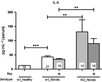
Increased IL-6 levels in cGKI-KO-mice
It has been shown that IL-6 promotes fibrosis [9].
UUO significantly increased the IL-6 concentration in serum of wt-mice. Administration of BAY tends to result in diminished IL-6 levels in serum of wt-mice compared with untreated wt-mice. Interestingly, the IL-6 concentration was significantly higher in untreated and treated cGKI-KO-mice and fluctuated much more than in wt-mice (Fig. 7).
Effect of BAY and role of cGKI on the renal function examining serum creatinine
The serum level of creatinine increased significantly after 7 days of UUO. Following BAY administration, serum creatinine was decreased, but there was no significant difference between BAY treated and untreated wt-mice. Conversely, in cGKI-KO-mice, BAY influenced in no way the serum creatinine (Fig. 8).
Discussion
In the present study, we have investigated the func- tional role of sGC stimulation in regulating renal fibrosis. BAY reduced the mRNA- and protein
Fig. 5. Effect of cGMP on the TGF b /smad signalling pathway in renal wt- and cGKI-KO-fibroblasts. Serum-starved fibroblasts which were isolated from wt- (left sided) and cGKI-KO-kidneys (right sided) were pretreated with 8Br-cGMP (1 m
M) or vehicle (control) for 1 h, followed by TGF b (2 ng mL
1) or vehicle for 1 h and stained with P-smad3 (Alexa647 anti-rabbit, shown in red) and DAPI (shown in blue) (A). The statistic of the fluorescence intensity of P-smad3 in nucleus or cytosol is demonstrated in (B) wt-fibroblasts and (C) cGKI-KO-fibroblasts. Wt- fibroblasts were stimulated with TGF b or cGMP/TGF b for the coimmunprecipitation (Co-IP) analysis which was performed with whole cell extracts using anti- b 2-tubulin antibody. The blot was probed with anti-cGKI a and smad3, then after stripping with anti-P-smad3 and then after stripping with anti- b 2-tubulin (D). Significant differences between two groups are indicated with asterisks ( *P < 0.05, ***P < 0.001).
The experiments were repeated five to seven times.
Fig. 6. Effect of BAY on the TGF b /Erk signalling pathway in wt- and cGKI-KO-mice (A) Representative western blots of Erk1/2 in healthy/
fibrotic kidney tissue of wt-mice untreated/treated with BAY. The graphs statistically compare the protein expression of P-Erk1 (B) and P- Erk2 (C) in fibrotic wt- and cGKI-KO-kidneys. P-Erk1 (B) and P-Erk2 (C) were significantly reduced by BAY in wt-, but not in cGKI-KO-kidneys.
Thereby, each value of P-Erk1/2 of wt- and cGKI-KO-kidneys is related to the mean value of untreated fibrotic wt-kidneys which was set to
one and normalized to the corresponding Erk1/2. Significant differences between two groups are indicated with asterisks ( *P < 0.05). The
columns show the number of animals which were used. The right columns illustrate the data of GKI-KO mice and patterned columns the
data of BAY-treated mice.
expression of different fibrosis marker. The antifibrotic impact of sGC stimulation was not observed in cGKI- KO-mice, suggesting that cGKI mediates the repair process of renal fibrosis.
Our study confirmed that the serum creatinine, which is a parameter for renal function, is increased after UUO [10]. However, it was not significantly reduced by BAY in wt-mice and unchanged in cGKI- KO-mice. Our results are in line with the nephropro- tective effects of PDE5 inhibitors which also enhance the cGMP pool [11 – 13]. cGKI-KO-mice have higher IL-6 levels [14,15] which exert profibrotic effects [9,16].
Conforming with our present study, the IL-6 levels were increased by UUO and treated, as well as untreated cGKI-KO-mice showed a higher IL-6 con- centration than wt-mice. However, cGKI-KO-kidneys revealed no more pronounced fibrosis compared to wt- kidneys suggesting that other signalling pathways as IL-6 are important for induction of renal fibrosis. The application of BAY reduced the IL-6 concentration, but the difference was not significant. Considering the effects of the MAPK signalling, the phosphorylation of Erk promotes fibrosis [17]. In cardiac fibrosis the inhibition of Erk phosphorylation by cGMP has already been discussed [18]. Our results confirmed the decrease in phosphorylation of Erk after BAY applica- tion. Consistent with our data, Beyer et al. [19] have also identified that the stimulation of sGC decreased TGF b signalling through the inhibition of Erk1/2 phosphorylation. Additionally, we observed that cGMP influenced via cGKI the phosphorylation of Erk because in cGKI-KO-mice, the effects of BAY were lower.
It is generally accepted that TGF b acts by stimula- tion of its downstream mediator smad2 and smad3.
Latest studies report that diminished smad2- as well as smad3 phosphorylation results in enhanced renal fibrosis [20–22]. However, it is also recently discussed that phosphorylation of smad2 and smad3 by TGF b exerts reverse effects in renal fibrosis. Smad2 maybe plays a protective role negatively regulating the smad3 signalling. TGF b activates smad2 which diminishes TGF b 1/smad3 signalling, including phos- phorylation, nuclear translocation and the binding of smad3 to the Col1 promoter, leading to augmented collagen synthesis [23,24]. In our study, phosphory- lated smad2 was unaffected by cGMP in renal fibrob- lasts (data not shown). However, nuclear translocation of P-smad3 was diminished by cGMP in the presence of TGFb in wt-, but not in cGKI- KO-fibroblasts. Interestingly cGMP inhibited only the translocation of P-smad3, but not the phosphoryla- tion of smad3 (Fig. S2). In contrast to our study, Beyer et al. [19] showed that nuclear P-smad2- and P-smad3 levels and smad reporter activity were unaf- fected by sGC stimulation in human fibroblasts. As already mentioned in pulmonary artery smooth
Fig. 7. Effect of BAY on the IL-6 levels in the serum of wt- and cGKI-KO-mice. The IL-6 levels in serum of both treated and untreated cGKI-KO-mice were significantly higher than in corresponding wt-mice. However, BAY itself revealed no significant effects in wt- and in cGKI-KO-mice. Significant differences between two groups are indicated with asterisks ( **P < 0.01, ***P < 0.001). The columns show the number of animals which were used. The right columns illustrate the data of GKI-KO mice and patterned columns the data of BAY-treated mice.
1.5
Creatinine
**
1.0
*
mg·L–1 (serum) 0.5
0.0 4 9 19 5 6 14
Bay
Genotype wt_healthy wt_fibrotic KO_healthy KO_fibrotic