Reconstitution of mammalian
m-AAA protease complexes with variable subunit composition in yeast mitochondria
I n a u g u r a l - D i s s e r t a t i o n
zur
Erlangung des Doktorgrades
der Mathematisch-Naturwissenschaftlichen Fakultät
der Universität zu Köln
vorgelegt von Mirko Koppen
aus Köln
Köln, 2007
Berichterstatter:
Professor Dr. Thomas Langer
Professor Dr. R. Jürgen Dohmen
Tag der mündlichen Prüfung: 13. Juni 2007
Zusammenfassung
Die m-AAA-Protease ist ein ATP-abhängiger proteolytischer Komplex in der inneren Mitochondrienmembran, der eine wichtige Rolle bei der Qualitätskontrolle mitochondrialer Proteine und bei der Regulation der Ribosomen-Assemblierung spielt. Mutationen in Paraplegin, einer Untereinheit der humanen m-AAA-Protease, führen zu hereditärer spastischer Paraplegie (HSP). Diese Krankheit ist durch eine zellspezifische Degeneration von Axonen gekennzeichnet, die auch in Paraplegin-defizienten Mäusen auftritt. Es wird vermutet, dass Paraplegin die proteolytische Prozessierung von OPA1 vermittelt. OPA1 ist für die Aufrechterhaltung der mitochondrialen Morphologie essentiell und wird mit dominanter optischer Atrophie, einer weiteren neurodegenerativen Erkrankung, in Verbindung gebracht.
Paraplegin bildet zusammen mit homologen Afg3l2-Untereinheiten einen heterooligomeren Proteinkomplex. Die Auswirkungen eines Verlustes von Paraplegin auf die Assemblierung und Funktion der m-AAA-Protease sind jedoch unklar. Des Weiteren wird eine dritte mutmaßliche Untereinheit der m-AAA-Protease, Afg3l1, in Mäusen exprimiert, aber ihre Rolle für die Aktivität der m-AAA-Protease ist noch nicht bestimmt worden.
Der Assemblierungszustand von humanem AFG3L2 wurde in Paraplegin-defizienten Mitochondrien aus humanen HSP-Zellen untersucht. Dadurch konnte die Existenz eines homooligomeren AFG3L2-Komplexes nachgewiesen werden, der auch bei heterologer Expression in Hefe gebildet wurde. Dieser AFG3L2-Komplex ist proteolytisch aktiv, da er die m-AAA-Protease der Hefe ersetzen konnte. Weitere Komplementationsstudien in der Hefe ergaben, dass die m-AAA-Protease-Untereinheiten der Maus in der inneren Mitochondrien- membran verschiedene proteolytische Komplexe mit unterschiedlicher Zusammensetzung bilden. Homooligomere Afg3l1- und Afg3l2-Komplexe sowie heterooligomere Komplexe beider Proteine zusammen mit Paraplegin konnten identifiziert werden. Afg3l1 stellt somit eine echte Untereinheit von m-AAA-Proteasen mit proteolytischer Aktivität dar. Alle Komplexe weisen konservierte und überlappende Substratspezifitäten auf, da sie grundlegende mitochondriale Funktionen aufrechterhalten konnten. Diese Ergebnisse lassen vermuten, dass die Abwesenheit von Paraplegin nicht zu einem vollständigen Verlust der m- AAA-Protease-Aktivität in betroffenen Mitochondrien führt. Stattdessen könnten m-AAA- Proteasen mit unterschiedlicher Untereinheiten-Zusammensetzung gebildet werden und m- AAA-Proteasen mit Paraplegin-Beteiligung ersetzen. Durch die Rekonstitution der OPA1- Prozessierung in Hefezellen, die verschiedene m-AAA-Protease-Komplexe aus Säugetieren enthielten, konnten erste Hinweise für unterschiedliche Substratspezifitäten von m-AAA-
Proteasen mit unterschiedlicher Zusammensetzung erhalten werden. Die Effizienz der OPA1- Prozessierung war abhängig von der Zusammensetzung der Untereinheiten der Säugetier-m- AAA-Proteasen. Homooligomere Komplexe bestehend aus murinem Afg3l1, Afg3l2 oder humanem AFG3L2 prozessierten OPA1 mit höherer Effizienz als heterooligomere Komplexe, die Paraplegin enthielten. Diese Ergebnisse bestätigen, dass OPA1 ein weiteres potentielles Substrat der m-AAA-Protease darstellt. Die Befunde dieser Arbeit offenbaren eine unerwartete Vielfalt an m-AAA-Proteasen in der inneren Mitochondrienmembran von Säugetieren und könnten dazu beitragen, die Pathogenese und Gewebespezifität von HSP zu verstehen.
Table of Contents
Zusammenfassung
1 Introduction 7
1.1 The proteolytic system of mitochondria 7
1.1.1 Processing peptidases 9
1.1.2 ATP-dependent proteases 10
1.1.3 Oligopeptidases 12
1.1.4 Other mitochondrial proteases 13
1.2 AAA proteases 14
1.2.1 Domain structure and assembly of AAA proteases 14 1.2.2 Functions of AAA proteases within mitochondria 17 1.2.3 Proteolytic processing by AAA proteases 20
1.2.4 Mammalian AAA proteases 22
1.3 Hereditary spastic paraplegia (HSP) 23 1.3.1 HSP is a clinically and genetically heterogenous disease 23
1.3.2 HSP due to lack of paraplegin 25
1.3.3 A possible mechanism for axonal degeneration in HSP 26
1.4 Mitochondrial dynamics 27
1.4.1 Mitochondrial fusion in yeast and mammals 27 1.4.2 Regulation of mitochondrial fusion by proteolytic processes 29
1.4.3 OPA1 and optic atrophy 30
1.5 Aims of the thesis 33
2 Material and Methods 34
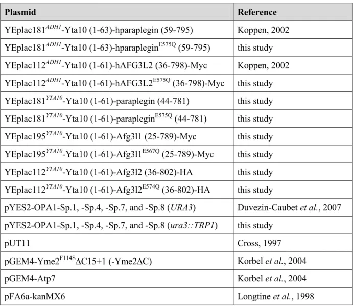
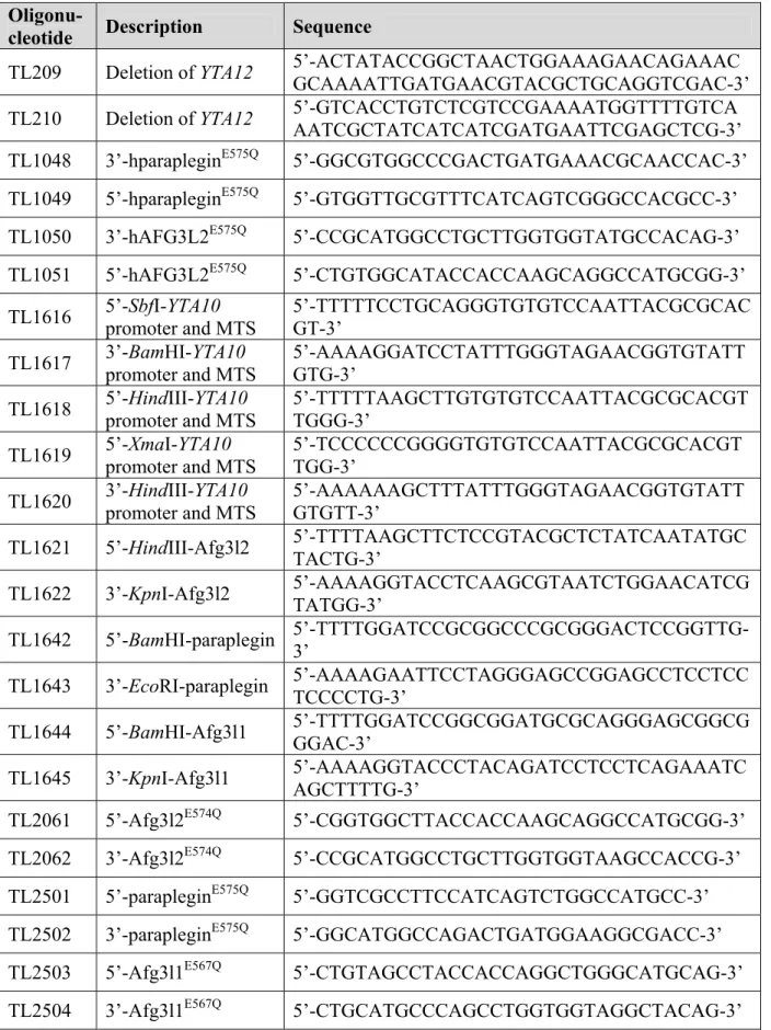
2.1 Expression plasmids and cloning procedures 34 2.2 Yeast strains and growth conditions 37
2.3 Cell culture 40
2.4 Preparation of total extracts from S. cerevisiae and HeLa cells 41 2.5 Preparation of cellular membranes from S. cerevisiae 41 2.6 Alkaline extraction of mitochondrial membranes 42 2.7 Isolation of mitochondria from human fibroblasts 42 2.8 Blue native polyacrylamide gel electrophoresis (BN-PAGE) 43 2.9 Gel filtration analysis of mitochondrial extracts from yeast 43 2.10 Degradation of newly imported, radiolabelled polypeptides in isolated yeast
mitochondria 44
2.11 Co-immunoprecipitation 45
2.12 Immunological detection of proteins 45
2.13 Miscellaneous 47
Table of contents
3 Results 48
3.1 Identification of the human AFG3L2 complex, a novel homo-oligomeric m-AAA
protease 48
3.1.1 A homo-oligomeric hAFG3L2 complex in HSP fibroblasts and upon expression in yeast 48 3.1.2 The hAFG3L2 complex can substitute for the yeast m-AAA protease 50 3.1.3 Proteolysis by homo-oligomeric hAFG3L2 complexes 52 3.1.4 hAFG3L2 is active upon homo-oligomerisation and assembly with hparaplegin 55 3.2 Murine m-AAA protease subunits assemble into protease complexes with variable
subunit composition 58 3.2.1 Murine m-AAA protease subunits show mitochondrial localisation and correct membrane
insertion when expressed in yeast 58 3.2.2 Murine Afg3l1 and Afg3l2 can assemble into homo-oligomeric complexes able to
substitute for the yeast m-AAA protease 60 3.2.3 Murine paraplegin can assemble with either Afg3l1 or Afg3l2 into hetero-oligomeric
protease complexes 63 3.2.4 Proteolysis by homo- and hetero-oligomeric murine m-AAA protease complexes 65 3.2.5 Evidence for autocatalytic processing of murine m-AAA protease subunits during
mitochondrial import 68 3.3 Mammalian homo-oligomeric m-AAA proteases mediate OPA1 processing in yeast 71
3.3.1 OPA1 processing can be reconstituted in yeast and is mediated by the endogenous m-
AAA protease 71
3.3.2 Deletion of yeast PHB1 can enhance m-AAA protease mediated OPA1 processing 74 3.3.3 Proteolytic processing of OPA1 splice variants by the human AFG3L2 complex 76 3.3.4 Variable efficiencies of OPA1 processing by homo- and hetero-oligomeric murine m-AAA
proteases 76
4 Discussion 81
4.1 Mammalian m-AAA protease subunits assemble into proteolytic complexes with variable subunit composition 81 4.2 Mammalian m-AAA proteases have conserved and over-lapping functions in
mitochondria 84
4.3 Mammalian homo-oligomeric m-AAA protease complexes mediate OPA1 processing reconstituted in yeast 86 4.4 OPA1 processing provides first evidence for different substrate specificities of m-
AAA protease isoenzymes 89 4.5 The versatile assembly of the mammalian m-AAA protease and its implications for the pathogenesis of HSP 91
5 Abstract 94 6 References 95 7 List of Abbreviations 110 8 Appendix 112 Danksagung 117 Eidesstattliche Erklärung 118 Lebenslauf 119
1 Introduction
1.1 The proteolytic system of mitochondria
Cell survival critically depends on the integrity and functionality of mitochondria.
These organelles evolved from an endosymbiontic relationship of aerobic bacteria and primordial eukaryotic cells and are the major energy production site in “modern” eukaryotic cells (Wallace, 2005). Besides oxidative phosphorylation, crucial activities in intermediary metabolism, calcium signalling, and cell death pathways highlight the central role of mitochondria for cellular physiology (Chan, 2006a; McBride et al., 2006). In agreement with the multitude of cellular functions, mitochondrial dysfunction is thought to contribute to cellular ageing and plays an important role in human diseases, such as diabetes, cancer, and prevalent neurodegenerative disorders (Chan, 2006a; Kwong et al., 2006; Lin and Beal, 2006). Moreover, a number of rare genetic diseases are caused by mutations in mitochondrial proteins (DiMauro, 2004; Taylor and Turnbull, 2005; Schapira, 2006).
It is therefore an important cellular task to ensure correct mitochondrial biogenesis and maintain mitochondrial activities under varying physiological conditions. Mitochondria are built up of ~1000 proteins (Mootha et al., 2003; Sickmann et al., 2003; Taylor et al., 2003).
The vast majority of them are nuclearly encoded, synthesised on cytosolic ribosomes, and imported into mitochondria. However, a small number of mitochondrial proteins, almost exclusively subunits of respiratory chain complexes in the inner membrane, are encoded by the mitochondrial genome (Anderson et al., 1981; Foury et al., 1998). They must assemble with nuclearly encoded subunits to constitute a functionally active respiratory chain and to allow oxidative phosphorylation. Maintenance of mitochondrial activities therefore depends on the coordinated expression of two cellular genomes. Moreover, this task is made even more difficult by the fact that mitochondria are highly dynamic organelles. They undergo constant fusion and fission and frequently change their shape in response to altered physiological conditions (Okamoto and Shaw, 2005).
Mitochondria must be able to eliminate excess, non-assembled polypeptides to avoid potential deleterious effects on organellar functions. Elaborate proteolytic systems which conduct the quality surveillance of mitochondrial proteins are present within different subcompartments of the organelle. A number of these proteases also exert essential housekeeping functions and control crucial steps during mitochondrial biogenesis. Many of
Introduction 8 them are highly conserved and ubiquitously distributed in eukaryotic cells. They can be categorised into several classes according to their main activities (Fig. 1; Tab. 1):
Table 1. Mitochondrial peptidases.
Name Loc. Yeast Function in yeast
Mammals Function in mammals Processing Peptidases
MPP M Mas1
Mas2
Presequence cleavage α-MPP β-MPP
See yeast
MIP M Oct1 Removal of
octapeptides
MIPEP (HMIP)
See yeast
IMP IM Imp1
Imp2 Presequence cleavage IMMP1L
IMMP2L Unknown IMS Atp23 Atp6 processing and
assembly
KUB3 Unknown Rhomboid IM Pcp1 Ccp1 and Mgm1
processing
PARL Protection against apoptosis
ATP-dependent proteases
i-AAA IM Yme1 Quality control YME1L1 Unknown
m-AAA IM Yta10
Yta12 Quality control, protein processing, membrane dislocation
paraplegin AFG3L1*
AFG3L2
Quality control, cleavage of MrpL32 and OPA1
Lon M Pim1 Quality control,
mtDNA maintenance and gene expression
LON Quality control,
mtDNA binding
ClpXP M - ClpP
ClpX
Quality control Oligopeptidases
IMS Mop112 Degradation of pep-
tides and presequences PreP** Aβ degradation1 IMS Prd1 Degradation of pep-
tides and presequences
Neurolysin Peptide degradation
M Lap3 Aminopeptidase,
protection against homocysteine2
Bleomycin hydrolase
See yeast
Other proteases
IM Oma1 Quality control OMA1 Unknown
IMS - HtrA2
(Omi) Pro-apoptotic, cleavage of β-APP
*AFG3L1 is only expressed in mice but encoded by a pseudogene in humans. **Human PreP has been localised to the matrix. 1Falkevall et al., 2006. 2Zimny et al., 2006.
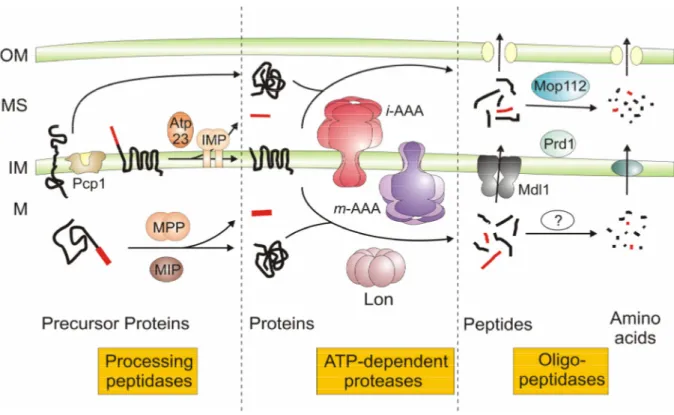
Figure 1. Proteolytic pathways within mitochondria of Saccharomyces. cerevisiae.
Mitochondrial proteases can be functionally classified into processing peptidases, ATP-dependent proteases, and oligopeptidases. Proteins residing in the intermembrane space (IMS), the inner membrane (IM), or the matrix (M) can be degraded by the consecutive action of ATP-dependent proteases and oligopeptidases to amino acids. The oligopeptidases Prd1 and Mop112 have been demonstrated to degrade peptides generated by the i-AAA protease. Although oligopeptidases are present in the matrix, it remains to be determined which of them act in concert with ATP-dependent proteases during protein turnover. See text for details. OM, outer membrane.
1.1.1 Processing peptidases
Sorting signals of nuclearly encoded proteins are removed by specific processing peptidases present within different subcompartments of mitochondria (Gakh et al., 2002).
These enzymes generally exhibit rather degenerate sequence specificity and, in case of composite targeting sequences, can act sequentially. The mitochondrial processing peptidase MPP, a conserved hetero-dimeric metallopeptidase, cleaves off sorting sequences in the matrix space (Hawlitschek et al., 1988; Yang et al., 1988; Ou et al., 1989). Some mitochondrial precursor proteins are cleaved by a second processing peptidase in the matrix space (Kalousek et al., 1988). The mitochondrial intermediate peptidase MIP, a monomeric metallopeptidase, removes an octapeptide from preproteins after their processing by MPP (Isaya et al., 1991; Isaya et al., 1992; Kalousek et al., 1992). The physiological role of MIP cleavage is currently poorly understood; however, severe phenotypes upon gene inactivation in yeast and mice suggest important regulatory functions (Isaya et al., 1994; Gakh
Introduction 10 et al., 2002). The so-called inner membrane peptidase IMP is located within the inner membrane but exposes its active sites to the intermembrane space of mitochondria (Gakh et al., 2002). This hetero-oligomeric complex is composed of a non-catalytic subunit, Som1, and two catalytic subunits containing serine/lysine dyads (Nunnari et al., 1993; Schneider et al., 1994; Jan et al., 2000). IMP mediates the maturation of intermediate forms of several nuclearly encoded proteins generated by MPP in the matrix space (Daum et al., 1982;
Nunnari et al., 1993; Burri et al., 2005), as well as mitochondrially encoded subunit 2 of cytochrome c oxidase (Cox2) (Pratje et al., 1983).
In addition to these generic enzymes, several other mitochondrial peptidases can act as processing peptidases and regulate the biogenesis of mitochondria. These include energy- dependent AAA proteases, which are discussed in detail below, but also enzymes with apparently more specialised functions. The conserved metallopeptidase Atp23 cleaves off the presequence of the mitochondrially encoded subunit Atp6 of the F1FO-ATP synthase complex in the intermembrane space after its insertion into the inner membrane (Osman et al., 2007;
Zeng et al., 2007). Independent of its proteolytic activity, Atp23 acts as a chaperone protein and promotes the assembly of Atp6 into the membrane-embedded FO-moiety of the ATP synthase complex (Osman et al., 2007; Zeng et al., 2007). A processing peptidase with a regulatory role for mitochondrial biogenesis is represented by the rhomboid protease in the inner membrane. Rhomboid proteases comprise a conserved family of membrane-embedded serine peptidases, which possess catalytic residues within their hydrophobic, membrane- spanning domains and which generate soluble, biologically active protein fragments by cleaving membrane-anchored proteins within transmembrane segments (Urban, 2006). The yeast rhomboid Pcp1 cleaves the dynamin-like GTPase Mgm1 (Herlan et al., 2003;
McQuibban et al., 2003; Sesaki et al., 2003a) and cytochrome c peroxidase (Ccp1), a mitochondrial scavenger of reactive oxygen species (ROS) (Esser et al., 2002). Mgm1 is a central component of the mitochondrial fusion machinery and accordingly Pcp1-mediated processing affects mitochondrial dynamics (Herlan et al., 2003).
1.1.2 ATP-dependent proteases
Proteases powered by ATP have been identified in various subcompartments of mitochondria. They generally form multimeric protein complexes, which constitute sequestered, proteolytic microcompartments, and contain P-loop ATPase domains characteristic of the AAA+-family of ATPases (Sauer et al., 2004; Hanson and Whiteheart, 2005). The energy derived from ATP-hydrolysis is utilised to unfold specific substrate
proteins and to transport them into this internal proteolytic cavity (Baker and Sauer, 2006).
The importance of ATP-dependent proteases for mitochondrial functions is illustrated by severe phenotypes associated with the loss of these proteases in yeast (Van Dyck and Langer, 1999). Although these phenotypes are understood on the molecular level only in a few cases, they appear to reflect two activities of the proteases: protein quality surveillance and proteolytic control of regulatory steps during mitochondrial biogenesis. Mitochondrial ATP- dependent proteases are closely related to bacterial enzymes and can be assigned to highly conserved protease families, the Lon proteases, the Clp proteases, and FtsH-like AAA proteases (Fig. 1; Tab. 1).
The Lon protease (or PIM1 protease in yeast) is localised in the mitochondrial matrix space (Suzuki et al., 1994; Van Dyck et al., 1994; Wang et al., 1994). This serine protease forms homo-oligomeric complexes which, based on cryoelectron microscopy studies on purified PIM1 protease, represent flexible, ring-shaped heptamers (Stahlberg et al., 1999).
The subunits are nuclearly encoded and synthesised in a prepro-form. After import into mitochondria, maturation and thereby activation of the subunits occurs in a two-step process:
the mitochondrial targeting sequence is removed by MPP followed by protease assembly and autocatalytic cleavage of the pro-region (Wagner et al., 1997). The PIM1/Lon protease conducts protein quality control in the matrix space, a function which is of particular importance under stress conditions, when thermally denatured or oxidatively damaged proteins accumulate (Bota and Davies, 2002). The protease recognises misfolded or damaged proteins, mediates their complete proteolysis, and thereby prevents their accumulation and deleterious effects on mitochondrial activities. In addition to its function in protein quality control, regulatory functions of the Lon protease during mitochondrial biogenesis are suggested by phenotypes associated with the loss of PIM1 protease in yeast. Deletion of the PIM1 gene causes the destabilisation of the mitochondrial genome, impairs mitochondrial gene expression, and results in respiratory deficiency (Suzuki et al., 1994; Van Dyck et al., 1994; Van Dyck et al., 1998a). Mitochondrial Lon proteases exhibit a peculiar binding affinity for nucleic acids pointing to a specific role of these proteases for DNA maintenance (Fu and Markovitz, 1998; Lu et al., 2003; Liu et al., 2004). It is conceivable that Lon proteases affect mitochondrial DNA (mtDNA) stability by complete proteolysis or by processing of a regulatory protein. The autocatalytic maturation of newly imported Pim1 subunits in yeast illustrates that Lon proteases, like the m-AAA protease (see below), can indeed act as a processing peptidase.
Introduction 12 A second ATP-dependent protease, the ClpXP protease, is present in the matrix of mammalian mitochondria (De Sagarra et al., 1999; Santagata et al., 1999). This hetero- oligomeric protease is built up of proteolytic ClpP subunits with serine peptidase activity and of ClpX subunits, which exert ATPase and, most likely, chaperone activity and confer substrate specificity to the complex (Kang et al., 2002; Kang et al., 2005). Notably, yeast mitochondria harbour only ATPase but no proteolytic ClpP subunits suggesting non- proteolytic functions (Van Dyck et al., 1998b). The role of Clp proteases within mitochondria is currently not understood. The ClpP-mediated turnover of a misfolded model substrate and the increased expression of ClpP under cellular stress in mammalian cells suggest a similar role of mitochondrial Clp and Lon proteases for protein quality control (Zhao et al., 2002).
Members of a third class of ATP-dependent proteases, the AAA proteases (AAA:
ATPases associated with a variety of cellular activities), are present within the inner membrane of mitochondria and expose their catalytic sites either to the matrix or the intermembrane space (Fig. 1) (Langer, 2000). AAA proteases conduct protein quality surveillance in the inner membrane and exert crucial functions during mitochondrial biogenesis. They are discussed in detail below.
1.1.3 Oligopeptidases
Oligopeptidases are, with few exceptions, the most poorly characterised mitochondrial proteases. Their existence was suggested by the observation that polypeptides can be completely degraded to amino acids within mitochondria (Desautels and Goldberg, 1982;
Young et al., 2001). As ATP-dependent proteases are known to degrade proteins processively to peptides, proteolysis was expected to be completed by other proteins with oligopeptidase activity (Fig. 1). Several proteins with oligopetidase activity in vitro have been identified, mostly localised in the intermembrane space. However, evidence for their in vivo function is scarce. Yeast mutant cells lacking mitochondrial oligopeptidases show usually no or only mild phenotypes, most likely due to the redundant activity of various enzymes (Garcia- Alvarez et al., 1987; Büchler et al., 1994; Kambacheld et al., 2005). Moreover, mitochondria contain a peptide export system which allows the efficient extrusion of peptides and prevents their accumulation in case of an impaired proteolysis. The ABC-transporter protein Mdl1 has been linked to this transport process (Fig. 1) (Young et al., 2001). Nevertheless, a quantitative assessment of mitochondrial peptide export and the mass-spectrometric identification of peptides released from yeast mitochondria provided first evidence for the function of oligopeptidases in vivo (Kambacheld et al., 2005). Two oligopeptidases were found to cleave
peptides generated by the i-AAA protease in the intermembrane space (Fig. 1) (Kambacheld et al., 2005): the thimet oligopeptidase Prd1 (or saccharolysin) and a second metallopeptidase Mop112/Cym1 (or PreP1 and PreP2 in Arabidopsis thaliana and PreP in human) (Jonson et al., 2004; Kambacheld et al., 2005; Falkevall et al., 2006, Glaser et al., 2006). Peptides degraded by this group of peptidases also include various mitochondrial targeting signals cleaved off by specific processing peptidases (Stahl et al., 2002; Moberg et al., 2003;
Kambacheld et al., 2005). Interestingly, the yeast oligopeptidases Prd1 and Mop112 also mediate the proteolytic breakdown of peptides, which were initially generated in the matrix and then transported into the intermembrane space (Kambacheld et al., 2005). They thus can contribute to the complete degradation of proteins localised in another mitochondrial subcompartment. Several enzymes with proposed oligopeptidase activity have also been identified in the matrix space including the bleomycin hydrolase Lap3. It is a cysteine peptidase which is localised both in the cytosol and the mitochondrial matrix space in yeast (Kambouris et al., 1992; Enenkel and Wolf, 1993; Magdolen et al., 1993; Huh et al., 2003;
Sickmann et al., 2003). A functional characterisation of the in vivo function of matrix- localised oligopeptidases, however, is still awaited. It is likely that they act in concert with ATP-dependent proteases in the proteolysis of matrix proteins and are thus part of a similar pathway as present in the intermembrane space (Fig. 1).
1.1.4 Other mitochondrial proteases
A number of additional proteases are present within mitochondria but their functional classification into the above mentioned categories remains unclear. The metallopeptidase Oma1 has been identified in a genome-wide screen for mitochondrial peptidases in yeast and was localised to the inner membrane of mitochondria with its catalytic site facing the matrix space (Käser et al., 2003). Oma1 is a member of a conserved and widespread family of membrane-integrated metallopeptidases and was shown to cleave a misfolded polytopic membrane protein at multiple sites (Käser et al., 2003). It therefore exerts a function similar to the m-AAA protease for quality control of inner membrane proteins and has been proposed to be part of a salvage system under conditions of limited AAA protease activity.
HtrA2 (Omi, Prss25) is localised in the intermembrane space of mammalian mitochondria (Suzuki et al., 2001; Hegde et al., 2002). It is a member of a conserved family of oligomeric serine peptidases, which were functionally linked to protein quality control in various organisms (Clausen et al., 2002; Kim and Kim, 2005). Mitochondrial HtrA2 has been identified as a pro-apoptotic XIAP-binding protein in the cytosol and was demonstrated to be
Introduction 14 released from mitochondria in apoptotic cells (Suzuki et al., 2001; Hegde et al., 2002; Martins et al., 2002; van Loo et al., 2002; Verhagen et al., 2002). A pro-apoptotic function of HtrA2, however, was recently challenged as targeted deletion of HtrA2 in mice did not impair the rate of apoptosis, but led to a selective loss of neurons in the striatum (Martins et al., 2004).
Consistently, a missense mutation inactivating HtrA2 in mice causes a neuromuscular disorder (Jones et al., 2003). The molecular basis of these phenotypes, as the function of HtrA2 within mitochondria, is currently not understood. Murine embryonic fibroblasts lacking HtrA2 exhibit an increased sensitivity to stress-induced cell death (Martins et al., 2004). It is therefore conceivable that, in analogy to homologous bacterial proteases, HtrA2 conducts protein quality control and protects mitochondrial activities under stress conditions (Clausen et al., 2002; Kim and Kim, 2005). Evidence for a processing activity of HtrA2 was recently provided analysing cleavage of the β-amyloid precursor protein in vivo and in vitro (Park et al., 2006).
1.2 AAA proteases
1.2.1 Domain structure and assembly of AAA proteases
AAA proteases are integral parts of the mitochondrial inner membrane and their catalytic domains are facing either the matrix or the intermembrane space (Langer, 2000).
Accordingly, they were termed m-AAA protease, active on the matrix side, and i-AAA protease, active on the intermembrane side (Fig. 2). AAA proteases form large complexes with a native molecular mass of ~1 MDa, which are composed of closely related or identical subunits of 70-80 kDa (Arlt et al., 1996; Leonhard et al., 1996; Klanner et al., 2001; Atorino et al., 2003; Urantowka et al., 2005). An N-terminal mitochondrial sorting signal targets the nuclearly encoded subunits to mitochondria. While m-AAA protease subunits are anchored to the inner membrane by two transmembrane segments present in the N-terminal part, i-AAA protease subunits span the inner membrane only once (Fig. 2). Accordingly, catalytic domains following the membrane-embedded domains are exposed to the intermembrane or matrix space. The AAA domain contains Walker A and Walker B motifs involved in ATP binding and hydrolysis, respectively, and the so-called second region of homology (SRH) (Hanson and Whiteheart, 2005). Two conserved arginine residues in this region protrude into the catalytic site of adjacent subunits and thereby stimulate ATP hydrolysis (Hanson and Whiteheart, 2005). Mutational alterations of these arginine fingers in bacterial and mitochondrial m-AAA protease subunits impaired ATPase activity (Karata et al., 1999;
Karata et al., 2001; Korbel et al., 2004). Therefore, assembly of AAA protease subunits is a prerequisite for ATP-dependent proteolysis. AAA proteases belong to the M41 family of metallopeptidases characterised by the consensus metal-binding motif HExxH (x represents a variable amino acid residue) (Rawlings and Barrett, 1995). Mutation of the glutamate residue within the proteolytic centre inhibits protein degradation by AAA proteases (Arlt et al., 1996;
Guélin et al., 1996; Leonhard et al., 1996; Weber et al., 1996; Atorino et al., 2003). Notably, proteolytic activity is only completely abolished if all subunits harbour mutations in their proteolytic centres. Hetero-oligomeric mutant m-AAA proteases still containing wild type subunits exhibit residual activity and are capable of processing protein substrates (Arlt et al., 1998).
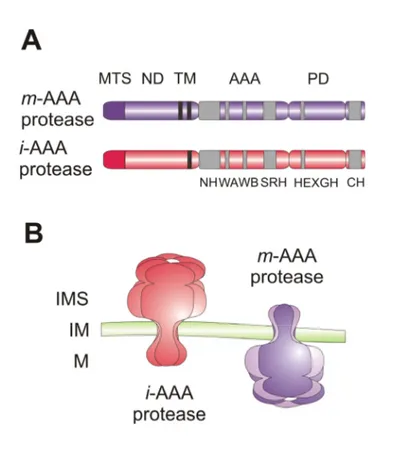
Figure 2. Mitochondrial AAA proteases.
(A) Domain structure of AAA protease subunits. Subunits of i- and m-AAA protease complexes harbour one or two transmembrane domains (TM), respectively. Conserved sequence motifs within domains are indicated with grey boxes. MTS, mitochondrial targeting sequence; ND, N-terminal domain; AAA, AAA domain; PD, proteolytic domain; NH, N-terminal helices; WA, Walker A-motif;
WB, Walker B-motif; SRH, second region of homology; HEXGH, proteolytic centre; CH, C-terminal helices.
(B) Membrane topology of AAA proteases. The stoichiometry and arrangement of mitochondrial AAA protease subunits within a complex is still speculative. IMS, intermembrane space; IM, inner membrane; M, matrix space.
Introduction 16 For two homologous eubacterial FtsH proteases, the crystal structure of the soluble cytosolic region containing the catalytic domains has recently been solved (Bieniossek et al., 2006; Suno et al., 2006). They form a hexameric structure which adopts a flat-cylinder-like shape and consists of two structurally separate rings composed of the proteolytic domains and the AAA domains, respectively (Fig. 3). The proteolytic centres are located within an inner cavity of the hexameric molecule and can be accessed via a narrow central pore formed by the AAA domains facing the membrane. A similar hexameric and ring-like assembly can also be suggested for mitochondrial AAA proteases.
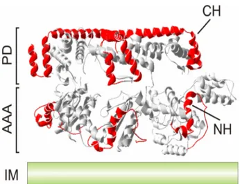
Figure 3. Substrate engagement by the yeast i-AAA protease.
A lattice-like arrangement of the substrate binding regions CH (C-terminal helices) and NH (N- terminal helices) at the surface of the proteolytic cylinder of an AAA protease. Helices homologous to CH and NH of yeast Yme1 are marked in red in the crystal structure of Thermotoga maritima FtsH (Bieniossek et al., 2006). Only three subunits are shown. AAA, AAA domain; PD, proteolytic domain;
IM, inner membrane.
Both the m- and the i-AAA protease recognise and degrade non-native and non- assembled polypeptides to peptides (Fig. 4A) (Arlt et al., 1996; Leonhard et al., 1996). These proteases exhibit degenerate substrate specificity and, similar to molecular chaperone proteins, recognise the folding state of solvent-exposed domains of membrane proteins (Leonhard et al., 1999). Studies on the i-AAA protease subunit Yme1 of yeast allowed the identification of two helical binding regions, which form a lattice-like structure at the surface of the proteolytic cylinder and mediate the initial encounter of substrate proteins with the protease (Fig. 3) (Leonhard et al., 1999; Graef et al., 2007): helices C-terminal of the proteolytic domain (CH) and N-terminal helices of the AAA domain (NH), which are located
in close proximity to the membrane surfaces and highly negatively charged. Thus, substrate proteins initially interact with the i-AAA protease at the outer surface of the proteolytic cylinder, before they enter the proteolytic chamber (Graef et al., 2007). Evidence for substrate translocation into a proteolytic chamber through the central pore of mitochondrial AAA proteases has been obtained by mutational analysis of a conserved loop motif YVG (aromatic- hydrophobic-glycine) in Yme1 (Graef and Langer, 2006), which has been localised to the central pore of other hexameric AAA+ ring complexes (Wang et al., 2001). Furthermore, AAA protease-mediated degradation of inner membrane proteins involves the extraction of the substrate from the membrane bilayer (Leonhard et al., 2000). Recently, the ability of the m-AAA protease to mediate vectorial membrane dislocation of proteins in an ATP-dependent reaction has been directly demonstrated (see below) (Tatsuta et al., 2007). This membrane extraction of substrate proteins is likely to be facilitated by the membrane-embedded parts of AAA protease subunits which might form a pore-like structure or provide at least a more hydrophilic environment (Korbel et al., 2004).
The yeast m-AAA protease assembles with the prohibin complex into a large supercomplex (Steglich et al., 1999). Two ubiquitous and highly conserved subunits, Phb1 and Phb2, assemble into a multimeric complex, which is exposed to the intermembrane space and anchored N-terminally to the inner membrane (Steglich et al., 1999; Nijtmans et al., 2000; Artal-Sanz et al., 2003). Prohibitins appear to modulate proteolysis by the m-AAA protease, since deletion of prohibitins in yeast results in accelerated protein degradation by the m-AAA protease (Steglich et al., 1999). However, the molecular mechanism of how prohibitins exert this function is not understood.
1.2.2 Functions of AAA proteases within mitochondria
AAA proteases in the inner mitochondrial membrane exert essential housekeeping functions like protein quality control (Fig. 4A) and have crucial roles in mitochondrial biogenesis. Therefore, their deletion or inactivation causes severe pleiotropic phenotypes in various organisms, which are best characterised in the yeast Saccharomyces cerevisiae. Here, all observed defects can be attributed to a loss of proteolytic activity as identical phenotypes were observed after deletion of an AAA protease subunit or after inactivation of the proteolytic sites of all subunits of AAA protease complexes (Leonhard et al., 1996; Weber et al., 1996; Arlt et al., 1998). Notably, yeast cells deficient in both the m- and the i-AAA protease are not viable (Lemaire et al., 2000; Leonhard et al., 2000).
Introduction 18
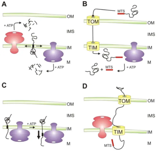
Figure 4. Versatile functions of AAA proteases within mitochondria.
A) Protein turnover. AAA proteases conduct protein quality surveillance and degrade excess and damaged proteins to peptides after their dislocation from the membrane.
B) Protein processing. AAA proteases can cleave mitochondrial proteins resulting in their activation.
This is exemplified by the maturation of newly imported MrpL32 by yeast and mammalian m-AAA proteases, but additional substrates are likely to exist.
C) ATP-dependent membrane dislocation. Independent of its proteolytic activity, the m-AAA protease mediates the vectorial dislocation of Ccp1 precursor proteins to allow intramembrane cleavage by rhomboid. It is conceivable that this activity is required for the correct sorting of additional mitochondrial proteins residing in the inner membrane or the intermembrane space.
D) Protein import into mitochondria. The yeast i-AAA protease is required for the import of heterologously expressed PNPase into the mitochondrial intermembrane space. The role of the proteolytic activity of the i-AAA protease in this process is currently not understood. OM, outer membrane; IMS, intermembrane space; IM, inner membrane; M, matrix space; TOM, translocase of the outer membrane; TIM, translocase of the inner membrane; MTS, mitochondrial targeting sequence.
The yeast i-AAA protease is a homo-oligomeric complex of Yme1 subunits (Leonhard et al., 1996). In the absence of a functional i-AAA protease, yeast cells are characterised by a respiratory deficiency at high temperature, an inability to grow on rich glucose medium at low temperature, an increased rate of mtDNA escape to the nucleus (Thorsness and Fox, 1993;
Thorsness et al., 1993), and a petite-negative phenotype, i.e. strongly impaired growth in the
absence of mtDNA (Weber et al., 1995). Moreover, ∆yme1 cells exhibit an accumulation of punctate mitochondria with grossly swollen compartments pointing to a potential role of Yme1 in maintaining normal mitochondrial morphology (Campbell et al., 1994). However, it remains an open question if these phenotypes are caused by the accumulation of misfolded polypeptides or the impaired proteolysis of specific substrate proteins. Only a limited number of protein quality control substrates of the yeast i-AAA protease has been identified including non-assembled Cox2 (Nakai et al., 1995; Pearce and Sherman, 1995; Weber et al., 1996), unassembled prohibitin subunits Phb1 and Phb2 (Kambacheld et al., 2005), as well as external NADH dehydrogenase (Nde1) (Augustin et al., 2005). In addition to protein quality surveillance (Fig. 4A), a novel function has recently been associated with the i-AAA protease.
Polynucleotide phosphorylase (PNPase) has been localised to the mitochondrial intermembrane space in mammals and was shown to be required for the maintenance of mitochondrial homeostasis (Chen et al., 2006). Import of mammalian PNPase into yeast mitochondria critically depends on the presence of the i-AAA protease Yme1 in mitochondria (Rainey et al., 2006). Yme1 binds to the precursor form of PNPase and mediates its translocation across the outer membrane, but does not degrade it suggesting a non-proteolytic function (Fig. 4D).
In yeast, the m-AAA protease is hetero-oligomeric and composed of two closely related subunits, Yta10 (Afg3) and Yta12 (Rca1), which assemble in an ATP-dependent manner into m-AAA protease complexes (Arlt et al., 1996). Yeast cells lacking either subunit or expressing proteolytically inactive variants of both subunits are respiratory deficient (Guélin et al., 1994; Tauer et al., 1994; Tzagoloff et al., 1994; Arlt et al., 1998) and lack assembled respiratory chain and ATP-synthase complexes in the inner membrane (Paul and Tzagoloff, 1995; Arlt et al., 1998; Galluhn and Langer, 2004). As substrates, non-assembled, mitochondrially encoded respiratory chain subunits of complexes III, IV and V (Cob, Cox1, Cox3, Atp6, Atp8, Atp9) were found to be degraded by the yeast m-AAA protease (Arlt et al., 1996; Guélin et al., 1996). In addition to these integral membrane proteins, the m-AAA protease has also been demonstrated to degrade peripheral membrane proteins such as Atp7 (Korbel et al., 2004). Recently, the processing of specific substrate proteins with regulatory functions has been recognised as a second housekeeping function of the m-AAA protease (Fig. 4B). Specific substrates for proteolytic cleavage include the ribosomal subunit MrpL32 (Nolden et al., 2005) and cytochrome c peroxidase (Ccp1), a heme-binding ROS scavenger in the intermembrane space (Esser et al., 2002; Tatsuta et al., 2007). These findings have provided a rationale for cellular defects associated with an m-AAA protease-deficiency in
Introduction 20 yeast. Moreover, they allowed the identification of ATP-dependent membrane dislocation as a novel non-proteolytic function of AAA proteases (Fig. 4C).
1.2.3 Proteolytic processing by AAA proteases
In order to purify specific substrate proteins biochemically, an m-AAA protease variant harbouring point mutations in the proteolytic centres was employed as substrate-trap (Nolden et al., 2005). This approach has led to the identification of MrpL32, a conserved nuclearly encoded subunit of mitochondrial ribosomes (Grohmann et al., 1991). Surprisingly, the m-AAA protease does not affect the stability of this protein, but cleaves off the N-terminal targeting sequence upon import into mitochondria (Fig. 5A) (Nolden et al., 2005). MrpL32 processing is a prerequisite for its assembly into ribosomes, which is thus completed in close proximity to the inner membrane. An impaired translation within mitochondria rationalises the loss of the respiratory competence of m-AAA protease-deficient yeast cells, as essential subunits of the respiratory chain are encoded by the mitochondrial DNA (Foury et al., 1998).
That the processing of MrpL32 indeed represents a key function of the m-AAA protease in yeast is demonstrated by a complementation experiment. Expression of an MrpL32 variant, which harbours an unrelated presequence and is matured by MPP, maintains respiratory growth of m-AAA protease-deficient cells (Nolden et al., 2005). Therefore, it can be concluded that cellular defects associated with an inactivation of the yeast m-AAA protease are largely explained by an impaired processing of a matrix protein rather than the deleterious effects of accumulating non-native membrane proteins in mitochondria.
Another substrate whose processing depends on the yeast m-AAA protease is the ROS-scavenger protein Ccp1 (Fig. 5B). A bipartite presequence targets this nuclearly encoded protein into the intermembrane space and is cleaved off by the consecutive action of m-AAA and rhomboid proteases in the inner membrane (Esser et al., 2002). Strikingly, although strictly dependent on the presence of the m-AAA protease, maturation of Ccp1 by the rhomboid protease Pcp1 was observed in yeast cells harbouring a proteolytically inactive variant of the m-AAA protease (Tatsuta et al., 2007). In contrast, Ccp1 cleavage by rhomboid was abolished if mutations were introduced into the AAA domains of m-AAA protease subunits. Subsequent experiments revealed that the m-AAA protease is required to mediate the ATP-dependent vectorial dislocation of Ccp1 from the membrane bilayer (Fig. 5B) (Tatsuta et al., 2007). This activity most likely ensures the correct positioning of Ccp1 relative to the membrane to allow intramembrane cleavage by Pcp1. It appears that the general
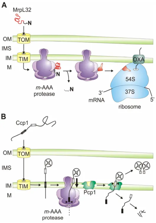
Figure 5. Protein processing by the yeast m-AAA protease.
A) Processing of MrpL32, a subunit of mitochondrial ribosomes. Newly imported MrpL32 is matured by the m-AAA protease, most likely accompanied by a conformational change. This allows its assembly with pre-assembled ribosomal particles at the inner surface of the inner membrane. The presequence of MrpL32 is either degraded within mitochondria or released from the organelle (Kambacheld et al., 2005).
B) Processing of cytochrome c peroxidase (Ccp1), a ROS scavenger in the intermembrane space. Ccp1 is targeted to mitochondria by a bipartite presequence which is cleaved off in a two-step process by the m-AAA protease and rhomboid (Pcp1). The first hydrophobic stretch triggers lateral sorting of the precursor protein in the inner membrane. The m-AAA protease mediates the ATP-dependent vectorial dislocation and positions the precursor protein within the lipid bilayer for intramembrane cleavage by rhomboid. Mature Ccp1 is released as a soluble protein into the intermembrane space. OM, outer membrane; IMS, intermembrane space; IM, inner membrane; M, matrix space; TOM, translocase of the outer membrane; TIM, translocase of the inner membrane; OXA, membrane insertion machinery containing Oxa1.
Introduction 22 activity of the m-AAA protease, namely to extract non-native substrate proteins from the membrane bilayer for proteolysis is recruited to ensure the biogenesis of Ccp1. Maturation of Ccp1 within mitochondria thus represents the first non-proteolytic function of the m-AAA protease within mitochondrial biogenesis. It is tempting to speculate that additional substrates of the m-AAA protease exist whose biogenesis depend on the ATP-dependent dislocase rather than the proteolytic activity of the m-AAA protease.
1.2.4 Mammalian AAA proteases
The presence of AAA proteases with their catalytic domains exposed to opposite membrane surfaces is conserved in mammals. AAA protease subunits with a high degree of homology to Yta10, Yta12, and Yme1 of yeast have been identified in humans and mice (Fig. 6) and several complementation studies in yeast have demonstrated a functional conservation of m- and i-AAA proteases (Shah et al., 2000; Atorino et al., 2003; Nolden et al., 2005).
A mammalian homologue of yeast Yme1 has been found in humans and mice, named YME1L1 (Coppola et al., 2000; Shah et al., 2000). Expression of human YME1L1 restores respiratory growth of ∆yme1 yeast cells at high temperature identifying it as the human i- AAA protease (Shah et al., 2000). Thus, the i-AAA protease in mammalian mitochondria apparently represents a homo-oligomeric assembly like in yeast. In contrast, next to nothing is known about its substrates and exact function in mammals and the generation of a Yme1l1- deficient mouse line is awaited.
In case of the m-AAA protease, three potential subunits have been identified in humans and mice, termed Afg3l1, Afg3l2, and paraplegin (Casari et al., 1998; Shah et al., 1998; Banfi et al., 1999). They are all mitochondrially localised and ubiquitously present in mammalian tissues. However, Afg3l1 which is highly homologous to Afg3l2 is only expressed in mice, whereas it is encoded by a pseudogene in humans (Kremmidiotis et al., 2001). Both in human and murine mitochondria, paraplegin assembles with Afg3l2 into a hetero-oligomeric complex which can functionally substitute for the yeast m-AAA protease when expressed in yeast (Atorino et al., 2003; Nolden et al., 2005). The assembly and function of Afg3l1 has not been analysed. Remarkably, paraplegin was initially identified by its association with a neurodegenerative disease which also provided first insights into the role of m-AAA proteases in mammalian mitochondria. Mutations in the gene encoding paraplegin (spastic-paraplegia-gene 7/SPG7) are causative for an autosomal recessive form of hereditary
spastic paraplegia (HSP) (Casari et al., 1998). It is noteworthy that up to date disease-causing mutations have not been reported for human AFG3L2 and YME1L1.
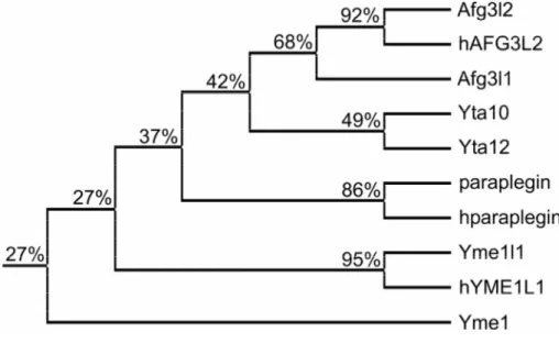
Figure 6. AAA protease subunits of yeast and mammals are evolutionary conserved.
Phylogenetic relationship between mitochondrial AAA protease subunits of yeast (Yta10, Yta12, Yme1), mice (paraplegin, Afg3l1, Afg3l2, Yme1l1), and humans (hparaplegin, hAFG3L2, hYME1L1), including their sequence identity.
1.3 Hereditary spastic paraplegia (HSP)
1.3.1 HSP is a clinically and genetically heterogenous disease
Hereditary spastic paraplegia comprises a diverse group of neurodegenerative disorders whose main clinical features include progressive weakness and spasticity of the lower limbs, loss of the vibratory sense, and urinary urgency (Rugarli and Langer, 2006;
Soderblom and Blackstone, 2006). The age of onset is variable, but usually lies between 10 and 40 years of age. A further characteristic of the “pure” forms of HSP, where these symptoms are isolated, is a remarkable tissue-specificity: only motor neurons of the cortico- spinal tracts and sensory neurons of the fasiculus gracilis are selectively affected by axonal degeneration (Rugarli and Langer, 2006; Soderblom and Blackstone, 2006). Interestingly, the axons of these neurons belong to the longest in the central nervous system reaching lengths of
~1 m. The axonal degeneration in HSP starts distally at the synaptic terminals and slowly proceeds towards the neuronal cell bodies which are preserved. Thus, HSP is a member of a new category of neurological disorders and can be classified as a “dying back” axonopathy
Introduction 24 (Züchner and Vance, 2005). In the “complicated” forms of HSP, patients have additional neurological abnormalities like cortical and cerebellar atrophy, amyotrophy, peripheral neuropathy, optic atrophy, deafness, short stature, and mental retardation (Harding, 1983).
The clinical heterogeneity of spastic paraplegias is paralleled by a genetic heterogeneity. The inheritance pattern can be X chromosome-linked, autosomal dominant, or autosomal recessive. In total, 33 genetic loci have been associated with different forms of HSP (SPG1 to SPG33). However, causative mutations have been identified only in 15 genes (Tab. 2), which can be divided into three functional categories: Firstly, genes whose products play a role in axonal targeting and pathfinding or myelination. These include the two X chromosome-linked genes SPG1 and SPG2 encoding the glycoprotein L1CAM and the proteolipid protein PLP1, respectively (Jouet et al., 1994; Saugier-Veber et al., 1994).
Secondly, genes whose products are involved in axonal transport and trafficking (Crosby and Proukakis, 2002; Soderblom and Blackstone, 2006). The kinesin heavy chain gene KIF5A (SPG10) is the paradigm of this group (Reid et al., 2002). KIF5A is part of a hetero-tetrameric protein complex, the neuronal kinesin-I, that acts as a plus-end-directed microtubule motor involved in anterograde transport of membranous organelles in nerve axons (Goldstein and Yang, 2000). The AAA protein spastin which is encoded by SPG4 interacts with microtubules and regulates microtubule dynamics (Errico et al., 2002; Errico et al., 2004; Evans et al., 2005). In addition, spastin interacts with one component of the endosomal sorting complex required for transport (ESCRT)-III and with the large GTPase atlastin (SPG3A), which also causative for HSP (Reid et al., 2005; Evans et al., 2006; Sanderson et al., 2006). Spartin (SPG20), maspardin (SPG21), and the recently identified protein ZFYVE27 (SPG33) have also been suggested to have a function in intracellular trafficking, but clear evidence is missing (Patel et al., 2002; Simpson et al., 2003; Mannan et al., 2006). The third functional group encompasses genes that have mitochondrial functions (Bross et al., 2004; Rugarli and Langer, 2006). Besides the m-AAA protease subunit paraplegin, mitochondrial Hsp60 (heat- shock protein 60; also known as chaperonin) which is encoded by SPG13 has been linked to HSP (Hansen et al., 2002). Hsp60 mediates the folding of newly imported proteins and protects mitochondrial proteins from heat denaturation (Voos and Röttgers, 2002). Recently, the protein product of the SPG31 gene, termed REEP1 (receptor expression-enhancing protein 1) has been localised to mitochondria but its function is unknown (Züchner et al., 2006). Conflicting results concerning the subcellular localisation of spartin have been reported (Lu et al., 2006; Robay et al., 2006). In one study, transfected and fluorescent-tagged
spartin was recently found to be mitochondrial and to co-localise with microtubules suggesting a possible role of spartin in transport of mitochondria (Lu et al., 2006).
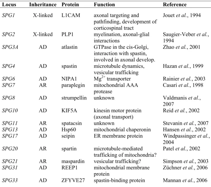
Table 2. Cloned genes which are linked to HSP.
Locus Inheritance Protein Function Reference
SPG1 X-linked L1CAM axonal targeting and
pathfinding, development of corticospinal tract
Jouet et al., 1994 SPG2 X-linked PLP1 myelination, axonal-glial
interactions Saugier-Veber et al., 1994
SPG3A AD atlastin GTPase in the cis-Golgi, interaction with spastin, involved in axonal develop.
Zhao et al., 2001
SPG4 AD spastin microtubule dynamics,
vesicular trafficking
Hazan et al., 1999 SPG6 AD NIPA1 Mg2+ transporter Rainier et al., 2003
SPG7 AR paraplegin mitochondrial AAA
protease
Casari et al., 1998
SPG8 AD strumpellin unknown Valdmanis et al.,
2007 SPG10 AD KIF5A kinesin motor protein
(axonal transport)
Reid et al., 2002
SPG11 AR spatacsin unknown Stevanin et al., 2007
SPG13 AD Hsp60 mitochondrial chaperonin Hansen et al., 2002 SPG17 AD seipin ER membrane protein Windpassinger et al.,
2004
SPG20 AR spartin microtubule-mediated
trafficking of mitochondria?
Patel et al., 2002 SPG21 AR maspardin vesicular trafficking? Simpson et al., 2003
SPG31 AD REEP1 mitochondrial membrane
protein
Züchner et al., 2006 SPG33 AD ZFYVE27 spastin-binding protein Mannan et al., 2006
AD, autosomal dominant; AR, autosomal recessive; X-linked, X chromosome-linked
1.3.2 HSP due to lack of paraplegin
Mutations in the SPG7 gene encoding paraplegin account for ~10% of recessive and sporadic cases of HSP (Casari and Rugarli, 2001). Analysis of the paraplegin mutations that were identified in human HSP patients suggests a complete loss of paraplegin function (McDermott et al., 2001; Wilkinson et al., 2004). What are the consequences of a paraplegin deficiency on mitochondrial activities in HSP patients? Primary fibroblasts from HSP patients showed defects in the assembly and activity of complex I of the respiratory chain, accompanied by decreased ATP levels (Atorino et al., 2003). An impaired activity of
Introduction 26 complex I can lead to an elevated production of reactive oxygen species (Barrientos and Moraes, 1999; Li et al., 2003). Consistently, an increased sensitivity of HSP fibroblasts towards oxidative stress was detected (Atorino et al., 2003).
Recently, a paraplegin-deficient mouse line has been generated which recapitulates central features of the human disease and thus can serve as a valuable model to further understand the molecular basis of mitochondrial dysfunction in HSP (Ferreirinha et al., 2004).
Similar to HSP patients, the long axons of paraplegin-deficient mice were specifically affected and found to contain enlarged and structurally abnormal mitochondria in the distal regions. These changes in mitochondrial morphology represented the first detectable defect whose appearance was followed by axonal swelling and finally progressive degeneration (Ferreirinha et al., 2004). Thus, the mammalian m-AAA protease might also play a role in the regulation of mitochondrial dynamics. Further studies revealed that, similar to m-AAA protease-deficient yeast cells, processing of murine MrpL32 and concomitantly mitochondrial translation were impaired in the absence of paraplegin (Nolden et al., 2005). When co- expressed with murine paraplegin and Afg3l2 in yeast, murine MrpL32 was matured identifying it as the first in vivo substrate of the mammalian m-AAA protease (Nolden et al., 2005). Since essential respiratory chain subunits are mitochondrially encoded (Anderson et al., 1981), mitochondrial-protein-synthesis defects should result in oxidative-phosphorylation deficiencies. However, the activities of respiratory chain complexes were not significantly affected and only moderately defective ATP synthesis became apparent in spinal cord mitochondria of old paraplegin-deficient mice (Ferreirinha et al., 2004).
1.3.3 A possible mechanism for axonal degeneration in HSP
Taken together, significant progress has been made in the recent years to unravel the pathogenetic mechanism of HSP due to a loss of paraplegin. However, it remains an open question whether accumulating non-native polypeptides, an impaired mitochondrial translation, or the impaired processing of another mitochondrial protein in the absence of paraplegin results in axonal degeneration. One can envision the following general model, which can also be reconciled with the genetic heterogeneity of this neurological disorder.
Axons depend on active anterograde transport for the supply of protein and lipid constituents, which are synthesised in the neuronal cell body and on retrograde transport for the degradation and replacement of old and damaged material (Hirokawa and Takemura, 2005).
Mitochondria are also transported in an anterograde manner mediated by kinesin motor proteins to the distal regions of axons (Hollenbeck and Saxton, 2005). Neurons with long
axons as those of the corticospinal tracts can be considered to be particularly vulnerable to defects in axonal trafficking as indicated by the axonal length-dependance in HSP. Thus, deficiencies in proteins directly involved in axonal transport such as KIF5A or in mitochondrial proteins like paraplegin and Hsp60 could both result in the absence of functional mitochondria from the synapses triggering degeneration. Consistently, it has been demonstrated that reduced mitochondria content leads to the loss of dendritic spines and synapses (Li et al., 2004). In the absence of paraplegin, synaptic mitochondria undergo morphological changes and accumulate which, in combination with a limited energy supply, might further affect axonal transport and eventually clog the axon. In line with this scenario, a delayed retrograde transport was indeed observed in motor neurons of paraplegin-deficient mice (Ferreirinha et al., 2004).
1.4 Mitochondrial dynamics
1.4.1 Mitochondrial fusion in yeast and mammals
Mitochondria are highly dynamic organelles whose morphology depends on the tissue, on the physiological condition of the cell, and in particular on the functional status of mitochondria (Okamoto and Shaw, 2005). This dynamic behaviour is crucial for a number of cellular processes, such as apoptosis, the inheritance of mtDNA, defence against oxidative stress, and development through spermatogenesis (Hales and Fuller, 1997; Chen and Chan, 2005; Chen and Butow, 2005; Cereghetti and Scorrano, 2006). Mitochondrial morphology is regulated by opposing but balanced fusion and fission events which maintain the normal mitochondrial network (Okamoto and Shaw, 2005). Loss of fusion results in mitochondrial fragmentation due to ongoing fission events. Conversely, loss of fission leads to the formation of elongated and highly interconnected mitochondria. The central components of the mitochondrial fusion and fission machineries have been identified, but the molecular mechanisms of both processes are not completely understood. Most proteins involved are conserved in yeast, flies, mice, and humans indicating that the fundamental mechanisms controlling mitochondrial dynamics have been maintained during evolution.
Mitochondrial fusion includes the fusion of the outer and the inner membrane of two organelles in a coordinated manner. Three proteins are essential for fusion in yeast and interact with each other to form a fusion complex: the two dynamin-related GTPases Fzo1 and Mgm1 as well as Ugo1 (Sesaki et al., 2003b; Wong et al., 2003; Sesaki and Jensen, 2004). Fzo1 is integrated into the outer membrane exposing its functional domains to the
Introduction 28 cytoplasm (Hermann et al., 1998; Rapaport et al., 1998; Fritz et al., 2001). Mgm1, on the other hand, resides in the intermembrane space but is present in two forms both of which are required for fusion (Wong et al., 2000; Herlan et al., 2003; Sesaki et al., 2003b; Wong et al., 2003): the large (l-) isoform, which is integrated into the inner membrane, and the short (s-) isoform, which is peripherally associated with inner and/or outer membrane and generated by proteolytic processing of the l-form, as discussed below. The third player, Ugo1, is also embedded into the outer membrane and exposes its N-terminal domain to the cytoplasm and the C-terminal domain to the intermembrane space (Sesaki and Jensen, 2001). Ugo1 has been shown to bind to both Fzo1 and Mgm1 via its N- and C-terminal domains thus linking the fusion components of the outer and inner membrane and possibly the fusion events of both membranes (Sesaki and Jensen, 2004). Outer- and inner-membrane fusion events are tightly coupled in vivo, but could be separated by the reconstitution of fusion in vitro (Meeusen et al., 2004). Furthermore, these in vitro studies revealed that Fzo1 is required for outer membrane fusion, whereas inner membrane fusion depends on Mgm1 (Meeusen et al., 2004; Meeusen et al., 2006). For both mitochondrial membranes, trans interactions of either Fzo1 or Mgm1 mediate tethering of opposing membranes and subsequent fusion.
Two homologues of Fzo1 have been identified in humans, called mitofusin (Mfn) 1 and 2, whereas OPA1 is the homologue of Mgm1 (Alexander et al., 2000; Delettre et al., 2000; Santel and Fuller, 2001; Rojo et al., 2002). Interestingly, Ugo1 does not have an obvious counterpart in mammals. The localisation of mitofusins and OPA1 in the outer membrane and intermembrane space, respectively, and their topology has been conserved in mammals (Rojo et al., 2002; Griparic et al., 2004). In addition, different isoforms of OPA1 are also present in mammalian mitochondria. The essential role of mitofusins and OPA1 in mitochondrial fusion has been established through the generation of knockout mice and by knockdown experiments using RNA intereference (RNAi). The absence of either Mfn1 or Mfn2 causes embryonic lethality in mice and isolated mouse embryonic fibroblasts only exhibit greatly reduced levels of mitochondrial fusion leading to a highly fragmented mitochondrial population (Chen et al., 2003; Chen et al., 2005). This indicates that Mfn1 and Mfn2 apparently play similar roles in mitochondrial fusion, which was supported by the finding that they can form both homo- and hetero-oligomeric complexes all competent for fusion (Chen et al., 2003; Eura et al., 2003; Chen et al., 2005). Similarly, depletion of OPA1 from mammalian cells also results in mitochondrial fragmentation due to loss of fusion (Olichon et al., 2003; Cipolat et al., 2004; Griparic et al., 2004; Chen et al., 2005).
1.4.2 Regulation of mitochondrial fusion by proteolytic processes
In yeast, two components of the mitochondrial fusion machinery, Fzo1 and Mgm1, are under proteolytic control. In the case of Fzo1, the maintenance of mitochondrial morphology depends on the tight control of its steady state concentration which is regulated by degradation of Fzo1 via two independent proteolytic pathways. On one hand, Fzo1 turnover can be induced by cell cycle arrest with the mating factor alpha which leads to mitochondrial fragmentation (Neutzner and Youle, 2005). This induced degradation of Fzo1 is mediated by the ubiquitin-proteasome system (UPS) (Escobar-Henriques et al., 2006), the central proteolytic system in the cytosol of eukaryotic cells (Ciechanover, 2005). On the other hand, Fzo1 is degraded in vegetatively growing yeast cells in a constitutive manner which depends on the F-box protein Mdm30 (Escobar-Henriques et al., 2006). In the absence of Mdm30, the steady state concentration of Fzo1 is increased and yeast cells accumulate aggregated and fragmented mitochondria (Fritz et al., 2003). F-box proteins often assemble into Skp1-Cdc53- F-box (SCF) E3 ubiquitin ligase complexes which ensure ubiquitin-dependent degradation by the 26S proteasome (Willems et al., 2004; Petroski and Deshaies, 2005). However, Mdm30- dependent turnover of Fzo1 does not involve SCF complexes or the UPS, but rather proceeds along an alternative proteolytic pathway which remains to be identified (Escobar-Henriques et al., 2006).
In contrast to control of Fzo1, Mgm1 is not subject to complete degradation but is proteolytically processed at its N-terminus yielding two isoforms within the mitochondrial intermembrane space: the large (l-) and the short (s-) isoform. Both Mgm1 isoforms are required for mitochondrial fusion and their balanced formation appears to be crucial for mitochondrial morphology (Herlan et al., 2003). According to the alternative topogenesis model, the ratio of both isoforms is dictated by the level of matrix ATP (Herlan et al., 2004).
The Mgm1 protein contains two N-terminal transmembrane segments of which the first one serves as a stop-transfer signal during mitochondrial import of the pre-protein. At low ATP levels, the subsequent removal of the MTS by MPP and lateral membrane insertion result in l- Mgm1, which is anchored to the inner membrane. At high ATP levels, however, Mgm1 can be pulled further into the matrix by the ATP-dependent mitochondrial import motor until the second hydrophobic segment reaches the inner membrane. Thereby, a second cleavage site within this segment gets accessible for the rhomboid protease Pcp1 in the inner membrane.
Pcp1-mediated cleavage generates s-Mgm1 (Herlan et al., 2003; McQuibban et al., 2003;
Sesaki et al., 2003a), which is peripherally associated with the inner and/or outer membrane.
This mechanism may link the bioenergetic state of mitochondria and their morphology.
Introduction 30 Furthermore, it would allow separating damaged, non-functional mitochondria from the intact mitochondrial network by preventing fusion due to impaired formation of s-Mgm1.
1.4.3 OPA1 and optic atrophy
Mammalian OPA1 has been originally identified by mutations causative for autosomal dominant optic atrophy (ADOA) type I, the most common form of inherited optic neuropathy (Alexander et al., 2000; Delettre et al., 2000). A degeneration of retinal ganglion cells leading to atrophy of the optic nerve is the underlying defect in this disease (Delettre et al., 2002).
Thus, optic atrophy is also characterised by an intriguing tissue-specificity, although the OPA1 mRNA and protein are widely distributed in mammalian tissues including various brain areas (Alexander et al., 2000; Delettre et al., 2000; Misaka et al., 2002; Olichon et al., 2002; Aijaz et al., 2004; Bette et al., 2005). Interestingly, mutations in Mfn2 cause a different neuropathy, called Charcot-Marie-Tooth type 2A (CMT2A), whose hallmarks are muscle weakness and sensory loss in the distal limbs (Züchner et al., 2004).
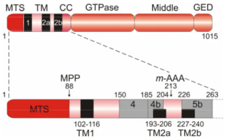
Figure 7. Domain structure of OPA1.
Schematic representation of the domain structure of splice variant 8 of human OPA1, which contains the complete sequence (1015 amino acids) and all three hydrophobic stretches called TM1, TM2a, and TM2b, indicated with black boxes (upper panel). OPA1 splice variants differ from each other by the presence or absence of protein segments encoded by alternatively spliced exons 4, 4b, and 5b, which are indicated by grey boxes (lower panel). Amino acid positions of the hydrophobic stretches and the alternative protein segments are given. The cleavage site of the mitochondrial processing peptidase (MPP) and the putative cleavage site of the m-AAA protease (m-AAA), as determined by Ishihara et al., 2006, are marked by arrows. In this study, the L1 and S3 isoforms were purified from HeLa cells expressing FLAG-tagged rat OPA1 splice variant 7, which lacks TM2a, and used for protein sequencing. MTS, mitochondrial targeting sequence; TM, transmembrane domain; CC, coiled-coil domain; GTPase, GTPase domain; Middle, middle domain; GED, GTPase effector domain.
OPA1 is a key player in regulating dynamic changes of mitochondrial morphology by promoting mitochondrial fusion (Olichon et al., 2003; Cipolat et al., 2004). Accordingly, downregulation of OPA1 results in fragmentation of the mitochondrial network owing to ongoing fission (Olichon et al., 2003; Cipolat et al., 2004; Griparic et al., 2004; Chen et al., 2005). In addition, cells depleted of OPA1 show an aberrant cristae structure (Olichon et al., 2003; Griparic et al., 2004), which is reminiscent of the role of yeast Mgm1 in maintenance of cristae morphology (Sesaki et al., 2003b; Amutha et al., 2004; Meeusen et al., 2006). Further phenotypes that have been linked to loss of OPA1 are reduction of mitochondrial membrane potential, defects in mitochondrial respiration, accelerated release of cytochrome c and concomitantly increased propensity for apoptosis (Olichon et al., 2003; Lee et al., 2004;
Arnoult et al., 2005; Chen et al., 2005). Thus, OPA1 seems to have additional functions in mitochondria which are independent from mitochondrial fusion. Indeed, evidence has been obtained for a direct role of OPA1 in the control of cristae remodelling during apoptosis and release of cytochrome c which is sequestered in the intra-cristae regions (Cipolat et al., 2006;
Frezza et al., 2006).
OPA1 is a dynamin-like GTPase which resides in the mitochondrial intermembrane space (Olichon et al., 2002; Satoh et al., 2003; Griparic et al., 2004) and features the following domain structure (Fig. 7): an N-terminal mitochondrial targeting sequence is followed by one transmembrane segment (TM1) and two further hydrophobic stretches (TM2a and TM2b). A coiled-coil region (or heptad repeat) is located immediately in front of the GTPase domain containing the consensus GTP binding sites. The GTPase domain is followed by a middle domain and a C-terminal coiled-coil domain, known as the GTPase effector domain, which both are generally involved in oligomerisation and regulation of GTPase activity (Praefcke and McMahon, 2004). The mRNAs transcribed from the OPA1 gene are subject to extensive alternative splicing resulting in eight different splice variants (Delettre et al., 2001; Satoh et al., 2003). The alternative splicing involves the exons 4, 4b, and 5b which encode protein segments of the N-terminal region between the first transmembrane domain and the GTPase domain. Therefore, OPA1 splice variants differ in the presence or absence of the two additional hydrophobic stretches TM2a and TM2b. In addition, the different splice variants are not uniformly expressed in human tissues and appear to have distinct roles in mitochondria such as mitochondrial fusion and apoptosis (Delettre et al., 2001; Olichon et al., 2006). Several protein isoforms of OPA1 can be detected in human cells, e.g. five different isoforms are apparently present in HeLa cells (Olichon et al., 2003;
Ishihara et al., 2006; Olichon et al., 2006), which have been designated L1 and L2 for the two