Neurodegeneration and the m-AAA protease:
pathogenic cascades in neurons and myelinating cells
Inaugural-Dissertation
zur
Erlangung des Doktorgrades
der Mathematisch-Naturwissenschaftlichen Fakultät der Universität zu Köln
vorgelegt von Shuaiyu Wang aus Hunan, China
Köln, 2016
Berichterstatter:
Prof. Dr. Elena I. Rugarli
Prof. Dr. Aleksandra Trifunovic
Tag der Mündlichen Prüfung:
01.07.2016
To my dear family, teachers and friends
Table of contents
Table of contents ... 1
Abstract ... 4
Abbreviations ... 5
1 Introduction ... 7
1.1 Mitochondria ... 8
1.1.1 Mitochondrial function ... 8
1.1.2 Mitochondrial quality control ... 8
1.2 The m-AAA protease ... 9
1.2.1 The m-AAA protease subunits and function ... 9
1.2.2 Human diseases causes by the m-AAA protease mutations ... 12
1.2.3 Available murine models of the m-AAA protease ... 14
1.3 Myelinating cells ... 18
1.3.1 Oligodendrocytes ... 20
1.3.2 Schwann cells ... 22
1.3.3 Pathology of myelin-forming cells ... 24
1.3.4 The role of mitochondria in myelinating cells ... 26
2 Aim of the thesis ... 28
3 Material and methods ... 30
3.1 Animal experiments ... 31
3.1.1 Mouse breedings ... 31
3.1.2 Genotyping ... 33
3.1.3 Tamoxifen administration ... 34
3.1.4 Behavioral tests ... 34
3.1.5 Fat content measurement ... 35
3.2 Tissue Collection ... 35
3.3 Histology and immunohistochemistry ... 35
3.3.1 Slice preparation ... 35
3.3.2 Immunostaining on vibratome sections ... 36
3.3.3 Immunostaining on paraffin sections ... 36
3.3.4 Nissl staining ... 37
3.3.5 Gallyas staining ... 37
3.3.6 Haematoxylin and eosin staining ... 38
3.3.7 TUNEL assay ... 38
3.4 Microscopy ... 39
3.4.1 Light microscopy ... 39
3.4.2 Electron microscopy ... 39
3.5 Cell counting and axon morphometry ... 40
3.5.1 Cell counting ... 40
3.5.2 g ratio and myelinated axon counting ... 40
3.6 Oligodendrocytes preparation ... 40
3.7 Biochemistry ... 41
3.7.1 Isolation of crude mitochondria ... 41
3.7.2 Myelin isolation ... 41
3.7.3 Protein extraction ... 41
3.7.4 Western blot ... 42
3.7.5 Oxyblot ... 44
3.8 Statistical analysis ... 45
3.8.1 Chi-square test ... 45
3.8.2 Unpaired Student’s t test ... 45
3.8.3 Two-way ANOVA test ... 45
4 Results ... 46
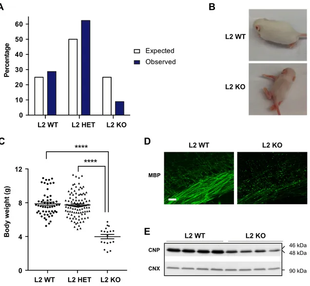
4.1 Afg3l2 full-body knockout mice show severe developmental phenotype ... 47
4.1.1 Phenotypic complexity of L2 KO mice ... 47
4.1.2 Loss of Afg3l2 unbalances mitochondrial fission and fusion ... 49
4.1.3 Lack of Afg3l2 triggers tauopathy ... 51
4.1.4 Investigation of possible kinases involved in tau hyperphosphorylation ... 51
4.2 Afg3l2 forebrain-neuron specific knockout mice show early-onset neurodegeneration ... 54
4.2.1 Signs of neurodegeneration in AFG3L2
NKOmice ... 54
4.2.2 Mitochondrial morphology aberration in AFG3L2
NKOmice ... 56
4.2.3 Tau hyperphosphorylation in AFG3L2
NKOmice ... 56
4.3 Afg3l2 oligodendrocytes-specific knockouts display late-onset myelin abnormalities ... 60
4.3.1 AFG3L2 is abundantly expressed in oligodendrocytes ... 60
4.3.2 The PLP1 promoter is efficiently and specifically expressed in
4.3.3 Deletion of Afg3l2 induces mitochondrial abnormalities in oligodendrocytes
... 65
4.3.4 Lack of AFG3L2 does not affect the survival of oligodendrocytes ... 65
4.3.5 Late-onset motor impairment and axonal dysmyelination in AFG3L2
MC-KOmice ... 66
4.4 Afg3l1 full-body knockout mice show no evident sign of neurodegeneration ... 70
4.4.1 Normal growth curve of Afg3l1 KO mice ... 70
4.4.2 Preserved axonal myelination and integrity in Afg3l1 KO mice ... 72
4.5 Ablation of the m-AAA protease in myelinating cells causes axonal demyelination and hair greying ... 74
4.5.1 DKO mice showed progressive motor impairment and hair greying ... 74
4.5.2 Genetic tracing of targeted cells in the skin ... 74
4.5.3 Loss of melanocytes and melanoblasts in the skin of DKO mice ... 78
4.5.4 Peripheral neuropathy in the DKO mice ... 80
4.5.5 Axonal demyelination and neuroinflammation in the DKO mice ... 82
4.5.6 Rapid loss of targeted cells in the DKO mice ... 88
5 Discussion ... 95
5.1 Mitochondrial morphology alteration upon loss of the m-AAA protease ... 96
5.2 Tau hyperphosphorylation evoked by AFG3L2 deficiency in neurons ... 99
5.3 Physiological role of AFG3L1 in the mouse ... 101
5.4 AFG3L2 is required for oligodendrocyte maturation but is dispensable for the survival of adult oligodendrocytes ... 102
5.5 Lack of the m-AAA protease causes death of myelinating cells ... 103
5.5.1 Oligodendrocyte death and axonal demyelination in the central nervous system ... 103
5.5.2 Peripheral neuropathy ... 105
5.5.3 Melanocytes reduction and hair greying in the skin ... 106
5.6 Conclusions ... 109
Zusammenfassung ... 110
References ... 112
Acknowledgements ... 127
Eidesstattliche Erklärung ... 129
Curriculum Vitae ... 130
Abstract
The m-AAA protease is a hexameric complex involved in processing of specific substrates and turnover of misfolded polypeptides in the mitochondrial inner membrane.
In humans, the m-AAA protease is composed of AFG3L2 and paraplegin. Mutations in AFG3L2 have been implicated in dominant spinocerebellar ataxia (SCA28) and recessive spastic ataxia-neuropathy syndrome (SPAX5). Mutations of SPG7, encoding paraplegin, are linked to hereditary spastic paraplegia. In the mouse, a third subunit AFG3L1 is expressed. Various mouse models recapitulate the phenotype of these neurodegenerative disorders, however, the pathogenic mechanism of neurodegeneration is not completely understood.
Here, we studied several mouse models and focused on cell-autonomous role of the m-
AAA protease in neurons and myelinating cells. We show that lack of Afg3l2 triggers
mitochondrial fragmentation and swelling, tau hyperphosphorylation and pathology in
Afg3l2 full-body and forebrain neuron-specific knockout mice. Moreover, deletion of
Afg3l2 in adult myelinating cells causes early-onset mitochondrial abnormalities as in
the neurons, but the survival of these cells is not affected, which is a contrast to early
neuronal death. Despite the fact that myelinating cells have been previously shown to
survive respiratory deficiency by glycolysis, total ablation of the m-AAA protease by
deleting Afg3l2 in an Afg3l1 null background (DKO), leads to myelinating cell demise
and subsequently progressive axonal demyelination. Interestingly, DKO mice show
premature hair greying due to loss of melanoblasts. Together, our data demonstrate cell-
autonomous survival thresholds to m-AAA protease deficiency, and an essential role of
the m-AAA protease to prevent cell death independent from mitochondrial dynamics
and the oxidative capacity of the cell. Thus, our findings provide novel insights to the
pathogenesis of diseases linked to m-AAA protease deficiency, and also establish
valuable mitochondrial dysfunctional mouse models to study other neurodegenerative
diseases, such as tauopathies and demyelinating diseases.
Abbreviations
ABCD1 ABC binding cassette family D member 1
AD Alzhermer’s diseases
AFG3L1 AFG-like protein 1
AFG3L2 AFG-like protein 2
APC Adenomatous Polyposis Coli
APS Ammoniumpersulfate
ASA Aryl Sulphatase A
BSA Bovine Serum Albumin
CECAD Cologne Cluster of Excellence in Cellular Stress Responses in Aging-associated Diseases
CMT Charcot-Marie-Tooth disease
CNP 2’, 3’-cyclic nucleotide 3’-phosphodiesterase
CNS Central Nervous System
CNX Calnexin
COX Cytochrome c oxidase
CTRL Control
DKO Double knockout
DNPH 2,4-dinitrophenylhydrazine
EAE Experimental Autoimmune Encephalomyelitis
ECL Enhanced chemiluminescent
GFAP Glial Fibrillary Acidic Protein
GJB1 Gap Junction Protein β1
GJC2 Gap Junction Protein γ2
h hour (s)
HBSS Hanks’ Balanced Salt Solution
HSP Hereditary Spastic Paraplegia
HET Heterozygous
IBA1 Ionized calcium Binding Adaptor molecule 1
KO (ko) Knockout
LHON Leber’s Hereditary Optic Neuropathy
m-AAA matrix ATPases Associated with diverse cellular Activities
MAG Myelin Associated Glycoprotein
MBP Myelin Basic Protein
MC Melanocytes
min minute (s)
MPTP Mitochondrial Permeability Transition Pore
MOG Myelin Oligodendrocyte Glycoprotein
MS Multiple Sclerosis
mt Mitochondrial
Ndufs4 NADH dehydrogenase-ubiquinone-FeS 4
NRG1 Neuregulin-1
OXPHOS Oxidative phosphorylation
OPA1 Optic atrophy type 1
OPC Oligodendrocyte Precursor Cells
PBS Phosphate Buffered Saline
PLP 1 Proteolipid protein 1
PNS Peripheral Nervous System
PDGFα Platelet-Derived Growth Factor receptor alpha
PFA Paraformaldehyde
PMD Pelizaeus–Merzbacher disease
PMLD Pelizaeus–Merzbacher-like disease
RIPA RadioImmunoPrecipitation Assay
ROS Reactive Oxygen Species
SCA28 Spinocerebellar ataxia type 28
SCP Schwann Cell Precursors
SDHA Succinate dehydrogenase A
SDS Sodium Dodecyl Sulfate
SPAX5 Spastic ataxia type 5
SPG Spastic paraplegia
Surf1 Surfeit locus protein 1
TE Tris-EDTA
TEMED N,N,N',N'-Tetramethylethylenediamine
UPR
mtMitochondrial unfolded protein response
WT (wt) Wildtype
1 Introduction
1.1 Mitochondria
About 2 billion years ago, ancient bacteria entered primordial eukaryotic cells with a symbiotic relationship and evolved into mitochondria, which are semi-autonomous and essential organelles in modern eukaryotic cells (Gerdes et al., 2012; Wallace, 2005). In humans, although 99% of mitochondrial proteins are encoded by the nuclear genome, synthesized in the cytosol and imported into mitochondria, mitochondria maintain a genome encoding 13 proteins that are core subunits of respiratory chain complexes I, III, IV, and V, but not complex II, which is exclusively composed of proteins encoded by nuclear genes (Chan, 2006; Gerdes et al., 2012; Wallace, 2005). The mitochondrial DNA (mtDNA) genome also encodes 2 rRNAs and 22 tRNAs that are required for translation of mtDNA transcripts (Chan, 2006; Wallace, 2005).
1.1.1 Mitochondrial function
The well-known function of mitochodondria is ATP production through the process of oxidative phosphorylation (OXPHOS). Moreover, mitochondria participate in diverse cellular processes, such as lipid biosynthesis, Ca
2+and iron homeostasis, autophagy and apoptosis (Chan, 2006; Rugarli and Langer, 2012). Endoplasmic reticulum (ER) regulate mitochondrial morphology and function through the ER-mitochondria contact sites (Friedman and Nunnari, 2014). Proper mitochondrial morphology and function are extremely required for neurons, which are polarized cells, need high amount of ATP and rely on OXPHOS to produce energy (Rugarli and Langer, 2012). However, OXPHOS generates reactive oxygen species (ROS) as a by-product, which may in turn impair mitochondrial function (Wallace, 2005).
1.1.2 Mitochondrial quality control
Cells have a hierarchical system of surveillance mechanisms to prevent mitochondrial
damage or selectively remove dysfunctional mitochondrial proteins or entire organelles
under stress (Rugarli and Langer, 2012). At molecular levels, the first line of defense is
conducted by mitochondrial chaperones and proteases, which promote folding of newly
imported polypeptides and degrade irreversibly misfolded or damaged proteins (Rugarli
and Langer, 2012; Tatsuta and Langer, 2008). During protein homeostasis and
mitochondrial stress, mitochondria communicate with nucleus to activate the expression
proteases, to perform intramitochondrial quality control (Quiros et al., 2016; Rugarli and Langer, 2012). The UPR
mthas been characterized extensively in C. elegans and also has been reported in mammalians (Dogan et al., 2014; Quiros et al., 2016). In addition, at organellar levels, sustained mitochondrial stress and damage, lead to mitochondrial morphological alterations, including hyperfusion and fragmentation, to alleviate mild stress or segregate severe damaged mitochondria, which might be eliminated by selective autophagy namely mitophagy (Rugarli and Langer, 2012).
Recently, a novel mitochondrial quality control pathway has been identified, in which mitochondria selectively degrade impaired proteins and lipids by generating small vesicular carriers that transport mitochondrial proteins and lipids to intracellular organelles, such as late endosome and peroxisomes (Sugiura et al., 2014).
Failure of mitochondrial quality control leads to cell death, and mitochondrial dysfunction has been implicated in various neurodegenerative diseases, metabolic disorders, ageing, and cancer (Burte et al., 2015; Chan, 2006; Rugarli and Langer, 2012;
Wallace, 2005).
1.2 The m-AAA protease
Mitochondrial proteases, termed mitoproteases, act as key regulators for mitochondrial biogenesis, protein quality control, dynamics, mitophagy and apoptosis (Figure 1.1) (Martinelli and Rugarli, 2010; Quiros et al., 2015; Rugarli and Langer, 2012; Tatsuta and Langer, 2008). Numerous mitoproteases have been identified, and they are classified into three categories- intrinsic, transient and pseudo mitoproteases, according to their location, function, and structural and proteolytic characteristics (Quiros et al., 2015). The mitochondrial matrix ATPases associated with diverse cellular activities (m- AAA) protease is an intrinsic metalloprotease in the inner membrane with the catalytic domain facing the matrix (Gerdes et al., 2012; Koppen and Langer, 2007; Martinelli and Rugarli, 2010; Quiros et al., 2015).
1.2.1 The m-AAA protease subunits and function
In the mouse, the m-AAA protease is a hexameric complex formed by AFG3L1,
AFG3L2 and paraplegin (Koppen et al., 2007). Whereas in humans, AFG3L1 is
pseudogene (Kremmidiotis et al., 2001) and the m-AAA protease is composed by
AFG3L2 and paraplegin. All subunits of the m-AAA protease have similar domains
including a mitochondrial targeting sequence, two transmembrane domains, an AAA domain and a metal-dependant proteolytic domain (Martinelli and Rugarli, 2010).
AFG3L1 and AFG3L2 form homo- and hetero-oligomeric complexes, but paraplegin can only assemble into hetero-hexamers (Koppen and Langer, 2007; Koppen et al., 2007) (Figure 1.2).
In the last decade, the function of m-AAA protease has been broadly explored in yeast, cell lines, murine models and human patients. These studies have shown that the m- AAA protease processes specific substrates and also regulates the turnover of misfolded mitochondrial respiratory chain complex subunits. The best-characterized substrate of the m-AAA protease is the mitochondrial ribosomal protein MrpL32, which mediates ribosome assembly and mitochondrial translation (Almajan et al., 2012; Bonn et al., 2011; Nolden et al., 2005). The m-AAA protease degrades a truncated form of cytochrome c oxidase subunit I (COX1) to maintain mitochondrial protein quality control (Hornig-Do et al., 2012). Depletion or mutations of the m-AAA protease harms mitochondrial function in multiple aspects, including mitochondrial respiratory chain complex deficiency, dynamic disturbance, Ca
2+handling defect, and anterograde transport impairment (Atorino et al., 2003; Ehses et al., 2009; Kondadi et al., 2014;
Maltecca et al., 2015; Maltecca et al., 2012).
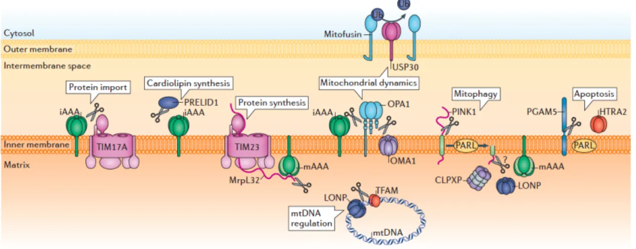
Figure 1.1 Proteolytic regulation of mitochondrial functions by mitoproteases
Mitoproteases perform proteolytic reactions to regulate mitochondrial protein import, cardiolipin synthesis, protein systhesis, dynamics, mitophagy, apoptosis and mitochondrial DNA (mtNDA) (Quiros et al., 2015).
Figure 1.2 Isoenzymes of the m-AAA protease
In humans, AFG3L2 and paraplegin assemble into homo-oligomeric AFG3L2 and hetero-oligomeric AFG3L2/paraplegin complexes. In the mouse, a third subunit AFG3L1 is present. Therefore, five isoenzymes of the m-AAA protease have been identified, and the hetero-oligomeric complex composed by AFG3L1, AFG3L2 and paraplegin is speculative (indicated by a question mark) (Koppen and Langer, 2007).
AFG3L2 paraplegin AFG3L1 AFG3L2 paraplegin
In humans In the mouse
?
1.2.2 Human diseases causes by the m-AAA protease mutations
Mutations of the m-AAA protease subunits have been linked to three rare inherited neurodegenerative disorders. In general, these diseases are clinically heterogeneous, and the phenotype is variable among affected individuals even within the same family.
SPG7 gene, maps on chromosome 16q24.3 and encodes for paraplegin, which mutation causes autosomal recessive hereditary spastic paraplegia (HSP) characterized by lower limb weakness and spasticity due to retrograde degeneration of corticospinal axons (Arnoldi et al., 2008; Brugman et al., 2008; Casari et al., 1998; De Michele et al., 1998;
Elleuch et al., 2006; McDermott et al., 2001; Warnecke et al., 2007; Wilkinson et al., 2004). So far, more than 30 mutations in SPG7 gene have been found, accounting for approximately 5% of autosomal recessive HSP (Salinas et al., 2008; Sánchez-Ferrero et al., 2013). Recently, familial analysis from a large cohort of Spanish HSP patients suggests a dominant inheritance of SPG7 mutations (Sánchez-Ferrero et al., 2013). The onset of HSP patients carrying SPG7 mutations is mostly in the adulthood (mean age, 30 years), and the range is from 1 year to 77 years (Arnoldi et al., 2008; Brugman et al., 2008; Casari et al., 1998; De Michele et al., 1998; Elleuch et al., 2006; McDermott et al., 2001; Sánchez-Ferrero et al., 2013; Warnecke et al., 2007; Wilkinson et al., 2004).
SPG7 affected individuals may show variable clinical features with both pure and complicated HSP (Casari and Marconi, 2006, updated 2010; Harding, 1983). The phenotype of pure HSP includes gait disturbance, hyperreflexia, extensor plantar responses, and decreased vibratory sense in the distal limbs (Casari et al., 1998; Casari and Marconi, 2006, updated 2010; Harding, 1983). Some patients show complicated form of HSP, which is characterized by additionally symptoms, such as mental retardation, optic atrophy, peripheral neuropathy, ptosis, decreased hearing, amyotrophy and ataxia (Casari et al., 1998; Casari and Marconi, 2006, updated 2010). Mitochondrial DNA deletion and mitochondrial respiratory chain defects in SPG7 patient muscles imply mitochondria dysfunction in the pathogenesis of HSP (Atorino et al., 2003;
Casari et al., 1998; McDermott et al., 2001; Wedding et al., 2014; Wilkinson et al., 2004).
Mutations in AFG3L2 on chromosome 18p11.21 (Banfi et al., 1999; Cagnoli et al., 2006;
Di Bella et al., 2010), cause two neurological diseases, autosomal dominant
Bella et al., 2010; Edener et al., 2010; Mariotti et al., 2008; Smets et al., 2014;
Svenstrup et al., 2016; Zuhlke et al., 2015) and recessive spastic ataxia 5 (SPAX5) (Pierson et al., 2011). Additionally, two unrelated subjects carrying homozygous missense mutation of AFG3L2, have been identified from a cohort of progressive myoclonus epilepsy by exome sequencing (Muona et al., 2015). Spinocerebellar ataxia (SCA), characterized by progressive gait, stance and limb ataxia, oculomotor disturbances, dysarthria, is mostly owing to degeneration of Purkinje cells in the cerebellum (Brussino et al., 2011, updated 2013; Di Bella et al., 2010; Schols et al., 2004). SCA28, one of the most recently characterized forms of SCA, is the first autosomal dominant spinocerebellar ataxia shown to be caused by mitochondrial gene mutations (Mariotti et al., 2012). The general onset of SCA28 is in young adulthood (24.4 ± 14.9 years) with the range from 3 years to 60 years (Brussino et al., 2011, updated 2013). Patients show slowly progressive gait disorder, upper limb ataxia, lower limb hyperreflexia, dysarthria, nystagmus and ophthalmoparesis (Brussino et al., 2011, updated 2013; Edener et al., 2010; Mariotti et al., 2012; Zuhlke et al., 2015). To date, 15 missense mutations of AFG3L2 have been identified (Cagnoli et al., 2006; Cagnoli et al., 2010; Di Bella et al., 2010; Lobbe et al., 2014; Svenstrup et al., 2016; Zuhlke et al., 2015), which represent around 3% of all SCAs and 1.5% of autosomal dominant cerebellar ataxias in Europe (Cagnoli et al., 2010). Recently, one partial deletion and one frameshift mutations of AFG3L2 also have been shown to cause SCA28 (Musova et al., 2014; Smets et al., 2014). Most mutations in the exon 15-16 of AFG3L2 corresponding the highly conserved proteolytic domain indicate specific substrates processing might be critical in the molecular pathology of SCA28 (Cagnoli et al., 2010;
Di Bella et al., 2010; Lobbe et al., 2014; Svenstrup et al., 2016; Zuhlke et al., 2015).
Likewise, two reported homozygous mutations of AFG3L2 are also in this mutational
hotspot exon 15-16 (Muona et al., 2015; Pierson et al., 2011). The two brothers
identified with AFG3L2 homozygous mutations from a consanguineous family, showed
early onset SPAX5, which is characterized clinically by lower extremity spasticity,
progressive myoclonic epilepsy, peripheral neuropathy, dysarthria, ptosis, oculomotor
apraxia and cerebellar atrophy (Pierson et al., 2011). This phenotype is reminiscent of a
combined syndrome of HSP and SCA28. The disease course of these two brothers was
similar, and the younger sibling was more severe and died at the age of 13 years due to
pneumonia-related complications (Pierson et al., 2011). The newly reported two cases
carrying homozygous mutations of AFG3L2, known to be unrelated, also displayed
severe progressive myoclonus, ataxia and mild cognitive decline (Muona et al., 2015).
Notably, their phenotype is not as severe as the two brothers’, suggesting clinical variability in SPAX5. Loss-of-function mutations in AFG3L2 cause proteolytic impairment and mitochondrial respiratory chain deficiency have been implicated in the pathogenesis of SCA28 and SPAX5 (Di Bella et al., 2010; Pierson et al., 2011).
1.2.3 Available murine models of the m-AAA protease
In order to better unravel the pathogenesis of human diseases associated with mutations of SPG7 and AFG3L2, multiple murine models of the m-AAA protease have been established. These models show dosage-dependant phenotype, probably owing to the fact that AFG3L2 forms both homo- and hetero-hexameric complexes and paraplegin only forms hetero-hexameric complex (Koppen et al., 2007).
Two murine models of SPAX5 have been reported: Afg3l2 null (Afg3l2
EMV66/EMV66) generated by integrating ecotropic murine leukemia virus insertion 66 (EMV66) within intron 14, and a spontaneous mutant carrying a missense mutation in exon 10 (Maltecca et al., 2008). Both Afg3l2 null and spontaneous missense mutant mice display a very severe neuromuscular syndrome with hindlimb paraparesis one week after birth and general die at P16 (Maltecca et al., 2008). Afg3l2 deletion or loss-of-function causes mitochondrial morphological alternation and respiratory chain complexes deficit, leading to an impairment of axonal development with myelination delay and radial growth defect (Kondadi et al., 2014; Maltecca et al., 2008). Notably, in these Afg3l2 mutant mice, non-neuronal tissues, such as muscle and liver, are not significantly affected, indicating that nervous system are solely affected by Afg3l2 deletion or mutation (Maltecca et al., 2008). Interestingly, studies from our group provided novel insights into pathogenic mechanism of neurodegeneration. We have shown that an impairment of mitochondrial ribosome assembly and a decrease of mitochondrial translation in the Afg3l2 null mice (Almajan et al., 2012). More recently, in these Afg3l2 null mice, we have found microtubular network disruption and accumulation of hypersphosphorylated tau in the neurons (Kondadi et al., 2014).
The murine model haploinsufficient for Afg3l2 (Afg3l2
+/EMV66), which represents model
of SCA28, shows progressive impairment in motor balance and coordination owing to
Purkinje cell dark degeneration (Martinelli et al., 2009). Mitochondrial respiratory chain
(Martinelli et al., 2009). Moreover, recent data have shown that the ataxic phenotype of Afg3l2
+/EMV66mice can be partial restored by reducing Ca
2+influx in Purkinje cells by either genetic silencing of the metabotropic glutamate receptor or administration of the β-lactam antibiotic ceftriaxone (Maltecca et al., 2015). Thus, mitochondrial Ca
2+handling inefficiency is also involved in the pathogenesis of SCA28 and might be a potential therapeutic target (Maltecca et al., 2015).
In comparison with severe developmental phenotype of Afg3l2 null and missense mutant mice, Spg7 knockout mice (Spg7
-/-) grow normally and show progressive axonal degeneration and motor impairment starting from 4 months of age (Ferreirinha et al., 2004), which models the disease of HSP with loss-of-function mutations in SPG7.
Interestingly, in the Spg7
-/-mice, abnormal mitochondrial morphology occurs before initial signs of axonopathy, and followed by axonal swelling with organelle and neurofilament accumulation, together, suggesting that primary mitochondrial dysfunction and secondary axonal transport defect may lead to corticospinal and peripheral axonal swelling and degeneration (Ferreirinha et al., 2004). Interestingly, alternative splicing of Spg7 produces a variant of paraplegin in the mouse, which localizes to the endoplasmic reticulum, still presents in the Spg7
-/-mice. The Spg7
-/-murine model is therefore an isoform-specific knockout (Mancuso et al., 2012).
Spg7
-/-Afg3l2
+/EMV66double mutant mice display an early-onset severe neurologic phenotype characterized by loss of balance, tremor, and ataxia (Martinelli et al., 2009).
These mice show an acceleration of the axonopathy observed in Spg7
-/-mice, and prominent Purkinje cell degeneration at earlier age comparing to Afg3l2
+/EMV66mice (Martinelli et al., 2009), revealing a genetic interaction between the m-AAA protease isoenzymes homo-oligomeric AFG3L2 complex and hetero-oligomeric complex composed of paraplegin and AFG3L2 (Martinelli et al., 2009).
Studies of Afg3l2 conditional murine models provide more insights into the pathogenic
mechanism. Deletion of Afg3l2 in Purkinje cells in the cerebellum causes mitochondrial
fragmentation and COX deficiency prior to neurodegeneration, suggesting that
mitochondrial morphological alternation and respiratory chain complex defect are early
events in the pathogenic cascades (Almajan et al., 2012). Notably, the number of COX-
deficient cells does not increase with time suggesting that Purkinje cells die shortly after
displaying this phenotype (Almajan et al., 2012). Similarly, postnatal ablation of Afg3l2
in forebrain neurons causes rapidly neuronal death (Kondadi et al., 2014). Interestingly, we have identified ERK1/2 and PKA activation and tau hyperphosphorylation in Afg3l2-deficient neurons (Kondadi et al., 2014). Thus, together with the finding of tau hyperphosphorylation in Afg3l2 null mice, a novel pathogenic mechanism in which tau pathology caused by AFG3L2 depletion leads to neurodegenration is proposed (Kondadi et al., 2014).
In summary, aforementioned murine models recapitulate the important
pathophysiological features of related human diseases. Mechanistically, these mouse
models show that AFG3L2 influences mitochondrial dynamics, respiration, ROS levels
and Ca
2+handling, it is therefore essential for neuronal survival (Figure 1.3). Besides,
different neuronal populations have variable thresholds of susceptibility to the m-AAA
protease deficiency (Martinelli et al., 2009) and Purkinje cells seem to be the most
vulnerable cells upon loss of the m-AAA protease. However, the cell-autonomous role
of the m-AAA protease in other cell types, such as glial cells, has not been extensively
investigated. Moreover, these studies have not considered that an additional subunit of
the m-AAA protease, AFG3L1, which is a high homologous to AFG3L2, is present in
the mouse.
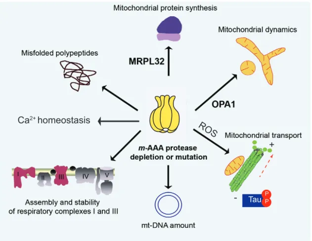
Figure 1.3 Pathogenic mechanisms of neurodegeneration caused by m-AAA protease deficiency Depletion or mutations of the m-AAA protease subunits cause several pathogenic pathways including impaired mitochondrial protein synthesis, accumulation of misfolded proteins, inefficient Ca
2+handling, decreased assembly and stability of respiratory chain complexes I and III, reduced mt-DNA, defective anterograde transport and disturbed mitochondrial dynamics. Adapted from (Martinelli and Rugarli, 2010).
Ca
2+homeostasis ROS
Tau
PP-
+
1.3 Myelinating cells
In vertebrates, myelinating cells are highly specialized glial cells that wrap axons with multi-layered myelin for rapid impulse propagation and axonal integrity (Nave and Trapp, 2008; Simons and Trotter, 2007). The myelin sheath, a specialized cell membrane, is formed by oligodendrocytes in the central nervous system (CNS) and Schwann cells in the peripheral nervous system (PNS) (Nave and Trapp, 2008; Simons and Trotter, 2007). The myelin membrane is composed of unique lipids and proteins that have to be assembled temporally and spatially (Figure 1.4) (Aggarwal et al., 2011;
Baumann and Pham-Dinh, 2001; Simons and Trotter, 2007). A recent study has shown
that myelin growth occurs by consecutive wrapping of the inner tongue around the axon
and simultaneous lateral extension of newly formed myelin layers (Snaidero et al.,
2014). Neuronal signals regulate myelinating cell development and myelin assembly
(Nave and Trapp, 2008; Simons and Trajkovic, 2006); in turn, deficiency of myelin
proteins or myelinating cells causes axonal demyelination and/or degeneration (Dewar
et al., 2003; Lee et al., 2012; Nave et al., 2007; Nave and Trapp, 2008; Olmos-Serrano
et al., 2016; Trapp and Nave, 2008; Viader et al., 2011).
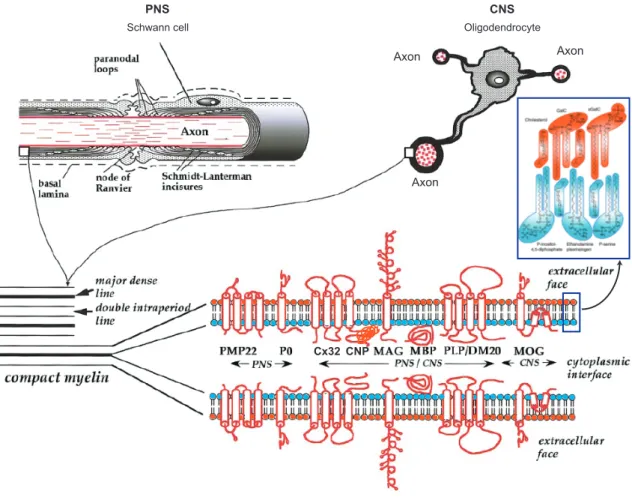
Figure 1.4 Schematic representation of myelinating cells, myelin structure and components.
While Schwann cells in the PNS only myelinate one axon, oligodendrocytes in the CNS are able to wrap several axons. The compact myelin is formed by the apposition of the external surfaces of the myelinating cell membrane, forming the “double intraperiod line”; the apposition of the internal surfaces followed by the extrusion of the cytoplasm, forming the “major dense line.” The lipid composition of myelin bilayer is asymmetric. Some major myelin proteins are indicated and classified according to the expression region.
Adapted from (Aggarwal et al., 2011; Baumann and Pham-Dinh, 2001).
Schwann cell Oligodendrocyte
PNS CNS
Axon
Axon
Axon
Cx32 CNP
1.3.1 Oligodendrocytes
During the development of CNS, oligodendrogenesis takes place in multiple foci. In the spinal cord, most oligodendrocyte precursor cells (OPC) arise from the ventral progenitor domain starting from E12.5, then migrate laterally and dorsally to occupy the whole spinal cord after specification (Fancy et al., 2011). Besides, commencing ~E15.5, dorsal-derived OPC contribute to 20-30% of total OPC (Fancy et al., 2011; Vallstedt et al., 2005). In the forebrain, multiple waves of OPC production and migration occur from embryonic to postnatal stages (El Waly et al., 2014; Fancy et al., 2011). At E12.5, the first wave of OPC production originates in the medial ganglionic eminence and anterior entopeduncular area in the ventral brain (Kessaris et al., 2006). However, the majority of adult oligodendrocytes in mouse arise from the second and third wave of OPCs emanating from the lateral and/or caudal ganglionic eminences at E15.5 and from the cortex after birth, respectively (El Waly et al., 2014; Kessaris et al., 2006).
Interestingly, genetical ablation of one population of oligodendrocytes in restricted areas in the CNS, triggers adjacent populations rapid migration and proliferation to maintain their normal density in adult mouse (Hughes et al., 2013; Kessaris et al., 2006).
While some OPC are reserved for myelin regeneration, others are specified and differentiate to myelinating oligodendrocytes (Figure 1.5) (Fancy et al., 2011).
Oligodendrocytes are capable to myelinate several axons selectively with diameter
above 0.2 µm (Simons and Trajkovic, 2006; Waxman and Sims, 1984). The process of
oligodendrocyte maturation and myelination is very complicated, because it is highly
coordinated with neurons/axons, and numerous signal pathways and regulatory factors
are involved (Bradl and Lassmann, 2010; Fancy et al., 2011) (Figure 1.5).
Figure 1.5 Schematic illustrating oligodendrocyte development and differentiation
In the spinal cord, oligodendrocyte precursor cells (OPC) are derived from the ventral progenitor domain, which also produces motor neurons. Oligodendrocytes express specific antigenic markers and follow a stepwise morphological transformation during development and differentiation. Solid black arrows indicate oligodendrocyte-lineage transitions that are dependent on specific transcription factors, and coloured gradient bars represent predicted temporal expression pattern of essential factors or regulators (Fancy et al., 2011).
PLP CNP MAG O4
1.3.2 Schwann cells
At early embryonic developmental stage of peripheral nerves, neural crest cells give rise to Schwann cell precursors (SCP) that are found among the axons of nascent nerves, and SCP develop into immature Schwann cells before birth (Jessen and Mirsky, 2005).
Postnatal immature Schwann cells differentiate into either myelinating Schwann cells or non-myelinating Schwann cells (Figure 1.6) (Jessen and Mirsky, 2005; Woodhoo and Sommer, 2008). Schwann cells produce basal lamina and start radial sorting of 1:1 relationship (one myelinating Schwann cell ensheaths one axon) with large-calibre axons (diameter > ~ 1 µm) (Jessen and Mirsky, 2005; Simons and Trotter, 2007).
Unmyelinated smaller axons (also known as C-fibre axons) forming Remak bundles are wrapped and segregated by non-myelinating Schwann cells. Multiple molecules and factors have been implicated in the regulation of Schwann cell development and myelination (Jessen and Mirsky, 2005). For instance, axon-derived signaling molecule Neuregulin-1 (NRG1), a EGF-like ligand for the ErbB family of tyrosine kinase receptors (Garratt et al., 2000; Van Raamsdonk and Deo, 2013), promotes Schwann cell lineage survival and development, and regulates myelin sheath thickness (Brinkmann et al., 2008; Garratt et al., 2000; Grinspan et al., 1996; Michailov et al., 2004; Taveggia et al., 2005).
One fascinating aspect of SCP is bipotentiality, not only having glial fate, but also giving rise to melanocytes that originate from neural crest cells as SCP. In the mouse at E10.5, shortly after neural crest delamination, the first wave of melanocyte precursor- melanoblasts migrate dorsolaterally between the dermamyotome and eventually reach the destination of the basal layer of the epidermis and the hair follicles (Adameyko et al., 2009; Erickson, 1993). At around E12.5, a second pathway, melanoblasts differentiated from SCP migrate ventrally along the nerves and contribute to a large number of melanocytes in the limbs, in the dorsal and lateral body wall (Adameyko et al., 2009;
Van Raamsdonk and Deo, 2013). However, the role of SCP in formation of
melanocytes in the adult or age-related hair greying remains elusive (Adameyko et al.,
2009).
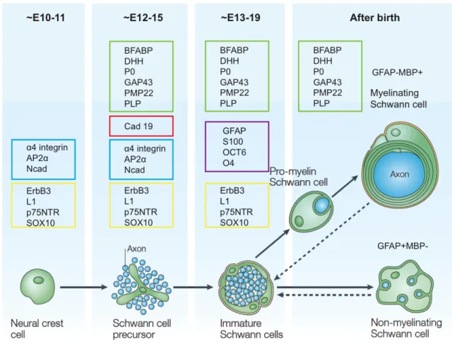
Figure 1.6 Development of Schwann cell lineage in mouse.
Schwann cells originate from the neural crest cells at early embryonic development. Markers of different
developmental Schwann cell lineages are in coloured frames. The same colour means shared antigen
expression. Dashed arrows indicate the reversible transformation of mature myelinating and
nonmyelinating Schwann cells to immature Schwann cells. Adapted from (Jessen and Mirsky, 2005).
1.3.3 Pathology of myelin-forming cells
One of the main functions of myelinating cells is to produce multi-layered membrane structure of myelin, which insulates the axon and clusters sodium channels into the nodes of Ranvier, thus enables the action potential to conduct from one node to the other (Aggarwal et al., 2011). During the peak of myelination, oligodendrocytes elaborate several times the cell weight in membrane per day and ultimately produce membranes up to 100 times the weight of the cell body (Bradl and Lassmann, 2010), and the process of myelin synthesis and assembly requires a great amount of energy.
Assuming that all basic components for lipid and proteins were available locally, the minimum energetic cost of myelin synthesis would be 3.30 x 10
23ATP molecules per gram myelin in the CNS (Harris and Attwell, 2012). Oligodendrocytes have very high metabolic rates to produce ATP, and this process also generates toxic by-product ROS (Bradl and Lassmann, 2010). Additionally, oligodendrocytes contain high amount of myelin synthetic enzyme cofactor-iron (Thorburne and Juurlink, 1996), which can provoke free radical formation and lipid peroxidation under adverse conditions (Bradl and Lassmann, 2010; Braughler et al., 1986; Juurlink, 1997). Given the fact that oligodendrocytes have low concentration of ROS scavenger glutathione and the susceptibility to inflammatory cytokines exposure, hypoxia, ischemia and pathogenic autoimmune, these cells are particularly vulnerable under pathological conditions (Bradl and Lassmann, 2010; Dewar et al., 2003; Thorburne and Juurlink, 1996).
In addition to myelin sheath formation, myelinating cells also metabolically support
axonal survival (Funfschilling et al., 2012; Lee et al., 2012). Oligodendrocytes highly
express monocarboxylate transporter 1 (MCT1) to transport glycolysis product lactate
to axons (Funfschilling et al., 2012; Lee et al., 2012; Rinholm et al., 2011), and MCT1
deficiency leads to axonal degeneration (Lee et al., 2012). Importantly, several human
neurodegenerative disorders, for instance, Multiple Sclerosis (MS), inherited
leukodystrophies and Charcot-Marie-Tooth disease (CMT), have been implicated with
myelinating cell pathology and myelin defects (Nave, 2010). Primary axonal
degeneration occurs in some forms of demyelinating diseases, but secondary axonal
degeneration is believed be the major cause of persistent neurological impairments
(Table 1.1) (Nave, 2010).
Table 1.1 Human diseases associated with myelin defects and secondary axonal degeneration Diseases Frequency and
causes
Glia and myelin pathology
Secondary axonal loss
Animal models
Multiple Sclerosis (MS)
• Relatively common autoimmune disease
• Primary cause unknown
• Viral origin and genetic risk factors suggested
• CNS-specific disease
• Inflammatory lesions in white-matter tracts cause oligodendrocyte death, extensive demyelination and macroscopic plaques
• Axonal swellings and transections in white- and grey- matter lesions
• Axon loss associated with permanent disability at later disease stages
• EAE for modelling autoimmunity against myelin epitopes and the inflammatory phase of MS
Inherited leukodyst rophies
• Very rare genetic disorders
• Several single- gene defects identified, such as in PLP1 (for PMD), GJC2 (for PMLD), ABCD1 (for adrenoleukodystrop hy) and ASA (for metachromatic leukodystrophy)
• Defects of terminal oligodendrocyte differentiation and myelin formation
• Defects of myelin maintenance with demyelination (also secondary
inflammation in adrenoleukodystroph)
• Perturbation of axonal transport followed by Wallerian degeneration
• Purely axonal forms of leukodystrophies : SPG2
(associated with PLP1 defects) and
adrenomyelo- neuropathy (associated with ABCD1 defects)
• Rodents with mutations corresponding to the human disease gene, such as a Plp1 point mutation or Plp1
overexpression (for PMD), Plp1 knockout (for SPG2) and Abcd1 knockout (for adrenomyeloneuro- path)
Inherited demyelina ting neuropat hies
• Rare genetic disorders, such as CMT
• Many disease genes identified, such as PMP22 (for CMT1A), P0 (for CMT1B) and GJB1 (for X-linked CMT)
• Defects of Schwann cell differentiation and peripheral myelination, or myelin maintenance
• Segmental demyelination with formation of ‘onion bulbs’
• Slowly progressive and length-dependent axon loss causing denervation, sensory deficits and muscle weakness — the clinical
hallmarks of CMT
• Rodents with mutations in or overexpression of genes
corresponding to human disease genes, such as Pmp22 (for CMT1A), Mpz (for CMT1B) and Gjb1 (for X-linked CMT)
ABCD1, ABC binding cassette family D member 1; ASA, aryl sulphatase A; EAE, Experimental allergic encephalomyelitis; GJB1, gap junction protein β1; GJC2, gap junction protein γ2 (also known as GJA12 and CX47); PLP1, proteolipid protein 1; PMD, Pelizaeus–Merzbacher disease; PMLD, Pelizaeus–
Merzbacher-like disease. This table is from (Nave, 2010).
1.3.4 The role of mitochondria in myelinating cells
Mitochondrial dysfunction has been implicated in inflammatory demyelinating diseases (Mahad et al., 2008; Mahad et al., 2015; Popescu and Lucchinetti, 2012; Witte et al., 2014), and axonal demyelination is associated with several mitochondrial diseases, such as autosomal dominant optic atrophy, caused by OPA1 mutations (Alexander et al., 2000; Johnston et al., 1979); dominant CMT type 2 disease, caused by MFN2 mutations (Niemann et al., 2006; Zuchner et al., 2004); inherited Leber’s hereditary optic neuropathy (LHON), caused by mitochondrial DNA mutations (Kovacs et al., 2005).
Therefore, more insights into the function of mitochondria in myelinating cells may be beneficial to understand the pathogenic cascades of axonal demyelination and neurodegeneration.
The roles of mitochondria in myelinating cells are distinct and depend on the developmental stages. In vitro studies have demonstrated that oligodendrocyte precursors are more resistant to mitochondrial respiratory chain complex inhibitors; in contrast, differentiated and mature oligodendrocytes are much more sensitive mitochondrial respiratory defects (Schoenfeld et al., 2010; Ziabreva et al., 2010).
Inhibition of mitochondrial complex I or complex IV impairs differentiated oligodendrocyte process formation and myelin protein expression (Schoenfeld et al., 2010; Ziabreva et al., 2010), despite the fact that the viability of oligodendrocytes under this circumstances needs further investigation. Moreover, differentiated oligodendrocytes upregulate globally the transcripts of mitochondrial genes to provide energy and metabolites for myelin synthesis (Schoenfeld et al., 2010). Nevertheless, limited in vivo studies have shown that mitochondrial respiratory deficiency does not affect the survival of Schwann cells nor oligodendrocytes, and an energy metabolism adaptation is proposed (Funfschilling et al., 2012; Viader et al., 2011). Interestingly, Schwann cell-specific deletion of mitochondrial transcription factor A (Tfam), which is essential for mitochondrial DNA maintenance and biogenesis (Larsson et al., 1998), activates heme-regulated inhibitor kinase and induces an integrated stress response maladaptation, thereby causes lipid metabolism shift from fatty acid synthesis toward oxidation and subsequently leads to progressive demyelination (Viader et al., 2011;
Viader et al., 2013). Cox10, encodes a heme A farnesyl transferase and is an assembly
factor of COX (Antonicka et al., 2003; Nobrega et al., 1990), depletion of which using
and oligodendrocytes leads to severe neuropathy (Funfschilling et al., 2012). By
contrast, using an inducible PLP1-CreERT2 line to delete Cox10 in oligodendrocytes in
adult mice does not cause any phenotype. This is because when myelination is complete,
mature oligodendrocytes utilize glycolysis to supply lactate for the maintenance of
myelin and axonal integrity (Funfschilling et al., 2012). Thus, mitochondria play crucial
roles for myelinating cell differentiation and myelination, but these glial cells do not
rely on mitochondria to produce ATP for the survival of these cells or axonal energy
requirement in the adulthood.
2 Aim of the thesis
Mitochondria are pivotal organelles to support cellular energy requirement, Ca
2+homeostasis, metabolism, and apoptosis regulation, especially in polarized and aerobic neurons. Mitoproteases degrade misfolded, non-assembled or damaged proteins as part of quality control surveillance system (Martinelli and Rugarli, 2010; Quiros et al., 2015;
Rugarli and Langer, 2012). Moreover, mitoproteases perform highly regulated proteolytic reactions, such as processing specific substrates that are crucial for mitochondrial biogenesis, translation, dynamics, mitophagy and apoptosis.
Mitoproteases therefore emerge as essential regulators of mitochondrial function and quality control at molecular, organellar and cellular levels (Anand et al., 2013; Quiros et al., 2015).
One of the known mitoproteases is the m-AAA protease, localized in the mitochondrial inner membrane, and composed of hexameric complexes formed by AFG3L2 and paraplegin in humans (Koppen et al., 2007). Mutations of AFG3L2 and paraplegin have been associated with three neurodegenerative diseases SCA28, SPAX5 and HSP (Rugarli and Langer, 2012). Previous studies have shown that m-AAA protease deficiency causes mitochondrial respiratory deficits, morphological alterations, anterograde transport impairment, and calcium handling failure (Almajan et al., 2012;
Atorino et al., 2003; Ehses et al., 2009; Ferreirinha et al., 2004; Kondadi et al., 2014;
Maltecca et al., 2008; Maltecca et al., 2015; Maltecca et al., 2012; Martinelli et al., 2009). However, the pathogenic cascades of neurodegenerative diseases linked to m- AAA protease mutations remain elusive. In addition, the cell-autonomous role of the m- AAA protease is poorly understood. Therefore, in this study, we have these two aims:
1. Investigation the pathogenic cascades of AFG3L2 deficiency in neurodegeneration Lack of AFG3L2 leads to mitochondrial fragmentation and dysfunction, and subsequently neuronal death, however, the pathogenic mechanism is not very clear.
Since Afg3l2 full-body knockout mice show phenotypic complexity and early death, we
generated forebrain neuron-specific mice in which Afg3l2 is depleted postnatally. We
studied these two murine models to investigate the molecular alterations or signaling
pathways triggered by Afg3l2 deletion.
2. Elucidation the role of the m-AAA protease in myelinating cells.
Oligodendrocytes and Schwann cells produce myelin sheaths to wrap axons, thus
facilitating the efficient and rapid propagation of nerve impulse (Nave and Trapp, 2008),
and metabolically support axonal long-term survival (Funfschilling et al., 2012). How
mitochondrial dysfunction in glial cells contributes to neurodegenerative diseases is
largely unknown. Moreover, the m-AAA protease plays key roles in multiple aspects of
mitochondria, but all published m-AAA protease related mouse studies have either used
constitutive mutant models or focused on the role of AFG3L2 in neurons. Whether
AFG3L2 deficiency in glial cells leads to mitochondrial dysfunction and contributes to
neurodegenerative phenotype has not been explored. To this end, we deleted Afg3l2 in
myelinating cells by crossing tamoxifen inducible PLP1-CreERT mice (Doerflinger et
al., 2003) with conditional Afg3l2
fl/flmice (Almajan et al., 2012). Moreover, considering
potential compensatory effect of AFG3L1 that is also expressed in the mouse, we
depleted the whole m-AAA protease in myelinating cells by crossing PLP1-
CreERT/Afg3l2
fl/flmice with unpublished Afg3l1 full-body knockout mice, which do not
show evident neurological phenotype. In addition, a reporter ROSA26
+/SmYline (Sterky
et al., 2011) was used to examine mitochondrial morphology and the fate of targeted
cells. With these murine models, we explored possible OXPHOS-independent function
of the m-AAA protease in myelinating cells, which in comparison with neurons, may
survive by glycolysis.
3 Material and methods
3.1 Animal experiments
All animals procedures were conducted in accordance with European, national and institutional guidelines and were approved by local authorities. Unless stated otherwise, animals were hosted in the CECAD in vivo research facility with 12:12 light/dark cycle.
Daily care of animals was performed by qualified caretakers in compliance with institutional animal welfare protocols. Mice of both female and male were used in this study.
3.1.1 Mouse breedings
Single transgenic mouse strains:
1. Afg3l2
+/EMV66mice were generated originally in FVB genetic background (Maltecca et al., 2008). Afg3l2
EMV66/EMV66(L2 KO) mice were produced by mating Afg3l2
+/EMV66(pure FVB background) mice with Afg3l2
+/EMV66(50% FVB + 50%
C57BL/6) mice (Figure 3.1).
Figure 3.1 L2 KO mice were obtained on a mixed FVB-C57BL/6 background.
2. The generation of CamkIIα-Cre mice was as previously described (Minichiello et al., 1999). Starting from P20, Cre recombinase is expressed in neurons in the forebrain including the neocortex, the hippocampus, and the striatum, but it is not expressed in the cerebellum (Merkwirth et al., 2012; Minichiello et al., 1999).
FVB Afg3l2+/EMV66
FVB Afg3l2+/EMV66
Afg3l2+/EMV66
Afg3l2EMV66/EMV66 (L2 KO)
X
C57BL/6 WT
50% FVB and 50% C57BL/6
75% FVB and 25% C57BL/6