Functional characterization of the mammalian iAAA protease
subunit, YME1L.
Inaugural - Dissertation zur
Erlangung des Doktorgrades der
Mathematisch-Naturwissenschaftlichen Fakultät der Universität zu Köln
vorgelegt von
Joanna Majczak
aus Tarnobrzeg, Polen
Köln, 2008
Berichterstatter:
Professor Dr. Thomas Langer Professor Dr. Jens C. Brüning
Tag der mündlichen Prüfung: November 2008
Abstract
The iAAA protease is an ATP-dependent proteolytic complex in the mitochondrial inner membrane and belongs to the highly conserved family of AAA proteins. In the yeast Saccharomyces cere- visiae, the iAAA protease is a homo-oligomeric complex composed of Yme1p subunits which are active in the intermembrane space and mediate protein quality control. Yeast cells lacking Yme1p are charac- terized by pleiotropic phenotypes including a respiratory deficiency at elevated temperature and an aberrant mitochondrial morphology.
However, the molecular basis of the different yeastyme1phenotypes has not been completely understood. Human YME1L was shown to be the ortholog of the yeast Yme1p, but its functions within mam- malian mitochondria and specific substrate proteins have not been identified so far. In order to define the roles of the mammalian iAAA protease two main approaches were carried out: (1) down regulation studies in mammalian cells using RNA interference, and (2) inducible overexpression of YME1L and several mutant variants in the mam- malian system.
The present study reports the functional conservation of the mam- malian YME1L by showing its involvement in the proteolysis of pro- hibitin 1 whose homolog is also degraded by the iAAA protease in yeast. Furthermore, it demonstrates that YME1L has a role in the maintenance of the tubular mitochondrial morphology and in the constitutive processing of OPA1, a component of the mitochondrial fusion machinery. Moreover, the degradation of TIM23, a core sub- unit of the translocase of the inner membrane, in caspase-independent apoptosis depends on YME1L identifying a novel substrate for the mammalian iAAA protease and a new pathway it is involved in. The overexpression of YME1L mutants has no dominant negative effect on the iAAA protease. Its functions in mitochondrial proteolysis, im- port, and mitochondrial morphology remain unaltered.
Taken together, this study reveals versatile roles of mammalian YME1L in proteolytic quality control, mitochondrial morphology
and apoptosis, thus demonstrating its importance for both the cel- lular viability and death.
Zusammenfassung
Die iAAA-Protease ist ein ATP-abhängiger proteolytischer Kom- plex in der inneren Mitochondrienmembran und gehört zu der hoch- konservierten Familie der AAA-Proteine. In der HefeSaccharomyces cerevisiaeist die iAAA-Protease ein homooligomerer Komplex beste- hend aus Yme1p-Untereinheiten, die im Intermembranraum aktiv sind und die Qualitätskontrolle von Proteinen vermitteln. Hefezel- len, denen Yme1p fehlt, sind durch pleiotrope Phänotypen charak- terisiert. Diese beinhalten einen Atmungsdefekt bei erhöhter Tempe- ratur und eine gestörte mitochondriale Morphologie. Jedoch ist die molekulare Grundlage der verschiedenenyme1-Phänotypen in Hefe bisher nicht vollständig verstanden. Das humane YME1L wurde als Ortholog von Yme1p der Hefe beschrieben, aber seine Funktionen in Säugetier-Mitochondrien und spezifische Substratproteine sind noch nicht identifiziert worden. Um die Rolle der iAAA-Protease in Säugetieren zu bestimmen, wurden zwei experimentelle Ansät- ze verfolgt: (1) Depletion von YME1L in Säugetier-Zellen mittels RNA-Interferenz und (2) induzierbare Überexpression von YME1L und verschiedenen mutierten Varianten im Säugetier-System.
Die vorliegende Arbeit demonstriert die funktionelle Konservie- rung von YME1L in Säugetieren, indem gezeigt wird, dass es an der Proteolyse von Prohibitin 1 beteiligt ist, dessen Homolog in der Hefe ebenfalls von der iAAA-Protease abgebaut wird. Deswei- teren, weist sie nach, dass YME1L eine Rolle bei der Aufrechter- haltung der tubulären mitochondrialen Morphologie und bei der konstitutiven Prozessierung von OPA1, einem Bestandteil der mit- ochondrialen Fusionsmaschinerie, spielt. Darüber hinaus ist der Ab- bau von Tim23, einer Kernuntereinheit der Translocase in der In- nenmembran, während der caspaseunabhängigen Apoptose abhän- gig von YME1L. Dieser Befund identifiziert ein neues Substrat der iAAA-Protease in Säugetieren und einen neuen zellulären Prozess, an dem sie beteiligt ist. Die Überexpression von YME1L-Mutanten hat keinen dominant-negativen Einfluss auf die iAAA-Protease. Ihre
Funktionen in mitochondrialer Proteolyse, Morphologie und Import bleiben unverändert.
Zusammengefasst offenbart diese Arbeit vielfältige Rollen von YME1L bei der Qualitätskontrolle von Proteinen, der mitochondria- len Morphologie und der Apoptose in Säugetieren und demonstriert somit die Bedeutung von YME1L für sowohl die zelluläre Lebensfä- higkeit als auch den zellulären Tod.
Contents
Abstract II
Zusammenfassung IV
1 Introduction 1
1.1 Mitochondrial dynamics . . . 3
1.2 Apoptosis and a link between apoptosis and a fusion/fission machinery . . . 5
1.3 AAA+superfamily and AAA proteins . . . 9
1.3.1 AAA+proteases . . . 10
1.4 Mitochondrial proteolytic quality control . . . 11
1.5 Mitochondrial ATP - dependent proteases . . . 12
1.5.1 ATP - dependent proteases in the mitochondrial matrix 12 1.5.2 ATP dependent proteases in the mitochondrial mem- branes . . . 14
2 Aim of the thesis 22 3 Results 23 3.1 Down regulation of YME1L using small interfering RNAs (siRNAs) . . . 23
3.1.1 Specific antibody against YME1L . . . 23
3.1.2 Depletion of YME1L in mammalian cells . . . 25
3.1.3 Mammalian YME1L role in proteolysis . . . 26
CONTENTS
3.1.4 YME1L role in mitochondrial biogenesis . . . 28
3.2 Overexpression of YME1L in the inducible system: HEK293 T-REx Flp-In cells . . . 34
3.2.1 Expression constructs . . . 34
3.2.2 Generation of stable cell lines . . . 36
3.2.3 Test of expression . . . 37
3.2.4 Characterization of mYME1L expressing cell lines . . 38
3.2.5 Functionality of the iAAA protease in the HEK293 cells expressingYME1Land YME1L . . . 41
4 Discussion 45 4.1 YME1L is specifically recognized by the antibody and effi- ciently down regulated with siRNAs . . . 46
4.2 YME1L is involved in the proteolysis of proteins in the mammalian mitochondria . . . 46
4.3 Mammalian YME1L has a role in the mitochondrial mor- phology . . . 48
4.4 Overexpression of YME1L in the cells . . . 52
5 Materials and methods 59 5.1 Materials . . . 59
5.1.1 Bacterial, yeast and mammalian strains and cell lines 59 5.1.2 Oligonucleotides . . . 60
5.1.3 Vectors and constructs . . . 61
5.1.4 Antibodies . . . 62
5.1.5 General reagents . . . 63
5.2 Methods . . . 63
5.2.1 Affinity purification of antisera . . . 63
5.2.2 Cell culture . . . 65
5.2.3 Transfection . . . 66
5.2.4 Protein isolation from tissue culture cells . . . 67
5.2.5 SDS-PAGE and Western Blotting . . . 67
CONTENTS
5.2.6 Fluorescence microscopy . . . 67 5.2.7 Transmembrane potential detection . . . 69 5.2.8 Cloning procedures . . . 70 5.2.9 Preparation of the mYME1L-StrepII-8HIS expression
constructs . . . 71 5.2.10 Isolation of mitochondria from tissue cultures . . . . 73 5.2.11 Blue-Native polyacrylamide gel electrophoresis . . . 74
6 List of abbreviations 76
Appendixes 81
Bibliography 86
Chapter 1
Introduction
The mitochondrion is a complex organelle that possesses a multitude of functions, some of which are essential for the cellular survival. Beside its role in respiration and energy supply, it is involved in the apoptotic signaling, aging, synthesis of metabolites, lipid metabolism free radical production, metal ion homeostasis and the assembly of iron-sulfur (Fe-S) clusters. The latter are present in more than 120 distinct types of enzymes and proteins having function in the electron transfer, substrate binding, regulation of gene expression and enzyme activity [38]. Interestingly, syn- thesis of Fe-S clusters is the sole function maintained in so called “relict mitochondria” present in amitochondriate protists like Giardia, [156] sug- gesting that this function is the basis for the evolutionarily essential nature of mitochondria.
It is widely accepted that a mitochondrion has a monophyletic origin from anα- proteobacterial ancestor being an endosymbiont of eukaryotic cells, and as such it has following special features: first, it can be only in- herited by the daughter cells, not synthesizedde novo; second, it possesses its own genome and transcription and translation apparatus; and third, it is dividing and fusing. During the evolution from α- proteobacteria to eukaryotic organelle, many bacterial genes were transferred into the nu- clear genome of eukaryotic cells [68]. As a consequence, targeting signals
Chapter 1. Introduction
on the nuclear encoded polypeptides as well as translocation machineries in both outer and inner membrane developed. An estimate is that the mi- tochondrial proteome contains around 1000 proteins [136; 151] but only 3 to 32 are mitochondrially encoded [60] depending on the organism. Those genes that resisted transfer to the nucleus encode mainly proteins inca- pable of translocation from the cytosol into the mitochondrion, for exam- ple extremely hydrophobic proteins like subunits of cytochromecoxidase or hydrophobic segments of cytochromeb. The proportion of hydrophilic proteins encoded by the mitochondrial genome declined with decreasing genome complexity, but the necessity to synthesize highly hydrophobic ones in the mitochondrial matrix was probably the force that made the eukaryotic cells keep mitochondrial DNA (mtDNA).
Proteins encoded in the nuclear genome and synthesized in the cytosol are imported into one of four mitochondrial locations [42]: (1) the outer membrane (OM), (2) the intermembrane space (IMS), (3) the inner mem- brane (IM), and (4) the matrix (M).
Translocation through the outer membrane is performed by TOM com- plex. Outer membrane proteins contain targeting information within and near the transmembrane domain [85]. Insertion into the inner mitochon- drial membrane proceeds in at least three different ways: (1) through the TIM23 complex and lateral insertion into the lipid bilayer; (2) through the TIM22 complex, and (3) translocation through the inner membrane pro- tein Oxa1, which belongs to a highly conserved protein family present in mitochondria, bacteria and chloroplasts [173]. Proteins translocated in this pathway carry presequences and are completely imported into the ma- trix and further into the inner membrane. Matrix proteins are translocated across the inner membrane by TIM23 complex in an ATP- and transmem- brane potential - dependent manner, where their presequences are pro- cessed and folding follows.
The outer mitochondrial membrane forms an envelope and presents a
Chapter 1. Introduction
barrier only for macromolecules as it contains pore forming proteins that allow the free passage of solutes up to few thousands Dalton in size [161].
Inner mitochondrial membrane encloses the matrix space. Even ions and metabolic substrates cannot pass it without the help of carrier pro- teins. It is the membrane, where multisubunit protein complexes reside, fulfilling various mitochondrial functions, like: oxidative phosphorylation (OXPHOS), translocation, metabolic exchange, protein assembly, Fe-S bio- genesis and proteolytic degradation [161]. The inner membrane is orga- nized in two morphologically distinct domains: (1) the inner boundary membrane, which is a second envelope, forms contact sites with the outer membrane and functionally interacts with it, and (2) cristae membrane, building up inner membrane invaginations connected to the boundary in- ner membrane through ring-shaped cristae junctions.
Mitochondrial cristae undergo morphological changes in response to changing metabolic requirements and/or matrix volume. An exchange of the inside content with the rest of intermembrane space was connected to metabolic and apoptotic pathways [81; 102].
1.1 Mitochondrial dynamics
Mitochondria form an interconnected tubular network, whose steady state morphology is a derivative of three different processes: First, fusion and fission which control mitochondrial shape and size; second, active transport inside a cell, controlling subcellular distribution of mitochon- dria; third, metabolic status of the mitochondrion itself. The balance be- tween fusion and fission regulates mitochondrial morphology, i.e.shape, length and number, which affects the ability of cells to distribute the mito- chondria to specific subcellular locations. Fusion and fission also allow ex- change of membrane lipids as well as mitochondrial content mixing. Such a dynamic behavior is crucial for a number of cellular processes, such as apoptosis, the inheritance of mtDNA, defense against oxidative stress, and
Chapter 1. Introduction
development through spermatogenesis [26; 30; 65]. It provides one of the ways for the quality control allowing constant mixing of potentially dam- aged mitochondria with healthy ones [147]. Hence, mitochondrial dynam- ics plays and important role in both cellular survival and death [73].
Mitochondrial fusion and fission are unique in two main aspects: first, because of double membrane boundary, they have to proceed in a strictly coordinated way to ensure integrity of both membranes plus two mito- chondrial subcompartments: matrix and intermembrane space; and sec- ond, there is no evidence for the involvement of SNARE proteins, having function in other cellular fusion events.
In mammalian cells fission requires dynamin-related protein 1 (Drp1) predominantly distributed in the cytosol and partially associated with the mitochondrial outer membrane [137; 175] together with hFis1, an outer membrane protein [142], endophilin B1/Bif1 [79], MTP18, GDAP1 and DAP3 [167]. A portion of cytosolic Drp1 can be recruited to mitochondria through an interaction with hFis1 [174].
Mitochondrial fission is counterbalanced by the fusion. Fzo1p was shown to be required for fusion in Drosophila melanogaster[65] and yeast [67]. Mammalian outer membrane proteins mitofusin 1 and mitofusin 2 are functional homologues of Fzo1p, involved in the mitochondrial fusion [27; 125; 130]. Mitofusins form homo- and hetero - oligomeric complexes and are required for adjacent mitochondria during the fusion process sug- gesting formation of trans complexes of the apposing mitochondria [89].
Another protein - yeast Mgm1, a dynamin-related GTPase, is essential for the mitochondrial inner membrane fusion, maintaining mtDNA and in- ner membrane structures in yeast [105; 135; 172]. Yeast outer membrane protein Ugo1 physically links Mgm1 and Fzo1, however no mammalian homologue has been discovered so far [115]. It was reported that the mam- malian homologue of Mgm1, OPA1, interacts physically with Mfn1/2 [64].
OPA1 is an IMS protein with soluble or closely associated with the inner membrane pools. It is thought to form oligomers involved in the
Chapter 1. Introduction
regulation of mitochondrial cristae morphology and complete release of cytochrome cwhich is sequestered in the intra - cristae regions [9; 33; 51;
116].OPA1 is a casual gene product of autosomal dominantopticatrophy which features a progressive loss of retinal ganglion cells that leads to legal blindness [4; 40].
In human cells there are eight splice variants of OPA1 each of which is subsequently processed to form several isoforms with distinct molec- ular sizes [41]. The combination of long and short OPA1 isoforms is im- portant for mitochondrial fusion activity [138]. Processing of OPA1 is acti- vated by the transmembrane potential dissipation [44] and strongly corre- lates with stimulation of the mitochondrial fragmentation [73]. There have been reports linking loss of OPA1 to defects in mitochondrial respiration, increased release of cytochrome c [9] and susceptibility towards apopto- sis [28; 92], as well as reduction of the mitochondrial transmembrane po- tential [116]. They point to additional functions of OPA1, independent of mitochondrial fusion. In yeast Mgm1 is required for oligomerization of F1F0ATP synthase, an inner membrane enzyme coupling proton pumping to ATP synthesis, essential for normal cristae structure [120]. This require- ment provides a link between these two modulators of cristae structure [5].
1.2 Apoptosis and a link between apoptosis and a fusion/fission machinery
The development of cytokine - mediated apoptosis programs in higher multicellular organisms provides a crucial way to coordinate the regula- tion of cell numbers at the organism level in response to the environmental stimuli [39]. InC.elegansand mice the most frequent form of developmen- tal cell death is apoptosis [1; 34]. It can proceed in three main pathways (summarized on Fig. 1.1): (1) intrinsic, (2) extrinsic, and (3) granzyme
Chapter 1. Introduction
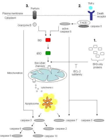
Figure 1.1: Caspase activation pathways
Intrinsic pathway(1): BH3-only proteins are activated by the cell stress or damage and overcome the inhibitory effect of BCL-2 family members. BAX-BAK assembly in the mitochondrial OM permit the efflux of intermem- brane space proteins, like cytochrome cinto the cytosol. Cytochromectriggers the apoptosome assembly:
caspase 9 and APAF1. Conformational activation of caspase 9 propagates the proteolytic cascade of further caspase activation events. In the extrinsic pathway(2)caspases are activated through the binding of extracel- lular death ligands, like TNFαto transmembrane death receptors. It provokes recruitment of adaptor proteins, such as FADD, which in turn recruit and aggregate several molecules of caspase 8, promoting its autoprocess- ing and activation. Active caspase 8 proteolytically processes and activates caspase 3 and 7, provoking further caspase activation events that culminate in substrate proteolysis and cell death. In some situations, extrinsic death signals can interact with the intrinsic pathway through caspase 8-mediated proteolysis of the BH3-only protein BID (BH3-interacting domain death antagonist). Truncated BID (tBID) can promote mitochondrial cy- tochromecrelease and assembly of the apoptosome. The granzyme B-dependent route to caspase activation (3)involves delivery of this protease into the target cell trough specialized granules that are released from cy- totoxic T lymphocytes (CTL) or natural killer (NK) cells. CTL and NK granules contain numerous granzymes as well as pore-forming protein, porfirin, which oligomerizes in the membranes f target cells to permit entry of the granzymes. Granzyme B, similar to caspases also cleaves its substrates after Asp residue, and can process BID and caspase 3 and 7 to initiate apoptosis. Reprinted with changes from [149].
Chapter 1. Introduction
apoptosis are: condensation of the nucleus and its fragmentation into smaller pieces [82], hydrolysis of nuclear DNA into multiple fragments [171], fragmentation of organelle‘s network: Golgi, endoplasmic reticulum and mitochondria, and protein release from the mitochondrial intermem- brane space [150].
During the early step of apoptosis the mitochondrial network disinte- grates. Apoptotic stimuli activate mitochondrial fission and block fusion.
It results in mitochondrial fragmentation and condensation [49; 81]. In this context it is closely correlated to the progression of apoptosis [92; 143].
Upon induction of apoptosis Drp1 translocates to the potential scission sites and becomes locked on the membrane during cell death in an hFis1 - independend and Bax/Bak - dependent manner [49]. It becomes stably sumoylated, which depends on the presence of Bax/Bak [167]. It occurs within the same time frame as activation of the proapoptotic Bcl-2 family member Bax and permeabilization of the mitochondrial outer membrane which in turn leads to the release of a multiple intermembrane space pro- teins and loss of the membrane potential [104]. Drp1 depletion blocks re- modelling of the mitochondrial cristae [55] where cytochrome cis mostly stored [9] and delays apoptosis [46]. In yeast, fission mediated by Drp1 ho- molog, Dnm1, is essential for the autophagic degradation of mitochondria [113]. Proapoptotic Bax protein co - localizes also with Mfn2 at distinct foci on mitochondria [109] and is required for the regulation of Mfn2 activity and lateral assembly into foci along the mitochondrial tubules [78]. This co - localization may account for a block in fusion observed during apop- tosis [80]. Recently, OPA1 has been shown to protect cells from apoptosis by controlling the remodelling of mitochondrial cristae, which is indepen- dent of its pro - fusion function [33; 51]. Out of eight OPA1 mRNA splice forms four have fusion activity. They produce a long isoform of OPA1 in addition to one or more further processed short forms [73]. It was demon- strated that those various forms are produced by processing at two dis- tinct sites: S1 encoded in all mRNA splice variants, and S2 encoded only
Chapter 1. Introduction
in some of them [73]. There is evidence for the involvement of rhomboid protease perselin - associated rhomboid like (PARL), mAAA and iAAA Zn2+-dependent metalloproteases belonging to the conserved AAA fam- ily of proteins, in the processing at those sites [61; 73; 138].
Chapter 1. Introduction
1.3 AAA
+superfamily and AAA proteins
ATPases play crucial roles in transforming chemical energy into bi- ological processes [114]. AAA+ superfamily of proteins (ATPases asso- ciated with diverse cellular activities) is based on careful and multiple alignments and crystallographic studies [110]. It contains proteins having extremely various functions in cells, and sharing common features [114].
They are involved in processes ranging from thermotolerance in bacteria, fungi and plants, through membrane fusion and microtubular movement in eukaryotes, to protein degradation and DNA replication [66].
AAA+ protein family is defined by the conserved AAA+ domain. It consists of N- and C - terminal subdomain, whose characteristics distin- guish AAA+ domain from other nucleotide-binding proteins [66]. Its ar- chitecture is more conserved than the underlying sequences. It is com- posed of Walker A (also called a P-loop, GxxxxGKT wherex= any amino acid residue) and Walker B (hhhhDExx where h = hydrophobic amino acid residue) motifs mediating ATP - binding and hydrolysis. They are fol- lowed by sensor 1, arginine fingers and sensor 2. Conserved polar residues of sensor 1 are physically located between Walker A and Walker B motifs and interact with important Walker B elements andγ- phosphate of ATP.
Arginine fingers are one or two conserved arginine residues which consti- tute part of the nucleotide - binding site of an adjacent subunit. Sensor 2 has a role in ATP hydrolysis and substrate unfolding. Its residues partici- pate in a nucleotide binding.
A conserved region that is positioned C - terminally from the Walker B motif named aSecondRegion ofHomology (SRH) is a characteristic for a subgroup of AAA+ superfamily, called AAA proteins. Its sequence is not strictly conserved, but the proteins share comparable structural features of SRH [74].
Chapter 1. Introduction
1.3.1 AAA
+proteases
AAA+ enzymes assemble mostly into hexameric complexes which is their biologically active form [66]. There has been a common mecha- nism proposed for ATP - dependent proteolysis by proteases belonging to the AAA+ superfamily. First, they oligomerize into barrel - shaped micro compartments allowing sequestered proteolysis [133], and second, ATP-dependent proteases need the energy from ATP hydrolysis to reg- ulate the accessibility of the proteolytic sites and to unfold substrates to drag them into the proteolytic chamber of the protease [8; 71]. The unfold- ing process is the rate - limiting step in proteolysis [84]. The initial sub- strate binding step does not require ATP hydrolysis, but for many AAA+ proteins ATP binding is required to generate the active, oligomeric form of the enzyme [114].
The mechanism of ATP hydrolysis involves the nucleophilic attack of an activated water molecule at the γ - phosphate of the ATP and the for- mation of a penta - coordinate transition - state [114]. Negative charge accumulating at the γ - phosphate is stabilized by the Mg2+ and by sur- rounding of positively charged groups and/or hydrogen bond donors.
Hence, the active sites of ATPases should contain a catalytic base able to activate nucleophilic water, and electrophilic groups able to stabilize the negatively charged transition state, plus groups coordinating the magne- sium ion which is an essential co - factor. The active sites contain “sensors”
which function is to “sense” the γ - phosphate and mediate the confor- mational changes that relay this information to remote sites. ATP binding pocket is located in between pairs of the hexamer subunits. Conserved residues that are functionally important for ATP binding and hydrolysis are: (1) in the Walker A motif: lysine (Lys, K) which forms ionic interactions withβ- andγ - phosphate oxygens, and threonine (Thr, T) which provides a metal ligand; (2) in the Walker B motif: aspartate (Asp, D) which is in- volved in the Mg2+ coordination sphere and glutamate (Glu, E) which is a catalytic residue important for ATP hydrolysis; (3) asparagine (Asn, N),
Chapter 1. Introduction
serine (Ser, S), threonine (Thr, T) or histidine (His, H) located at the N - terminal end of SRH in AAA proteases forming a polar contact with γ - phosphate of ATP and functioning as a sensor (sensor 1 [77]); (4) arginine (Arg, R) residue at the beginning of the third α-helix in the C-domain of the AAA+ module which in AAA proteins corresponds to the C - termi- nal part of SRH. It mediates relative movement of the C - domain to the N - domain during the ATP hydrolysis cycle. There are two conserved Arg residues in AAA proteins and one in the other AAA+family proteins.
They function as an arginine finger that transduces the chemical event of ATP hydrolysis into the conformational changes of the neighboring sub- unit of a hexamer. Such a coordinated cooperation of subunits greatly en- hances ATPase activity of a complex comparing to the singular ATPase activity of each subunit [98].
As a consequence of oligomerization, AAA proteases form a central cavity/pore lined with residues from each subunit [66]. Most of the pore surface is provided by the loop with three conserved residues (aromatic - hydrophobic - glycine [163]) which have a role in a function of several AAA proteins.
In the mitochondrial compartment both soluble and membrane ATP - dependent proteases have been found (Fig. 1.2).
1.4 Mitochondrial proteolytic quality control
A highly conserved proteolytic system conducts the surveillance of protein quality control within mitochondria, which has to cope with the diverse challenges imposed on mitochondrial integrity [147]. Molecular chaperones and energy - dependent proteases monitor the folding and as- sembly of mitochondrial proteins and selectively remove excess and dam- aged proteins from the organelle [87]. Key components are ATP - depen- dent proteases, which are derived from bacterial proteases and highly con- served in eukaryotes [87]. ATP - dependent proteases sense the folding
Chapter 1. Introduction
state of substrates by exerting chaperone - like properties and trigger the proteolysis of non - native proteins [147]. A central role in this process is exerted by conserved ATPase modules, which are characteristic of the AAA+ family of ATPases and present in all ATP - dependent proteases [147].
1.5 Mitochondrial ATP - dependent proteases
1.5.1 ATP - dependent proteases in the mitochondrial ma- trix
Two mitochondrial matrix proteases have been identified in various organisms: Lon and ClpXP. Lon proteases have been identified in the mitochondrial matrix of yeast and mammals [145; 159; 166; 165; 168]. They harbor a catalytic serine - lysine dyad and as such are classified as serine proteases [21; 126]. Lon forms presumably homooligomeric, ring - shaped complexes with hexameric or heptameric structure [140]. In most cases impaired folding appears to trigger protein degradation by Lon protease [159]. The yeast Lon ortholog, PIM1 has been shown to degrade various misfolded and non - assembled polypeptides, like thermally denatured and aggregated or oxidatively damaged proteins, such as aconitase [20;
100].
Molecular chaperones of the Hsp70 and Hsp100 family co - oper- ate with Lon protease during proteolysis by stabilizing misfolded pro- teins against aggregation or by dissolving already aggregated proteins [14; 124; 162]. Yeast∆pim1cells show inhibited growth on glycerol. Elec- tron dense inclusions in the mitochondrial matrix could be observed pre- sumably as a result of accumulation of non - degraded mitochondrial ma- trix proteins [145]. Similarly, down regulation of the human Lon protease leads to accumulation of protein inclusions, impaired mitochondrial func- tion and apoptotic cell death [19]. It was demonstrated that Pim1p was
Chapter 1. Introduction
required for the maintenance of mtDNA in yeast and present in the mito- chondrial nucleoids, and that it was able to bind GT - rich DNA sequences [31; 53; 52; 97]. Cellular effects of Lon deletion as well as known physi- ologic functions of this protease suggest its regulatory function, but the substrates and probable mechanisms it could be involved in are still un- known.
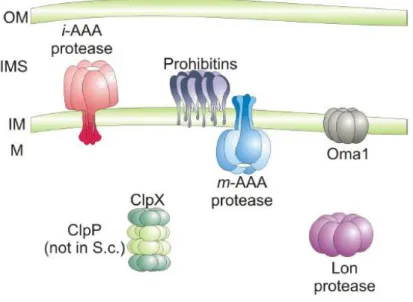
Figure 1.2: ATP-dependent proteases in the mitochondrial matrix and in the inner mitochondrial membrane. The Lon - protease and ClpXP (ClpP not present in yeast) are responsible for proteolytic breakdown of misfolded polypeptides in the mitochondrial matrix. In- tegral membrane and peripherally associated proteins are degraded by proteases of the inner mitochondrial membrane, the iAAA protease, active on the intermembrane side of the inner membrane; the mAAA protease, active in the mitochondrial matrix and the ATP - independent metallopeptidase Oma1, which is thought to pos- sess catalytic domains at the side of the inner membrane. Prohibitins built up a supercomplex with the mAAA protease.OM- outer membrane;IMS- intermembrane space;IM- inner membrane;M- mitochondrial matrix.
Reprinted with modifications from ([112]
The other mitochondrial matrix protease ClpX (caseino-lyticprotease) was found in all organisms whereas ClpP only in mammals and plants, but not yeast [37; 76; 160]. In contrast to Lon- and AAA proteases, their ATPase domain and the proteolytic domain are expressed as separate gene
Chapter 1. Introduction
products. The ATPase domains (ClpX, ClpA) assemble into hexameric, ring complexes with ATPase and chaperone activity, while the proteolytic domains (ClpP) form heptameric, double ring complexes. The ATPase domains determine the substrate specificity of the Clp protease and also exert regulatory functions during proteolysis [17; 117; 133; 169].
1.5.2 ATP dependent proteases in the mitochondrial mem- branes
Two membrane bound AAA proteolytic complexes have been iden- tified in mitochondria. They are integrated into the inner mitochondrial membrane and build up of orthologs of FtsH - membrane bound AAA protease essential for cell viability inEscherichia coli[2; 154; 155].
In yeast there are three FtsH orthologues: Yta10p and Yta12p forming a heterohexameric mAAA protease complex active at the matrix side, and Yme1p which is the only enzymatic component of iAAA protease active in the intermembrane space. They share several common features: N - ter- minal targeting signal followed by the AAA consensus and HEXXH motif characteristic for metallopeptidases of the thermolysin family M41 [123].
Mutation of the glutamate residue within the proteolytic centre inhibits protein degradation by AAA proteases [7]. Opposite orientation of cat- alytic domains of iAAA and mAAA proteases is a consequence of having one or two transmembrane domains respectively (Fig. 1.3). AAA protease - mediated degradation of membrane proteins involves dislocation or ex- traction of the substrate from the membrane [95]. This process requires around 20 residues to protrude from the membrane surface [32] probably to reach deep into the ATPase domain and establish productive binding.
Mutational analysis of a conserved loop motif YVG (aromatic - hydropho- bic - glycine) present in the central pore of hexameric AAA+ ring com- plexes [164], indicates substrate translocation into the proteolytic chamber through the central pore of mitochondrial AAA proteases [59]. Recently,
Chapter 1. Introduction
e1.3:DomainstructureofthemAAAandiAAAproteasesubunits ThenuclearencodedsubunitsareimportedintomitochondriaviaanN-terminalmitochondrialtargetingsequence(MTS)whichisremovedafterimport.One ortwo(Yta10p,Yta12p,FtsH)transmembrane(TM)domainsarepresentineachsubunit.TowardstheC-terminusareAAAdomainscontainingconserved characteristicfortheAAAproteinfamily;WalkerA(WA)andWalkerB(WB)motifsaswellasthesecondregionofhomology(SRH).TheAAAdomainsare byproteolyticdomainsharboringtheHEXXHmetalbindingmotif.AtthemostC-terminalendisthecoiled-coilregion(CC,(Reprintedwithmodificationsfrom B.AmultisequencealignmentoftheaminoacidregionscontainingtheconservedmotifsfromyeastmammalianandbacterialAAAproteasesubunits.Sequence dinatesoftheaminoacidsareindicated.ThecolorcodeaswellasthewholesequencealignmentispresentedintheAppendix1onpage81
Chapter 1. Introduction
Tatsutaet. al.has shown the ability of the mAAA protease to mediate vec- torial membrane dislocation of proteins in an ATP - dependent reaction [146]. This membrane extraction of substrate proteins is likely to be facili- tated by the membrane - embedded parts of AAA protease subunits which might form a pore - like structure or provide at least a more hydrophilic environment [88].
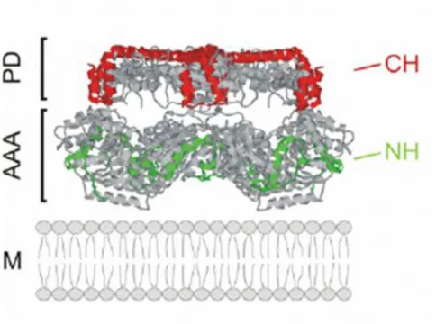
Figure 1.4: Folding of the yeast iAAA protease and substrate engagement
Side view of crystal structure of Thermotoga maritima FtsH [15] showing three subunits was used as a model for mapping the substrate binding regions of the yeast iAAA. Two identified regions CH and NH (red and green helices respectively) of the yeast Yme1p responsible for substrate engagement are located on the surface of proteolytic cylinder (CH helices marked on PD) and AAA domain (NH helices marked on AAA). AAA, AAA domain; PD, proteolytic domain; IM, inner membrane. Reprinted with modifications from [87].
Two FtsH structures from two eubacterial organisms have been solved recently [15; 90; 144] which facilitated studies on the yeast iAAA protease subunit Yme1p. It allowed the localization of two helical binding regions.
The C - terminal helices of the proteolytic domain (CH - region) and the
Chapter 1. Introduction
N-terminal helix (NH-region) in the AAA domain form a lattice-like struc- ture at the surface of the proteolytic cylinder and mediate the initial en- counter of substrate proteins with the protease (Fig. 1.4, [58; 87; 94]). The NH - region is located in a close proximity to the membrane surfaces and highly negatively charged. Thus, substrate proteins initially interact with the iAAA protease at the outer surface of the proteolytic cylinder, before they enter the proteolytic chamber. Interestingly, the binding properties of the surface - exposed interaction sites vary suggesting that alternative pathways for substrate entry into the proteolytic chamber of iAAA pro- teases exist. The CH - region is only required for the binding and degra- dation of a subset of proteins. The distance of an unfolded domain from the membrane surface, might be one parameter determining the involve- ment of the CH - region for substrate binding. In case of a non - assembled subunit 2 of the cytochrome c oxidase, Cox2p, destabilization of the sol- vent - exposed Cox2 domain at high temperatures renders CH - dependent proteolysis by the iAAA protease [58]. In contrast to the CH - region, the NH - region appears to be generally involved in proteolysis. It is therefore likely that CH - dependent substrates bind in a sequential manner at CH- and NH - regions before they enter the proteolytic chamber through the central pore formed by the AAA domains [87].
Growing evidence suggests that substrate binding and proteolysis by AAA proteases is modulated by additional factors within mitochondria [87]. It can appear in a substrate - specific manner. Like Cox20p which affects proteolysis of the Cox2p by the iAAA protease modulating recog- nition of a substrate [58]. Proteolysis of the non - assembled Cox2p was demonstrated to be strictly dependent on the CH - region in Cox20 - defi- cient mitochondria, but not in the presence of Cox20p [58] indicating over- lapping functions of Cox20 and CH - region of Yme1p. Another co - factor for iAAA protease is also not essential for proteolysis [42]. Together with Yme1p, Mgr1 is part of the iAAA proteolytic complex in yeast [42]. It was
Chapter 1. Introduction
suggested to act as an adaptor - like protein which targets specific sub- strates in the IMS for degradation by iAAA protease [87].
No substrate-specific co-factors of mAAA protease have been identi- fied so far. However, the yeast mAAA protease is part of a large super- complex with prohibitins, which modulate proteolysis [141]. Prohibitin 1 (Phb1p) and prohibitin 2 (Phb2p) are ubiquitous and highly conserved subunits of those supercomplexes. They assemble in a multimeric complex which is exposed in the intermembrane space and anchored N-terminally to the inner membrane [11; 111; 141]. Deletion of prohibitins results in ac- celerated protein degradation by the mAAA protease [141]. However it is not yet understood how they affect the proteolysis by the mAAA protease.
Both the m- and the iAAA protease recognize and degrade non-native and non-assembled polypeptides to peptides [7; 96]. These proteases ex- hibit degenerate substrate specificity and, similar to molecular chaper- one proteins, recognize the folding state of solvent-exposed domains of membrane proteins [94]. mAAA and iAAA proteases are the main compo- nents of mitochondrial quality control. They degrade proteins to peptides, which are subsequently either exported from the organelle or degraded further to amino acids by various oligopeptidases [147].
Yeast cells lacking both m- and iAAA proteases are not viable demon- strating the crucial function of this proteolytic system for cellular home- ostasis [93; 95]. Their depletion or inactivation causes severe pleiotropic phenotypes in various organisms, which are best characterized in the yeast Saccharomyces cerevisiae. Here, all observed defects can be attributed to a loss of the proteolytic activity as identical phenotypes were observed after deletion of an AAA protease subunit or after inactivation of the proteolytic sites of all subunits of AAA protease complexes [6; 96; 170]. Both pro- teases exert overlapping functions within mitochondria, having overlap- ping substrates; however different phenotypes resulting from mutations indicate substantial differences.
YTA10 and YTA12 yeast mutants exhibit respiratory incompetence
Chapter 1. Introduction
[62; 148] and lack assembled respiratory chain and ATP-synthase com- plexes in the inner membrane [54; 118; 158]. Yeast mAAA protease was found to degrade non assembled mitochondrialy encoded respiratory chain subunits. Also peripheral membrane protein such as Atp7 was iden- tified as a mAAA substrate [88]. Recently, a subset of specific substrates was identified proving a regulatory, housekeeping function of mAAA protease. That includes proteolytic cleavage of a conserved, nuclearly en- coded subunit of mitochondrial ribosomes MrpL32 [112] and cytochromec peroxidase (Ccp1), a heme-binding ROS scavenger in the intermembrane space [45; 146]. Maturation of Ccp1 within mitochondria represents the first non-proteolytic function of the mAAA protease in the mitochondrial biogenesis, and rather severe mutant phenotypes of YTA10/YTA12could be explained not only by loss of respiration but also by the overall mito- chondrial malfunction.
SPG7 - a mammalian homologue of the yeast mAAA protease sub- units was identified in the genetic screen of patients affected with an au- tosomal, recessive, neurodegenerative disorder called Hereditary Spastic Paraplegia (HSP) [24]. Further identification of mammalian AAA pro- teases homologues followed. Complementation studies identified a com- plex of mammalian paraplegin and AFG3L2 as the functional orthologue of the yeast mAAA protease [12; 112] and a knock-out mouse model was generated [47] where the human phenotype of the disease was repro- duced. It has been proposed that mAAA mutations mapped in the HSP patients led to the defects in an axonal transport of mitochondria, result- ing in the energy insufficiency and a neuronal decay [47]. AFG3L2 was shown to homooligomerize, which was proposed to be responsible for in- creased severity of the Afg3l2 mutants [87; 101].
The mammalian mAAA protease was linked to the transmembrane potential-dependent proteolysis of OPA1 [43; 73] together with a mam- malian rhomboid protease PARL [33] and prohibitin 2 (PHB2) - a part
Chapter 1. Introduction
of a prohibitin-mAAA supercomplex, is involved in the OPA1-dependent cristae remodeling in mitochondria [106].
Yeast lacking YME1 lose mtDNA at an accelerated rate forming pe- tite negative colonies, i.e. unable to grow on glucose in the absence of mtDNA. ∆yme1cells fail to grow on a non fermentable carbon source at 37°C , as well as on glucose at 14°C [23; 152]. Theyme1mutant cells show deficient oxygen consumption and mitochondrial morphological abnor- malities: from abbreviated branched structures to swollen forms, together with alterations in protein/lipid composition. Mitochondrial abnormali- ties could be partially reversed in S.cerevisiaeby mutations ofYNT1 gene encoding a 26S protease subunit suggesting similar functions [23]. The Yme1p is immunologically detectable as an 80 kDa protein present in the mitochondria [153]. It has a role in the degradation of unassembled in- ner membrane protein complexes, like cytochrome c oxidase subunit 2 [121] and Phb1p and Phb2p [75] as well as external NADH dehydroge- nase (Nde1) [13]. Raineyet al.reported crucial non-proteolytic function of Yme1p in the import of an exogenously expressed human PNPase [122] - mitochondrial intermembrane space protein required for the maintenance of mitochondrial homeostasis [29]. Selective removal of malfunctional mi- tochondria in the autophagic process, named mitophagy [83] suppresses apoptosis and has a cytoprotective function [99]. Interestingly, recent re- ports suggest a new function of Yme1p, namely the proteolytic turnover of a mitochondrial inner membrane protein phosphatidylserine decarboxy- lase 1 (Psd1p) responsible for the synthesis of phosphatidylethanolamine - an essential mitochondrial component, having function in the autophagy [108].
Human ortholog of Yme1p has been identified in the complementa- tion studies [134]. It has been expressed in the ∆yme1 yeast cells rescu- ing the temperature-sensitive phenotype, and demonstrating functional complementation and conservation of the iAAA complex [134]. Human YME1L1 (Yme1-like 1, further related to as YME1L) and paraplegin have
Chapter 1. Introduction
42% and 33% homology with the yeast Yme1p respectively. Expression and immunofluorescence studies revealed that YME1L and paraplegin share similar expression pattern and the same subcellular localization [36].
Two transcript variants encoding different isoforms have been found for the human gene. Transcript variant 3 lacks an internal coding exon 3 compared to transcript variant 1. However, it maintains the same read- ing frame, and encodes an isoform 3 (716 aa; 80kDa) that is missing a 57 aa segment compared to isoform 1 (773 aa; 86.5kDa). The exon 3 en- codes an N-terminal part of the protein, which is presumably processed off upon import of YME1L into the mitochondria. There is only one tran- script variant annotated for the murineYme1l1encoding only one form of YME1L: 715 aa, 80kDa. Because of the high conservation and homology (see alignment and homology tree in the Appendix 1 on page 81) possi- ble involvement in spastic-like disorders has been suggested for the mam- malian iAAA protease [36; 134]. However, Coenenet al.[35] failed to iden- tify YME1L mutations in patients with combined defects in the oxidative phosphorylation system. Only recently, it has been suggested that YME1L may have a function in a novel apoptotic pathway independent of cas- pases [57]. Thus, the mammalian iAAA protease is believed to have an additional role in the regulation of mitochondrial dynamics and morphol- ogy, independent of its proteolytic quality controlling in the mitochondria.
Chapter 2
Aim of the thesis
Identification of the YME1L as an ortholog of the yeast iAAA pro- tease subunit opened a question about its action and its importance for the mammalian cell. Little is known about the mammalian iAAA protease complex as no functional studies were done on that protein. The question was, whether evolutionary conservation of the protein results in a func- tional conservation within the mitochondria. To address it and to examine the relevance of the mammalian iAAA protease for mitochondrial mor- phology, proteolysis and function two approaches were carried out:
• YME1L down regulation studies in the mammalian cells and
• overexpression studies of the wild-type and various mutant variants of the YME1L protein in the mammalian inducible system.
Chapter 3 Results
3.1 Down regulation of YME1L using small in- terfering RNAs (siRNAs)
3.1.1 Specific antibody against YME1L
The proper recognition of the protein by the antibodies used through- out the study was verified prior to the verification of the efficiency and specificity of YME1L down regulation and its overexpression in mam- malian cells. As a reference for the running behavior on SDS-PAGE of the mature YME1L data were used from the in vitroprocessing of the human YME1L by thematrixprocessingprotease (MPP; Figure 3.1A; courtesy of Dr Mirko Koppen). The cloned construct was encoding human isoform 3, i.e.having 716 amino acids in length (80kDa) which was also used by Shah et al.in complementation studies [134]. The hYME1L was transcribed and radiolabeled in a cell-free system and incubated at 30°C for 20 min with the MPP purified fromE.coli. In the next experiments three different anti- bodies were checked for the recognition specificity:
• polyclonal antiserum raised against hYME1L protein (Figure 3.1B and C, kind gift of Dr Carla Koehler);
Chapter 3. Results
• affinity purified polyclonal antiserum SPY531 raised against two peptides common for the murine and human protein sequence:
(152-166aa) TLKSRTRRLQSTSER and (517-531aa) DKILMGPERRS- VEID (Fig. 3.1D);
• commercial polyclonal antibody (ptglab; Fig. 3.1E).
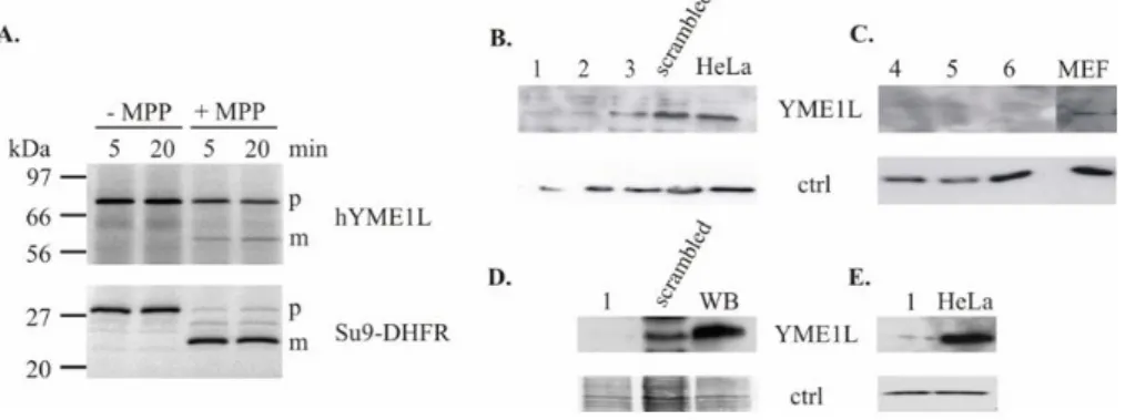
Figure 3.1: Recognition of the mature YME1L by different antibodies used in the present work
A. Human YME1L processingin vitro by the matrix MPP protease. Mature form of the hYME1L runs at the size of∼60kDa. Efficiency of the processing was controlled by Su9-DHFR, a construct cleaved by MPP (courtesy of Dr Mirko Koppen). Four different antibodies used in the present work were checked for specific recognition of YME1L by comparison of the size of bands recognized in Western blotting. As a reference for the proper band size,in vitroprocessing of hYME1L was used(A.). With numbers 1-3 are marked cell lysates from HeLa cells transfected with siRNA1-3; and 4-6 show lysates form MEF cells transfected with siRNA4-6.
Extracts from the scrambled negative siRNA control and non-transfected cells were used as protein down regulation reference. WB refers to cell extract from HEK293 Flp-In clone expressing mYME1LE381Qinduced with 1µg/ml tetracycline for 24h.β-actin was used as a loading control (antiβ-actin 1:5000, Sigma, clone AC15);
B.-C.polyclonal antiserum raised against hYME1L protein (kind gift of C.Koehler) specifically recognized both human(B.)and murine(C.)YME1L;D.polyclonal antiserum SPY531 raised in the present study against two peptides from the murine YME1L recognized specifically both human (scrambled) and murine (WB) YME1L E.
commercial polyclonal antibody (ptglab, 1:500 dilution) recognized human YME1L protein.
In all of the cases the antibodies recognized specifically a band running on SDS-PAGE at the size of ∼60kDa, which disappeared upon siRNA transfection i.e.down regulation of YME1L protein in all of the used cell lines: HeLa (Fig. 3.1B and E) MEF (Fig. 3.1C) and HEK293 (Fig. 3.1D). As reference cell extracts were used from cells transfected with a scrambled
Chapter 3. Results
negative siRNA control (Invitrogen) as well as from non-transfected cells.
The size of the protein running at that position is in agreement with the size of the mature hYME1L processed in vitro (Fig. 3.1A.). The exclusive disappearance of the 60 kDa band was observed only upon siRNA1-6 transfection which demonstrates specificity of the antibody recognition.
3.1.2 Depletion of YME1L in mammalian cells
Three different siRNAs were used to down regulate the YME1L expres- sion on the mRNA level. For human YME1L (hYME1L) that were siRNA1, siRNA2 and siRNA3, whereas siRNA4, siRNA5 and siRNA6 were de- signed against murine YME1L (mYME1L). All sequences hybridize within the coding sequence. Scrambled negative control (Invitrogen) i.e. siRNA duplexes with no match to the mRNA of YME1L, was used as a control.
It allowed verification of a knock down specificity on YME1L. Western blot analysis revealed the most efficient down regulation in the case of siRNA1 and 2 for hYME1L (Fig. 3.1B lane 1 and 2) and siRNA4 and 6 for mYME1L (Fig. 3.1C and not shown data). The protein was reduced to a level which was not detectable by immunoblotting with any of the usedα YME1L antibodies. siRNA 1, 2 and siRNA 4 and 6 were chosen for further experiments.
Chapter 3. Results
3.1.3 Mammalian YME1L role in proteolysis
Proteolysis of PHB1 upon depletion of its assembly partner PHB2 Depletion of PHB2 in mammalian cells is accompanied by the loss of its assembly partner PHB1; however, transcription ofPhb1was shown not to change [106]. This suggests proteolysis of PHB1 while the PHB2/PHB1 complex is destabilized upon absence of PHB2; an effect that was observed previously in yeast [75]. Therefore, the PHB1 steady-state level was as-
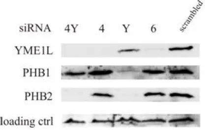
Figure 3.2: YME1L degrades PHB1 upon destabilization of the prohibitin complex
Cell extracts from MEFs transfected with siRNA designed against mYME1L (4and6) and against mPHB2(Y) or both of them(4Y)were analyzed after 48-60h post transfection by immunoblotting withαYME1L (ptglab),α PHB1 (NeoMarkers) andαPHB2 (BioLegend). Cells transfected with scrambled siRNA were used as reference control. Immunoblotting withαcomplexII antibody (Molecular Probes) was used as a loading control.
sessed in PHB2 deficient MEF cells by single (siRNA4 and 6 againstYme1l, siRNAY against Phb2) or double siRNA transfection (siRNA4 and Y; Fig.
3.2) to verify the potential role of YME1L in PHB1 degradation. In the case of single transfections Western blotting showed an efficient and specific down regulation of YME1L (Fig. 3.2, lane 2 and 4) while neither the level
Chapter 3. Results
of PHB1 and PHB2, nor complex II used as a loading control, changed.
Similarly, efficient depletion of PHB2 (Fig. 3.2, lane 3) did not lead to de- pletion of YME1L or complex II. It led however to destabilization of PHB1.
Simultaneous knock down of PHB2 and YME1L (Fig. 3.2, lane 1) stabilized PHB1 to the wild-type level (scrambled control). This indicates a role of the YME1L protein in the degradation of the unassembled prohibitin complex subunit - PHB1. This finding clearly shows that structural conservation of Yme1p throughout the evolution (compare with Fig. 1.3) results also in the functional conservation of the protein, as Phb1p was shown before to be a specific substrate for the yeast iAAA protease [75].
YME1L role in the new apoptotic pathway
The molecular bases of phenotypes associated with a deletion ofYME1 in yeast as well as the phenotypes observed upon down regulation of YME1L in mammalian cells are poorly understood. It appears likely that they are connected with the diverse roles of the so far undiscovered substrates of the iAAA protease. Given those pleiotropic functions, we wanted to examine further effects of YME1L down regulation in the mam- malian cells. Recently, Goemanset al.have reported a new apoptotic path- way activated by mitochondrial outer membrane permeabilization and in- hibition of caspases [57]. It involves a specific TIM23 degradation which impairs mitochondrial import and inhibits cell proliferation. Tim23p is a central component of the TIM23 translocase complex in the inner mi- tochondrial membrane, crucial for the pore formation [107]. It was fur- ther shown that TIM23 degradation depends on factors residing in the mitochondria. The authors proposed this novel apoptotic pathway as an
“emergency exit” for the apoptosis to occur upon inhibition of caspases.
To investigate the specific function of YME1L in TIM23 degradation, apop- tosis was induced with 200µM or 50µM etoposide (Eto, Fig. 3.3A, B) in the cells transfected with siRNA against YME1L and scrambled negative control (scr). Cells were treated with 10µM caspase inhibitor Q-VD-OPH
Chapter 3. Results
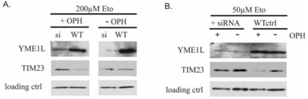
Figure 3.3: Stabilization of TIM23 upon YME1L down regulation
HeLa cells transfected with siRNA and scrambled negative control (scr) were treated with 200µM(A.)and 50µM(B.)of an apoptotic stimulus (Eto) in the presence or absence of caspases inhibitor (OPH) for 24h and 48h respectively. Destabilization of TIM23 (αTIM23, BD Biosciences) observed upon simultaneous apoptosis stimulation and inhibition of caspases can be rescued by siRNA knock down of YME1L (αYME1L, ptglab).
Immunoblotting withαcomplexII antibody (Molecular Probes) served as a loading control.
(OPH). Inhibition of caspases simultaneous with the apoptosis induction resulted in TIM23 destabilization as shown on Fig. 3.3A lane 2, and 3.2B lane 3. Knock down of YME1L stabilized the protein, revealing YME1L role in the apoptosis- and caspases inhibition-induced specific degrada- tion of TIM23. The effect was stronger with 50µM etoposide treatment for 48h, than 200µM etoposide for 24h, suggesting that this pathway is rather late reacting.
3.1.4 YME1L role in mitochondrial biogenesis
Mitochondrial morphology of the mammalian cells
Next, the effects of YME1L depletion on the mitochondrial morphol- ogy were addresses in human HeLa cells and murine MEFs. To this end both types of cells were transfected with siRNA against YME1L to knock down the protein. Subsequently, cells were transfected with a plasmid en- coding mitochondrialy targeted red fluorescent protein (pDsRed2-Mito) for the visualization of mitochondria by fluorescent microscopy. We used cells transfected with the scrambled negative control as controls (Fig. 3.4B)
Chapter 3. Results
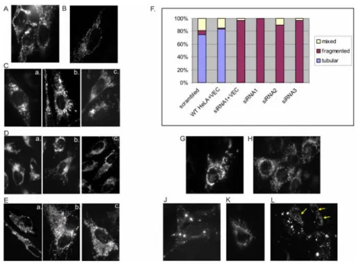
Figure 3.4: Aberrant mitochondrial morphology in the absence of YME1L
A. - E.HeLa cells were transfected with: siRNA1 (C. a, b, c) siRNA2 (D. a, b, c) siRNA3 (E. a, b, c) designed against human YME1L mRNA, and scrambled negative control(B.) or no siRNA(A.) followed by pDsRed-Mito (1µg) transfection and analyzed by fluorescence microscopy after 48-60 h post-transfection. Different morpho- logical types of mitochondria were quantified(F.) and compared to controls: HeLa transfected with scrambled negative control siRNA (scrambled) and pDsRed-Mito (1µg), HeLa cotransfected with pcDNA3.1Hygro empty plasmid and pDsRed-Mito (4:1, 1µg of total plasmid DNA) (WT+VEC), HeLa transfected with siRNA1 and co-transfected with pcDNA3.1Hygro:pDsRed-Mito (4:1, 1µg of total plasmid DNA) (siRNA1+VEC). 100 cells were calculated for each sample.G. - L.MEF cells transfected asA. - E.with no siRNA (WT control;G.); scram- bled negative control(H.)and siRNAs designed against murine YME1L mRNA: siRNA4(J.)siRNA5(K.)and siRNA6(L.). Yellow arrows point to the non affected cells with mitochondrial morphology as in the control cells.
as well as cells not transfected with siRNA (WT, Fig. 3.4A). In all of tar- get siRNA-transfected cells we could see aberrant mitochondria. From swollen but still interconnected branches (Fig. 3.4Cb, Ea-c) to the com- plete lack of the network (Fig. 3.4Ca, Dc, J, K, L). HeLa cells were quanti- fied: 100% cells transfected with siRNA1 had fragmented/aggregated mi- tochondria; 90% transfected with siRNA2 and 97% with siRNA3. None of the transfected HeLa cells had wild-type mitochondrial tubular net-
Chapter 3. Results
work. Only 6% of the cells transfected with scrambled control had frag- mented mitochondria. WT and siRNA1 transfected cells were addition- ally co-transfected with an empty pcDNA3.1Hygro vector (VEC) to ex- clude deleterious effect of plasmid transfection. They had 2% and 97%
of fragmented mitochondria respectively. Even in the populations, where Western blotting analysis still revealed residual levels of the YME1L pro- tein (Fig.3.1B lane siRNA3) almost all of the cells showed abnormal mi- tochondrial morphology. That could indicate the primary effect of down regulation of YME1L on mitochondrial network not tightly related to its high enzymatic/proteolytic activity. Similar mitochondrial phenotypes in the MEFs transfected with siRNA4-6 were observed (Fig. 3.4J-L). The frag- mentation/aggregation was even more striking than in HeLa cells (Fig.
3.4J, K). However, the transfection efficiency was lower as mixed popu- lations of cells having aberrant and wild-type morphology could be seen (Fig. 3.4L).
OPA1 processing in the YME1L knocked down cells
The dynamin-related GTPase OPA1 is essential for the maintenance of cristae in the mitochondrial inner membrane and for mitochondrial fusion [116; 138]. There are eight OPA1 splice variants which are proteolytically processed to two long isoforms (L1 and L2) which can be in turn processed to three short isoforms (S3-S5). S4 is a form that appears to be generated by YME1L [61; 73; 138]. In further experiments a relation of mitochondrial abnormalities in MEFs (Fig. 3.5A) to accumulation of the different OPA1 forms (Fig. 3.5B) was examined. Immunoblotting of the MEF cells trans- fected with siRNA4 revealed alterations in OPA1 cleavage (Fig. 3.5B, lane 2). Longer isoforms are absent (L1) or present in the smaller amount (L2) and the shorter isoforms accumulate. This effect is in line with the aber- rant morphology visible by fluorescent microscopy (Fig. 3.5A). Notewor- thy is the difference in mitochondrial morphology of cells transfected with siRNA against PHB2 (siRNA Y), and YME1L (siRNA 4). Mitochondria in
Chapter 3. Results
Figure 3.5: Processing of OPA1 depends on the presence of YME1L
Lysates from the MEF cells transfected with siRNA against murine YME1L(4), murine PHB2(Y)or both of them(4Y) were checked for OPA1 expression pattern by immunoblotting with the anti-OPA1 antibody (BD Biosciences). Ponceau S staining (PS) serves as a loading control. Fluorecence microscopy(A.)of indicated cell samples shows mitochondrial morphology effect in relation to OPA1 processing(B.). Scrambled transfection control (scrambled, SCR) and wild-type cells (MEF WT) were used as reference controls.
cells lacking PHB2 are small and dispersed, no elongated forms are visible.
No interconnections are present. Mitochondria from cells lacking YME1L are bigger, swollen with visible interconnections. Therefore they resemble more aggregated than fragmented tubular network. Recently, PHB2 was shown to influence the OPA1 processing and cristae remodeling, with the mitochondrial fragmentation as PHB2-depletion effect [106]. Apparently, PHB2 down regulation has an epistatic effect over YME1L down regula- tion, since double knock down of the proteins (siRNA 4 and Y) results in the morphology resembling single PHB2 depletion (Fig. 3.5A).
Chapter 3. Results
Mitochondrial transmembrane potential maintenance in the YME1L knocked down cells
Mitochondrial dysfunction and dissipation of the membrane potential across the inner membrane can induce OPA1 processing and lead to mito- chondrial fragmentation [44; 73; 106]. The YME1L knock down could also
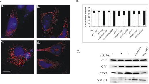
Figure 3.6: YME1L affects mitochondrial morphology but not respiration
A.Aberration in the mitochondrial morphology upon down regulation of YME1L by 3 different siRNAs: 1 (a.)2(b.)and 3(c.)in HeLa cells transfected with pDsRed-Mito and analyzed after 48-60 h by fluorescence microscopy. Scale bar 2µm.B.Maintenance of mitochondrial transmembrane potential in YME1L-deficient HeLa cells. Indicated cell samples were stained with the fluorescent dye JC1 and analyzed by flow cytometry at 590 nm. Dissipation of the membrane potential with CCCP both in wild-type HeLa cells and transfected with siRNAs 1 and 2 or scrambled control, together with DMSO treated cells were used as controls. White bars represent the percentage of cells giving red and green signal whereas black bars show percentage of cells giving only green signal, e.g. with the dissipated membrane potential.C.Immunoblot analysis of siRNA 1-3 transfected HeLa cells. Steady state levels of respiratory chain proteins: complex II (Molecular Probes), complex V (Molecular Probes) and complex IV subunit 2 (COX2; Molecular Probes) were analyzed by SDS-PAGE of total cell lysates and immunoblotting with the specific antibodies. Scrambled siRNA transfected and non transfected cells serve as reference controls. Immunoblotting with an anti-YME1L antibody (Dr. C. Koehler) shows YME1L-specific siRNA down regulation efficiency.
impair the transmembrane potential. That would explain induced OPA1 processing and mitochondrial morphology abnormalities (Fig. 3.6A). To examine this possibility, siRNA transfection was repeated. Subsequently cells were stained with JC1 (Molecular Probes). This is a fluorescent dye,
Chapter 3. Results
whose oligomerization in the living cell takes place in mitochondria and depends on the presence of the mitochondrial potential. JC1 aggregates in the mitochondria emitting a red fluorescence, or/and stays in the cy- tosol as monomer, emitting a green signal. Thus, in normal cells with transmembrane potential present, both red and green signals are present, while no red signal can be detected upon dissipation of the membrane potential. siRNA1 and siRNA2 were used, as the most efficient in down regulating YME1L levels. Wild-type HeLa cells and the negative siRNA control were used to exclude deleterious effect of the transfection. CCCP (carbonyl cyanide m-chloro-phenyl-hydrazone) treatment was used as a positive control for the transmembrane potential dissipation. CCCP is an uncoupling agent which abolishes the obligatory linkage between the res- piratory chain and the phosphorylation system observed with intact mi- tochondria. Cells were subjected for fluorescence-activated cell sorting (FACS) and quantified (Fig. 3.6B). The membrane potential was main- tained in YME1L depleted HeLa cells. Furthermore, respiratory chain complex II, complex V and cytochrome c oxidase subunit 2 (COX2) steady state levels were not affected (Fig. 3.6C). Taken together, increased OPA1 processing and mitochondrial fragmentation in YME1L-depleted cells is not caused by an impaired transmembrane potential or respiratory activ- ity. These findings are in agreement with the previous reports showing ab- normalities in the mitochondrial network upon YME1L down regulation [61] proposing YME1L role in the processing of OPA1 in a constitutive and transmembrane potential-independent manner at the S2 processing site [63; 138].
Chapter 3. Results
3.2 Overexpression of YME1L in the inducible system: HEK293 T-REx Flp-In cells
3.2.1 Expression constructs
Mitochondria depend on the functional iAAA and mAAA inner mem- brane proteases. The mAAA protease subunits paraplegin and AFG3L2 were reported in the pathogenesis of Hereditary Spastic Paraplegia [24;
25; 127], axonal development [101], in the maintenance of mitochondrial morphology [43; 73] and in the proteolytic cleavage/maturation of other mitochondrial proteins [112; 146]. Similarly, the present study reports a role of the iAAA protease in mitochondrial morphology, apoptosis and specific degradation. However, little is known about the mechanisms that could carry out those functions. Recent mutational studies on the yeast Yme1p revealed different modes of substrate binding [58] which could participate in different effects of gene deletion or Yme1 protein mutation on its different functions. Furthermore, studies on the mAAA protease have shown that a mutation of the conserved glutamate residue in the Walker B motif, required for ATP hydrolysis has a dominant negative ef- fect on the mAAA complex (personal communication: Ines Raschke, Flo- rian Gerdes, Dr Takashi Tatsuta and Dr Steffen Augustin). It is therefore conceivable, that similar mutation in iAAA could lead to the similar ef- fect. To test that possibility we constructed several expression constructs encoding different variants of murine YME1L (Fig. 3.7A). Mutations were introduced into:
• the lysine residue required for ATP binding in the Walker A motif (mYME1K327A;WA),
• the glutamate residue of the Walker B motif required for ATP hydrol- ysis (mYME1LE381Q;WB),