of Membrane-associated Proteins of the Arabidopsis Female Gametophyte
DISSERTATION ZUR ERLANGUNG DES
DOKTORGRADES DER NATURWISSENSCHAFT (DR. RER. NAT.) DER FAKULTÄT FÜR BIOLOGIE UND VORKLINISCHE MEDIZIN
DER UNIVERSITÄT REGENSBURG
vorgelegt von Thomas Hackenberg
Bad Reichenhall aus im Jahr
2017
Die Arbeit wurde angeleitet von:
PD Dr. Stefanie Sprunck
Unterschrift:
1
ContentsContents
Summary ... 7
1 – Introduction ... 9
1-1 The Life Cycle of Angiosperms ...9
1-2 Development of the Polygonum-type Female Gametophyte in Arabidopsis ...9
1-3 Cell-fate determination within the mature embryo sac ...12
1-4 Double Fertilization in Angiosperms ...12
1-4-1 Gametophytic Interactions during Double Fertilization ...13
1-4-2 Direct Cell-Cell Interactions during Double Fertilization ...14
1-5 Key Molecular Players during Gamete Interactions and Fusion ...15
1-6 Tetraspanins, Membrane Microdomains and Sphingolipids ...17
1-7 Aims of this Work ...20
2 – Results ... 21
2-1 Acquisition, Processing, and Evaluation of the Transcriptomic Data of Arabidopsis Gametophytic Cells ... 21
2-1-1 Previous Work: Isolation of Arabidopsis Female Gametophytic Cells by Micromanipulation, and ATH1 GeneChip Hybridization ...21
2-1-2 Identification of Differentially Expressed Genes encoding Membrane-associated and Secreted Proteins ... 21
2-1-3 Statistical and Gene Ontology-based Evaluation of the obtained Transcriptomic Data ...24
2-1-4 In silico Analysis of the Gametophytic Transcriptome Data ...26
2-1-4-1 In silico Analysis of the Gametophytic DEGs obtained from the Sporophytic Contrast ...26
2-1-4-2 In silico Analysis of the Gametophytic DEGs obtained from the Gametophytic Contrast ...30
2-1-4-3 The DEGs identified by either Sporophytic or Gametophytic Contrasts show little Overlap ....33
2
Contents2-2 The RKD2-induced EC-like Cell Line as Tool for EC Membrane-Proteomics ...35
2-2-1 Characterization of the EC-like Cell Line ...35
2-2-2 Acquisition of Proteomic Data of the EC-like Cell Line ...36
2-2-3 In silico Evaluation of Proteomic Data of the EC-like Cell Line ...38
2-3 Selection of Candidate Genes for Data Validation, and in-depth Functional Studies ...41
2-3-1 Relative Quantification of Gene Expression of selected Candidate Genes ...42
2-3-2 In situ Detection of Candidate Gene Transcripts in the Arabidopsis Ovule ...45
2-3-3 Verification of the Promoter Activities of Selected Genes in Arabidopsis Ovules ...47
2-4 Functional Studies of the Selected Genes: Overview ...50
2-5 DEGs Encoding Putative Embryo Sac-secreted Proteins ...52
2-5-1 DUF239, and X8 domain-containing Proteins are Secreted by the CC ...53
2-5-2 SSPR is an Ovule-specific Secreted Protein ...54
2-5-3 The SSPR Knock Out arrested during FG Development ...56
2-6 DEGs encoding Membrane-localized Proteins ...58
2-6-1 The DUF962-containing Gene Family: Phylogeny ...59
2-6-2 DUF962 Proteins of Arabidopsis and Yeast are Conserved ...60
2-6-3 DUF962 Genes are expressed in the Sprophyte and the Gametophyte ...61
2-6-4 The DUF962 Gene Family is expressed in the Female Gametes and the Sporophyte ...62
2-6-5 The DUF962_1 genomic locus and available T-DNA insertion lines ...64
2-6-6 Knock Down of DUF962 Genes by amiRNA ...65
2-6-7 The Seed Sets of Arabidopsis DUF962 amiRNA-Knock Down Lines ...66
2-6-8 The FG of EC1.1p:amiR
DUF962-expressing Plant Lines Arrest in Early Developmental Stages ...68
2-6-9 The S. cerevisiae Δmpo1 Mutant is Complemented by Arabidopsis DUF962_1 ...70
2-6-10 The Role of Tetraspanins in Arabidopsis Gametophytic Cells ...73
2-6-11 Two Subclades of the Tetraspanin family are Preferentially Expressed in Gametophytic Cells ..73
2-6-12 A tet8 tet11 Double Knock has a WT-like Seed Set ...77
3
Contents2-6-13 Simultaneous Tetraspanin Knock Outs by the CRISPR/Cas9 System ...78
2-6-14 Seed Sets of CRISPR/Cas9-mediated Tetraspanin Knock Out Lines ...79
2-6-15 Female Gametophytes of tet8 tet11 tet12 Mutant Lines Arrest in FG1 ...81
2-6-16 Female Gametophytes of the tet8 tet11 tet12 tet9
+/-Mutant Line #25 Arrest in FG4 ...82
3 – Discussion ... 86
3-1 Female Gametophytic Cells in the Focus: What We Know and What We Don’t Know ...86
3-2 Bridging the Gap: Transcriptomics ...87
3-3 What’s the Hold-up? Data Validation! ...88
3-4 Gametophytic Cells Show Distinct Expression Profiles of Genes Encoding Cell Surface Proteins ...90
3-5 I’m Gonna Send Them to Outta Space: Secreted Proteins of the Female Gametophyte ...91
3-6 SSPR, Ready for Development or Defense? ...93
3-7 I Sense a Disturbance in the Apoplast: Gamete-expressed RLKs ...95
3-8 Like a Stick to the Head: Reverse genetics of DUF962...98
3-9 DUF962_1 is a True Ortholog of Yeast MPO1 ...100
3-10 The Fat and the Furious: Sphingolipid Signaling and MPO1 ...101
3-11 The Egg Cell-like Callus, a Tool Worth Exploiting! ...103
3-12 Putative Protein Interaction Partners of TET9 ...104
3-13 The Role of Tetraspanin Proteins in Development ...106
3-14 Conclusions and Future Issues ...108
4 – Experimental Procedures ... 110
4-1 Computer-based Methods ...110
4-1-1 Databases, Online Tools, and Software ...110
4-1-2 Transcriptome Analysis (by Dr. Maxim Messerer) ...111
4-1-3 Manual Transcriptome Data Annotation and Processing ...113
4-1-4 GO-term Analysis ...113
4-1-5 Heatmap Generation ...114
4
Contents4-1-6 Reconstruction of Phylogenic Relationships ...114
4-1-7 Designing CRISPR/Cas9 sgRNA ...114
4-1-8 Manual Proteome Data Processing ...114
4-1-9 Plotting and Statistics ...114
4-1-10 Oligonucleotide Design ...115
4-2 Nucleic Acid-based Methods ...115
4-2-1 Isolation of Plant DNA ...115
4-2-2 Isolation of Plasmid DNA ...115
4-2-3 Isolation of DNA from Yeast ...115
4-2-4 Isolation of mRNA ...115
4-2-5 Degrading of DNA ...115
4-2-6 Synthesis of First-Strand cDNA ...115
4-2-7 Rapid amplification of cDNA ends of AT1G74440 ...116
4-2-8 Taq Polymerase-based PCR ...116
4-2-9 Colony PCR ... 116
4-2-10 Reverse-transcription PCR ...117
4-2-11 Genotyping of Transgenic Plants, and Determination of T-DNA Insertion Positions ...117
4-2-12 Phusion® High-Fidelity DNA Polymerase-based PCR...117
4-2-13 Overlap extension PCR ...117
4-2-14 qPCR ... 119
4-2-15 Agarose Gel Electrophoresis ...119
4-3 Cloning Procedures ...120
4-3-1 Restriction-Ligation-based Cloning ...120
4-3-2 Gateway Cloning® ...120
4-3-3 amiRNA Construction ...120
4-3-4 CRISPR/Cas9 Construct Preparation ...121
4-4 Organism-based Methods ...122
5
Contents4-4-1 Cultivation and Strains of Bacteria ...122
4-4-2 Transformation and Generation of Competent Cells ...122
4-4-4 Yeast Transformation ...123
4-4-5 Sterile Plant Growth Conditions ...123
4-4-6 Callus Line Generation and Propagation ...123
4-4-7 Plant Growth Conditions on Soil ...123
4-4-8 Generation of Stable Transgenic Lines of Arabidopsis ...123
4-4-9 In vitro Germination of Pollen Tubes ...124
4-4-10 Hand Pollinations ...124
4-5 Lipid Assays ... 124
4-5-1 Radioactivity Measurements ...124
4-5-2 Lipid Handling ...124
4-5-3 Conversion of [9,10]
3H PA in
3H PHS and Breakdown Products ...124
4-5-4 Lipid Extraction from Cell Pellets ...125
4-5-5 Lipid Extraction from the Growth Medium ...125
4-5-6 Lipid Separation by Thin-layer Chromatography and Detection ...126
4-5-7 Retardation Factor Calculation ...126
4-6 Microscopy-based Methods ...127
4-6-1 Flower, Ovule and Female Gametophyte Developmental Stage Classification ...127
4-6-2 CLSM ... 127
4-6-3 DIC Microscopy ...127
4-6-4 Silique Clearing ...127
4-6-5 Clearing of Ovules ...127
4-6-6 Feulgen Staining ...127
4-6-7 GUS Staining ...128
4-7 Protein-based Methods ...128
4-7-1 Extraction of Proteins from Plant Tissue ...128
6
Contents4-7-2 Microsomal Fractionation of Callus Cell Extracts for Mass Spectrometric Analysis ...128
4-7-3 Protein Precipitations of Callus Cells for Full Proteomic Analysis by Mass Spectrometry ...129
4-7-4 Mass Spectrometric Analysis (by Dr. Julia Mergner) ...129
4-7-5 Measurement of Protein Concentrations ...130
4-7-6 SDS PAGE ... 130
4-7-7 Staining of SDS gels ...130
4-7-8 Western Blot ...130
4-8 WISH ... 131
4-9 The TET9 Protein Interactor Screen using the mbSUS Assay ...131
5 – Supplement ... 133
5-1 Supplemental Figures ...133
5-2 Supplemental Tables ...139
6 – References ... 152
7 – Abbreviations ... 171
8 – List of Figures ... 172
9 – List of Tables ... 174
10 – Publications ... 175
11 – Acknowledgements ... 176
12 – Eidesstattliche Erklärung ... 177
7
SummarySummary
In the angiosperm life cycle, successful double fertilization marks the transition of the gametophytic to the sporophytic phase. The formation of the male and the female gametophytes and the subsequent interactions of the gametophytes and the male and the female gametes require cell-cell communication which heavily relies on molecules acting on the cell surface.
In this work, a transcriptomic dataset of manually microdissected Arabidopsis female gametophytic cells (egg cells, central cells, and synergid cells) was bioinformatically processed to enable for a selection of differentially upregulated candidate genes encoding for cell surface-associated, or extracellular localized proteins. After exhaustive in silico analyses the transcriptome data were validated in vitro by quantitative PCR, promoter:reporter studies, and whole mount in situ hybridization, confirming the array-based data in the majority of cases.
A subset of candidate genes was selected and functionally analyzed by reverse genetics, relying on T-DNA insertion lines, artificial microRNA, and CRISPR/Cas9-mediated genome editing.
Moreover, subcellular localization and expression of candidate gene-reporter fusion proteins were determined in stable transgenic Arabidopsis lines.
The expression of the secreted SPOROZOITE SURFACE PROTEIN-RELATED (SSPR) started early during megagametogenesis and remained in all cell types of the female gametophyte. CRISPR/
Cas9-mediated genome editing suggests SSPR to be functional during progression of the female gametophyte and awaits further investigations.
The role of the membrane-localized family of domain of unknown function 962 (DUF962) proteins in plants was successfully linked to phytosphingosine signal recycling on the molecular level, as the egg cell-expressed member of this family (DUF962_1) was verified to be a true ortholog of Saccharomyces cerevisiae MPO1: a key player in the catabolism of 2-hydroxylated C
16fatty acids to odd-numbered fatty acids. However, the role of DUF962_1 in the egg cell remains elusive as upon amiRNA-mediated downregulation of DUF962_1 in the egg cell no effect on this cell was observed but a high frequency of early developmental arrests in the coenocytic female gametophyte.
Expression studies of Tetraspanin GFP fusion proteins in the Arabidopsis gametophytes
revealed TET8 expression in the mature egg cell, while TET9 was detected in stage FG4 and in
the egg cells, the central cells, and weakly in the antipodal cells. Hand pollination with a sperm
8
Summarycell marker line series suggests that in contrast to some mammalian tetraspanin proteins, TET8 and TET9 are unlikely to contribute to the events of male and female gamete interactions.
In a combined approach of T-DNA insertion lines and CRISPR/Cas9-mediated genome editing
a quadruple knock out mutant for the egg cell-expressed Tetraspanins TET8 and TET9 and the
sperm cell-expressed TET11 and TET12 was established, and phenotypically characterized. This
revealed a possibly essential function of TET9 in the female gametophyte prior to embryo sac
cellularization. Moreover, in a split ubiquitin-based interactor screen for TET9 conducted on
an previously established EC-like cell line revealed MEMBRANE STEROID BINDING PROTEIN2
to be a possible membrane-based interactor of TET9.
9 Development of the Polygonum-type Female Gametophyte in Arabidopsis
1 – Introduction
1-1 The Life Cycle of Angiosperms
The life cycle of plants is characterized by an alternating gametophytic, and sporophytic phase.
In flowering plants (angiosperms), the visible plant represents the sporophyte that dominates over the gametophyte, which is differentiated from the sporophyte by meiosis and is specialized in sexual reproduction. While the male gametophyte consists of two cell types, one vegetative cell (the pollen grain) in which two sperm cells are enclosed, the female gametophyte consists of four distinct cell types: one egg cell, one central cell, two synergid cells, and three antipodal cells. Successful double fertilization marks the end of the gametophytic phase. During double fertilization one sperm cell fuses with the egg cell forming a zygote and the second sperm cell fuses with the central cell forming the endosperm, respectively. While the zygote develops into the embryo which will give rise to the successive generation, the endosperm assumes a supplementary role in providing nutrients for the embryo but perishes later (McCormick 1993;
Yadegari et al., 2004).
1-2 Development of the Polygonum-type Female Gametophyte in Arabidopsis
The Arabidopsis female gametophyte development can be divided into megasporogenesis
and megagametogenesis, which result in the Polygonum-type gametophytic pattern that
predominates in angiosperms (Figure 1-1) (Drews et al., 2011). Development of the female
gametophyte takes place in the ovule primordium, which emerges as protrusion from the inner
ovary wall of the placenta (Robinson-Beers et al., 1992). Within the distal end of the developing
ovule, a single subepidermal nucellus cell gives rise to the archesporial cell that elongates,
polarizes, and finally differentiates into the megaspore mother cell (MMC) (Grossniklaus et
al., 1998). In rice, the assignment of germline fate was shown to be restricted by the nucellus-
expressed Leucine-rich repeat receptor-like kinase (LRR-RLK) MULTIPLE SPOROCYTES1 (MSP1)
(Nonomura et al., 2003), and its coexpressed ligand TAPETUM DETERMINANT-LIKE1A (Zhao
et al., 2008). The MSP1 ortholog in Arabidopsis is EXCESS MICROSPOROCYTES1/EXTRA
SPOROGENOUS CELLS/ (EMS1/EXS), another LRR-RLK which is required for cytokinesis during
microsporogenesis (Zhao 2002). Moreover, in order to achieve this the interaction of EMS1/EXS
with TAPETUM DETERMINANT1 (TPD1) is required (Jia et al., 2008). TPD1 is a small cysteine-
10 Development of the Polygonum-type Female Gametophyte in Arabidopsis
rich peptide that is secreted from the microsporocyte (PMC) (Grelon et al., 2016). Upon EMS1/
EXS-TPD1 binding, periclinal cell divisions of the parietal cells are initiated, which subsequently leads to tapetum cell fate determination (Grelon et al., 2016). Furthermore, the tapetum cells now suppress PMC cell fate assumption (Grelon et al., 2016). The MMC and PMC undergo heterochromatin decondensation to allow for a permissive transcriptional state, in a possibly conserved scenario leading to somatic-to-reproductive cell fate transition (She et al., 2013;
She et al., 2014). During the process of megasporogenesis the outer and inner integument layers are initiated from the epidermal cell layer and start to enclose the nucellus (Schneitz et al., 1995).
Megasporogenesis concludes when, accompanied by callose deposition, the MMC undergoes meiosis and gives rise to four haploid megaspores, of which three execute programmed cell death (Rodkiewicz 1970; Webb et al., 1990). The survival of the remaining megaspore was demonstrated to rely on the plasma membrane-localized ARABINOGALACTAN PROTEIN 18 (AGP18) (Zhang et al., 2011; Demesa-Arevalo et al., 2013). Moreover, survival of the megaspore was linked to local callose degradation, which was retained around dying megaspores in Tillandsia (Papini et al., 2011).
During the following process of megagametogenesis the functional megaspore undergoes three rounds of karyokineses. Furthermore, the rapidly developing embryo sac is increasingly enclosed by the inner and outer integument layers in an asymmetric manner, thereby establishing a micropylar and chalazal pole (Schneitz et al., 1995) (Figure 1-1 A).
In ovule stage 3-I, the one-nucleate female gametophyte (stage FG1) passes through one round of karyokinesis into stage FG2 where the resulting nuclei are arranged along a longitudinal axis from the chalazal to microyplar pole (Schneitz et al., 1995). Now, the outer integuments enclose the nucellus and inside the developing embryo sac a central vacuole forms from several small vacuoles which marks ovule stage 3-III (FG3) (Schneitz et al., 1995; Christensen et al., 1997).
Stage FG4 is achieved after another round of karyokinesis resulting in four nuclei and the
inner integument now enveloping the nucellus as well (Schneitz et al., 1995). During stage FG4
progression, the newly formed nuclei of the chalazal pole rearrange in a longitudinal orientation
while the micropylar nuclei remain in orthogonal orientation to the chalazal micropylar axis
(Webb et al., 1994; Schneitz et al., 1995; Christensen et al., 1997).
11 Development of the Polygonum-type Female Gametophyte in Arabidopsis
Now, the coenocytial embryo sac undergoes a last round of karyokinesis and shortly afterwards the most centrally localized polar nuclei migrate towards each other, with the chalazal polar nucleus migrating further than the micropylar nucleus (Christensen et al., 1997). Phragmoblast- mediated cell wall formation between sister and non-sister nuclei completes cellularization and leads to the seven-celled eight-nuclate embryo sac in ovule stage 3-V (FG5) (Webb et al., 1994; Schneitz et al., 1995). Stage FG5 concludes when the polar nuclei fuse with each other to give rise to the homodiploid central cell nucleus (Schneitz et al., 1995). The resulting mature embryo sac contains seven cells – the egg cell and the two synergid cells, forming the egg apparatus, the central cell, and the three chalazal pole-localized antipodal cells (Schneitz et al., 1995) (Figure 1-1 B).
B
Figure 1-1 Female gametophyte development of the Polygonum-type.
(A) Schematic overview of the developing female gametophyte from ovule stage 3-I (FG1), with a functional megaspore and short integuments, via ovule stage 3-III (FG3) with a two-celled embryo sac, completely enclosed by the integuments, to the fully cellularized mature ovule stage 3-VI (FG6) with two synergid cells (yellow), and the egg cell (green) located closer to the micropylar pole, the central cell (orange), and at the chalazal pole the three antipodal cells (blue).
(B) Schematic overview of megagametogenesis, starting from the functional megaspore in FG1 stage
via two consecutive karyokineses to FG4 stage. FG5 stage is reached by mitotic division of all FG4
stage nuclei including cellularization. Finally FG6, the mature stage, is reached after fusion of the polar
nuclei to form the homodiploid central cell. Additionally, the seven-celled mature embryo sac contains
one haploid egg cell, two haploid synergid cells, and three haploid antipodal cells. ac = antipodal cell,
cc = central cell, ccn = central cell nucleus, ch = chalazal pole, ec = egg cell, f = funiculus, fg = female
gametophyte, fm = functional megaspore, ii = inner integuments, mp = micropylar pole, oi = outer
integuments, pn = polar nuclei, syn = synergid cell, (Schneitz et al., 1995; Drews and Koltunov 2011
with minor modifications).
12 Cell-fate determination within the mature embryo sac
1-3 Cell-fate determination within the mature embryo sac
For embryo sac development, a variety of indispensable genes with association to processes involving chromatin remodeling, mitosis, nuclear migration, intracellular protein sorting, cell expansion, proteolysis, and cellularization were identified (Christensen et al., 1998; Siddiqi et al., 2000; Park et al., 2004; Pagnussat et al., 2005; Blanvillain et al., 2008). These genes are regularly essential for both, male and female gametophyte development (Boavida et al., 2009).
However, in contrast to megasporogenesis, so far no cell surface, or extracellular localized proteins were determined crucial for megagametogenesis, with the exception of maize EGG APPARATUS-LIKE1 (EAL1), an egg cell secreted peptide which represses central cell identity in antipodal cells (Krohn et al., 2012).
Furthermore, cell fate decisions at the chalazal pole were shown to depend on the Histidine kinase CKI1-mediated signaling and, connected to CKI1, MYB119 and MYB64 expression (Rabiger et al., 2013; Yuan et al., 2016). The determination of egg cell identity relies on the RWP-RK DOMAIN CONTAINING1 (RKD1) and RWP-RK DOMAIN CONTAINING2 (RKD2) transcription factors (Koszegi et al., 2011). Moreover, the type I MADS-box transcription factors AGAMOUS-LIKE80 (AGL80) and AGAMOUS-LIKE61 (AGL61) function together and are essential for central cell identity assumtion (Portereiko et al., 2006; Bemer et al., 2008; Steffen et al., 2008). Furthermore, the RNA splicing-associated protein LACHESIS is known to restrict egg cell fate assumption in accessory cells like the synergid cells (Gross-Hardt et al., 2007). LACHESIS is regulated by CLOTHO/GAMETOPHYTIC FACTOR1, also a component of the spliceosome, that furthermore regulates restriction of central cell fate (Moll et al., 2008).
However, apart from MSP1 (Nonomura et al., 2003), TDL1 (Zhao et al., 2008), EMS1/EXS (Zhao 2002), TPD1 (Jia et al., 2008), AGP18 (Zhang et al., 2011), and EAL1 (Krohn et al., 2012), almost nothing is known about membrane-associated or secreted proteins involved in signaling during female gametophyte development
1-4 Double Fertilization in Angiosperms
Although apomixis can prove a beneficial short term adaption under specific enviromental
conditions like habitat fragmentation or loss of pollinators (Jacquemyn et al., 2012), and was
reported for numerous species (Bicknell et al., 2004; Barcaccia et al., 2013), in the long term,
sexual reproduction is evolutionary favored in diploid organisms (Bai 2015), as recessive
13 Gametophytic Interactions during Double Fertilization
mutations will be masked less in haploid generations and consequently harmful alleles will be purged (Hojsgaard et al., 2015). Flowering plant sexual reproduction via double fertilization is achieved in a multi-step process which involves the successful germination of the pollen on the stigma, the growth and guidance of the pollen tube through the style, followed by ingrowth into the micropylar region of the ovule, sperm cell delivery by pollen tube burst and receptive synergid degeneration (Dresselhaus et al., 2013). The last step of double fertilization involves sperm cell adhesion to the female gametes, gamete activation and finally plasmogamy followed by karyogamy (Dresselhaus et al., 2013).
1-4-1 Gametophytic Interactions during Double Fertilization
The short distance attraction of the pollen tube to grow through the micropyle towards the female gametophyte is mediated by synergid cell-secreted LURE proteins (Takeuchi et al., 2012).
Recently LUREs were found to interact with the pollen-specific and tube tip-localized receptor- like kinase PRK6, which subsequently activates the molecular switch ROP1, a Rho GTPase, and directs the pollen tube growth direction via Rho GTPase signaling in a LURE concentration- dependent manner (Takeuchi et al., 2016). Furthermore, LURE sensing is achieved by the cooperation of PRK6 with PRK1 and PRK3 (Takeuchi et al., 2016). Also, the pollen tube- expressed cell surface-localized leucine-rich repeat receptor-like kinase heteromer MDIS1-MIK confers LURE1 sensing (Wang et al., 2016). The LURE-sensing competency of the pollen tube was achieved by the arabinogalactan polysaccharide AMOR (Mizukami et al., 2016), which also enhances pollen germination (Jiao et al., 2017). In addition, several AGPs were found pistil- expressed along the growth route of the pollen tube towards the ovule and considered to be mediators of female-male crosstalk (Pereira et al., 2016). The pollen tube, carrying two sperm cells tethered to the vegative nucleus, enters the ovule at the micropylar pole and grows close to, and occasionally around, one receptive synergid cell (Denninger et al., 2014) (Figure 1-2 A).
The cell surface recognition of the synergid cell and the pollen tube is mediated by FERONIA
(FER), a member of the Catharanthus roseus subfamily of LRR-RLKs (Huck et al., 2003),
(Escobar-Restrepo et al., 2007). In the ovule, FER is specifically expressed in the synergid cells
and accumulates asymmetrically in the synergid membrane at the filiform apparatus (Escobar-
Restrepo et al., 2007). In the sporophyte, however, FER is ubiquitously expressed and in roots
14 Gametophytic Interactions during Double Fertilization
it was shown that FER interacts with the extracellular peptide RAPID ALKANIZATION FACTOR1 (RALF1) (Haruta et al., 2014). Upon RALF1-binding, FER functions as scaffold for formation of a complex composed of multiple LRR-RLKs (Stegmann et al., 2017). In the ovule, the pollen tube perception depends on the interaction of FER with LORELEI, a GPI-anchored, synergid cell-expressed protein (Capron et al., 2008; Li et al., 2015), and on EARLY NODULIN FACTOR14, another GPI-anchored protein with an arabinogalactan glycomodule that was also verified as FER interactor and found essential for pollen tube reception (Hou et al., 2016).
During the phase of physical synergid cell – pollen tube interaction of approximately 30 to 50 minutes, high oscillations in cytosolic Ca
2+levels within the receptive synergid cell and the pollen tube were observed, leading, after the strongest increase in cytosolic Ca
2+levels, to pollen tube burst and receptive synergid degeneration (Denninger et al., 2014). Reactive oxygen species (ROS), a key component of many signaling pathways including defense response, were shown to be required for pollen tube burst in FER-mediated cytoplasmatic Ca
2+-dependent processes, finally delivering the sperm cells to the fusion site (Duan et al., 2014; Ngo et al., 2014; Figure 1-2 B).
1-4-2 Direct Cell-Cell Interactions during Double Fertilization
Once the sperm cells are released from the pollen tube, the egg cell and the central cell were reported to spike in cytosolic calcium levels in a well-timed manner (Denninger et al., 2014;
Hamamura et al., 2014). After sperm cell discharge into the fusion site of discontinued cell walls
A B
rSYN nSYN
SC
CC EC
nSYN
CC EC
SC
PT PT
VN MGU
VN
Figure 1-2 Pollen tube arrival, attraction, and perception in Angiosperms.
(A) Schematic overview of the micropylar pole of the ovule and the entering pollen tube, containing
the male germ unit consisting of two sperm cells tethered to the vegetative nucleus, in contact with the
receptive synergid cell. (B) Schematic representation of the burst pollen tube and sperm cell discharge
towards the fusion site between egg cell, and central cell. EC = egg cell, CC = central cell, ii = inner
integument, MGU = male germ unit, nSYN = non receptive synergid cell, oi = outer integument, PT =
pollen tube, rSYN = receptive synergid cell, SC = sperm cell, VN = vegetative nucleus (Dresselhaus et al.,
2016 with minor additions).
15 Key Molecular Players during Gamete Interactions and Fusion
an average time span of 7.4 minutes passes (Hamamura et al., 2011). During this time span the two connected sperm cells reorient and rearrange towards the respective female gametes until one sperm cell is attached to central cell and egg cell, respectively (Huang et al., 2015).
Then the sperm cells separate and plasmogamy occurs with the female gametes, accompanied by another spike in cytosolic Ca
2+levels of the egg cell (Hamamura et al., 2011; Denninger et al., 2014; Hamamura et al., 2014). After successful gamete fusion, the endosperm starts to undergo rapid karyokinesis. In between the first and second nuclear division the persistent synergid fuses with the developing endosperm, and the synergid nucleus degenerates in an ethylene-dependent manner to prevent from polytubey (Völz et al., 2013; Maruyama et al., 2015). Elimination of the persistent synergid cell was shown to depend on yet another Arabinogalactan protein (AGP4), termed JAGGER, in a still unknown mechanism (Pereira et al., 2016). Meanwhile, the freshly formed zygote elongates along the future apical-basal axis, establishes polarity, and asymmetrically divides (ten Hove et al., 2015). This asymmetrical division gives rise to a large basal cell which will, with one exception, form the suspensor after a series of transverse divisions, and a small apical cell which will form the embryo (ten Hove et al., 2015).
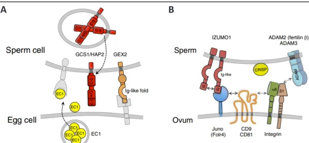
1-5 Key Molecular Players during Gamete Interactions and Fusion
The final stages of double fertilization are still incompletely understood on the molecular
level. GAMETE EXPRESSED 2 (GEX2) a single pass transmembrane protein with extracellular
filamin domains forming an immunoglobulin (Ig)-like fold was found essential for attachment
of the gametes to each other (Figure 1-3 A; Mori et al., 2014). EGG CELL1.1 (EC1.1), an egg
cell-specifically expressed small cysteine-rich protein under transcriptional regulation of
SUPPRESSOR OF FRIGIDA (Resentini et al., 2017), needs to be secreted in order to initiate
a preferential redistribution of GENERATIVE CELL SPECIFIC1/HAPLESS 2 (GCS1/HAP2) from
the endomembrane system of the sperm cells to the plasma membrane, and subsequently
enable for gamete fusion (Sprunck et al., 2012). GCS1/HAP2 is a single transmembrane protein
required for membrane fusion and fertility not only in angiosperms but also in algae like
Chlamydomonas, and in parasites like Plasmodium (Johnson et al., 2004; von Besser et al.,
2006; Mori et al., 2006; Wong et al., 2010) (Figure 1-3 A).
16 Key Molecular Players during Gamete Interactions and Fusion
Furthermore, it contains a large extracellular designated GCS1/HAP2 domain which was shown to trimerize once inserted into the target lipid bilayer in a similar fashion as viral class II fusogens, or the C.elegans fusogen EFF-1, and thereby mediates merging of the membranes (Fedry et al., 2017). Hence, it was postulated that these species share a common ancestral gene, as GCS1/HAP2 assumes the role of an ancient fusogen (Wong et al., 2010; Fedry et al., 2017).
Interestingly, GCS1/HAP2, is absent from mammals. During the final stage of mammalian fertilization, the sperm attaches to the egg plasma membrane by the interaction of IZUMO1 (Inoue et al., 2005), which is only exposed to the sperm plasma membrane upon sperm activation (Satouh et al., 2012), and the GPI-anchored folate receptor homolog JUNO (Spiegelstein et al., 2000; Bianchi et al., 2014) (Figure 1-3 B). Notably, JUNO is also expressed and essential for a subpopulation of T-cells in the lyme (Yamaguchi et al., 2007). Upon established IZUMO1-JUNO binding, IZUMO1 undergoes a conformational shift, dimerizes and no longer binds JUNO (Inoue et al., 2015). The resolved IZUMO1 crystal structure (Ohto et al., 2016) revealed structural similarities to domains found in Plasmodium proteins named SPECT (sporozoite microneme protein essential for cell traversal) and TRAP1 (thrombospondin repeat anonymous protein
A B
Figure 1-3 Key players of gamete interaction and fusion in flowering plants and in mammals.
(A) In Arabidopsis, upon sperm cell arrival at the fusion site the small cysteine-riche peptide EGG
CELL1 (EC1.1) is secreted from the egg cell and initiates preferential relocalization of GENERATIVE CELL
SPECIFIC1/HAPLESS2 (GCS1/HAP2) to the sperm cell plasma membrane, which subsequently acts as
fusogen and mediates merging of the membranes. GAMETE EXPRESSED2 (GEX2) is also exposed on
the sperm cell plasma membrane and mediates attachment to the female gametes. (B) Adhesion and
recogniction during mammalian fertilization is mediated by IZUMO1 on the sperm and JUNO on the
egg plasma membrane. Upon IZUMO1-JUNO binding, IZUMO1 undergoes a conformational shift, and
dimerizes which abolishes JUNO binding capacity. Furthermore, Tetraspanin proteins CD81, and CD9
were determined essential for fertility, and CD9 mediates assembly of fusion competent sites. Also
ADAM proteins and integrins are important mediators of attachment (Dresselhaus et al., 2016).
17 Tetraspanins, Membrane Microdomains and Sphingolipids
1), two proteins expressed by the invasive sporozoite stage of Plasmodium berghei parasites (Nishimura et al., 2016). This shared domain, an extensible beta ribbon domain, can undergo, as shown for TRAP1, a conformational shift and thereby, in combination with additional protein-binding domains, facilitates the gliding motility of TRAP1 (Song et al., 2012).
Until now the exact molecular mechanism of the final sperm egg fusion is unknown. In addition to the JUNO/IZUMO1-mediated recognition and adhesion of mammalian gametes, ADAM proteins, and, partly by interaction with ADAMs, the integrins alpha6 beta1, and alpha9 beta1 are known mediators of sperm egg adhesion (Georgadaki et al., 2016). Furthermore, alpha6 beta1 integrin interacts with the Tetraspanin protein CD9 (Ziyyat et al., 2006) (Figure 1-3 B), which concomitantly accumulates in the contact zone of egg and sperm prior to fusion (Chalbi et al., 2014). Thus, CD9 is considered to laterally organize the gamete fusion machinery on the egg membrane by recruiting necessary cis-partners, as it is known to generate fusion competent sites on the egg membrane (Jegou et al., 2011).
1-6 Tetraspanins, Membrane Microdomains and Sphingolipids
Tetraspanins are a large evolutionary highly conserved family of four pass transmembrane proteins, present in plants and mammals, with their respective N-, and C-termini facing the cytosol, a small occasionally glycosylated extracellular domain, and a large extracellular domain (Wang et al., 2012; Charrin et al., 2014). The large extracellular domain contains several conserve cysteine residues and mediates protein interactions and oligomerization (Schmidt et al., 2016), which was also strongly dependent on palmitoylation of TET9 at the cytosolic side of the transmembrane domains (Figure 1-4; Seigneuret et al., 2001; Berditchevski et al., 2002).
Moreover, Tetraspanin proteins are known to indirectly interact with the cytoskeleton via their
cytosolic C-termini (Sala-Valdes et al., 2006), and facilitate assembly of supramolecular plasma
membrane-based complexes, mostly with integrins (Rubinstein et al., 1994; Yanez-Mo et al.,
2009). In mice, Tetraspanin CD81, and CD9 were determined essential for fertilization (Miyado
et al., 2000; Rubinstein et al., 2006), and shown to promote muscle cell fusion (Charrin et
al., 2013), although they are also known to prevent the fusion of mononuclear phagocytes
(Takeda et al., 2003). Additionally, CD9 is known to generate fusion competent sites on the
egg membrane (Jegou et al., 2011), in accordance with the concept of Tetraspanin-enriched
18 Tetraspanins, Membrane Microdomains and Sphingolipids
microdomains (Hemler 2003), which pose platforms for mediating processes such as adhesion or exocytosis (Bailey et al., 2011; Perez-Hernandez et al., 2013).
Initially, these microdomains were identified due to their detergent insolubility similar to those of the lipid rafts (Hemler 2003). Lipids rafts constitute hypothetical organizing centers of the plasma membrane with lipid-based sorting mechanisms that in-, and exclude certain proteins (Hancock 2006). Meanwhile, increasing amounts of eukaryotic, and prokaryotic lipid raft-associated proteomic datasets, derived of the analysis of detergent-resistant membrane fractions, are becoming available, revealing lipoproteins, and in eukaryotic datasets tetraspanins (Bae et al., 2004; Dubois et al., 2015; Shah et al., 2015; Toledo et al., 2015).
Notably, these detergent-resistant membrane fractions are predominantly assembled of cholesterol and sphingolipids (Hancock 2006). Sphingolipids arise from the irreversible condensation of palmitoyl-CoA with serine performed by the endoplasmic reticulum-localized serine palmitoyltransferase, thereby forming, after a successive reduction step, Sphinganine (Hanada 2003). This long chain base (LCB) can be N-acylated, then hydroxylated and glycosylated to gain complex glycosphingolipids like phytoceramide (Figure 1-4). Glycosphingolipids are major constituents of the plasma membrane or serve as component of the GPI-anchor (Breslow et al., 2010; Gault et al., 2010). Alternatively, the LCB can be hydroxylated, desaturated, phosphorylated and thereby give rise to a variety of bioactive molecules serving as second messengers or secreted ligands for cell-surface receptors (Breslow et al., 2010; Gault et al.,
N-glycosyla�on palmitoyla�on
small ECD
large ECD
Figure 1-4 The schematic Tetraspanin structure.
A representative Tetraspanin structure with conserved amino acid residues displayed in circles, conserved transmembrane helices 1 - 4, and three conserved helices in the large extracellular domain.
Furthermore, palmitoylation, and glycosylation sites, as well as disulfide briges are indicated. ECD =
extracellular domain, TM = transmembrane domain (Skaar et al., 2015 with minor additions).
19 Tetraspanins, Membrane Microdomains and Sphingolipids
2010). Interestingly, sphingolipids are also actively metabolized in the nucleus, where, for example, sphingosine-1-phosphate was shown to inhibit histone deacetylases and thereby suppress transcription (Hait et al., 2009).
The entire sphingolipid breakdown is mediated via phosphorylation of the LCB, like phytosphingosine (PHS) to phytosphingosine-1-phosphate (PHS1P) and successive irreversible degradation by the mostly endoplasmic reticulum-localized phospholyase to ethanolamine and the respective acyl aldehydes like 2-hydroxyhexadecanal (Tsegaye et al., 2007; Aguilera- Romero et al., 2014) (Figure 1-4). Disturbed sphingolipid homeostasis was reported to have pleiotropic negative effects (Nishikawa et al., 2008) with toxic outcome (Han et al., 2010). Especially accumulation of free LCBs and their phosphorylated counterparts due to necrotrophic funghi-derived fumonisin B1-induced inhibition of the ceramide synthase was shown to be lethal (Wright et al., 2003), as these molecules have a severe impact on cellular survival and pro-death signaling (Pata et al., 2010).
Figure 1-5 Sphingolipid metabolism.
The first and irreversible step in sphingolipid biosynthesis is the condensation of palmitoyl-CoA and serine to 3-ketosphinganine, which, after subsequent reduction, results in the long chain base (LCB) (sphinganine). Subsequently, various modifications of sphinganine are possible: N-acylation (a) yields ceramide; hydroxlation (b) yields phytosphingosine (PHS), the major plant and yeast LCB; desaturation (c) yields sphingosine, the major mammalian LCB; phosphorylation (d) yields (phyto)sphingosine- 1-phosphate, an important signaling molecule. Furthermore ceramides vary in fatty acid (FA) chain length (e) (yellow box). The head group position (f) of the LCB part of ceramides (blue box) can be phosphorylated, or derivatized with inositol or mannose moieties, phosphorylcholine, or complex glycans yielding highly diverse glycosphingolipids. Complex sphingolipids are catabolized by deacylation to LCB and FA-aldehyde moieties, here exemplary to PHS. PHS is phosphorylated, then irreversible degraded to phosphorylethanolamine and 2-hydroxyhexadecanal, which can be oxidized further to 2-hydroxyhexadecanal. 2-OH = 2-hydroxy, FA = fatty acid, LCB = long chain base, PHS = phytosphingosine, PHS1P = phytosphingosine-1-phosphate, (with modifications from Breslow and Weissman 2010, and Kondo et al., 2014).
sphinganine
c a d
b
complex sphingolipids
NH2 OH
OH
PHS
PHS1P
2-OH hexadecanal
NH2 OH
OH OH
NH2 OH
OH
OPO3H
OH CHO
2-OH palmi�c acid
OH CO2H O-
O NH3+ HO O
S-CoA +
ceramide
f OH
c b OH FA
LCB
d
+R-CoA
-Ethanolamin-P
NH e b
( )n
O
20 Aims of this Work
1-7 Aims of this Work
The importance of cell surface-exposed and extracellular-localized proteins during gametophyte and gamete interactions has been emphasized. However, the knowledge about the proteins acting on the surface of the female gametophytic cells and on the sperm cells is scarce and much remains to be investigated.
The aim of this work was to examine the composition of cell surface proteins on female gametophytic cells and on sperm cells, and to select candidate proteins for functional characterization with respect to female gametophyte development and double fertilization.
To achieve this, own and publicly available microarray-based transcriptome data from Arabidopsis female gametophytic cells (egg cells, central cells, synergid cells) and sperm cells were subjected to comprehensive bioinformatics studies. The data were processed and analyzed together with microarray data from diverse sporophytic tissues to extract those genes encoding membrane proteins as well as membrane-associated and secreted proteins which are differentially upregulated (enriched) in either one or a combination of female gametophytic cells. This was achieved by comparing (contrasting) the female gametophytic cells and sperm cells with the sporophytic tissues and with each other.
However, the presence of a transcript does not necessarily imply the presence of the translated protein. Therefore, an Arabidopsis cell line with an egg cell-like transcriptional profile was used to generate full proteome and membrane proteome data, and to compare them with those of a root-derived callus, to find out whether this approach may serve as a tool to overcome the quantitative restrictions limiting the use of egg cells for proteomics.
Candidate genes, derived from transcriptome studies, were selected to verify the tissue- and/
or cell type-specific expression pattern by quantitative PCR, whole mount in situ hybridization, and microscopic analyses of transgenic Arabidopsis plants expressing either promoter:reporter or genomic:reporter constructs.
Based on these data, a subset of candidate proteins was selected to study their subcellular localization in the developing female gametophyte and during double fertilization. The function of three candidate protein families was investigated by reverse genetics, including the CRISPR/
Cas9 approach, and protein-protein interaction studies.
21 Identification of Differentially Expressed Genes encoding Membrane-associated and Secreted Proteins
2 – Results
2-1 Acquisition, Processing, and Evaluation of the Transcriptomic Data of Arabidopsis Gametophytic Cells
2-1-1 Previous Work: Isolation of Arabidopsis Female Gametophytic Cells by Micromanipulation, and ATH1 GeneChip Hybridization
In a previous effort by Lucija Soljic (Soljic 2012), the three major cell types of the Arabidopsis female gametophyte were isolated by manual microdissection (MM) as described in Englhart et al., 2017 (in press), and subjected to single cell transcriptome analysis using the GeneChip®
Arabidopsis ATH1 Genome Array. Hybridization was performed in four batches as provided by the “Kompetenzzentrum für Fluoreszente Bioanalytik” (Regensburg). Four CEL datasets were obtained, containing four replicates for the egg cell (EC), and three replicates each for the central cell (CC), and the synergid cells (SYN). Soljic excluded the second (E1) egg cell replicate from further studies. The remaining CEL data were processed, including sperm cell (SC)-derived data (Borges et al., 2008), as described in Soljic 2012, and resulted in 10065 EC, 11641 CC, 8728 SYN, and 5829 sperm cells expressed genes called „present” in three of three replicates (Soljic 2012).
2-1-2 Identification of Differentially Expressed Genes encoding Membrane- associated and Secreted Proteins
To acquire a more in-depth transcriptome analysis a combined approach was realized in
collaboration with Dr. Maxim Messerer and Dr. Daniel Lang (Helmholtz Center Munich). Here,
the CEL data of all ten replicates of female gametophytic cells isolated by MM (Solijc 2012), and
published laser-assisted micro dissected (LAM) female gametophytic cells (Wuest et al., 2010),
sperm cells (Borges et al., 2008), globular embryo (Spencer et al., 2007; Slane et al., 2014),
endosperm of germinating seeds (Penfield et al., 2006; Dekkers et al., 2013), leaves (Usadel
et al., 2008; Tang et al., 2012), roots (Stepanova et al., 2007; Lin et al., 2011), and seedlings
(Zheng et al., 2009; He et al., 2016) were included in a comprehensive analysis. Also the
same analysis was performed while excluding the LAM-derived data of female gameotphytic
cells (Wuest et al., 2010). Expression data from the developing endosperm four days post
22 Identification of Differentially Expressed Genes encoding Membrane-associated and Secreted Proteins
fertilization (Day et al., 2007; Day et al., 2008) were non includable into the analysis due to platform compatibility issues between [ATH1-121501] Affymetrix Arabidopsis ATH1 Genome Array and HortResearch_Arabidopsis_27K_Operon long oligo set_V1.0. For both analyses (MM+LAM and MM) all respective 78 and 69 CEL files (Supplemental Table 1) were subjected to frozen Robust-Multi-array Analysis (fRMA) from the Bioconductor package frma (McCall et al., 2010) for normalization, background subtraction, and batch-effect correction, which revealed good clustering of the respective replicates in multi dimensional scale (MDS) analysis, and identified the root- and the SC-derived replicates to be most distant from the remaining samples (Figure 2-1 A, B).
Intensity value cutoff was defined as mean value of the negative controls, times two the standard deviation. The individual overlap of probe sets with signal intensities above the cutoff value (5.599934), excluding internal controls, between the replicates of MM-isolated female gametophytic cells is depicted in Figure 2-1 C – F. The maximum overlap ranged from 60% (EC), via 68% (SYN), and 72% (SC), to 78% (CC). These probe sets corresponded to 11509 genes in the EC, 13186 genes in the CC, 12044 genes in the SYN, and 14056 genes in the SC, above the predefine cutoff value in all replicates. In total, the MM+LAM and MM-only analyses identified 19059 genes (19684 probe sets) and 19259 genes (19907 probe sets) above cutoff, respectively.
Arabidopsis thaliana gene descriptions and gene ontology (GO) terms were downloaded from TAIR v10. Prediction of transmembrane domains, and subcellular protein localizations were obtained from Dr. Rainer Schwacke directly as referenced in ARAMEMNON v8.1 (Schwacke et al., 2003), and SUBAcon (Tanz et al., 2013; Hooper et al., 2014), respectively. To address transcriptomic differences between gametophytic cells (EC, CC, SYN, SC) and the sporophytic tissues (endosperm, embryo, leaf, root, seedling) differential gene expression was calculated using the GLM functionality of the limma package (Ritchie et al., 2015), also including a corrective term for possible batch effects (Supplemental Table 5-1).
Differentially expressed genes (DEGs) were determined by contrasting of the female
gametophytic cells and male gametes against sporophytic tissues, and by contrasting the
gametophytic cells and gametes against each other. Furthermore, DEG annotation was
extended by TAIR10-Subcellular_Predictions.xlsx, and TAIR10_functional_descriptions.
23 Identification of Differentially Expressed Genes encoding Membrane-associated and Secreted Proteins
M̂
M̂
M̂
−1000 −500 0 500 1000
−400−2000200
M̂
M̂
M̂
M̂
M̂
M̂
M̂
M̂
M̂
central_cell egg_cell endosperm globular.embryo leafroot seedling sperm_cell synergid_cell
M̂M̂M̂
−100 0 100 200 300 400 500
−100−50050100
60242 GSE10522 29771 3045 25171 7432 GSE5618 GSE5617 GSE5641
M̂
M̂
M̂
M̂
M̂
M̂
M̂
M̂
egg_cell endosperm globular.embryo leaf root seedling sperm_cell synergid_cell
C D
E F
Figure 2-1 Transcriptomic data of female gametophytic cells.
69 ATH1-121501 CEL files of manual microdissected (MM) female gametophytic cells (egg cells, central cells, synergid cells), male gametes (sperm cells), and sporophytic tissues (endosperm, embryo, leaf, root, seedling) were subjected to frozen robust Multi-Array analysis. Multidimensional scale plot depiction before (A) and after (B) normalization, background subtraction, and batch effect correction revealed good replicate clustering. The MDS plots (A, B) were contributed by Dr. Maxim Messerer.
The detected probe sets of MM female gametophytic cells and sperm cells after background subtraction
and their distribution onto the individual replicates of the egg cell (C), the central cell (D), the synergid
cells (E), and the sperm cells (F). EC = egg cell, CC = central cell, SC = sperm cell, SYN = synergid cell.
24 Statistical and Gene Ontology-based Evaluation of the obtained Transcriptomic Data
txt obtained from TAIR v10. Gene functional descriptions, cellular compartments (CO), and localization predictions were assigned to the respective AGIs. Also, data derived of probe sets corresponding to multiple AGIs were removed.
As the aim of this study was to address cell-to-cell signaling and cell-surface interactions, all mitochondrial and chloroplastidial-derived gene identifiers were removed along all genes identifiers corresponding to putatively neither membrane-associated nor extracellular localized proteins. After calculating sporophytic and gametophytic contrasts in total 8506 (MM+LAM) and 8608 (MM-only) DEGs encoding putative memrane-associated, or secreted proteins remained.
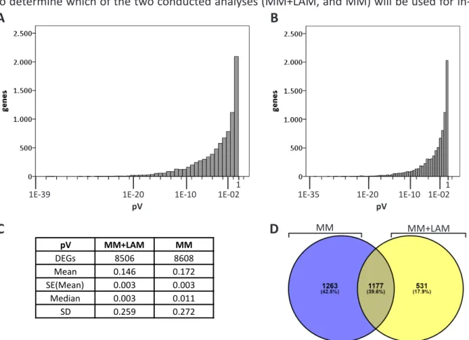
2-1-3 Statistical and Gene Ontology-based Evaluation of the obtained Transcriptomic Data
To determine which of the two conducted analyses (MM+LAM, and MM) will be used for in-
MM MM+LAM
pV MM+LAM MM
DEGs 8506 8608
Mean 0.146 0.172
SE(Mean) 0.003 0.003
Median 0.003 0.011
SD 0.259 0.272
A B
C D
1E-10 1E-02 1E-39
1E-20pV 1
1E-10 1E-02 1E-20
1E-35 1
pV
Figure 2-2 Statistical analysis of the LAM+MM and MM analyses.
Probability value frequencies of 8506 DEGs of the MM+LAM (A) and 8608 DEGs of the MM-only analysis
(B), and the respective statistical summary depicting mean and median values (C) showed a lower
median value for the LAM+MM analysis. (D) This diagram contains all DEGs of the EC, the CC, and the
SYN with an FC of 1.5 or higher, from either of the reference contrasts (sporophytic and gametophytic),
excluding SC-derived DEGs, and shows the 2440 DEGs identified in the dataset derived of the MM
analysis (blue subset), and the 1708 DEGs identified in the dataset derived of the LAM+MM analysis
(yellow subset). The non intersection subsets were used for GO term analyses (Table 2-1).
25 Statistical and Gene Ontology-based Evaluation of the obtained Transcriptomic Data
detail bioinformatic characterization, both were compared by descriptive statistics and GO term overrepresentation.
First of all, the obtained adjusted probability value (pV) frequencies of the “EC versus sporophytic tissue” contrast of the MM+LAM and MM analyses were compared. By calculating the median, a higher value (0.011) for the four EC replicate number analysis (MM) was obtained than for the seven EC replicate number analysis (MM+LAM) which was at 0.003 (Figure 2-2 A - C).
Based on these findings, the MM+LAM analysis should be preferred over the MM analysis.
To limit the chance of including non-DEGs into further studies, the maximum valid pV was set to 0.001, consequently DEGs with higher pVs were excluded. To assess the differences in DEG composure of the MM+LAM and MM analyses in a GO-dependent, biological processes- related context and in line with the working hypothesis that relatively strong expressed genes will participate in cell-to-cell communications and gamete interactions, a log2 fold change (FC) cut off, to represent upregulated genes, was specified to 1.5 or higher. Upregulated DEGs from sporophytic, and gametophytic contrasts were pooled and SC-only derived DEGs were temporarily excluded to estimate the overlap and non intersection subsets of identified DEGs in LAM+MM and MM analyses. 2440 DEGs originated from the MM analysis and 1708 DEGs originated from the MM+LAM analysis, which overlapped in 1177 DEGs (40%) (Figure 2-2 D).
The individual subsets of the DEGs (1263 MM-only, 1177 intersection, and 531 MM+LAM-only) Table 2-1 Overrepresented biological processes of the DEGs encoding membrane-associated or secreted proteins that were identified only in the MM-LAM analysis or only in the MM analysis but not in both.
All 531 DEGs of the MM+LAM-only non intersection subset (A), and all 1263 DEGs of the MM-only non intersection subset (B) were subjected to PANTHER-based statistical overrepresentation test.
Fold overrepresentation was calculated relative to the Arabidopsis full GO term reference database (Arabidopsis Ref). Results were sorted by ascending pV, the pV threshold was set to ≤ 0.05. Only overrepresented processes are shown. pV = probability value.
GO biological process complete Arabidopsis
Ref(27352) MM+LAM
-only (531) expected fold overrep. pV
photosynthesis (GO:0015979) 159 16 3.09 5.18 3.41E-04
establishment of localiza�on (GO:0051234) 1776 66 34.48 1.91 9.40E-04
transport (GO:0006810) 1758 65 34.13 1.90 1.38E-03
localiza�on (GO:0051179) 1846 66 35.84 1.84 3.50E-03
single-organism transport (GO:0044765) 791 35 15.36 2.28 1.78E-02
GO biological process complete Arabidopsis
Ref(27352) MM-only
(1263) expected fold overrep. pV establishment of localiza�on (GO:0051234) 1776 215 82.27 2.61 3.82E-34
transport (GO:0006810) 1758 213 81.43 2.62 7.87E-34
localiza�on (GO:0051179) 1846 219 85.51 2.56 1.31E-33 vesicle-mediated transport (GO:0016192) 284 56 13.16 4.26 1.75E-15 organic substance transport (GO:0071702) 943 111 43.68 2.54 3.94E-15
A
B
26 In silico Analysis of the Gametophytic DEGs obtained from the Sporophytic Contrast
were used for PANTHER-based GO term biological process statistical overrepresentation tests, with a predefined pV cutoff of 0.05 and revealed differences in DEG composure (Table 2-1).
Notably, “photosynthesis” (GO:0015979) was found most overrepresented by factor 5.18 in the non intersection subset corresponding to the MM-LAM-only DEGs (Table 2-1 A). The remaining overrepresented biological processes like “establishment of localization” (GO:0051234), and
“transport” (GO:0006810) were identified in both non intersection subsets, while “vesicle- mediated transport” (GO:0016192) was restricted to the MM-only non intersection subset (Table 2-1 B). Despite the pre-defined pV, and FC cutoffs, the MM+LAM analysis would likely include false positive DEGs derived of photosynthetic tissue and exclude potentially valuable DEGs and was discarded consequently. Therefore, an in-depth in silico transcriptome analysis was restricted to the data from SCs and female gametophytic cells, acquired by MM.
2-1-4 In silico Analysis of the Gametophytic Transcriptome Data
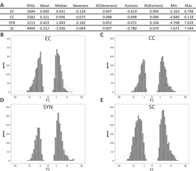
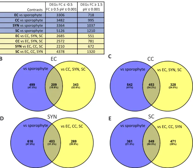
The gametophytic transcriptome data were either compared to sporophytic tissues, or the respective other gametophytic cells. The resulting DEGs, with a FC ≥ 0.05 or FC ≤ -0.05, and a pV ≤ 0.001, encoding for membrane-associated or secreted proteins, obtained by these two comparisons, were characterized by descriptive statistics and statistical GO term enrichment analysis.
2-1-4-1 In silico Analysis of the Gametophytic DEGs obtained from the Sporophytic Contrast
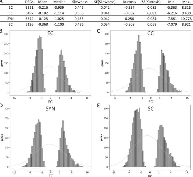
To assess differences and similarities between the gametophytic cells on the level of differential
gene expression, basic descriptive statistics were applied to the 3306, 3482, 3364, and 5126
DEGs of the EC, the CC, the SYN, and the SC, respectively after contrasting to sporophytic
tissues. The FC median values for all four gametophytic cell types were determined negative
between -0.939 (EC), and -1.114 (CC). Similar findings applied to the FC mean values, which
were slightly negative in all four cases ranging from -0.125 (SYN) to -0.368 (SC). The skewness,
which defines the asymmetry of the distribution around its mean value (Brys et al., 2004), was
positive for all four cell types. Median and skewness clearly demonstrated a higher frequency
of genes on the left side of the distribution, which resembled downregulation. Finally, kurtosis,
which determines the tailedness (Westfall 2014) was found negative for the EC (-0.397), and
27 In silico Analysis of the Gametophytic DEGs obtained from the Sporophytic Contrast
the SC (-0.308) indicating fewer than expected, by comparison to the normal distribution, strongly regulated genes. Kurtosis was even for the CC (-0.032), and positive for the SYN (0.256), indicating more strongly regulated genes in the SYN. Also, minimum and maximum values for differential gene expression were SYN-derived. These statistic values are all summed up in Figure 2-3 A, and gene expression frequency histograms for all gametophytic cells are depicted in Figure 2-3 B – E. Judging from gene expression frequencies, the synergid cell was found slightly more different from the gametes than the gametes from each other.
Figure 2-3 Descriptive statistics applied to the DEGs of the female gametophytic cells and male gametes identified by sporophytic contrasting.
Basis descriptive statistics were applied to the DEGs, with pV ≤ 0.001 of the EC, the CC, the SYN, and the SC after contrasting to the sporophyte and summed up (A). Log2 fold change frequencies of the DEGs for the EC (B), the CC (C), the SYN (D), and the SC (E) depicted as histograms. Grey lines indicate the normal distribution. For all four cell types less DEGs were upregulated than downregulated, which was determined by positive skewness and negative median values. Furthermore, only the SYN showed positive kurtosis thereby demonstrating more than expected strongly regulated genes and a difference from the gametes which had negative kurtosis (EC, SC) and almost even kurtosis (CC). EC = egg cell, CC
= central cell, SC = sperm cell, SE = standard error, SYN = synergid cell.
DEGs
EC 3321 -0.216 -0.939 0.445 0.042 -0.397 0.085 -5.363 8.316
CC 3497 -0.182 -1.114 0.556 0.041 -0.032 0,083 -6.216 9.430
SYN 3372 -0.125 -1.025 0.455 0.042 0.256 0.084 -7.881 10.778
SC 5126 -0.368 -1.100 0.426 0.034 -0.308 0.068 -7.079 8.921
Kurtosis SE(Kurtosis) Min. Max.
Mean Median Skewness SE(Skewness)
C
D E
A
B EC
SYN
CC
SC
FC FC
FC FC
28 In silico Analysis of the Gametophytic DEGs obtained from the Sporophytic Contrast
By utilizing GO term enrichment analysis, the possible biological and molecular differences between the gametophytic cells, encoded by the identified DEGs, were addressed. Again the same DEGs, previously utilized for the descriptive statistic analyses, were transformed into GeneEntrezIDs, affiliated with their respective expression values as determined by sporophytic contrasting and subjected to PANTHER-based statistical GO term enrichment analysis. The maximum pV for statistically valid enrichment or depletion of processes was set to 0.05.
Similarities in enrichment of biological processes, depicted as PANTHER-slim unified terms, were identified between all female gametophytic cells, especially regarding transport-associated processes like “intracellular protein transport” (GO:0006886), and “protein transport”
(GO:0015031) (Table 2-2 A). Also, “RNA metabolic process” (GO:0016070) was found enriched in all female gametophytic cells, while “protein glycosylation” (GO:0006486), and “exocytosis”
(GO:0006887) was exclusively found enriched in the SYN. Neither of these processes were found enriched in the SC, here only “DNA metabolic process” (GO:0006259), “DNA repair”
(GO:0006281), and “DNA recombination” (GO:0006310) were identified as enriched (Table 2-2 A). GO term-depleted biological processes were related to “carbohydrate transport”
(GO:0008643) in the EC and the CC, “defense response to bacterium” (GO:0042742) in the SYN, and a number of metabolic processes like “protein metabolic process” (GO:0019538),
“rRNA metabolic process” (GO:0016072), or “translation” (GO:0006412) and “protein folding”
(GO:0006457) in the SC (Table 2-2 B). These findings demonstrated, on transcriptomic level, the distance between the SCs, and the cells of the female gametophyte (FG), which had already been observed earlier by MDS plot analysis and moreover indicated slight differences between the SYN, and the female gametes.
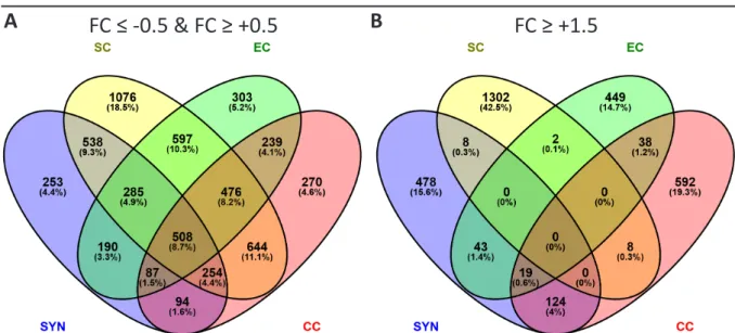
The identified 7168 DEGs with an FC ≥ 0.05 or FC ≤ -0.05, (pV ≤ 0.001), after sporophytic
contrasting, were distributed relatively evenly onto the individual intersection subsets of
gametophytic cells types and male gametes, with the exception of the SC non intersection
subset which contained 1874 DEGs (26%), and the EC, CC, SYN, SC intersection subset with
1102 DEGs (15%; Figure 2-4 A). When applying an FC cutoff for upregulation of 1.5 or higher to
all identified DEGs, 2438 DEGs remained. Here, the SC non intersection subset still was largest
with 798 DEGs (33%), while the EC, CC, SYN, SC intersection had diminished to 120 DEGs (5%),
indicating that gametophytic cells shared many downregulated, or slightly upregulated genes
29 In silico Analysis of the Gametophytic DEGs obtained from the Sporophytic Contrast
Table 2-2 Enriched and depleted biological processes in gametophytic cells after contrasting to sporophytic tissues.
All identified DEGs with a pV ≤ 0.001 and their affiliated expression values per respective gametophytic cell type were subjected to PANTHER-based GO term enrichment analysis, depicted as enriched (A), and depleted (B) GO-slim biological processes and sorted by ascending pV (cutoff pV ≤ 0.05). Enriched biological processes found in all female gametophytic cells are depicted in bold letters.
cell type Panther GO-slim Biological process_depleted genes pV
EC carbohydrate transport (GO:0008643) 45 1.6E-02
EC ion transport (GO:0006811) 97 4.1E-02
CC homeosta�c process (GO:0042592) 67 3.7E-03
CC carbohydrate transport (GO:0008643) 57 2.6E-02
SYN defense response to bacterium (GO:0042742) 49 1.7E-03
SYN secondary metabolic process (GO:0019748) 43 1.1E-02
SYN homeosta�c process (GO:0042592) 56 4.1E-02
SC transla�on (GO:0006412) 152 1.7E-14
SC protein metabolic process (GO:0019538) 556 1.2E-05
SC cellular component biogenesis (GO:0044085) 120 1.3E-05 SC cellular amino acid metabolic process (GO:0006520) 76 1.5E-04
SC rRNA metabolic process (GO:0016072) 22 1.6E-04
SC biosynthe�c process (GO:0009058) 301 1.3E-03
SC primary metabolic process (GO:0044238) 1122 6.7E-03
SC protein folding (GO:0006457) 41 9.9E-03
SC cellular amino acid catabolic process (GO:0009063) 12 1.0E-02
SC glycolysis (GO:0006096) 16 4.1E-02
cell type Panther GO-slim Biological process_enriched genes pV
EC RNA metabolic process (GO:0016070) 63 4.5E-06
EC intracellular protein transport (GO:0006886) 166 9.8E-06
EC nucleobase-containing compound metabolic process (GO:0006139) 199 1.1E-05
EC protein transport (GO:0015031) 171 2.1E-05
EC vesicle-mediated transport (GO:0016192) 133 3.9E-05
EC phosphate-containing compound metabolic process (GO:0006796) 189 1.9E-02
CC RNA metabolic process (GO:0016070) 42 1.6E-04
CC nucleobase-containing compound metabolic process (GO:0006139) 168 9.3E-04
CC primary metabolic process (GO:0044238) 692 4.1E-03
CC intracellular protein transport (GO:0006886) 139 9.9E-03
CC protein transport (GO:0015031) 142 1.1E-02
CC metabolic process (GO:0008152) 890 3.9E-02
SYN protein transport (GO:0015031) 187 4.4E-10
SYN intracellular protein transport (GO:0006886) 181 5.9E-10
SYN vesicle-mediated transport (GO:0016192) 141 5.9E-10
SYN RNA metabolic process (GO:0016070) 48 1.2E-03
SYN transport (GO:0006810) 402 3.4E-03
SYN localiza�on (GO:0051179) 408 4.5E-03
SYN nucleobase-containing compound metabolic process (GO:0006139) 164 9.0E-03
SYN protein glycosyla�on (GO:0006486) 59 1.6E-02
SYN exocytosis (GO:0006887) 30 3.8E-02
SC DNA metabolic process (GO:0006259) 43 6.6E-04
SC DNA repair (GO:0006281) 20 4.6E-03
SC DNA recombina�on (GO:0006310) 9 1.3E-02