Structural, physical and genetic interactions of the i -AAA protease Yme1 in
Saccharomyces cerevisiae
I n a u g u r a l - D i s s e r t a t i o n
zur
Erlangung des Doktorgrades
der Mathematisch-Naturwissenschaftlichen Fakultät der Universität zu Köln
vorgelegt von Tanja Engmann
aus Iserlohn
Köln, 2009
Berichterstatter:
Professor Dr. Thomas Langer Professor Dr. R. Jürgen Dohmen
Tag der mündlichen Prüfung: 20. Mai 2009
Abstract
The i-AAA protease Yme1 is a highly conserved ATP-dependent AAA (ATPase Associated with various cellular Activites) protease anchored to the inner mitochondrial membrane where it mediates protein quality surveillance that is crucial for cell survival. In yeast, deletion of YME1 is associated with pleitropic phenotypes. However, the few known proteolytic substrates and interaction partners of the i-AAA protease cannot explain the molecular basis of these phenotypes. Therefore, different approaches were used to define the function of the i-AAA protease. First, affinity purification of a proteolytic inactive variant of Yme1 working as a substrate trap was employed and led to the identification of eight novel Yme1-interacting proteins that localise to different submitochondrial compartments.
Subsequent analysis revealed that two interactors, Mcr1 and Mpm1, represent new proteolytic substrates of the i-AAA protease. Hence, additional functions of the i-AAA protease should be responsible for the other identified interactions. Second, insights into processes that require the function of the i-AAA protease were obtained by a synthetic genetic array (SGA) approach using an assorted library of 96 non-essential mitochondrial gene deletions. 34 identified synthetic lethal interaction potentially link Yme1 to new functions like mitochondrial morphology, protein processing and lipid metabolism. Finally, the synthetic lethal interaction of IMP1 and YME1 was analysed by a high copy suppressor screening. This interaction is of particular interest, as all substrates of the Imp1 catalytic subunit of the IMP processing peptidase could be co-purified with the i-AAA protease. The identified suppressor Pgk1, a key enzyme in glycolysis and gluconeogenesis, suggests a severe impairment of Δyme1Δimp1 cells in energy metabolism.
Moreover, in this thesis the substrate recognition by the i-AAA protease was examined by mutational analysis. Recently, two substrate binding regions have been identified within Yme1: the CH-(C-terminal helices) and the NH-(N-terminal helices) region. In contrast to the NH-region, the molecular mechanism of substrate binding to the CH-region has not been studied so far. Here, the CH-region was not only identified to have a role in substrate binding and transfer to the proteolytic cavity, but also for the stabilisation of the i-AAA protease complex.
Taken together, novel substrates and interaction partners of the i-AAA protease were identified, pointing to additional functions of Yme1 independent of its proteolytic activity. As only few approaches have addressed the function of the i-AAA protease in higher eukaryotes so far, it will be interesting to examine the relevance of these findings for mammals.
Table of contents
1 Introduction ...1
1.1 Cellular compartmentation... 1
1.2 Metabolic requirements of mitochondria ... 1
1.2.1 Role of mitochondria for the cellular energy metabolism ... 2
1.2.2 Mitochondrial lipids and ergosterol ...3
1.3 Proteolytic systems controlling mitochondrial maintenance ... 9
1.4 ATP-dependent proteases...11
1.4.1 AAA+-proteases ... 12
1.4.2 Recognition and handling of substrate by AAA+-proteases... 13
1.4.3 Mitochondrial AAA+-proteases... 15
1.4.4 Substrate recognition by the i-AAA-proteases... 16
1.4.5 Functions of the i- and m-AAA proteases in mitochondria... 18
1.4.6 The conserved i-AAA protease Yme1 ... 20
1.5 Mitochondrial peptidases and mitochondrial protein import ...22
1.6 Aims of the thesis ...25
2 Materials and Methods ...26
2.1 Molecular Biology Methods ...26
2.1.1 Yeast expression plasmid ... 26
2.1.2 Plasmids for in vitro transcription and translation ... 28
2.2 Cell biology methods ...29
2.2.1 Yeast strains and growth conditions ... 29
2.2.2 Yeast genetic procedures ... 35
2.3 Biochemistry Methods ...36
2.3.1 Total protein isolation from S. cerevisiae ... 36
2.3.2 Preparation of cellular membranes from S. cerevisiae ... 36
2.3.3 Immunological detection of proteins... 36
2.3.4 Procedures employing S35-radiolabelled polypeptides ... 37
2.3.4.1 In vitro MPP-cleavage assay...37
2.3.4.2 Import and import-chase of S35-radioalabled polypeptides into isolated mitochondria ...38
2.3.5 Co-immunoprecipitation ... 38
2.3.6 Blue native polyacrylamide gel electrophoresis (BN-PAGE) ... 39
2.3.7 Ni-NTA affinity chromatography of mitochondrial extracts ... 39
2.3.8 Lipid analysis ... 40
2.3.8.1 Lipid isolation ...40
2.3.8.2 Thin Layer Chromatography (TLC)...41
2.3.9 Miscellaneous... 41
3 Results ...42
3.1 Initial substrate binding to the CH-region of the i-AAA protease Yme1...42
3.1.1 Mutational analysis of the α-17 and α-18 helices of the CH-region of Yme1 in regard to substrate binding and degradation ... 43
3.1.1.1 In vivo activity of CH-mutant variants of Yme1...44
3.1.1.2 Cox2 binding to Yme1 CH-mutant variants...46
3.1.1.3 Degradation of Cox2 in Δsco1Δyme1 harbouring CH-mutant variants of Yme1 ...47
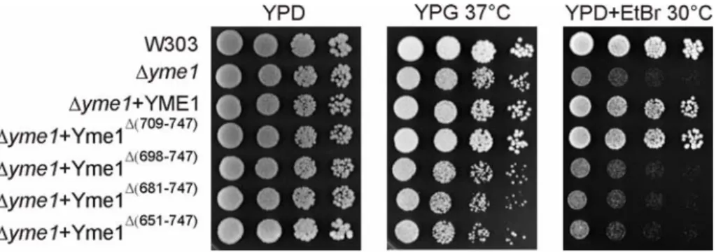
3.1.2 Effects of C-terminal truncations of the CH-region of Yme1 on substrate degradation and complex formation of the i-AAA protease... 48
3.1.2.1 In vivo activity of C-terminal truncation mutants of Yme1 ...49
3.1.2.2 Degradation of Cox2 in Δsco1Δyme1 and Δimp1Δyme1 harbouring C-terminal truncation mutants of Yme1...50
3.1.2.3 Complex formation of C-terminally truncated Yme1 mutants ...51
3.2 Identification of binding partners of the i-AAA protease Yme1...53
3.2.1 Maturation of the i-AAA protease subunits Yme1 from different species... 53
3.2.1.1 In vivo processing of Yme1 in mutants of different processing peptidases...54
3.2.1.2 Processing of in vitro translated 35S- radiolabelled Yme1 orthologs by purified MPP ...55
3.2.1.3 In vivo activity of N-terminally tagged i-AAA protease...56
3.2.2 Affinity purification of a proteolytically inactive variant of Yme1 ... 57
3.2.3 Co-immunoprecipitation of Yme1 from strains harbouring HA-tagged versions of Yme1- interacting proteins... 59
3.2.4 Correlation of Δyme1 phenotypes to the phenotypes of Yme1-interacting proteins ... 62
3.2.5 Effects of the i-AAA protease on Yme1-interacting proteins ... 63
3.2.5.1 Steady state levels of Yme1-interacting proteins in the presence or absence of Yme1...63
3.2.5.2 In organello import of in vitro translated Mcr1, Mpm1, Gep1 and Qcr2 into isolated wild type and Δyme1 mitochondria...64
3.2.5.3 i-AAA protease dependent degradation of Mcr1, Mpm1 and Phb1...66
3.2.5.4 In organello import of in vitro translated Pda1 and Pdb1 into isolated wild type and Δyme1 mitochondria...68
3.2.6 Variations in expression of Yme1-interacting proteins and their influence on Δyme1 ... 69
3.2.6.1 Effects of Gep1 overexpression in wild type and Δyme1 cells ...69
3.2.6.2 Overexpression of Mcr1 in wild type and Δyme1...70
3.2.7 Effects of Δyme1 on cellular ergosterol levels... 71
3.3 Analysis of genetic interactions of the i-AAA protease Yme1 ...74
3.3.1 Analysis of genetic interactions of Δyme1 with a deletion library of assorted mitochondrial proteins ... 74
3.3.1.1 Phenotypic analysis of Δyme1 synthetic lethal interactors ...77
3.3.2 High copy suppressor screening for synthetic lethal interactors of Yme1... 78
3.3.2.1 Identification of genes suppressing the synthetic lethality of Δyme1Δimp1...79
3.3.2.2 Suppression of Δyme1 phenotypes by suppressors of Δyme1Δimp1...81
4 Discussion...82
4.1.1 Initial substrate binding by the CH-region of the i-AAA protease Yme1 ... 82
4.1.2 CH-dependent degradation of substrates by the i-AAA protease Yme1 ... 84
4.1.3 Structural importance of the CH-region of Yme1... 86
4.2 Processing of the i-AAA protease Yme1 ...86
4.3 Novel interaction partners of the i-AAA protease Yme1...88
4.3.1 Possible function of the i-AAA protease Yme1 in sorting and assembly of the mitochondrial proteins Qcr2, Pda1 and Pdb1 ... 89
4.3.2 New substrates of the i-AAA protease: Mpm1 and Mcr1 ... 91
4.3.3 The i-AAA protease Yme1 functionally interacts with Gep1 ... 93
4.4 Genetic interaction of the i-AAA protease Yme1 ...95
4.4.1 Synthetic lethality of the i-AAA protease Yme1 – more than just an consequence of mtDNA loss... 95
4.4.2 New implications for Yme1 function ... 96
4.4.3 High copy suppressor screen of Δyme1Δimp1 ... 98
4.5 Impact of the i-AAA protease Yme1 on cellular and mitochondrial lipid levels....99
4.5.1 Depletion of Yme1 changes mitochondrial phospholipids levels... 99
4.5.2 A potential influence of Yme1 on cellular ergosterol distribution... 100
5 Zusammenfassung...103
6 References ...105
7 List of abbreviations ...123
8 Attachment ...124
9 Danksagung...125
10 Eidesstattliche Erkärung ...126
11 Lebenslauf ...127
1 Introduction
1.1 Cellular compartmentation
The concept of eukaryotic compartmentation enables the segregation of specific biochemical reactions to distinct environments for the purpose of biochemical efficiency and restricted dispersion of intermediate reaction products. This is of particular importance for subsequent conversions of substrates, accomplished during metabolic processes and proteolytic events.
Local enrichments are either achieved by formation of complexes that cluster specific sets of enzymes or enclosure of reactions by membranes resulting in defined aqueous environments. While prokaryotic cells do not show enclosed membrane structures, eukaryotic cells comprise a complex intracellular compartmentalisation. One of these intracellular compartments is the mitochondrium. Mitochondria, as chloroplasts, are surrounded by two membranes which, according to the endosymbiotic theory, originate from invasion of a α-proteobacterium into a primordial eukaryotic cell (Wallace, 2007). These two membranes configure four mitochondrial subcompartments: Outer mitochondrial membrane, intermembrane space, inner mitochondrial membrane and the matrix space. Within the mitochondria, fundamental cellular processes are conducted which render this organelle essential for most eukaryotic cells. In fact, several diseases are associated with defects in mitochondria (Chan, 2006; Lin and Beal, 2006; Wallace, 2007) underlining the essential function of this organelle. Consequently, maintenance of mitochondrial integrity and function is crucial for an eukaryotic cell. It is therefore not surprising, that the inherited proteolytic quality control system of mitochondrial ancestors is conserved (Koppen and Langer, 2007) and that a number of accessory defence systems have evolved (Tatsuta and Langer, 2008).
1.2 Metabolic requirements of mitochondria
Mitochondria do not only adapt their activity to different physiological demands, but they are in addition able to modulate their subcellular distribution to local cellular requirements. The later is accomplished by the formation of a reticulated mitochondrial network which responses to cellular needs by constant fission and fusion events, thereby optimising mitochondrial performance (Cerveny et al., 2007; Detmer and Chan, 2007; Hoppins et al., 2007). The most prominent metabolic reactions within mitochondria of S. cerevisiae are the
and the biosynthesis of amino acids (in mammals also β-oxidation of fatty acids). Moreover, mitochondria are the site of heme and Fe/S cluster biosynthesis (Hamza, 2006; Lill and Muhlenhoff, 2006; Lill and Muhlenhoff, 2008) and involved in the intermediary metabolism, calcium homeostasis and apoptosis (Chan, 2006; McBride et al., 2006). The maintenance of all of these processes is crucial for the survival of an organism.
1.2.1 Role of mitochondria for the cellular energy metabolism
Under aerobic conditions, mitochondria can serve as the major site of ATP production of the cell, harbouring the constituents of the citric acid cycle and the oxidative phosphorylation within the matrix space and the inner mitochondrial membrane, respectively. The mitochondria dependent energy production is also called respiration. Conversely, in the absence of oxygen cells undergo fermentation, a process that does not require the function of mitochondria. Glucose is the main source of energy utilised in both processes. However, yeast and other eukaryotic cells do not strictly depend on the availability of glucose; they are able to adapt their metabolic system to different carbon sources, but only under aerobic conditions. In the presence of glucose, most energy is generated by cytosolic glycolysis. Only upon utilisation of non-fermentable carbon sources, such as glycerol or lactate, the mitochondrial oxidative metabolism is fully activated. Non-fermentable carbon sources usually need to be remodeled before they can serve as a source of energy. Such enzymatic activities are partially present within mitochondria. Glycerol is processed within the cytosol to glycerol-3-phosphate by Gut1 (Pavlik et al., 1993). After glycerol-3-phosphate is internalised into mitochondria, it is converted into dihydroxy-acetate-phosphate (DAH-phosphate) that can enter the cytosolic glycolysis or gluconeogenesis processes (Sprague and Cronan, 1977).
The mitochondrial enzyme producing DAH-phosphate is known as glycerol 3-phosphate dehydrogenase, Gut2, and is localized to the inner mitochondrial membrane (Rijken et al., 2007; Ronnow and Kielland-Brandt, 1993). Lactate, another non-fermentable carbon source, is converted within the mitochondria to pyruvate that can enter glycolysis or gluconeogenesis. Within yeast mitochondria, one L-lactate dehydrogenase (cytochrome b2) Cyb2 (Lodi and Ferrero, 1993) and two D-lactate dehydrogenase, Dld1 and Dld2 (Chelstowska et al., 1999; Rojo et al., 1998) are present. In addition, mitochondria contain enzymes that are able to connect different metabolic pathways. One important representative is the multienzyme pyruvate dehydrogenase complex that resides in the matrix space of mitochondria. The enzyme is composed of two E1 proteins Pda1 and Pdb1, the E2 core protein Lat1, the E3 protein Lpd1 and the protein X component Pdx1 (Pronk et al., 1996). The important function of this mitochondrial complex is the oxidative
decarboxylation of internalised pyruvate into acetyl CoA and CO2, thereby linking glycolysis to the citric acid cycle. In general, the citric acid cycle is responsible for the complete conversion of nutrients, thus producing NADH and FADH2 molecules that are utilised during oxidative phosphorylation, as well as GTP. Furthermore, biosynthetic components, like amino acids, relevant for diverse cellular elements are initialised via the citric acid cycle.
Therefore, the mitochondria do not only present the most efficient site of cellular ATP production, but are also involved in utilisation of alternative carbon sources. In addition, pathways within the mitochondria enable the supply/allocation of components originally used for energy production in the biogenesis of diverse cellular constituents like amino acid, fatty acids and heme (porphyrine) precursors.
1.2.2 Mitochondrial lipids and ergosterol
Although mitochondrial functions have been analysed extensively over the past decades, the role of lipids within this organelle has only more recently attracted the attention of research.
Just like proteins, lipids are also important for the biogenesis and maintenance of mitochondria. In respect to cellular lipid supply, mitochondria are of particular interest, as they are involved in the biogenesis of some lipid species (Voelker, 2004). Yeast mitochondria usually comprise glycerophospholipids (phospholipids) and to a lower extent the yeast specific sterol ergosterol (Zinser and Daum, 1995). However, in contrast to other cellular membranes no sphingolipids are found in mitochondrial membranes.
Commonly, phospholipids are composed of a phosphatidic acid element that is attached to variable head groups. The phosphatidic acid element contains a glycerol backbone which attaches to one phosphate and two fatty acids through ester bonds (Fig. 1.1). The variable head groups of phospholipids include choline, ethanolamine, serine, inositol and glycerol (van Meer et al., 2008). A phospholipid exclusively present within mitochondria is cardiolipin (Jakovcic et al., 1971). In contrast to other phospholipids, cardiolipin is a polyglycerophospholipid and contains two phosphatidic acid moieties that are linked via a glycerol bridge and harbour altogether four fatty acids (Fig. 1.1) (Schlame, 2008).
The two most abundant phospholipids within yeast mitochondria are phosphatidyl- ethanolamine (PE) and phosphatidycholine (PC) (Zinser and Daum, 1995). Whereas PC is only imported into mitochondria, PE can also be generated within mitochondria. The inner mitochondrial membrane protein Psd1 produces the majority of cellular PE by conversion of phosphatidyserine (PS) to PE (Clancey et al., 1993). Alternatively PE is produced from PS at the golgi-apparatus by Psd2 (Trotter and Voelker, 1995). Both PE and PC can also be synthesised via the Kennedy-pathway (Kent, 1995). However, the PE and PC species
synthesised through this pathway make only minor contributions to mitochondrial phospholipids (Birner et al., 2001). Another cellular compartment essential for the biogenesis of phospholipids is a specialised fraction of the endoplasmic reticulum (ER) called the MAM (mitochondria associated membrane) fraction. Here, the two MAM proteins Pem1 and Pem2 are required for conversion of PE to PC (Zinser et al., 1991). Furthermore, Cds1 produces CDP-diacylglycerol (CDP-DAG) from phosphatidic acid (PA) that is required for the generation of phosphatidylserine (PS) and phosphatidylinositol (PI) in the MAMs (Achleitner et al., 1999;
Gaigg et al., 1995). Cds1 is also present in the inner mitochondrial membrane where it plays a role in the production of cardiolipin (Kuchler et al., 1986). PA is synthesised in mitochondria by a two-step acylation of glycerol-3-phosphate at the outer membrane of mitochondria (Athenstaedt and Daum, 1999).
Cardiolipin (CL) is the third most abundant phospholipid (14,6%) present in mitochondria.
Unlike PE and PC, CL is enriched in the inner mitochondrial membrane and not distributed equally between the two mitochondrial membranes (Schlame, 2008). Consistently, the synthesis of cardiolipin takes place in the inner membrane of mitochondria. After CDP-DAG is produced by Cds1, it is further processed by Pgs1 to phosphatidylglycerolphosphate (PGP) (Chang et al., 1998). Then, dephosphorylation occurs by a still unknown enzyme. The resulting phosphatidylglycerol (PG) is converted by the cardiolipin synthase, Crd1, to CL (Chang et al., 1998; Tuller et al., 1998). In addition, so-called remodelling processes postsynthetically modify the acylchain composition of CL. One enzyme responsible for such remodelling is the transacetylase Taz1 that is localised to the inner leaflet of the outer mitochondrial membrane (Claypool et al., 2006). Interest in the functional role of Taz1 has increased when frequent mutations in the human homolog have been identified in patients suffering from Barth Syndrome (Bione et al., 1996). In yeast, CL is also required for a variety of cellular functions like ageing, apoptosis, mitochondrial protein import, mitochondrial bioenergetics, translational regulation and cell wall biosynthesis (Joshi et al., 2008).
Although phospholipids produced in mitochondria are often transferred to different sites within the cell before they can fulfil their function, not much is known about the exact transport mechanisms of these lipids within and from mitochondria. It is conceivable that most phospholipids laterally diffuse through membrane continuities. Further, Met30 has been demonstrated to be required for transport of PS from MAMs into mitochondria (Choi et al., 2006). Thus, distinct transport molecules might exist. In general, most membrane transports are facilitated by the vesicular transport system. However, mitochondria are not connected to this system and might thus have different mechanisms for the allocation of these phospholipids to the rest of the cell. The unique lipid composition of mitochondria could have resulted from the detachment of the organelle form the vesicular transport system. Instead
of depending on the internalisation of certain lipid species, the mitochondria might rather rely on the lipids produced by themselves.
Another lipid found in mitochondria but originating from synthesis in the ER is ergosterol.
Sterols are composed of a cholestane basis to which different site chains can be attached at the C-17 residue (Fig. 1.1) (Moss, 1989). Biosynthesis of ergosterol is a fairly complex procedure that involves 22 different enzymatic activities (Daum et al., 1998). The biosynthetic pathway of ergosterol formation can be split into two parts. The first part, commonly called the mevalonate or isoprenoid pathway, is initiated with two acetyl CoA molecules that are processed to farnesyl pyrophosphate. In the second part of ergosterol biosynthesis, farnesyl pyrophosphate is converted to ergosterol. Deletions of the enzymes required for the earlier step of the ergosterol biosynthesis are lethal. Conversely, the absence of the enzymes facilitating the last eight steps is not lethal, although the produced intermediates cannot substitute for ergosterol (Daum et al., 1998). The reason for this discrepancy is not understood. Most steps within the biosynthetic pathway of ergosterol are performed within the ER (Zinser et al., 1993) and only a subset of enzymatic activities is found in mitochondria. Erg10, an acetoacetyl CoA thiolase facilitating the first step of the ergosterol biosynthesis, is localised to both the cytoplasm and mitochondria (Kornblatt and Rudney, 1971). Additionally, the two outer mitochondrial membrane proteins Ncp1 and Mcr1 are involved in the reduction of the cytochrome P450 family member Erg11 (Lamb et al., 1999; Sutter and Loper, 1989), for which an ER localisation is proposed (Ott et al., 2005).
Hence, no direct participation of mitochondria in ergosterol biosynthesis is evident.
Nevertheless, ergosterol biosynthesis requires molecular oxygen and is thus depending on Figure 1.1 Lipid components within mitochondria. (A) Glycerophospholipids. The glycerol backbone is bound to two variable fatty acids (R1 and R2) and a phosphate. Variable headgroup are choline, ethanolamine, serine, inositol and glycerol. (B) Polyglycero- phospholipids. Two glycerol backbones are linked with two phosphates by a glycerol bridge between the two phosphate moieties. Four variable fatty acids can be attached here. The predominant polyglycerophospholipid in mitochondria is cardiolipin. (C) Sterols. Ergosterol, the sterol found in yeast like S. cerevisiae, is shown.
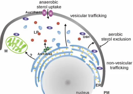
oxygen dependent steps in the ergosterol biosynthesis are the epoxidation of squalene and the desaturation and demethylation of lanosterol (Hata et al., 1981; Lorenz et al., 1986). As a result, yeast is only able to produce ergosterol under aerobic conditions. Under anaerobic conditions, however, yeast is sterol auxotroph and depends on the exogenous supply of this essential cellular compound (Lewis et al., 1985; Lorenz and Parks, 1991). Accordingly, uptake mechanisms of ergosterol have been identified (Fig. 1.2). This uptake depends on an impaired heme biosynthesis and is hence restricted to anaerobic conditions, a phenomenon also referred to as ‘aerobic sterol exclusion’ (Lorenz et al., 1986) that is not well understood.
Consequently, under aerobic conditions ergosterol is synthesised endogenously and no uptake is possible, whereas under anaerobic conditions endogenous synthesis is impaired and the essential uptake of exogenous ergosterol is induced via the transcription factor Ucp2 (Crowley et al., 1998). Resulting expression of Pdr11 and Aus1, two ATP-binding cassette transporters, leads to the ATP-dependent uptake of the exogenous ergosterol (Wilcox et al., 2002) and thereby ensures cell survival.
Ergosterol is unequally distributed throughout different membranes and the incorporation of sterol intermediates is not favoured (Daum et al., 1998). Ergosterol is most abundant in the plasma membrane and in secretory particles (Zinser and Daum, 1995). Further, lipid particles, microsomes and the inner membrane of mitochondria contain ergosterol (Schneiter et al., 1999). Surprisingly, the site of ergosterol biosynthesis, the ER, contains a relatively low ergosterol content implying an efficient postsynthetic depletion and trafficking of the sterol to a distinct location (Fig. 1.2). Ergosterol resides in cellular compartments in form of free ergosterol (plasma membrane). Another form of ergosterol, steryl ester, resembles the storage form of ergosterol and its intermediates and is predominantly found together with triacylglycerol (TAG) in lipid particles (Czabany et al., 2007; Czabany et al., 2008).
Conversion of ergosterol and its intermediates to the storable ester form is achieved by Are1 and Are2 at the expense of acetyl CoA (Zweytick et al., 2000). Stored steryl ester are mobilised upon requirement by the action of the steryl ester hydrolases Tgl1, Yeh1 and Yeh2 (Köffel et al., 2005; Wagner et al., 2009). As hydrolysis of steryl esters occurs at different cellular localisations, a role of lipid particles in trafficking of ergosterol cannot be excluded.
However, the major sterol trafficking between the ER and the plasma membrane has been demonstrated to occur via vesicular or non-vesicular pathways (Raychaudhuri and Prinz, 2006; Sullivan et al., 2006). In the non-vesicular pathway, translocation of sterols from the ER to the plasma membrane is based on the decreased chemical potential of sterol after its binding to other kinds of lipids within the plasma membrane. As these sterol moieties are not able to diffuse within the gradient between the sterol rich plasma membrane and the sterol poor ER, successive incorporation of ergosterol into the already sterol enriched
compartment is achieved. The non-vesicular pathway was identified in experiments utilising Brefeldin A for disruption of the vesicular pathway where no impairment of sterol trafficking was obvious (Baumann et al., 2005). Within this pathway, oxysterol binding proteins (OSH) are responsible for the transport of sterols through the hydrophilic environment (Beh et al., 2009; Wang et al., 2005). Additional lipid transfer proteins have been identified in mammals (Prinz, 2007). In yeast, additional factors are found to influence the cellular distribution of sterols. Altered intracellular sterol is observed in the absence of Arv1, Ptc1 and Plc1 (Fei et al., 2008; Tinkelenberg et al., 2000). Their exact function, however, is not understood.
As mitochondria are not connected to the vesicular transport system of the cell, import of sterols into the organelle must also be achieved by a non-vesicular pathway. In yeast, no components involved in this process have been identified yet. The movement of sterols between the inner and outer mitochondrial membrane has in any case been shown to
Figure 1.2 Uptake and trafficking of cellular ergosterol. Uptake of exogenous sterol in yeast is only facilitated under anaerobic conditions by the two ATP-binding cassette transporters Aus1 and Pdr11 localised to the plasma membrane (PM). Under aerobic conditions the supply of ergosterol is restricted to intracellular synthesis, a phenomenon called
“aerobic sterol exclusion”. The major site of sterol synthesis is the endoplasmic reticulum (ER). Ergosterol trafficking occurs over vesicular and non-vesicular pathways. The later involves the action of oxysterol binding proteins (OSH). Both trafficking pathways mainly mediate the translocation of ergosterol between ER and PM. The mechanism by which ergosterol is transported to mitochondria (M) is, however, not understood. Storage of ergosterol and intermediates in their steryl ester form is mediated by Are1 and Are2 which compose lipid particles (LP) in the ER. PM, plasma membrane; ER, endoplasmic reticulum; M, mitochondria; LP, lipid particle; OSH, oxysterol binding proteins.
contrast, adrenal mammalian tissue contains two mitochondrial outer membrane proteins responsible for sterol transport into mitochondria. The peripheral benzodiazepine receptor (PBR) contains a cholesterol binding domain and is highly expressed in steroidogenic cells (Papadopoulos et al., 1997; Papadopoulos et al., 1997). The steroid acute regulatory protein (STAR) mediates the translocation of cholesterol from the outer mitochondrial membrane to the inner mitochondrial membrane (Miller, 2007). The exact mode of action by which STAR facilitates sterol transport is still under debate.
Although sterols represent extremely expensive metabolic components, they account for a substantial amount of the cellular lipids. As cells acquiesce the complexity and cost of sterol biosynthesis, the function of sterols must be crucial for the viability of eukaryotic cells, and irreplaceable by any other cellular lipid component (Parks et al., 1995). Most studies defining the role of sterol in eukaryotic membranes have been performed in yeast. Here, ergosterol functions in bulk membrane formation influencing the rigidity and permeability of the membrane (Abe and Hiraki, 2009; Kleinhans et al., 1979; Lees et al., 1979). In this line, sterols participate in the formation of detergent resistant membranes (DRM) (Cerneus et al., 1993). Ergosterol is implicated in a variety of other function in yeast, that will not be discussed in detail here. Moreover, a function in membrane packing of phospholipids by the mammalian sterol cholesterol is described (Ikonen, 2008; McConnell and Radhakrishnan, 2003). Further, coinciding ergosterol has a protective effect against phospholipid peroxidation (Wiseman et al., 1993; Wiseman et al., 1993) occurring for example upon stroke.
Most of the proposed functions of ergosterol are based on the analysis of the plasma membrane moiety of this lipid. However, similar roles in sorting and organisation by ergosterol may be expected in mitochondria where the majority of lipid is represented by phospholipids. One proposed regulatory role of ergosterol is the maintenance and/or biogenesis of mitochondrial morphology (Altmann and Westermann, 2005). Whether the underlying events leading to the disturbed mitochondrial morphology are based on alterations of mitochondrial or cellular ergosterol levels is not clear. The exact function of ergosterol in mitochondria remains up to date unknown. In contrast, mammalian cells are depending on mitochondria for the synthesis of steroids (steroidogenesis). For this purpose, conversion of internalised cholesterol to pregnenolone, the rate limiting step of steroidogenesis, is achieved by the action of cytochrome P450scc (CPY11A1) (Miller, 2007).
The STAR lipid transfer proteins play a crucial role in this process by delivering cholesterol from the outer to the inner mitochondrial membrane. The essential role of this mitochondrial process is evident in mammals that suffer from congenital lipoid adrenal hyperplasia that is associated with mutations in STAR or P450scc (Miller, 1997; Yang et al., 1993). Additional
metabolic reactions are performed on internalised 11-decorticosterone (DOC) and 11- deoxycortisol (DOCHL). Nevertheless, other functions of mitochondrial sterol in mammals and in yeast remain to be elucidated, as well as the mitochondrial transport of sterols in yeast.
1.3 Proteolytic systems controlling mitochondrial maintenance
Different lines of defence assuring the integrity of mitochondria have evolved (Tatsuta and Langer, 2008). On tissue level, cells with damaged mitochondria can undergo apoptosis to prevent for example subsequent damage of adjacent cells. On an organellar level, the mitochondrial integrity can be controlled by complete removal of damaged organelles by mitophagy (Kim et al., 2007), an autophagy related process. This event will lead to the turnover of the ~1000 known mitochondrial proteins (Mootha et al., 2003; Sickmann et al., 2003; Taylor et al., 2003). A more economic process is the restoration of function by fusion of damaged mitochondria with intact neighbouring mitochondria (Detmer and Chan, 2007).
Mitochondria display a dynamic network structure that is subjected to constant fission and fusion events accomplished by intricate molecular machines (Hoppins et al., 2007). Although restoring mitochondrial function, the fusion process does not ensure removal of damaged structures and does not explain the different turnover rates seen for mitochondrial proteins (Augustin et al., 2005; Russell et al., 1980). Hence, a different defence system has to exist on the level of mitochondria. Indeed, mitochondria contain an elaborate proteolytic system distributed throughout the different compartments of the organelle (Koppen and Langer, 2007). In S. cerevisiae, as well as in higher eukaryotes, this system is mainly built up by ATP-dependent proteases and oligopeptidases (Fig. 1.3). Representative of the yeast ATP- dependent proteases are Pim1 that acts together with Hsp78 (Röttgers et al., 2002) and the two AAA-proteases, the i-AAA and the m-AAA protease (Van Dyck and Langer, 1999). Yeast oligopeptidases are Mop112 and Prd1. In general, the term oligopeptidases refers to the ability of those peptidases to degrade peptides that result from degradation of polypeptides by other proteases further to amino acids (Desautels and Goldberg, 1982; Young et al., 2001). In case of Mop112 and Prd1 this protease is predominantly the i-AAA protease (Kambacheld et al., 2005).
Furthermore, peptide export has been described for yeast mitochondria depending on the ABC-transporter Mdl1 (Young et al., 2001) that is localised to the inner mitochondrial membrane. A signalling function of released peptides on nuclear gene expression of mitochondrial genes has been proposed (Arnold et al., 2006; Young et al., 2001). Within the mitochondria Mdl1 transports peptides from the matrix site to the intermembrane space, where both Mop112 and Prd1 reside. Therefore, the complete breakdown of cleaved mitochondrial targeting sequences and other peptides originally generated in the matrix space by the two oligopeptidases is plausible (Kambacheld et al., 2005; Moberg et al., 2003;
Stahl et al., 2002). As deletion of both oligopeptidases is associated with mild phenotypes (Kambacheld et al., 2005), the two oligopeptidases might not be essential for breakdown of the vast majority of peptides generated by processing of mitochondrial targeting sequences in the matrix. Besides, peptides in the intermembrane space can be released freely and additional matrix oligopeptidases might exist. Furthermore, the two oligopeptidases are connected to the quality control of mitochondrial proteins since turnover occurs for 5% to 10% of all mitochondrial proteins (Augustin et al., 2005; Kambacheld et al., 2005).
Moreover, yeast contains the metallopeptidase Oma1, an integral inner membrane protein Figure 1.3 The proteolytic system of mitochondria in S. cerevisiae. Two classes of peptidases can be distinguished within mitochondria: ATP-dependent proteases and oligo- peptidases. Additional peptidases like Oma1 exist. Both ATP-dependent proteases and the metallopeptidase Oma1 generate peptides within the mitochondrial matrix space (M) that are either directly released over the inner mitochondrial membrane (IM) into the intermembrane space (IMS) via the peptide transporter Mdl1 or are degraded further to amino acids by a so far unknown peptidase. Peptides can also be generated within the intermembrane space by the i-AAA protease. Then, peptides can either be released from mitochondria through the outer mitochondrial membrane (OM) or their breakdown to amino acids is facilitated by Mop112 and Prd1. See text for details.
with a catalytic site that is facing the matrix space (Käser et al., 2003). Oma1 shows overlapping function with the m-AAA protease in quality control of inner membrane proteins by cleavage of misfolded polytopic membrane proteins at multiple sites (Käser et al., 2003).
In higher eukaryotes, an additional intermembrane space serine proteases exist, HtrA2 (Omi) (Hegde et al., 2002). Diseases are not only related to mutations within the respective genes of HtrA2, but the serine protease has been linked to Parkinson disease (Martins et al., 2004;
Plun-Favreau et al., 2007). Besides, mutations within the human ortholog of the m-AAA protease cause an autosomal recessive form of hereditary spastic paraplegia (HSP) (Casari et al., 1998). In conclusion, a concerted working mode of all proteases, the ATP-dependent proteases, oligopeptidases and other proteasese like Oma1 or HtrA will facilitate quality surveillance within the mitochondria and therefore assures their function and maintenance.
1.4 ATP-dependent proteases
ATP-dependent proteases are conserved from bacteria to man. In eukaryotes, ATP- dependent proteases are required for a variety of cellular processes (Ogura and Wilkinson, 2001; Tucker and Sallai, 2007). In Escherichia coli four of ATP-dependent proteases, Lon, FtsH, Clp and HslUV can be distinguished (Gottesman, 2003). ATP-dependent proteases are members of the AAA+ (ATPases associated with various cellular activities) family of Walker type P-loop ATPases (Ammelburg et al., 2006; Hanson and Whiteheart, 2005). Proteins belonging to that class are characterised by the presence of a homologous ATPase domain referred to as AAA domain and function as molecular chaperones. The AAA domain is often accompanied by a proteolytic domain responsible for subsequent degradation of the unfolded substrate. Depending on the type of protease, the AAA domain and the proteolytic domain are expressed as one or two polypeptide chains. Furthermore, different proteolytic classes of the ATP-dependent proteases are described: Lon and Clp proteases for example represent serine proteases (Van Dyck et al., 1994; Wang et al., 1997), whereas FtsH is a metalloprotease (Tomoyasu et al., 1993). The functional protease is usually assembled into a hexameric or heptameric core ring structure that buries the proteolytic sites inside a proteolytic chamber (Sauer et al., 2004; Schmidt et al., 1999; Zwickl et al., 2000). This kind of assembly produces a high local density of proteolytic sites beneficial for substrate degradation, but also restricts the access to the proteolytic site and thereby restrains proteolysis by the ATP-dependent proteases (Prakash and Matouschek, 2004; Sauer et al., 2004; Schmidt et al., 1999; Singh et al., 2000). Moreover, functions independent of the
proteolytic activity have been indicated for the AAA domains of ATP-dependent proteases (Suzuki et al., 1997; Tatsuta et al., 2007).
1.4.1 AAA
+-proteases
The active form of AAA+-proteases is established by assembly of their subunits into a hexameric or heptameric ring structure (Hanson and Whiteheart, 2005; Schmidt et al., 1999). This ring shaped structure creates an inner proteolytic cavity and is required for a coordinated function of AAA domains (Ishikawa et al., 2004; Kim and Kim, 2003; Wang et al., 2001; Xia et al., 2004; Zhang et al., 2000). In general, the AAA domain of each oligomer contributes the following functional key elements: the Walker A and B motif, the sensor-1 and sensor-2 motif and the arginine finger (Hanson and Whiteheart, 2005; Tucker and Sallai, 2007). Within the protein sequence of a subclass of the AAA+-proteases, the so-called AAA proteases, the sensor-1 motif and the arginine finger are mapped to the highly conserved region, called the second region of homology (SRH) (Lupas and Martin, 2002) (Fig. 1.4).
Functionally, the Walker A motif is required for binding of ATP by directly interacting with the phosphate moiety of ATP (Neuwald et al., 1999). The bound ATP is then subsequently hydrolysed to ADP by residues of the Walker B motif, where an asparte coordinates the essential Mg2+ ion while a glutamate activates a water molecule for the nucleophilic attack (Hanson and Whiteheart, 2005; Iyer et al., 2004). Sensor-1 and sensor-2 motif, as well as the arginine finger, also play a role in ATP binding and hydrolysis (Hanson and Whiteheart, 2005; Hishida et al., 2004; Karata et al., 1999; Ogura et al., 2004). For the action of the arginine finger and the sensor-2 motif, the formation of the ring structure is essential, because only in this configuration these elements are able to protrude into the ATPase domain of the neighbouring subunit and contact the bound nucleotide (Ogura et al., 2004).
The universal function of the AAA domain is the implementation of energy derived from ATP hydrolysis into a conformational change of the molecule, driving substrate unfolding and translocation (Martin et al., 2008). Apparently, not just hydrolysis of ATP, but also binding and release of the nucleotide seem to generate the driving force for the conformational change (Wang et al., 2001). Different modes of ATP hydrolysis within the ring structure are discussed (Martin et al., 2005; Moffitt et al., 2009; Ogura and Wilkinson, 2001).
The concept of a proteolytic chamber also enables selective and entire degradation of substrates by AAA+-protease. Depending on the type of AAA+-protease the proteolytic chamber is either exclusively built up of the proteolytic domain or a participation of the AAA domain in the formation of the chamber is seen (Bieniossek et al., 2006).
Furthermore, the specific properties of prokaryotic Clp, Lon and FtsH proteases are introduced here, as orthologs of these proteases are found in mitochondria (Chapter 1.4.3).
The caseinolytic protease ClpP resembles only a proteolytic subunit that is found to assemble with variable AAA subunits (mostly ClpA and ClpX) to different AAA+-protease constellations (Yu and Houry, 2007). Both Lon and FtsH proteases express the proteolytic and the AAA subunit in one polypeptide chain. All three proteases assemble into ring structures and a substrate entry and degradation mechanism similar to that of other AAA+-proteases is conceivable. Distinct structural features of Lon are the amino terminal N-domain and the proposed substrate sensor and disriminatroy domain (SSD) (Licht and Lee, 2008). FtsH proteases contain an SRH domain and are therefore the only AAA protease of the three compared AAA+-proteases (Lupas and Martin, 2002). Moreover, FtsH proteases are integral membrane protein with two transmembrane domains situated within the N-terminal region of the protein adjacent to the AAA domain (Ito and Akiyama, 2005). The membrane bound state of FtsH proteases raises the question of how substrates are recognised and degraded.
Substrate binding and translocation are crucial processes for the functionality of all AAA+- proteases and will be addressed in the following paragraph.
1.4.2 Recognition and handling of substrate by AAA
+-proteases
Substrate engagement by the different AAA+-proteases occurs by a certain substrate recognition motif on the substrate, over an adaptor protein of the protease or to a specific site within the protease. Then, binding and hydrolysis of ATP will induce conformational changes that lead to an unfolding of the substrate. Finally, translocation of the substrate will
Figure 1.4 Structure of AAA protease domains and their orientation relative to the membrane. Conserved domains and motifs are indicated on the left side. Domains of the FtsH protease are projected into the predicted hexameric structure based on the crystal structure of Thermus thermophilus (Suno et al., 2006). ND, N-terminal domain; TM, transmembrane region; AAA, AAA domain; NH, N-terminal helix; WA, Walker A motif; YVG, central pore loop; WB, Walker B motif; SRH, second region of homology; PD, proteolytic domain; HEXGH, proteolytic center; CH, C-terminal helix; M, membrane. The figure is adapted from (Graef et al., 2008).
enable its subsequent degradation within the proteolytic cavity. The proposed rate limiting steps during the whole process are the unfolding and translocation of the substrate (Kenniston et al., 2003).
Here, the mechanism for substrate engagement of prokaryotic Clp, Lon and FtsH are described. Recognition elements within the substrate mediate its first contact with the protease. Accordingly, terminal or internal consensus sequences have been identified in Clp and FtsH substrates. One of the best characterised consensus sequences is the ssrA tag. It consists of 11 amino acids that are added C-terminally to proteins that are arrested during translation (Gottesman, 2003; Keiler et al., 1996). Sequence comparison of additional Clp substrates allowed the identification of five terminal recognition elements (Flynn et al., 2003). Also, latent terminal recognition sequences are known; for example the LexA repressor generates new C-terminal and N-terminal ends by an autocatalytic process that serve as recognition sequences for ClpXP (Neher et al., 2003). A consensus sequence has not been identified for Lon proteases, but substrate engagement is also initiated within certain recognition elements (Nishii et al., 2002; Ondrovicova et al., 2005; von Janowsky et al., 2005). Further, internal sequences can act as recognition elements for substrates engagement by the Clp and FtsH proteases under certain conditions (Hoskins et al., 2002;
Okuno et al., 2006). Protease adaptor proteins are best known for Clp proteases, were the AAA subunit (ClpA or ClpX) has to be connected to the proteolytic subunit ClpP to form the functional AAA+-protease (Yu and Houry, 2007). The function of the AAA+-protease ClpAP is inhibited by ClpS (Guo et al., 2002; Zeth et al., 2002); in contrast ClpXP function is stimulated by SspB (Bolon et al., 2004; Levchenko et al., 2000). Functional stimulation of FtsH is achieved by the DnaK/DnaJ/GroE chaperone system (Tatsuta et al., 2000). No such adaptors are known for bacterial Lon. Lastly, substrate engagement can be influenced by recognition elements within the protease itself. Within the Clp proteases the substrate recognition elements are localised to the AAA subunits, ClpA or ClpX. Both proteins harbour an accessory N-terminal domain (N-domain) that is activated upon assembly of the ring-like structure. Zn2+ binding elements of one subunit have to dimerise with the element in the adjacent subunit (Hinnerwisch et al., 2005; Singh et al., 2001; Wojtyra et al., 2003; Xia et al., 2004). Moreover, an involvement of the central pore structure of the AAA domain in substrate binding is suggested (Hinnerwisch et al., 2005). Within the sequence of FtsH substrate binding element have not been identified.
One striking feature of AAA+-proteases is their ability to unfold a bound substrate with the help of the AAA domain prior to its degradation by the proteolytic domain. This process is referred to as ‘protein unfolding-coupled translocation’ and is based on studies in HslUV (Wang et al., 2001; Wang et al., 2001). A threading mode of substrate translocation is
proposed in which local denaturation of the substrate protein is caused by a pulling force that originates from conformational changes of the AAA domain induced by ATP hydrolysis.
Crucial for this process are conserved residues (XVG) within a loop structure that resides in the pore of the AAA domain of the AAA+-proteases (Wang et al., 2001). In respect to substrate unfolding, an intriguing property of FtsH proteases is their capability to extract polypeptide chains from the lipid bilayer for degradation (Ito and Akiyama, 2005). However, the unfoldase activity of FtsH has been shown to be limited (Herman et al., 2003). Hence, AAA+-proteases have not only developed efficient systems for substrate engagement but also for substrate translocation.
1.4.3 Mitochondrial AAA
+-proteases
Mitochondria contain representatives of Clp, Lon and FtsH proteases of which here the respective yeast proteins are described. Mcx1 is a ClpX homolog that is localised to the matrix space of mitochondria (Fig.1.1), where it is proposed to function as a chaperone (Van Dyck et al., 1998) as no ClpP homologs are present in yeast mitochondria. Additional studies, however, reveal only a minor impact of Mcx1 on the aggregation and disaggregation of proteins (von Janowsky et al., 2006).
Pim1 belongs to the Lon family of proteases and resides within the matrix space of mitochondria (Suzuki et al., 1994; Van Dyck et al., 1994; Wagner et al., 1994) were it forms a heptameric ring structure (Stahlberg et al., 1999). Most substrates are subjected to Pim1 degradation after exposure of denatured protein segments (Ondrovicova et al., 2005; von Janowsky et al., 2005). Subsequent degradation depends on the substrates solubility that is assured by the assisting chaperone Hsp78 under heat stress (Röttgers et al., 2002; von Janowsky et al., 2006). A function of Pim1 is the quality control of mitochondrial matrix proteins under stress conditions like heat stress or oxidative damage (Bota and Davies, 2002). However, this function does not directly explain the loss of respiration of a Δpim1 strain (Suzuki et al., 1994). Elucidation of this phenotype is provided by the potential function of Pim1 in mtDNA maintenance that might be based on the non-selective binding of mitochondrial Lon proteases to DNA (Liu et al., 2004).
Two FtsH family members are present within the inner membrane of yeast mitochondria: the i-AAA proteases active in the intermembrane space and the m-AAA proteases active in the matrix space (Fig.1.3). Based on structural analysis of bacterial homologs, both proteases constitute a typical barrel-shaped membrane embedded ring structure of an FtsH protease and exhibit a narrow axial pore and a proteolytic cavity (Ito and Akiyama, 2005). A unique
m
closely related subunits, Yta10 and Yta12. In contrast, the i-AAA protease comprises only one subunit, Yme1. Similar to other FtsH proteases, both the i-AAA and m-AAA protease harbour an HEXGH motif typical for members of the M41 family of metallopeptidase (Rawlings and Barrett, 1995). Within the proteolytic center of FtsH the two histidines coordinate the Zn2+ metal ion together with a structurally proximal aspartate residue (Bieniossek et al., 2006). Nucleophilic cleavage of the peptide bonds is facilitated by activation of a water molecule by the glutamate residue. In addition, the ability of FtsH proteases to extract proteins from a lipid bilayer is conserved for the i- and m-AAA protease.
First, both proteases are capable of degrading integral membrane proteins that expose at least an unfolded 20 amino acid segment on either site of the inner mitochondrial membrane (Leonhard et al., 2000); moreover, direct evidence for a membrane dislocation mechanism of the m-AAA protease was recently provided (Tatsuta et al., 2007). Though, the molecular details of this dislocation mechanism are elusive, they point to a crucial role of the transmembrane domains of the proteases (Korbel et al., 2004). Elements within the substrate sequence responsible for its binding to the protease seem to be only characterised by their unfolding state representing a rather degenerate strategy of substrate recognition.
Within the sequence of the protease the already mentioned distinct regions, the NH- and CH- region, seem to regulate efficient, but specific binding of substrates to the i-AAA proteases.
1.4.4 Substrate recognition by the i -AAA-proteases
A sophisticated substrate recognition system is responsible for defined degradation of substrates by AAA proteases in mitochondria. Plainly relying on unstructured, exposed sequence elements of the substrate, the AAA proteases appear to be able to identify specific substrates. This is of particular importance as both AAA proteases are ATP-dependent proteases which are to a certain extent able to actively unfold substrates which might enable unselective and potentially harmful proteolysis. On the other hand, mitochondrial AAA proteases are embedded into the lipid bilayer and hence, restricted in their orientation toward a substrate protein. Therefore, substrate recognition by these proteases most likely requires a substantially different mechanism than the one described for their soluble counterparts.
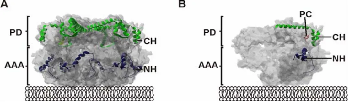
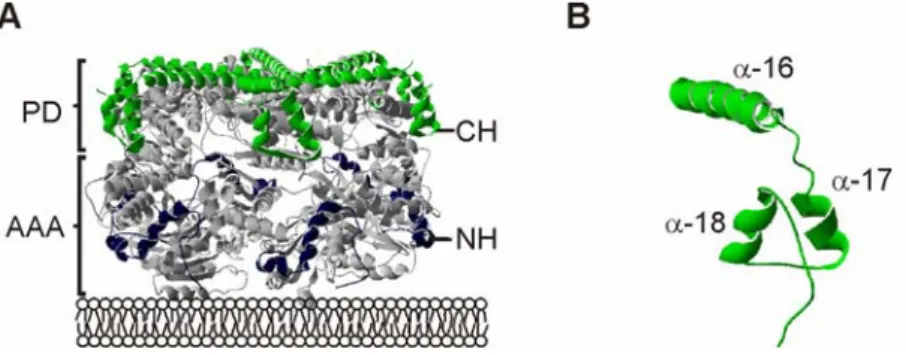
Within the structure of the i-AAA protease two distinct substrate binding sites have been identified: the NH-(N-terminal helices) region, located at the surface of the AAA domain, and the CH-(C-terminal helices) region, exposed at the surface of the proteolytic domain and by that on top of the cylindrical structure of the i-AAA protease (Graef et al., 2008) (Fig. 1.5).
The ~ 40 amino acid long NH-region is positioned in close proximity to the inner
mitochondrial membrane, making it an ideal element to encounter substrates protruding from the membrane. Structurally, it consists of helical structure build up by highly negatively charged amino acids that are required for substrate binding to this region (Graef and Langer, 2006). The independent/autonomous action of the NH-region in substrate binding is supported by in vitro studies of C-terminally truncated Yme1 molecules (Leonhard et al., 1999). Within the process of degradation, substrates are reported to first bind to the NH- region, before they are encountered by the central pore loops to be subsequently transferred to the proteolytic center (Graef and Langer, 2006).
The second substrate binding site the CH-region appears more suitable for binding of soluble substrates to the i-AAA protease, as it is located rather distantly from the inner mitochondrial membrane. The structure of the CH-region is built up by three helices the α-16, α-17 and α- 18 helix of Yme1 at the top and side of the catalytic chamber of the i-AAA protease (Graef et al., 2007). In respect to substrate binding and degradation, two different mechanisms are described for the CH-region, a CH-dependent and a CH-independent mechanism.
As an example, the degradation of cytochrome c oxidase 2 (Cox2) is described which can take place in a CH-dependent as well as in a CH-independent manner. CH-independent degradation of Cox2 is observed for fully folded Cox2 where the NH-region serves as a backup for initial substrate binding site (Graef et al., 2007). Any interference with the folding status of Cox2 renders its degradation CH-dependent (Graef et al., 2007). So, whether the degradation of a substrate occurs in a CH-dependent or CH-independent manner seems to be influenced by the folding state of a substrate and its localisation relative to the membrane. In this line, soluble or peripherally attached substrate proteins are thought to be
Figure 1.5 Initial substrate binding sites of the i-AAA protease. (A) Within the cylindrical structure of the AAA protease (grey) the substrate binding sites are exposed in a lattice like assembly at the outer surface of the protease. The NH-region (blue) resides at the surface of the AAA domain, whereas the CH-region (green) is part of the proteolytic domain.
(B) Open structure revealing the relative position of CH-region (green), NH-region (blue) and the proteolytic center (red). Here the different domains within the crystal structure of the related AAA protease of Thermotoga maritima are shown (U. Baumann personal communication). PD, proteolytic domain; AAA, AAA domain; NH, N-terminal helices; CH, C- terminal helices, PC, proteolytic center.
folded and tightly attached or inserted into the membrane might be degraded CH- independent. The exact mechanism determining the CH-dependent or CH-independent degradation of a substrate remains elusive. Autonomous substrate binding to the CH-region has been determined by in vitro binding studies (Graef et al., 2007).
Although the existence of two independent substrate binding sites might have evolved to achieve an enhanced recognition of substrates with different properties, the relevance of these two binding sites within the degradation process is far from being understood. During degradation, substrates first have to bind to the protease before being encountered by the central pore loops for further translocation to the inner cavity for degradation. For the NH- region such a mechanism is already implied (Graef and Langer, 2006). In contrast, there is no evidence for substrate translocation from the CH-region to the central pore loops. As the CH- region is situated rather distant to the central pore loops substrates might first bind to the NH-region before they are encountered by the central pore loops. Alternatively, additional substrate entry pathways to the proteolytic center could exist for substrates that are bound to this region. Therefore, analysis of substrate transition mechanisms after binding of substrates to the CH-region is awaited. The fact that the i-AAA protease is membrane embedded could again make the mechanism more complicated in comparison to soluble AAA proteases.
1.4.5 Functions of the i - and m -AAA proteases in mitochondria
Various functions have been described for the two mitochondrial AAA proteases (Fig. 1.6).
Both the i-AAA and the m-AAA protease are required for mitochondrial protein quality surveillance of inner membrane proteins (Leonhard et al., 2000) on which they show overlapping function. One argument for this overlap of function is the synthetic lethality seen for simultaneous deletion of both proteases (Lemaire et al., 2000; Leonhard et al., 2000) that is also underlining the essential role of the proteases for cellular function. Moreover, the presence of either of the proteases is demonstrated to be sufficient for complete degradation of a certain substrate protein (Leonhard et al., 2000). Furthermore, prohibitins are found to assemble with the m-AAA protease into a high molecular weight complex in the inner mitochondrial membrane, where they negatively regulate the quality control function of the m-AAA protease (Steglich et al., 1999).
In addition to its quality control function, the m-AAA protease is involved in the processing of nuclearly encoded mitochondrial proteins (Esser et al., 2002; Nolden et al., 2005). One such mitochondrial protein is MrpL32, a subunit of the large particle of the mitochondrial ribosome. After mitochondrial import of MrpL32, the m-AAA protease processes the protein
to its mature form and by that allows the completion of a functional mitochondrial ribosome formation. Why the proteolytic action of the m-AAA protease on MrpL32 does not lead to complete degradation of the protein remains to be elucidated. The function of the m-AAA protease in processing of MrpL32 explains the respiratory deficiency of a strain depleted of the m-AAA protease subunits, as a lack of mitochondrial translation results in a loss of respiratory complexes (Nolden et al., 2005).
Moreover, membrane dislocation events are demonstrated for the m-AAA protease (Tatsuta
m c
Figure 1.6 Versatile functions of the i- and m-AAA proteases of mitochondria. (A) Quality control surveillance. Misfolded polypeptides are degraded to peptides after their dislocation from the membrane. The membrane topology of substrates determines the involvement of either i-AAA (red) or m-AAA protease (blue) which exert overlapping substrate specificity (Leonhard et al., 2000). (B) Protein processing. The m-AAA protease mediates processing of nuclear encoded mitochondrial proteins resulting in their activation (Esser et al., 2002; Nolden et al., 2005). Maturation of the ribosomal protein MrpL32 by the m-AAA protease enables ribosomal assembly within mitochondria (Nolden et al., 2005). (C) Membrane dislocation. Ccp1 is dislocated by the m-AAA protease in an ATP-dependent manner allowing its intramembrane cleavage by the rhomboid protease Pcp1 (Tatsuta et al., 2007). (D) Protein import. The i-AAA protease is required for import of heterologously expressed, mammalian PNPase into the mitochondrial intermembrane space (Rainey et al., 2006). OM, outer mitochondrial membrane; IMS, intermembrane space; IM, inner mitochondrial membrane; M, matrix space. The figure is adapted from (Graef et al., 2008).
(Ccp1) (Esser et al., 2002), a reactive oxygen scavenger in the intermembrane space that contains a bipartite import sequence for its posttranslational import into mitochondria (Kaput et al., 1982). Upon import into mitochondria, Ccp1 is matured by subsequent action of the m-AAA protease and the ATP-dependent rhomboid protease Pcp1 (Esser et al., 2002;
Tatsuta et al., 2007). This process strictly depends on the presence of the m-AAA protease, as intramembrane cleavage by Pcp1 can only occur after membrane dislocation of Ccp1 by the m-AAA protease which renders the cleavage site of Ccp1 accessible for Pcp1 (Tatsuta et al., 2007). Also for the i-AAA protease a non-proteolytic function is suggested. Mitochondrial import of mammalian PNPase expressed heterologously in yeast requires the i-AAA protease (Rainey et al., 2006). PNPase binding to the i-AAA protease subunit Yme1 promotes the translocation of PNPase across the outer mitochondrial membrane. This function is impaired if the proteolytic activity of Yme1 is inhibited, although Yme1 is not able to degrade PNPase under these conditions. Whether the action of Yme1 in import of PNPase resembles the membrane dislocation event seen for the m-AAA protease in case of Ccp1 remains unclear at this point.
Thus, additional functions independent of the proteolytic activity of the mitochondrial AAA proteases have been identified (Graef et al., 2008) highlighting the relevance of these proteases for additional mitochondrial processes involving housekeeping and regulatory roles during mitochondrial biogenesis. Nevertheless, they are required for quality control of mitochondrial membrane proteins. Therefore, combination of the activity of Pim1 and the two AAA proteases defines an elaborate system of mitochondrial quality control that assures maintenance of mitochondrial function.
1.4.6 The conserved i -AAA protease Yme1
The i-AAA protease is best studied in the yeast S. cerevisiae. Here, the homo-oligomer is composed of Yme1 subunits that assemble into a proposed hexameric structure localised to the inner mitochondrial membrane with their catalytic domains facing the intermembrane space (Leonhard et al., 1999). Unexpectedly, analysis of the complex by gelfiltration experiment and blue native gel electrophoresis reveals a complex size that does not resemble an assembly of only six subunits. It is therefore conceivable that other components are involved in the assembly of the native complex. Two proteins influencing the complex size of the i-AAA protease are the yeast specific adaptors Mgr1 and Mgr3 (Dunn et al., 2008). The complex size of the i-AAA protease is reduced in the absence of either MGR1 or MGR3 and degradation of model substrates is decreased. However, both proteins are not shown to be involved in degradation of the substrate Cox2 and their deletion resembles only
one of the known Δyme1 phenotypes, the dependence on mitochondrial DNA (mtDNA) (Dunn et al., 2006). In general, the deletion of YME1 is connected to pleiotrophic phenotypes. The already noted dependence of cells on mtDNA in the absence of Yme1 is also described as ‘petite negative’ phenotype (Chen and Clark-Walker, 1999; Thorsness and Fox, 1993). Furthermore, Δyme1 cells are respiratory incompetent at elevated temperature and not able to grown on glucose-rich medium at lower temperature (Thorsness et al., 1993). Deletion of YME1 also induces an aberrant mitochondrial morphology and an increased mitochondrial turnover via the vacuole which also results in transfer of mtDNA to the nucleus (Campbell et al., 1994; Campbell and Thorsness, 1998; Thorsness and Fox, 1993). More recently, the function of Yme1 has been linked to ergosterol and longevity.
Δyme1 cells are deficient for uptake of ergosterol under anaerobic growth condition (Reiner et al., 2006). As ergosterol biosynthesis requires molecular oxygen uptake of ergosterol is essential under those conditions (Parks et al., 1995). The connection of Yme1 to longevity is based on studies monitoring the replicative lifespan of cells in the absence of YME1 (Francis et al., 2007; Palermo et al., 2007). Further, YME1 is deleted in life-extending mutants of which many show a decreased cytosolic protein synthesis (Wang et al., 2008). The resulting double mutants exhibit a loss of extended lifespan, linking the function of Yme1 to a role in longevity.
According to the vast number of phenotypes associated with the loss of the i-AAA protease, this protein, although present within mitochondria, participates in many cellular processes.
Nonetheless, the described phenotypes cannot be directly linked to so far identified Table 1.1 Homology of Yme1 orthologs. Yme1 orthologs exists in Homo sapiens, Mus musculus, Rattus norvegicus, Caenorhabditis elegans, Schizosaccharomyces pompe and Canadida alibicans. There respective protein names and relative homology to Yme1 from S.
cerevisiae are depicted here. Source: BIOBASE Knowledge Library.
Species Name
% identity to S. cerevisiae Yme1
H. sapiens YME1L1 52%
M. musculus Yme1l1 51.8%
R. norvegicus Yme1l1 51.8%
C. elegans M03C11.5 51.6%
S. pombe SPCC965.04c 47.8%
C. albicans YME1 67.3%
protease. Besides Cox2 (Leonhard et al., 1996; Nakai et al., 1995; Pearce and Sherman, 1995; Weber et al., 1996), the i-AAA protease is also involved in the degradation of Nde1 (Augustin et al., 2005) and Phb1/2 (Kambacheld et al., 2005). Hence, in order to explain the molecular mechanisms causing the pleitropic phenotypes of a Δyme1 strain, the identification of additional interaction partners and substrates is required.
Yme1 is expressed throughout the eukaryotic kingdom and described orthologs show a high sequence identity (Tab. 1.1), speaking for a general importance of the i-AAA protease in cellular function. The high degree of similarity is also supported by complementation studies of yeast, where expression of YME1L1 restored the respiratory deficiency of Δyme1 at elevated temperature (Shah et al., 2000). The human homolog YME1L1 has been demonstrated to process the mitochondrial fusion component OPA1 at a specific site (Griparic et al., 2007; Song et al., 2007). Furthermore, stabilisation of another OPA1 isoform is apparent upon downregulation of YME1L1, pointing to its degradation by the protease (Guillery et al., 2008). OPA1 is the human homolog of Mgm1. In yeast, processing of Mgm1 is less complex and shown to be mediated by Pcp1 (Sesaki et al., 2006; Sesaki et al., 2003).
Although yeast cells exhibit a mitochondrial morphology defect in the absence of Yme1, the i-AAA protease seems not to be involved in the processing of Mgm1 (Campbell et al., 1994).
It would be interesting to see if other phenotypes found for the yeast homolog Yme1 can be observed in higher eukaryotes.
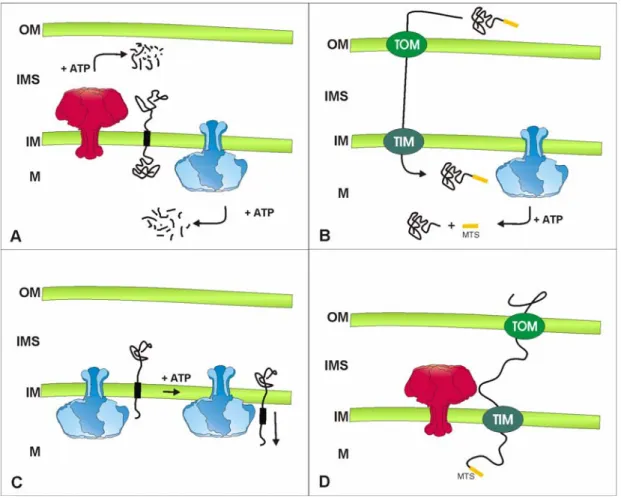
1.5 Mitochondrial peptidases and mitochondrial protein import
Mitochondrial processing peptidases are predominantly important for the cleavage of mitochondrial targeting sequences generating the active, matured form of a protein in different mitochondrial subcompartments (Koppen and Langer, 2007). Usually targeting sequences consist of 10-80 amino acids at the N-terminus of a protein. These sequences are rich in positively charged amino acids and are able to form amphipatic helical structures (Song et al., 1998). In addition, internal targeting sequences exist, but their nature remains largely elusive (Folsch et al., 1996). Further, bipartite import sequences can be found, that require that action of two peptidases for full maturation (Hartl et al., 1987). Prior to their processing, the respective precursor proteins have to be imported into mitochondria. The import of proteins into mitochondria is achieved by an elaborate system of translocases located in inner and outer mitochondrial membrane (reviewed in (Bolender et al., 2008;
Neupert and Herrmann, 2007)) (Fig. 1.6).
Three major mitochondrial processing peptidases exist in the matrix space and the inner mitochondrial membrane of mitochondria (Gakh et al., 2002). Within the matrix space the peptidases MPP (mitochondrial processing peptidase) and MIP (mitochondrial intermediate peptidase) ensure maturation of imported precursor proteins (Hawlitschek et al., 1988;
Kalousek et al., 1988; Yang et al., 1988). The yeast MPP peptidase is a hetero-dimer of Mas1 and Mas2 that is essential for cell survival (Yaffe et al., 1985). The conserved metallopeptidase is highly homologous to non-catalytic subunits of the cytochrome c reductase (Gencic et al., 1991; Schulte et al., 1989). Therefore, it is not surprising that MPP is a bi-functional protein in some organisms (Glaser and Dessi, 1999). The monomeric metallopeptidase MIP is composed of Oct1 in yeast (Chew et al., 1996; Kalousek et al., 1988) and requires octapeptides for the cleavage of precursor proteins (Isaya et al., 1992).
The severe phenotypes associated with inactivation of the gene point to an important Figure 1.6 The mitochondrial protein import machinery and its connection to mitochondrial processing peptidases in S. cerevisiae. After translocation of proteins through the TOM (translocase of the outer membrane) complex proteins enter different pathways. β-barrel proteins are assembled into the outer membrane (OM) by the action of the SAM (sorting and assembly machinery) and the MDM (mitochondrial distribution morphology) complex. Intermembrane space proteins can be oxidised and assembled by Mia40 (mitochondrial intermembrane space import and assembly). Inner mitochondrial membrane and carrier proteins are translocated through the inner mitochondrial membrane (IM) and are assembled by the TIM23 or TIM22 (translocase of the inner membrane) complex, respectively. Import of matrix proteins is facilitated by the Tim23 complex. After import into the matrix (M) proteins are either translocated through the inner mitochondrial membrane via Oxa1 or are processed by MPP (mitochondrial processing peptidase) or MIP (mitochondrial intermediate peptidase). Proteins imported into the intermembrane space (IMS) are processed by IMP (inner membrane protease), Pcp1 or Atp23.