Mechanistic and structural characterization of the SUMO-specific protease
Ulp2
Inaugural-Dissertation zur
Erlangung des Doktorgrades
der Mathematisch-Naturwissenschaftlichen Fakultät der Universität zu Köln
vorgelegt von Julia Eckhoff
aus Buchholz in der Nordheide
Köln, 2016
1. Berichterstatter: Prof. Dr. R. Jürgen Dohmen 2. Berichterstatter: Prof. Dr. Kay Hofmann
Tag der mündlichen Prüfung: 05.07.2016
This is for everyone who loves science, the moment it
kisses you after it has just beat you up for month… And
then kicks you in the face again. Once you accept that
this is the dynamic of your relationship, you’ll have a fun
ride together.
Abstract ... 1
Zusammenfassung ... 2
1 Introduction ... 3
1.1 SUMO genes ... 3
1.2 SUMO protein ... 4
1.2.1 SUMO isoforms ... 4
1.3 The sumoylation cycle ... 6
1.4 Polysumoylation ... 7
1.5 Sumoylation motif ... 8
1.6 SUMO interacting motif ... 9
1.7 The ubiquitin-proteasome system ... 10
1.8 SUMO-targeted ubiquitin ligases ... 11
1.9 Desumoylating enzymes ... 12
1.9.1 Structures of SUMO proteases and catalytic mechanism ... 13
1.9.2 Regulation of SUMO proteases ... 14
1.9.3 Misregulated SUMO proteases ... 15
1.9.4 Proteases ... 16
1.9.4.1 In mammals ... 16
1.9.4.2 In budding yeast ... 17
1.9.4.2.1 Ulp1 ... 17
1.9.4.2.2 Ulp2 ... 18
1.9.4.2.3 Wss1 ... 21
1.10 Thesis objectives ... 21
2 Material and Methods ... 23
2.1 Material ... 23
2.1.1 Escherichia coli strains ... 23
2.1.2 Saccharomyces cerevisiae strains ... 23
2.1.3 Plasmids ... 24
2.1.4 Material ... 26
2.1.4.1 Chemicals and consumables ... 26
2.1.4.2 Electrical equipment ... 28
2.1.5 Enzymes ... 29
2.1.6 Antibodies ... 29
2.1.7 Media ... 30
2.1.7.1 Media for E. coli ... 30
2.1.7.2 Media for S. cerevisiae ... 30
2.1.7.2.1 Standard yeast media ... 30
2.1.7.2.2 Special yeast media ... 31
2.2 Methods ... 32
2.2.1 Cell cultivation ... 32
2.2.1.1 Standard conditions ... 32
2.2.1.1.1 Escherichia coli ... 32
2.2.1.1.2 Saccharomyces cerevisiae ... 32
2.2.1.2 Special conditions ... 32
2.2.1.2.1 Protein expression in E.coli ... 32
2.2.1.2.2 Growth assays of S. cerevisiae ... 33
2.2.2 Biomolecular techniques ... 34
2.2.2.2 PCR amplification ... 34
2.2.2.2.1 …for cloning purposes ... 35
2.2.2.2.2 …for screening purposes ... 37
2.2.2.3 Agarose gel electrophoresis ... 37
2.2.2.4 DNA restriction digestion ... 38
2.2.2.5 Vector dephosphorylation ... 38
2.2.2.6 Ligation of DNA fragments ... 38
2.2.2.7 Generation of transformation-competent E. coli cells ... 39
2.2.2.8 Transformation of E. coli cells ... 39
2.2.2.9 Transformation of S. cerevisiae cells ... 39
2.2.3 Genetic techniques ... 40
2.2.3.1 Mating and sporulation of S.cerevisiae strains ... 40
2.2.4 Biochemical techniques ... 41
2.2.4.1 SDS polyacrylamide gel electrophoresis (SDS-PAGE) ... 41
2.2.4.2 Coomassie staining of proteins in SDS polyacrylamide gels ... 42
2.2.4.3 Detection of proteins by immunoblotting (Western Blot) ... 42
2.2.4.4 Antibody removal ... 43
2.2.4.5 Protein purification ... 44
2.2.4.5.1 Purification of Ulp substrates ... 44
2.2.4.5.2 Purification of sumoylated Ubc9 ... 45
2.2.4.5.3 Purification of Ubp41 ... 46
2.2.4.5.4 Purification of full-length Ulp2 ... 46
2.2.4.5.5 Purification of the Ulp2 active domain ... 47
2.2.4.6 In vitro desumoylation assay ... 48
2.2.4.7 Ulp2 binding assay ... 49
2.2.4.8 Preparation of crude extracts from S. cerevisiae ... 50
2.2.5 Crystallization and crystal analysis ... 50
2.2.5.1 Crystallization and data collection ... 50
2.2.5.2 Data processing, structure determination and refinement ... 51
3 Results ... 52
3.1 Characterizing Ulp2 cleavage mechanism ... 52
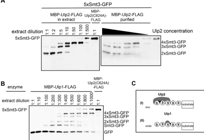
3.1.1 Ulp2 dismantles poly-Smt3 chains in a sequential manner ... 52
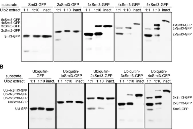
3.1.2 Three linked Smt3 moieties are the minimum Ulp2-target requirement ... 58
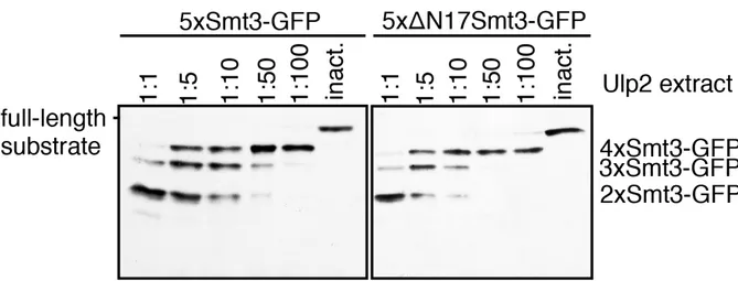
3.1.3 Efficient cleavage by Ulp2 requires free access to N-terminal region or surrounding surfaces of the distal Smt3 moiety ... 59
3.1.4 Ulp2 needs at least three consecutive Smt3 moieties to capture a target ... 62
3.1.5 The active domain of Ulp2 ... 63
3.1.5.1 Characteristics of the Ulp2 mechanism are grounded in the active domain .. 63
3.1.5.2 A SIM close to the active domain might be controlled by the NTD ... 65
3.1.6 Ulp2 hardly recognizes SUMO2 ... 66
3.1.7 Ulp2 recognizes different surface areas of each of its three binding partners in trimeric Smt3 ... 67
3.1.7.1 Exchanging four defined residues in Smt3 creates a version to which Ulps exhibit a very high affinity ... 75
3.2 The crystal structure of the Ulp2 active domain ... 75
3.2.1 Crystallization of the Ulp2 active domain ... 75
3.2.2 The structure of the Ulp2 active domain underlines its classification ... 77
3.2.3 Putative Smt3 binding sites in the Ulp2 active domain ... 80
3.3.1 HMW Smt3 conjugates are the causing agent of a proteolysis defect in ulp2-∆ 83
3.3.2 Ulp2 is the major player in SUMO recycling ... 84
4 Discussion ... 86
4.1 Ulp2 in-vitro-characterization setup ... 86
4.2 Ulp2 mechanism ... 87
4.2.1 Ulp2 cleaves polymeric SUMO chains from their distal ends ... 87
4.2.2 Its exo behavior differentiates Ulp2 from its subfamily members ... 88
4.2.3 Structure of the Ulp2 active domain ... 89
4.2.3.1 Determinants for the exo mode of Ulp2 are not known ... 90
4.2.3.2 Ulp2 and SENP7 are well superimposable ... 90
4.2.4 Ulp2 targets polymeric Smt3 chains ... 90
4.2.4.1 Ulp2 recognizes different regions in the three most distal members of polymeric Smt3 ... 91
4.2.4.2 The active site of the UD has a comparatively low surface charge ... 92
4.2.4.3 Co-crystallization of Ulp2 and its target was not possible ... 93
4.2.5 SIMs in Ulp2 ... 93
4.2.5.1 The SIM close to the active site must be subject to control in vivo ... 93
4.2.5.2 Possible role of SIMs in the non-catalytic domains of Ulp2 ... 94
4.2.6 Ulp2 maintains short Smt3 chains ... 94
4.2.7 Ulp2 and STUbLs ... 95
4.2.8 Poly-ubiquitin does not interfere with target recognition by Ulp2 ... 97
4.2.9 A high-affinity Smt3 variant ... 97
4.2.10 Ulp2 location ... 98
5 Outlook ... 100
6 References ... 101
List of Oligonucleotides ... 115
Appendix ... 118
Abbreviations ... 118
Acknowledgements ... 121
Eidesstattliche Erklärung ... 122
Abstract
Posttranslational attachment of the small ubiquitin-related modifier (SUMO) to a protein, commonly known as sumoylation, is a highly dynamic process of conjugation and
deconjugation. This work is concerned with the latter. Via different approaches it characterizes the S. cerevisiae SUMO-specific protease Ulp2, giving insight into its mechanism, its structure, as well as its role in vivo. It was found that Ulp2 dismantles poly- SUMO sequentially starting from the distal end (exo mode) down to two linked SUMO moieties. This differentiates Ulp2 from all other members of the Ulp/SENP family of proteases that have been analyzed, so far. Previous studies suggested rather stochastic mechanisms for all of them. Apparently, Ulp2 recognizes a region at the N-terminus or surrounding surfaces of SUMO that are not accessible in moieties of the chain other than the most distal one. Full accessibility to the N-terminus needs to be granted in order to achieve full cleavage efficiency. Additionally, Ulp2 requires at least tri-SUMO to bind and/or to process a target chain. Binding seems to happen in a cooperative manner, meaning that all three binding sites have to be occupied in order to achieve interaction. It was found that in each one of the units of a trimeric SUMO chain a different surface area is involved in SUMO/Ulp2 interactions in the event of cleaving off the most distal unit. The entire
mechanism and preferences of Ulp2 are contained in its catalytic domain (UD). In the course of this work, the crystal structure of this domain was solved. The structural insights confirmed Ulp2’s family affiliation with Ulp1, and underscored its close relation to SENP7.
Nevertheless, concrete unique regions in the structure open up opportunities for discovering the reasons for the distinct mechanistic characteristics Ulp2 exhibits. Also, surface charge and hydrophobicity of the UD is significantly different to Ulp1 as well as SENP7 active domains.
In vivo studies in the course of this work showed that polymeric SUMO is not subjected to degradation alongside the anchor protein it is attached to, but rather liberated beforehand.
Ulp2 was identified as the major, if not only, SUMO-specific protease involved in this SUMO recycling process. However, it is yet to be clarified whether mono- and di-SUMO
modifications are subjected to degradation, since Ulp2 would not process that. In the light of the mechanistic findings, the in vivo analysis led to a model in which Ulp2 is in an
antagonistic relationship with STUbLs: While it prevents SUMO chains from getting long enough for STUbLs to capture them, once a STUbLs has bound polymeric SUMO, it
obstructs the chain in such a way that it is not possible for Ulp2 to attack it, thus the substrate
inevitably files into degradation by the proteasome.
Zusammenfassung
Sumoylierung, die posttranslationale Modifikation eines Proteins mit SUMO (small ubiquitin- related modifier), ist ein hoch dynamisches Wechselspiel aus Konjugation und De-
konjugation. Im folgenden Text liegt der Fokus auf letzterem, genauer noch auf der SUMO- spezifische Protease Ulp2. Diese Arbeit charakterisiert Ulp2 sowohl mechanistisch als auch strukturell und erlaubte durch gezielte In-vivo-Experimente, den mechanistischen
Gegebenheiten eine Funktion im größeren, zellulären Kontext zuzuordnen. Wie sich zeigte, baut Ulp2 poly-SUMO-Ketten von ihrem distalen Ende her ab. Das unterscheidet Ulp2 signifikant von sowohl Ulp1, der einzigen anderen bekannten S. cerevisiae SUMO-Protease, als auch seinen humanen Orthologen, SENP6 und SENP7, die Ketten eher ungeordnet, stochastisch prozessieren.
Bei einer Kettenlänge von zwei SUMOs stoppt der Abbau durch Ulp2. Weder mono- noch di- SUMO-Modifikationen werden von Ulp2 als Substrate erkannt, dementsprechend weder dekonjugiert noch gebunden. Die Resultate aus unterschiedlichen experimentellen Ansätzen deuten darauf hin, dass die Oberfläche von Ulp2 drei verschiedene SUMO-Bindestellen aufweist, die simultan besetzt sein müssen, um eine ausreichend starke Interaktion zwischen Enzym und Kette zu erhalten. Sterisch unbeschränkter Zugang zum Aminoterminus des distalen SUMO erwies sich als Grundvoraussetzung für maximale Prozessierungseffizienz.
Scheinbar erkennt Ulp2 eine Region am N-Terminus oder umliegende Oberflächen von SUMO, die in anderen Glieder einer poly-SUMO-Kette weniger zugänglich sind. Es zeigte sich, dass Ulp2 in jedem seiner drei Bindungspartner eine andere Oberfläche erkennt.
Sämtliche genannte Eigenschaften sind in der aktiven Domäne angelegt; die nicht-
katalytischen Domänen haben mechanistisch keine Relevanz. Im Zuge dieser Arbeit wurde die Kristallstruktur der aktiven Domäne von Ulp2 (UD) gelöst. Sie zeigte klare Ähnlichkeiten, aber auch signifikante Unterschiede zwischen der Tertiärstruktur und der
Oberflächenbeschaffenheit (Ladung/Hydrophilie) von Ulp2 und Ulp1. Die Ähnlichkeit zu
SENP7 erwies sich als wesentlich größer, doch auch hier gab es prägnante Divergenzen, die
Ansätze für weitere mechanistische Aufklärung bieten. In-vivo-Experimente in S. cerevisiae
zeigten, dass polymeres SUMO nicht gemeinsam mit seinem Substrat abgebaut, sondern
vorab von Ulp2 dekonjugiert wird. Vor dem Hintergrund der mechanistischen Erkenntnisse
dieser Arbeit ergibt sich ein Modell, in dem Ulp2 die Kettenlänge von SUMO-Konjugaten in
einem gewissen dynamischen Rahmen hält und damit auch eine antagonistische Rolle zu
STUbLs (SUMO-targeted Ubiquitin ligases) einnimmt, die ausschließlich poly-SUMO-Ketten
binden können, um das an ihnen haftende Substratprotein seinem Abbau zuzuführen.
1 Introduction
This year (2016) marks 20 years since SUMO was first reported on. It was found as a
covalently linked attachment to RanGAP1, a GTPase activating protein in humans (Mahajan et al. 1997; Matunis et al. 1996). The name SUMO, small ubiquitin-related modifier, was chosen as a tribute to its similarity to ubiquitin. Ubiquitin and its kinsman, collectively termed ubiquitin-like proteins (Ubls), are posttranslational modifiers. Upon conjugation to proteins, they alter the properties of their targets, thereby increasing the variety of the proteome in eukaryotic cells. So far, hundreds of proteins have been found to be sumoylated at some point during the cell cycle (Wohlschlegel et al. 2004; Zhou et al. 2004; Denison et al. 2005; Hannich et al. 2005; Panse et al. 2004; Wykoff et al. 2005; Vertegaal et al. 2006;
Golebiowski et al. 2009; Schou et al. 2014; Hendriks et al. 2014). This makes SUMO the Ubl modifying the second larges pool of proteins, only outnumbered by ubiquitin itself.
SUMO-conjugated proteins are frequently found in the nucleus, pointing towards sumoylation being an important nuclear process (Rodriguez et al. 2001).
Many sumoylated proteins are tumor suppressors, transcription factors and nuclear body proteins, a fact that accounts for a fast-growing interest in the understanding of the SUMO system. Since usually only a small fraction, frequently less than 1 %, of a substrate is sumoylated at any given time (Johnson 2004), studying sumoylation is not trivial, and there might be many more SUMO targets yet to be discovered.
1.1 SUMO genes
SUMO has been shown to be essential in most eukaryotes (Johnson et al. 1997; Nacerddine et al. 2005). Schizosaccharomyces pombe represents a known exception from this general view. Its sole SUMO-encoding gene, pmt3, is not essential, but a knockout mutant suffers from severe genome maintenance defects and is barely viable (Tanaka et al. 1999). Another exception is the filamentous fungus Aspergillus nidulans (Wong et al. 2008). In
Saccharomyces cerevisiae (S. cerevisiae), SUMO is expressed from a single gene, SMT3 (suppressor of mif two), which encodes a 101-amino acid molecule (Meluh et al. 1995). Like S. cerevisiae, Caenorhabditis elegans features only a single SUMO gene (Choudhury et al.
1997). In contrast, plants have several; e.g. Arabidopsis thaliana expresses SUMO from 8
different genes (Kurepa et al. 2003; Novatchkova et al. 2012). In higher eukaryotes, so far, 4
isoforms have been found: SUMO1-4 (Mahajan et al. 1997; Matunis et al. 1996; Johnson 2004; Shen et al. 1996; Kamitani et al. 1998; Mukhopadhyay et al. 2007; Bohren et al.
2004).
1.2 SUMO protein
SUMO has a molecular weight of ~11 kDa, but on an SDS-PAGE adds a ~20 kDa increment in size to the protein it is linked to. While SUMO shares only ~18 % sequence identity with ubiquitin, the two structures are for the most part superimposable (Fig. 1) (Johnson 2004;
Bayer et al. 1998). Like all Ubls, SUMO features the ubiquitin superfold, a β-grasp fold in which four β-strands surround an α-helix which diagonally transverses the molecule (Bayer et al. 1998; Welchman et al. 2005; Vijay-Kumar et al. 1987). But unlike ubiquitin, SUMO has an N-terminal extension of ~20 amino acids, which is flexible in solution (Bayer et al. 1998).
This region is rich in proline, glycine, and charged residues. Notably, this flexible part has been shown to be dispensable, as a corresponding N-terminally truncated SUMO mutant can still serve its essential functions (Bylebyl et al. 2003).
FIGURE 1: Structures of SUMO and ubiquitin. Graphics were prepared with Chimera using PDB 1L2N (Smt3) and PDB 1UBQ (ubiquitin) in ribbon representation. N-t. = amino-terminus; C-t. = carboxy-terminus.
1.2.1 SUMO isoforms
The four different SUMO genes in mammalian cells translate to four different SUMO
isoforms. The human SUMO1 (also known as sentrin, Ubl1 and Smt3c) was the first one to
be discovered in a homology screen to yeast SMT3 (Shen et al. 1996). SUMO4, the latest
discovered member, is a special case and will be discussed separately below.
SUMO1 shares only ~45 % sequence identity with SUMO2 and SUMO3, which are virtually identical (~95 % similarity), hence frequently referred to as SUMO2/3 (Johnson 2004; Flotho et al. 2013). Despite the fact that SUMO1 and SUMO2/3 are activated and conjugated by the same E1 and E2 enzymes, they have significantly different features (Johnson 2004;
Mukhopadhyay and Dasso 2007; Flotho and Melchior 2013). To start with, they show unique intracellular abundance: The cell cycle-dependent distribution of SUMO2/3 has been shown to be distinct from SUMO1 location, mobility and dynamics (Ayaydin et al. 2004).
Additionally, conjugation of SUMO2/3 is strongly induced upon various stresses; SUMO1 modification, in contrast, dominates under non-stress conditions (Seifert et al. 2015). Another noticeable difference between SUMO1 and the other two paralogues is its inability to
efficiently form chains (Tatham et al. 2001). While SUMO2/3 efficiently form polymers on their substrates, the existence of poly-SUMO1 has been debated (Flotho and Melchior 2013;
Tatham et al. 2001; Saitoh et al. 2000). Even though, SUMO1 polymerization has been observed in vitro (Pichler et al. 2002; Yang et al. 2006a; Pedrioli et al. 2006), the prevalent role of SUMO1 in in vivo poly-SUMO conjugates seems to be terminating SUMO2/3 chain elongation by capping (Matic et al. 2008). Yet another difference of the SUMO paralogues lies in the abundance of their free forms. There is a large pool of free, unconjugated
SUMO2/3, but SUMO1 exists predominantly in its conjugated form (Saitoh and Hinchey
2000). That does not implicate SUMO1 as being favored by the conjugation machinery, but
might find its origin partly in the fact that SUMO2/3 is expressed ten times more than
SUMO1 (Saitoh and Hinchey 2000). The difference in abundance is counterbalanced by
significantly faster cleavage of SUMO2/3- than SUMO1-conjugates (Flotho and Melchior
2013; Kolli et al. 2010). Although targets and downstream effectors of the SUMO paralogues
significantly overlap, some proteins are preferentially conjugated to a certain subfamily. So
far, two distinct mechanisms that account for this have been reported on. One of them is
based on intrinsic preferences of the substrate protein itself (Meulmeester et al. 2008; Zhu et
al. 2008); the other one has been described in the course of RanGAP1 studies. It was found
that while being efficiently modified with either of the paralogues, only RanGAP1-SUMO1
was stably incorporated in a Ubc9-RanBP1-RanGAP1 complex, and that this complex
formation prevents desumoylation of RanGAP1 (Zhu et al. 2009; Werner et al. 2012). Other
mechanisms –possibly involving E3 ligases– have been subject to speculation, but are yet to
be discovered (Flotho and Melchior 2013).
While SUMO1 and SUMO2/3 are ubiquitously distributed in most tissues, SUMO4
apparently is expressed in a tissue- or organ-specific way. So far, it has been detected only in discrete areas like spleen, pancreatic islets, kidney or lymph node (Guo et al. 2004; Wang et al. 2008). Several important transcription factors involved in immune response have been found to be modified by SUMO4, suggesting a regulatory role in that field (Wang and She 2008). However, its maturation is not yet understood. Despite 86 % identity to SUMO2/3 (Bohren et al. 2004), none of the known SUMO-specific proteases recognizes SUMO4 as a target (Mukhopadhyay and Dasso 2007). Interestingly, a single-site mutation modulates pre- SUMO4 amenable to processing by SENP2 (Wang and She 2008; Liu et al. 2014).
Nevertheless, its biological relevance is still unclear.
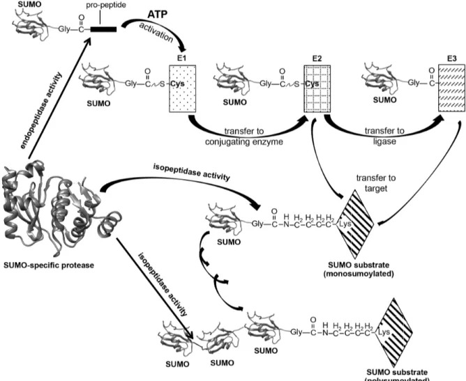
1.3 The sumoylation cycle
SUMO proteins are translated as immature precursors that require processing to become conjugation competent. The proteolytic reaction trims off several C-terminal residues to expose a diglycine motif. Attachment of SUMO happens via an ATP-dependent enzymatic cascade strongly reminiscent of ubiquitination (Komander et al. 2012). In general,
sumoylation involves three different classes of enzymes: A SUMO-activating enzyme (E1), which renders the C-terminal carboxyl group of SUMO active for reaction, and then forms a thioester with it; a SUMO-conjugating enzyme (E2), and a SUMO ligase (E3) (Gareau et al.
2010; Hickey et al. 2012). Chemically speaking, the result of the sumoylation cascade is the formation of an isopeptide bond between the C-terminal carboxyl group of SUMO and an ε- amino group of an acceptor lysine on the surface of a substrate molecule (Johnson 2004;
Geiss-Friedlander et al. 2007; Kerscher et al. 2006; Capili et al. 2007). A general overview of the SUMO cycle is given in Fig. 2. In the first step, the SUMO-specific E1 activating
heterodimer consisting of SUMO-activating enzyme subunit 1 (SAE1; also known as Aos1)
and SUMO-activating enzyme subunit 2 (SAE2; also known as Uba2) activates the C-
terminus of SUMO in a two-step reaction: First, it forms a SUMO adenylate with it. Then,
after a dramatic remodeling of the E1 active site, a conserved cysteine residue on the E1
attacks the adenylate to result in an SAE2~SUMO thioester bond (Olsen et al. 2010; Dye et
al. 2007; Schulman et al. 2009). The E2 conjugating enzyme Ubc9 binds to a ubiquitin fold-
domain of SAE2. Upon interaction of the thioester-charged E1 enzyme with Ubc9, SUMO is
transferred to the catalytic Cys residue of the E2 enzyme, again forming a thioester bond. At
this point, there are two possibilities: Either Ubc9 directly interacts with the SUMO target to
modify an acceptor lysine on its surface, or E3 protein ligases facilitate this process. The latter is usually the case and much more efficient. Basically, there are two mechanisms:
Either, the SUMO E3 enzymes recruit the substrate protein into a complex with the Ubc9~SUMO thioester, thereby promoting specificity, or in case of direct E2-substrate interaction, they expedite sumoylation by stimulating the E2 enzyme to transfer SUMO to the target protein. Either way, the result is a SUMO moiety covalently attached to a Lys residue on the surface of an acceptor.
FIGURE 2: Schematic representation of the SUMO cycle. SUMO precursors are processed by specific proteases to expose their C-terminal di-glycine motives. The mature SUMO is activated and conjugated to a target protein via an E1-E2(-E3) cascade. In an ATP-dependent step it forms a thioester bond to the active Cys of the E1. This is then transesterified to the Cys of the E2 enzyme. Then, directly or via an E3 ligase, the conjugation-competent SUMO is transferred to an ε-amino group of an acceptor lysine on the surface of a SUMO substrate. The SUMO modification itself can then serve as a target, leading to SUMO chain formation.
SUMO-specific proteases are able to dismantle the chains again.
1.4 Polysumoylation
Notably, not only mono- and multisumoylation (several SUMO molecules attached to
different Lys residues on the same protein), but also polysumoylation, meaning formation of
SUMO chains, is possible. The finding of SUMO chains came as a surprise, as in vivo most
substrates are decorated with single SUMO moieties –frequently at multiple sites, though (Johnson et al. 1999; Mahajan et al. 1998). Polymeric SUMO is possible, because the SUMO sequence itself contains lysine residues, which can serve as a docking site for another SUMO moiety. These residues lie in a favoring environment, a so-called sumoylation motif (see section 1.5) (Bylebyl et al. 2003; Tatham et al. 2001; Johnson et al. 2001; Bencsath et al.
2002). Generally, they are located in the flexible N-terminal appendix. While SUMO2/3 have only one such lysine, Lys11, Smt3 features three of them: Lys11, Lys15, and Lys19 (Johnson 2004; Tatham et al. 2001; Ulrich 2008). They are known as the canonical attachment sites.
However, in the last few years it has become obvious that formation of polymeric SUMO chains is by far not restricted to these prominent lysine residues. For instance, rendering them inoperable as linkage sites by replacing them with arginine does not significantly reduce buildup of SUMO chains. Only exchange of all lysine residues present in SUMO inhibits polysumoylation (amongst other reports of this, it was confirmed in this work, though data not shown). Poly-SUMO chains accumulate upon heat shock, osmotic- and replicative stress (Vertegaal 2010). However, the relevance of SUMO chains is still not clear. In yeast,
polysumoylation can be eliminated without notable effects on SUMO function or conjugates pattern (Bylebyl et al. 2003; Takahashi et al. 2003). Many endeavors have been made to unravel this puzzle, and for instance the discovery of SUMO-targeted ubiquitin ligases (see section 1.8) gave the explanation of polymeric SUMO one plausible direction, but the exact function of SUMO chains still awaits elucidation. The fact that they are rapidly turned over, hence scarce in vivo, is a major obstacle to investigations in that direction.
1.5 Sumoylation motif
Usually, SUMO is attached to a lysine residue embedded in the consensus motif ΨKxE on the surface of a target protein. In this recognition pattern, Ψ stands for a hydrophobic residue, while “x” can be any residue. Of note, the glutamate is the most highly conserved position (Rodriguez et al. 2001; Sapetschnig et al. 2002). ΨKxE is the recognition site for the E2 Ubc9 (Sampson et al. 2001), and the interaction between motif and enzyme are key to the conjugation mechanism (Bernier-Villamor et al. 2002; Lin et al. 2002; Reverter et al. 2005).
Obviously, this motif is too short and too undefined to confer specificity on the sumoylation
process. Indeed, more than one third of all proteins characterized to date contain such a patch,
but only a small amount of them are sumoylated (Yang et al. 2006b). Consequently, there
must be further determinants to account for accurate sumoylation. It has been shown that the
steric environment is another determinant for SUMO attachment signals (Pichler et al. 2005), likely, because the target lysine residue must be accessible to the SUMO-conjugating
machinery. Apart from secondary structure elements factoring in, extended variants of the core consensus motif were described. Among them were a negatively charged amino acid- dependent sumoylation motif (NDSM) (Yang et al. 2006b), and a phosphorylation-dependent sumoylation motif (PDSM). For both of them, clusters of residues downstream of the ΨKxE core were found to strengthen the interaction between substrate and Ubc9, thereby promoting SUMO conjugation (Yang et al. 2006b; Mohideen et al. 2009; Hietakangas et al. 2006).
There are also reports of sumoylation of the inverted motif [(E/D)xKΨ] (Ivanov et al. 2007;
Matic et al. 2010). In addition to that, several proteins have been found to be modified on sites that do not conform to ΨKxE (Hendriks et al. 2014; Adamson et al. 2001; Ishov et al.
1999; Lin et al. 2003; Chakrabarti et al. 2000; Picard et al. 2012). Indeed, this seems to be quite frequently the case (Hendriks et al. 2014). Taken together, the exact set of determinants for sumoylation remains elusive.
1.6 SUMO interacting motif
At its basis, units of SUMO attached to a target protein create a new interaction platform on the surface of this protein. In order for this role to be fulfilled, there must be patches in the respective interaction partners that recognize SUMO. In 2000, the first conserved SUMO- interacting motifs (SIMs) were reported (Minty et al. 2000). SIMs are typically composed of a hydrophobic core with the loose consensus sequence V/L/I-x-V/L/I-V/L/I (x = any amino acid) and several acidic amino acids, frequently juxtaposing the core (Minty et al. 2000; Song et al. 2004; Hecker et al. 2006; Kerscher 2007). Frequently, SIMs are located in unstructured protein regions. Upon binding, the hydrophobic core arranges itself in a β-strand
conformation that fits in a pocket of SUMO formed by its α
1-helix and its β
2-strand (Hecker et al. 2006; Song et al. 2005). The core motif binds SUMO in a parallel or anti-parallel orientation, depending on the distribution of the negatively charged residues, that extends the SUMO β-sheet (Gareau and Lima 2010; Reverter and Lima 2005; Song et al. 2005). This allows interactions between the aliphatic residues of the SIM and the hydrophobic pocket of SUMO. The negatively charged residues can be decisive for the affinity, polarity, as well as for the paralogue-specificity of the SUMO/SIM interaction, probably by salt bridging and hydrogen bonding with conserved basic residues on the SUMO surface (Meulmeester et al.
2008; Hecker et al. 2006; Chang et al. 2011). So far, three different types of SIMs are known:
SIMa (ΨΨxΨAc
n), SIMb (ΨΨDLT) and SIMr, (Ac
nΨxΨΨ) (Praefcke et al. 2012;
Sriramachandran et al. 2014). In the bracketed description, Ψ is a hydrophobic residue (V, I or L), n is a number between two and five, and Ac stands for Asp, Glu or Ser. Even though it is not an acidic amino acid, Ser is frequently found in SIMs, probably due to it being a target for phosphorylation (Sriramachandran and Dohmen 2014). Compared to the 16 known ubiquitin-binding domains (Grabbe et al. 2009), the diversity of SIMs as it is to date is small.
This might indicate that there are additional types of SIMs, still awaiting their discovery.
1.7 The ubiquitin-proteasome system
A eukaryotic cell has two main systems for protein degradation: The lysosomal pathway and the ubiquitin-proteasome system (UPS) (Haas et al. 2008; Laney et al. 2001). While the former is autophagy-based, degrading extracellular proteins that have been imported into the cell by pinocytosis or endocytosis, the UPS operates the degradation of intracellular proteins (Hochstrasser 1995; Goldberg 1995). It degrades approximately 80-90 % of the intracellular proteins involved in every cellular function and process (Chen et al. 2010). Compared to the autophagy-lysosome system, this degradation pathway is highly selective.
The UPS consists of many different molecules, with the main players being ubiquitin, ubiquitinating enzymes, deubiquitinating enzymes (DUBs), and proteasomes (Zhong et al.
2016). The 26S proteasome is a 673-kDa multi-catalytic proteinase complex, which is found in the nucleus as well as in the cytosol (Chen and Dou 2010; Lowe et al. 1995). It is
composed of several subunits: The 20S core particle consists of 28 subunits, 14 α-subunits, each weighting 25.8 kDa, and 14 β-subunits (22.3 kDa each), which form a barrel-shaped structure of four stacked rings arranged in a α
7β
7β
7α
7-manner (Lowe et al. 1995). This is the proteolytic heart of the proteasome. The core is capped at one or both ends by the 19S
regulatory particle. The 19S regulatory particle can be further subdivided into the lid- and the
base subcomplexes. The former consists of ~10 subunits; the latter features 6 AAA-type
ATPases plus 2 non-ATPase subunits (Sauer et al. 2011). A simplified overview of the UPS
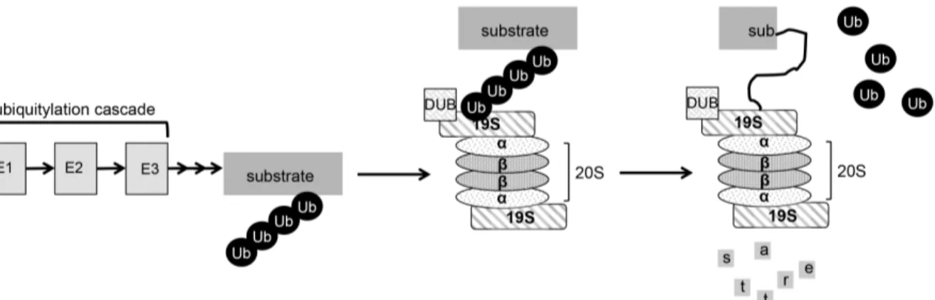
is given in Figure 3.
FIGURE 3: Schematic representation of the UPS pathway of degradation. Sizes of the individual components are not proportionate. A substrate is polyubiquitinated via an enzymatic cascade analogous to the sumoylation cascade presented in Fig. 2. Thereby, it is targeted to the 26S proteasome, a barrel-shaped protease consisting of multiple subunits. Before fed into the proteolytic core (20S), deubiquitinating enzymes dismantle the ubiquitin chain and the 19S regulatory particle unfolds the substrate protein.
The degradation signal is ubiquitin, a highly conserved 76-amino acid protein (Goldknopf et al. 1977). Ubiquitinated proteins are recognized by receptors in the 19S particle. Proteasome- attached DUBs remove ubiquitin from the target protein, before it is unfolded by ATPases in the 19S base. From there, the polypeptide is translocated into the 20S core for destruction.
Three different proteolytic activities act there: Chymotryptic-, tryptic- and caspase-like activities attack the target, and typically cleave it into peptides of 4-25 amino acids (Voges et al. 1999; Geng et al. 2012; Groll et al. 2000; Köhler et al. 2001a; Köhler et al. 2001b). The ubiquitin molecules liberated before the destruction process add to the pool of free ubiquitin competent to be used in another round of target protein ubiquitination.
1.8 SUMO-targeted ubiquitin ligases
The SUMO- and the ubiquitin pathways maintain a certain level of crosstalk. For long, it has been thought that this was of purely antagonistic nature (Ulrich 2005). Therefore, it came as a surprise when it was found that the two also work hand in hand. SUMO-targeted ubiquitin ligases (STUbLs) [also known as ubiquitin ligases for sumoylated proteins (ULS)] recognize poly-SUMO chains and decorate them with ubiquitin (Uzunova et al. 2007; Perry et al. 2008;
Lallemand-Breitenbach et al. 2008). This files the target protein in the UPS pathway of degradation (Praefcke et al. 2012; Sriramachandran and Dohmen 2014; Uzunova et al. 2007;
Perry et al. 2008; Heideker et al. 2009; Geoffroy et al. 2009; Hunter et al. 2009). In the last decade, several STUbLs have been identified by the presence of SIMs, their interaction with SUMO, or via homology studies (Weisshaar Stefan R. 2008; Sun et al. 2007; Prudden et al.
2007; Xie et al. 2007; Køhler et al. 2015; Westerbeck et al. 2014). The proteins distinguish
themselves by combining two features in their structures: A RING-finger domain (variants of zinc-fingers that coordinate two zinc atoms in a cross-brace structure; mediates protein- protein interaction (Borden et al. 1995; Saurin et al. 1996)) that allows for interaction with a ubiquitin-conjugating enzyme, and several SIMs. The former characterizes them as ubiquitin ligases, the latter as SUMO-binding proteins (Sriramachandran and Dohmen 2014; Uzunova et al. 2007; Perry et al. 2008). The existence of more than one SIM in STUbLs enables them to selectively ubiquitinate target proteins that carry poly-SUMO chains (Hannich et al. 2005;
Uzunova et al. 2007; Lallemand-Breitenbach et al. 2008; Sun et al. 2007; Prudden et al.
2007; Xie et al. 2007; Weisshaar et al. 2008; Tatham et al. 2008). In S. cerevisiae, to date two STUbLs have been identified, named Uls1 and Uls2. Uls1 (also known as Ris1, Dis1 and Tid4) is a 1612-amino acid protein accommodating a RING finger motif, a Swi2/Snf2-like translocase domain, and four predicted SIMs (Hannich et al. 2005; Sriramachandran and Dohmen 2014; Uzunova et al. 2007). Uls2, in contrast, is a heterodimer consisting of Slx5 and Slx8. Slx5 (synthetic lethal of unknown function 5; also known as Hex3) contains two SIMs and a RING finger domain (Uzunova et al. 2007; Xie et al. 2007; Ii et al. 2007; Wang et al. 2006). Aberrant levels of the human STUbL Rnf4 have been implicated in a number of diseases including cancer (Perry et al. 2008; Pero et al. 2001). This highlights the importance of STUbLs for cellular homeostasis.
In summary, linking ubiquitin and SUMO pathways, STUbLs represent another regulatory mechanism for levels of sumoylated forms of a substrate.
1.9 Desumoylating enzymes
Posttranslational modification in general offers the opportunity to provoke much faster responses than regulation via transcription alteration. Sumoylation is not an exception: It provokes an immediate cellular response to various intracellular signals and extracellular stimuli. Obviously, for this regulatory mechanism detachment of the modification is as important as conjugation. Deconjugation is carried out by specific enzymes broadly referred to as SUMO proteases. They cleave between the terminal Gly of SUMO and the Lys residue on the substrate it is attached to. Some SUMO proteases are also capable of processing SUMO precursors, thereby making it competent for conjugation in the first place.
SUMO proteases are not only responsible for maturation of nascent SUMO, and
deconjugation of the modifier from its target, but by doing so they also regulate the pool of
conjugation-competent SUMO in the cell (Xu et al. 2009). All known SUMO-specific
proteases are cysteine proteases featuring a papain-like proteinase fold (Hickey et al. 2012).
Although they share some catalytic features, the different superfamilies distinguish
themselves by the specific fold of their catalytic domains (Hickey et al. 2012). All SUMO- specific peptidases of the Ulp/SENP superfamily known to date belong to the C48 family of thiol proteases (Hickey et al. 2012; Shin et al. 2012; Schulz et al. 2012).
The first SUMO-specific protease to be described was the budding yeast enzyme ubiquitin- like protein-specific protease 1 (Ulp1) (Li et al. 1999). Shortly thereafter, sequence
comparison of its catalytic domain to databases uncovered the existence of a second protease of that kind in S. cerevisiae, Ulp2 (Li et al. 2000a), as well as the first mammalian SUMO protease SENP1 (Gong et al. 2000), and many others in various organisms (Mukhopadhyay and Dasso 2007).
1.9.1 Structures of SUMO proteases and catalytic mechanism
In 2001, the first structure of a SUMO-specific protease was solved. It was the catalytic domain of Ulp1 in complex with Smt3 trapped in a transition-state-like conformation (Mossessova et al. 2001). The high-resolution insight into its active site confirmed Ulp1 to belong to the cysteine proteases, as its sequence similarity to the adenovirus processing protease (AVP) had previously suggested (Li and Hochstrasser 1999). The structure revealed that Ulp1 makes contact with Smt3 via an extensive surface area, and that unique hydrophilic interactions and a high number of salt bridges account for its specificity for SUMO. Notably, Ulp/SENP proteases bind SUMO via surfaces distinct from SIMs (Hickey et al. 2012;
Mossessova and Lima 2001). The active-site pocket harbors the catalytic triad (His-Asp-Cys) and an additional fixed Gln residue in close proximity, which stabilizes the transition state during catalysis (Mossessova and Lima 2001; Reverter et al. 2004, 2006). The SUMO C- terminal diglycine motif is positioned in a shallow tunnel formed by the side chains of two Trp residues. This tunnel is so narrow that insertion of any residue other than glycine is sterically hindered (Mossessova and Lima 2001). Structures of SENP1 and SENP2 in complex with SUMO or sumoylated substrates showed many similarities, including the Trp tunnel and SUMO-enzyme interfaces (Reverter and Lima 2004, 2006; Shen et al. 2006a;
Shen et al. 2006b). The structures of SENP1 and -2 bound to real substrates revealed that the
scissile bond (= bond that is subject to cleavage) is oriented in a cis configuration with
respect to the amide nitrogens for cleavage. It is kinked in a right angle to the C-terminus of
SUMO (Reverter and Lima 2006; Shen et al. 2006a). If in trans, the nitrogen atoms would
cause a clash between the residue C-terminal to the scissile peptide bond and the active site
loop of the protease. A cis arrangement positions the carbonyl group of the scissile bond in an optimal position for catalysis (Hay 2007).
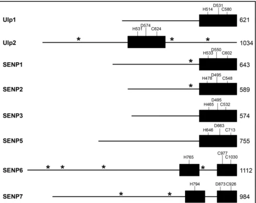
FIGURE 4: Domain architecture of Ulp/SENP family members. Black boxes = catalytic domains; line = non-catalytic domain; residues of catalytic triad are marked in one-letter code; total length of protease (in amino acids) is given on the far right of every schematic. Putative SIMs are indicated by asterisk (*).
Unlike other members of the Ulp/SENP family, Ulp2, SENP6 and SENP7 carry large loop insertions within their catalytic domains (Hickey et al. 2012) (Fig.4). SENP6 and SENP7 both have four of these. Resolution of the structure of the active domain of SENP7 showed that its loop1, which is not conserved in Ulp2, projects towards the putative SUMO binding interface, and mutant analysis indicated that it is decisive for SUMO paralogue specificity (Lima et al. 2008). So far, no structure information is available for SENP6.
1.9.2 Regulation of SUMO proteases
SUMO proteases are regulated in a number of different ways. Firstly, localization limits their
activity radius (Kolli et al. 2010). Secondly, transcriptional regulation controls the abundance
of e.g. specific SENPs. For instance, the promoter region of SENP1 contains androgen
response elements as well as a hypoxia response element, thus is stimulated under such
conditions. Additionally, it has been proposed that SENP1 itself triggers its own transcription
via a positive feedback loop (Bawa-Khalfe et al. 2007; Xu et al. 2010; Cheng et al. 2007).
SENP2, in contrast, participates in a negative feedback loop (Lee et al. 2011). Thirdly, SUMO proteases are frequently subject to post-translational modification, influencing their activity and/or stability. For example, under normal conditions, SENP3 is ubiquitinated and turned over via the UPS. Upon oxidative stress, its ubiquitination is attenuated (Kuo et al.
2008; Yan et al. 2010). Ulp2, in turn, is subject to cell cycle-dependent phosphorylation (Baldwin et al. 2009). Fourthly, protease activity can be directly affected by specific cellular stresses. To name just one, heat shock was found to render several SENPs irreversibly inactive (Pinto et al. 2012). Another very interesting activity-regulation mechanism has been suggested: SENP1 has been found to dimerize via an intersubunit disulphide bond in
response to oxidative stress (Xu et al. 2008). Since the active-site cysteine was involved, the authors of that study proposed that the transient disulphide bond might protect the enzyme from irreversible oxidation of the catalytic cysteine residue.
Last but not least, shielding may play a role for the rate of desumoylation of a given substrate. Still not completely understood, substrate binding by other, competing cellular proteins can hinder SUMO proteases to process a target (Hickey et al. 2012; Xu et al. 2005).
1.9.3 Misregulated SUMO proteases
In the last decade, the SUMO cycle has attracted a considerable degree of attention since its deregulation has been associated with various pathological conditions including pathogen proliferation, neurological disorders or cancer (reviewed in e.g. (Droescher et al. 2013;
Wimmer et al. 2012; Sarge et al. 2009)). At any given time during the cell cycle, a distinctive pattern of SUMO conjugates is observed (Kolli et al. 2010). Disturbance of the crosstalk between sumoylation and other post-translational modifications (e.g. ubiquitin) can easily tip over the finely tuned levels of sumoylated proteins and thereby severely impair homeostasis (shown for specific proteins by e.g. (Song et al. 2015; Dou et al. 2010; Román González- Prieto 2015; Liu et al. 2013; Mukhopadhyay et al. 2010a)). Unsurprisingly, SUMO-specific proteases have been found to be implicated in the development of several important
conditions (Chun-Jie Huang 2015). In the last few years, misregulation of the two mammalian SUMO proteases SENP6 and SENP7 have been identified as a factor in
development of certain types of cancer and other disorders. For example, recently, elevated
SENP6 activity was reported to promote gastric cancer by liberating SUMO1 from the
transcription factor FoxM1 (Song et al. 2015). SENP6 also plays a role in toll-like receptor
(TLR) inflammatory signaling. SENP7, in turn, was found to modulate the stability of c-Myc,
an oncogene, by balancing out the STUbL pathway of degradation (Román González-Prieto 2015).
1.9.4 Proteases 1.9.4.1 In mammals
SUMO is also known as sentrin. This explains the naming of the first-discovered family of SUMO deconjugation enzymes in mammals, sentrin-specific proteases (SENPs). So far, six different, highly specific SUMO proteases have been identified: SENP1-3 and SENP 5-7 (Mukhopadhyay and Dasso 2007). They are tightly controlled, and are equipped with distinct features and biological significance in cells; accordingly, they are not redundant (Chun-Jie Huang 2015). On a side note: For some time, another protease identified in human cells was counted among them, and accordingly termed SENP8. However, SENP8 represents a special case: It is non-specific for SUMO, but, in fact, it is specific for neddylation. Neural-
precursor-cell-expressed developmentally downregulated protein-8 (NEDD8) is a Ubl and conjugated to substrates in a process called neddylation (Shin et al. 2011; Kamitani et al.
1997). Appropriately, SENP8 is often referred to as human deneddylase 1 (DEN1) or
NEDD8-specific protease (NEDP1) (Gan-Erdene et al. 2003; Wu et al. 2003; Mendoza et al.
2003). In vitro, desumoylation of specific substrates by individual SENPs is generally not strongly paralogue specific, even though the activity levels have been found to be different for some SENPs depending on the SUMO isoform they are confronted with. However, some of the SENPs have clear preferences for certain isoforms in vivo (Hickey et al. 2012).
Due to their early connection to Ulps, SENPs have been grouped into two branches: the Ulp1 branch and the Ulp2 branch. The classification is made according to the architecture of their active domains. Those SENPs featuring unconventional elements, specifically big loop insertions, fall into the Ulp2 branch, all others are assigned to the Ulp1 branch
(Mukhopadhyay and Dasso 2007; Hickey et al. 2012; Hay 2007; Nayak et al. 2014).
Most SENPs localize in the nucleus or colocalize in distinguishable subnuclear compartments (Kolli et al. 2010). Since many cytoplasmic proteins, e.g. FAK, NEMO and IκBα, have been found to be sumoylated at some point (Shin et al. 2012; Kadaré et al. 2003; Mabb et al. 2006;
Takahashi et al. 2008), it has long been speculated on how their respective SUMO modifications would be removed. Recently, a new class of desumoylation enzymes was discovered in mammalian cells and characterized to some degree: Desumoylating
isopeptidases (DeSI), of which currently two members (DeSI-1 and DeSI-2) are known (Shin
et al. 2012). They belong to the C97 family of proteases. DeSIs recognize substrates distinct from SENP targets, suggesting that they are highly selective for specific substrates. As just one example, DeSI-1 specifically desumoylates the transcription repressor BZEL (Shin et al.
2012). Interestingly, DeSI-1 and DeSI-2 have been suggested to feature only a catalytic dyad (Suh et al. 2012). However, this hypothesis requires structural analysis of a DeSI in complex with SUMO, which is not available yet. Shortly after the identification of DeSIs, a third type of SUMO proteases was found. Ubiquitin-specific protease-like 1 (USPL1), a low-abundance protein, localizes exclusively in cajal bodies, where it plays an essential role (Schulz et al.
2012). Of note, this role is separate from its catalytic activity. USPL1 belongs to the C98 family of proteases and is neither related to the Ulp/SENP-, nor to the DeSI family. However, its catalytic domain shows some homology to the C19 family of ubiquitin-specific proteases (Schulz et al. 2012). Taken together, to date there are three distinct classes of SUMO
proteases in humans. This is reminiscent of the diversification seen for deubiquitinating enzymes (DUBs), which are each filed into one of five structural classes (Komander et al.
2009). However, the paralogue diversity within the DUB superfamily exceeds by far the number of SUMO-specific enzymes, with ~100 predicted DUBs, none of these resembling Ulps or SENPs. Though, as its name indicates, USPL1 shows remote similarity to the USP class of DUBs (Schulz et al. 2012). Considering the plethora of sumoylated proteins which has already been found in mammals (Li et al. 2004; Zhao et al. 2004b; Yang et al. 2015), it is likely that there are even more SUMO protease families, yet to be identified.
1.9.4.2 In budding yeast
To date, in S. cerevisiae two SUMO-specific proteases are known, Ulp1 and Ulp2. Genetic studies with yeast strains carrying mutations in either of the two revealed distinct phenotypic defects in the absence of each protease, as well as unique sumoylation patterns in the
different strains, indicating distinct substrate specificity (Li et al. 2000b). Localization in the cell strongly influences this selectivity.
1.9.4.2.1 Ulp1
As mentioned above, the 621-amino acid enzyme Ulp1 was the first SUMO-specific protease
to be discovered. It is essential, and the temperature sensitive mutants, with whom it has been
studied in vivo, exhibit a severely sick phenotype even at permissive temperature (Li and
Hochstrasser 1999; Li et al. 2003; Soustelle et al. 2004). Despite their common classification,
Ulp1 distinguishes itself from Ulp2 by its ability to process SUMO precursors in addition to
general SUMO deconjugation (Li and Hochstrasser 1999, 2003). Unlike Ulp2, Ulp1 lends itself very well to recombinant expression in E. coli and in vitro analysis (Li and
Hochstrasser 1999; Mossessova and Lima 2001). Even high-definition structural information is available for Ulp1 –alone and in complex with Smt3 (Mossessova and Lima 2001; Xu et al. 2008). This, however, only allows limited insight into what might be true for Ulp2, since the catalytic domains of the two Ulps are only ~27 % identical, and there is no obvious similarity at all between their non-catalytic domains (Li and Hochstrasser 2000a;
Schwienhorst 2000). Despite all this information that has been collected about Ulp1, its substrate selectivity, like for Ulp2, is still poorly understood. However, as correct localization of Ulp1 seems to be highly important for cell homeostasis, its spatial sequestration might be a crucial part of that. Ulp1 localizes at the nuclear periphery, primarily to the inner surface of the nuclear pore complex (NPC) (Li and Hochstrasser 2003; Panse et al. 2003; Palancade et al. 2007; Lewis et al. 2007; Zhao et al. 2004a). Precisely, it interacts with Pse1, Kap95 and Kap60 via its NPC-targeting domain, that comprises the first 340 residues of the enzyme (Panse et al. 2003). A Ulp1 mutant, in which this domain is deleted, localizes throughout the cell (Li and Hochstrasser 2003), and causes accumulation of Rad52 foci, which indicate endogenous DNA damage and repair (Palancade et al. 2007). Notably, when this truncated version of Ulp1 is expressed in a Ulp2-deficient strain, it partially suppresses several growth defects coming along with a ULP2 knockout, and reduces the levels of sumoylated Ulp2 substrates (Li and Hochstrasser 2003). This, as well, highlights the importance of a tight restriction of Ulp1 localization in order to control its in vivo activity. Even more so, high levels of Ulp1 activity mislocalized to the nucleoplasms have been found to be lethal (Li and Hochstrasser 2003; Panse et al. 2003). Interestingly, transiently during M-phase a fraction of Ulp1 is exported to the cytoplasm, where it desumoylates septin proteins (Takahashi et al.
2000; Elmore et al. 2011; Makhnevych et al. 2007). Septins are heavily sumoylated in a cell- cycle dependent manner and arrange into filaments at the bud neck of dividing cells
(Takahashi et al. 2008). The details of this temporary Ulp1 delocation still await their discovery.
1.9.4.2.2 Ulp2
The second known SUMO-specific protease in S. cerevisiae, Ulp2, is far less studied than Ulp1. At least in parts, this is because this 1034-amino acid protein is not easy to
recombinantly express and has exhibited very weak enzymatic activity in vitro in previous
studies (Li and Hochstrasser 2000b). This is just one of many features it shares with its mammalian orthologs SENP6 and SENP7 (Lima and Reverter 2008; Drag et al. 2008). In vivo, Ulp2 has been studied to a much larger extent.
1.9.4.2.2.1 Ulp2 regulation and localization
Ulp2 is a low-abundance, cell cycle-regulated phospho-protein (Baldwin et al. 2009;
Strunnikov et al. 2001). It is phosphorylated during mitosis at at least two sites in the C- terminal non-catalytic domain (CTD), and this transient modification is dependent on Cdc5 and cyclin-dependent kinase 1 (Cdk1) (Baldwin et al. 2009). Indeed, Cdc5 revealed itself as a negative regulator of Ulp2 when it was found that two known Ulp2 targets, Top5 and Pds5, fail to accumulate in a sumoylated state in the absence of Cdc5 (Baldwin et al. 2009). Apart from this, Ulp2 is spatially controlled. Ulp2 localizes throughout the nucleus and
occasionally the nucleolus (Kroetz et al. 2009; Srikumar et al. 2013). Its nuclear localization is critical for most, if not all, functions of the protease (Kroetz et al. 2009). A strain in which Kap95, a nuclear import receptor, is mutated, is defective in nuclear import of Ulp2, which results in aberrant accumulation of poly-SUMO structures (Kroetz et al. 2009). Ulp2 binds chromatin and has been suggested to desumoylate chromatin-associated proteins such as histones and topoisomerase II (Hannich et al. 2005; Strunnikov et al. 2001; Bachant et al.
2002; Nathan et al. 2006). Overexpression of it suppresses defects in chromosome
condensation and segregation, pointing towards a role in regulating these processes (Meluh and Koshland 1995; Strunnikov et al. 2001). This function is conserved in the analogous enzymes in C. elegans and humans (Mukhopadhyay et al. 2010a; Pelisch et al. 2014).
1.9.4.2.2.2 The ULP2 knockout mutant
A ULP2 knockout mutant has a very distinct phenotype: Apart from abnormal cell
morphology and a severe growth- and sporulation defect, ulp2-∆ exhibits elevated levels of chromosome loss and delayed recovery after DNA-damage induced cell-cycle checkpoint arrest. What is more, that strain is highly sensitive to various stresses like hydroxyurea, temperature and DNA-damaging agents (Li and Hochstrasser 2000a; Schwienhorst 2000;
Strunnikov et al. 2001; Bachant et al. 2002; Schwartz et al. 2007). Another very prominent feature of the ulp2 knockout is the accumulation of high-molecular weight (HMW) SUMO conjugates, so big they hardly penetrate the resolving gel upon SDS-PAGE analysis (Bylebyl et al. 2003).
Like its mammalian orthologs SENP6 and SENP7, Ulp2 has been found to be capable of
dismantling long poly-SUMO chains (Bylebyl et al. 2003; Lima and Reverter 2008;
Mukhopadhyay et al. 2006). Indeed, previous studies established a correlation between loss of SENP6 or Ulp2 and a failure to disassemble poly-SUMO chains (Bylebyl et al. 2003;
Mukhopadhyay et al. 2006).
1.9.4.2.2.3 Ulp2 domain architecture and functions
Apart from its catalytic domain, Ulp2 has large C- and N-terminal non-catalytic domains.
Utilizing truncation mutants, the CTD has been identified as a requirement for efficient depolymerization of large poly-SUMO conjugates (Kroetz et al. 2009). However, that mutant showed an only minor growth defect, suggesting that factors other than the HMW SUMO species also account for the growth defects of ulp2-∆ cells, or that such conjugates can be tolerated to some extent. It has been suggested that, in addition to SUMO chain
deconjugation, the growth-regulating roles of Ulp2 involve removal of SUMO from specific target proteins (Kroetz et al. 2009). Very recently, a study using a quantitative mass
spectrometry approach to analyze the effects of ulp1 and ulp2 mutations on the intracellular sumoylation pattern found that Ulp2 has a highly specific desumoylation activity in vivo, while Ulp1 exhibits a broader specificity towards many substrates (de Albuquerque et al.
2016). This fortified previous findings, which indicated that Ulp2 has critical functions in the depolymerization of poly-SUMO modification on specific substrates (Bylebyl et al. 2003;
Kroetz et al. 2009).
In general, Ulp-class SUMO proteases have extensive, poorly conserved N-terminal non- catalytic domains (NTDs). These NTDs are associated with subcellular localization and regulation of activity (Hickey et al. 2012; Kroetz et al. 2009). Previous studies showed that the NTD of Ulp2 is necessary and sufficient for nuclear localization of the enzyme (Kroetz et al. 2009). It contains two short nuclear-localization signal (NLS)-like motifs, either of which alone is sufficient to sustain nuclear localization and function of Ulp2 (Kroetz et al. 2009).
Indeed, in that study it was found that a single 7-amino acid long NLS makes the entire NTD obsolete. Fusing it to a corresponding ∆NTD mutant of Ulp2 restored Ulp2 in vivo
functionality almost completely, suggesting that the low sequence conservation of the NTDs might be due to the fact that only the NLS in this part of the protein are relevant for its proper cellular function (Kroetz et al. 2009).
Both the NTD and CTD of Ulp2 are predicted to lack stable tertiary fold to large extents (Kroetz et al. 2009). Intrinsically disordered protein regions are frequently implicated in low- affinity but high-specificity protein-protein interactions (Dyson et al. 2005). Ulp2 non-
catalytic domains contain three predicted SIMs: one in the NTD, and two in the CTD (Kroetz
et al. 2009). One of the two C-terminal SIMs is located directly adjacent to the catalytic domain and has already been subject to several studies (Baldwin et al. 2009; Kroetz et al.
2009). Still, the specific function of any of the three motifs is unknown.
1.9.4.2.3 Wss1
The list of SUMO-specific proteases in budding yeast would not be complete without mentioning Wss1. Originally, weak suppressor of smt3-33 (Wss1) was identified as a suppressor of the temperature sensitive phenotype produced by an L26S SUMO mutation (Biggins et al. 2001), and has been implicated in the response to genotoxic stress (O'Neill et al. 2004).
For a short period of time, Wss1 was regarded as the possible missing link between SUMO- and ubiquitin metabolisms, as it exhibited some deubiquitination activity to ubiquitin-SUMO mixed species and depolymerized SUMO chains in vitro, and physically associated with the 26S proteasome (Mullen et al. 2010). However, these findings were under debate ever since they had been published, and further studies could not confirm the SUMO- or ubiquitin- isopeptidase activity (Su et al. 2010; Stingele et al. 2014; Balakirev et al. 2015). As it turns out, rightfully so. In 2014, Wss1 was newly classified as a DNA-dependent protease involved in repair of DNA-protein crosslinks (Stingele et al. 2014). A later study identified Wss1 as an inactive metalloprotease under normal conditions that is activated by single-strand DNA upon genotoxic stress via a cysteine-switch regulatory mechanism. Downregulation of Wss1 activity was found to be mediated by autoproteolysis. Contrasting the aforementioned study, it was shown to have a SUMO ligase-like activity and to promote poly-sumoylation of proteins with which it interacts. This includes Cdc48. Indeed, Wss1 forms a ternary complex with the AAA ATPase Cdc48 and the adaptor protein Doa1. Upon DNA damage, this complex is recruited to already sumoylated targets and promotes elongation of their SUMO chains (Balakirev et al. 2015). Taken together, Wss1 seems to be a highly interesting protein, but in terms of activity profile it is not to be named in one line with the Ulps.
1.10 Thesis objectives
The main interest of this work was to characterize Ulp2 on the level of the molecule. Data on the enzyme itself was scarce, since it was fractious as a study object. However, in order to assume structure-function relationships, this hurdle had to be overcome.
This study set out to analyze the following aspects:
1) What are the requirements for a SUMO chain to serve as a Ulp2 target?
2) How does Ulp2 process a target chain? Randomly or in an ordered manner?
3) What is the molecular/structural basis on which the Ulp2/SUMO interactions are based?
4) What is the basis of the proteolysis defect in ulp2-∆?
5) How do its preferences and mechanistic conditions influence Ulp2’s activity in vivo?
Previous work had shown that the absence of Ulp2 causes a severe proteolysis defect in S.
cerevisiae, adding up to an already highly interesting phenotype of the ulp2-∆ mutant. This
was the starting point for further functional characterization of Ulp2 in vivo, which is
addressed in the last part of this work.
2 Material and Methods
2.1 Material
2.1.1 Escherichia coli strains Tab.1: E. coli strains used in this study.
Strain Genotype/relevant genotypic characteristics Source XL1 Blue
recA1 endA1 gyrA96 thi-1 hsdR17 supE44 relA1lac [F´ proAB lacIqZΔM15 Tn10 (Tetr)]
Stratagene
BL21-CodonPlus
E. coli B F– dcm ompT hsdS(rB– mB–) gal λ(DE3)[pLysS Camr]Novagene
BL21(DE3) E. coli str. B F
–ompT gal dcm lon hsdS
B(r
B–m
B–) λ(DE3 [lacI lacUV5-T7 gene 1 ind1 sam7 nin5]) [malB
+]
K-12(λ
S)
Novagene
2.1.2 Saccharomyces cerevisiae strains Tab.2: S. cerevisiae strains used in this study.
Strain Relevant genotype Source
JD47-13C MATa leu2-∆1 trp1-∆63 his3-∆200 ura3-52 lys2-801 ade2-101
Dohmen et al. (1995)
yJE1 MATa ulp2-∆::HIS3 smt3-KallR::TRP1 This work
yJE2 MATa pdr5-∆::nat smt3-HA ulp1-I615N This work
MS84 MATa pdr5-∆::nat smt3-HA lab collection
MS87 MATa pdr5-∆::nat ulp2-∆::HIS3 smt3-HA lab collection MS91 MATa pdr5-∆::nat smt3-HA wss1::hygB lab collection
yKU1 MATa ulp2-∆::HIS3 lab collection
sul25-2 MATa ulp2-∆::HIS3 uba2-ts9 lab collection
smt3-KallR MATα smt3-KallR::TRP1 Erica Johnson
2.1.3 Plasmids
Tab.3: Plasmids used in this study.
Plasmids for yeast are described as such. For all others, the description indicates the recombinant protein(s) encoded in them. TEVp.r.s. = TEV protease recognition site; His6 = hexahistidine tag; T = terminator; P = promoter; CYC1 = cytochrome c-1; CUP1 = metallothionein.
Plasmid name Description Source
pACYC-Duet- Aos1p/Uba2p
His6-Aos1, Uba2-Stag J. Wohlschlegel
(Wohlschlegel et al. 2006) pRSF-Duet-
Ubc9/Smt3p
His6-Smt3, Ubc9-Stag J. Wohlschlegel
(Wohlschlegel et al. 2006) pRSF-Duet-
Ubc9/Smt3p- K11R,K15R,K19R
His6-Smt3(K11R,K15,K19R), Ubc9-Stag M. Schnellhardt
pHUbp41 His6-Ubp41 R. Baker
pMAF17 PCUP1-UB-R-eK-HA-Ura3-TCYC1; yeast expression plasmid M. Froehlich pMAF60 PCUP1-UB-V76-eK-DHFR-2xHA-TCYC1; yeast expression plasmid M. Froehlich PCA129 PCUP1-PRS316-stp-102-3xHA-TCYC1; yeast expression plasmid C. Andreasson pLS1 FLAG-Smt3-∆N17Smt3(M0)-3xΔN17Smt3-eGFP-HA-His6 L. Schürholz pLS2 FLAG-Smt3-∆N17Smt3(M1)-3xΔN17Smt3-eGFP-HA-His6 L. Schürholz pLS3 FLAG-Smt3-∆N17Smt3(M2)-3xΔN17Smt3-eGFP-HA-His6 L. Schürholz pLS5 FLAG-Smt3-∆N17Smt3(M4)-3xΔN17Smt3-eGFP-HA-His6 L. Schürholz pLS6 FLAG-Smt3-∆N17Smt3(M5)-3xΔN17Smt3-eGFP-HA-His6 L. Schürholz pLS7 FLAG-Smt3-∆N17Smt3(M6)-3xΔN17Smt3-eGFP-HA-His6 L. Schürholz
pJE2 FLAG-Ubiquitin-1xΔN17Smt3-eGFP-HA-His6 This work
pJE3 FLAG-Ubiquitin-2xΔN17Smt3-eGFP-HA-His6 This work
pJE4 FLAG-Ubiquitin-3xΔN17Smt3-eGFP-HA-His6 This work
pJE5 FLAG-Ubiquitin-4xΔN17Smt3-eGFP-HA-His6 This work
pJE6 FLAG-Smt3-eGFP-HA-His6 This work
pJE7 FLAG-Smt3-ΔN17Smt3-eGFP-HA-His6 This work
pJE8 FLAG-Smt3-2xΔN17Smt3-eGFP-HA-His6 This work
pJE9 FLAG-Smt3-3xΔN17Smt3-eGFP-HA-His6 This work
pJE10 FLAG-Smt3-4xΔN17Smt3-eGFP-HA-His6 This work
pJE12 MBP~TEVp.r.s.~Ulp2-FLAG This work
pJE14 FLAG-eGFP-HA-His6 This work
pJE18 MBP~TEVp.r.s~Ulp1-FLAG This work
pJE20 FLAG-Smt3(G98A)-4xΔN17Smt3-eGFP-HA-His6 This work
pJE22 FLAG-5xΔN17Smt3-eGFP-HA-His6 This work
pJE23 MBP~TEVp.r.s~Ulp2(C624A)-FLAG This work
pJE26 FLAG-Smt3(G98A)-ΔN17Smt3(G98A)-3xΔN17Smt3-eGFP- HA-His6
This work
pJE28 FLAG-Smt3(G98A)-His6 This work
pJE30 FLAG-Smt3(G98A)-ΔN17Smt3(G98A)-His6 This work
pJE31 FLAG-Smt3(G98A)-2xΔN17Smt3(G98A)-His6 This work
pJE32 FLAG-MBP-4xΔN17Smt3-eGFP-HA-His6 This work
pJE33 FLAG~TEVp.r.s.~Smt3-4x∆N17Smt3-eGFP-HA-His6 This work
pJE34 FLAG-SUMO1-4xΔN17Smt3-eGFP-HA-His6 This work
pJE37 FLAG-2xUbiquitin-4xΔN17Smt3-eGFP-HA-His6 This work
pJE38 FLAG-3xUbiquitin-4xΔN17Smt3-eGFP-HA-His6 This work
pJE42 FLAG-SUMO2-4xΔN17Smt3-eGFP-HA-His6 This work
pJE43 FLAG-Smt3(G98A)-Ubiquitin-3xΔN17Smt3-eGFP-HA-His6 This work
pJE44 FLAG-Smt3(G98A)-SUMO2-3xΔN17Smt3-eGFP-HA-His6 This work
pJE45 FLAG-Smt3(M0)-4xΔN17Smt3-eGFP-HA-His6 This work
pJE50 FLAG-Smt3(M2)-4xΔN17Smt3-eGFP-HA-His6 This work
pJE51 FLAG-Smt3(MEnd)-4xΔN17Smt3-eGFP-HA-His6 This work
pJE54 FLAG-Smt3-ubiquitin-3xΔN17Smt3-eGFP-HA-His6 This work