The role of caseinolytic mitochondrial matrix peptidase proteolytic subunit (CLPP)
in regulation of
mitochondrial ribosome biogenesis in mammals
Inaugural–Dissertation
zur
Erlangung des Doktorgrades
der Mathematisch-Naturwissenschaftlichen Fakultät der Universität zu Köln
vorgelegt von
Priyanka Maiti
aus Kolkata, India
Köln 2015
Berichterstatter: Prof. Dr. Aleksandra Trifunovic Prof. Dr. Thomas Langer
Tag der mündlichen Prüfung: 15.06.2015
To My Beloved Family and Friends…
Table of Contents
Table of Contents ... iv
List of Figures ... vii
List of Tables ... ix
Abbreviations ... x
Abstract ... xiii
Zusammenfassung ... xv
1. Introduction ... 1
1.1. Mitochondria ... 1
1.2. Mitochondrial Genetics ... 3
1.3. Oxidative Phosphorylation (OXPHOS) system ... 4
1.4. Replication of mtDNA ... 8
1.5. Mitochondrial transcription ... 9
1.6. Mitochondrial translation ... 10
1.7. Mitochondrial quality control (MQC) ... 15
1.7.1 MQC in the matrix: ... 18
1.7.2 MQC in the IMM: ... 20
1.7.3 MQC in the IMS: ... 23
1.7.4 MQC in the OMM: ... 24
1.7.5 Mitochondrial dynamics -‐ fusion & fission: ... 25
1.7.6 Mitochondrial autophagy (Mitophagy): ... 26
1.7.7 Mitochondrial derived vesicles (MDV): ... 28
1.7.8 Apoptosis: ... 29
1.8. CLPXP: ... 31
1.9. Objectives: ... 35
2. Materials and Methods ... 36
2.1 Mouse Experiments ... 36
2.1.1 Animal Care ... 36
2.1.2 Mouse handling and breeding ... 36
2.1.3 Mice-‐ Genetic ablation of Clpp gene by homologous recombination ... 36
2.1.4 Body weight ... 37
2.1.5 Analysis of body composition (NMR) ... 37
2.1.6 Food intake and indirect calorimetry ... 37
2.1.7 Determination of blood glucose and lactate levels ... 37
2.1.8 Glucose Tolerance Test ... 37
2.1.9 Insulin Tolerance Test ... 38
2.1.10 Measurement of rectal body temperature ... 38
2.2 Molecular Biology ... 38
2.2.1 Isolation of genomic DNA from mice tails ... 38
2.2.2 Isolation of genomic DNA from mice tissues ... 38
2.2.3 Isolation of total RNA from mice tissues ... 39
2.2.4 Quantification of nucleic acids ... 39
2.2.5 Polymerase chain reaction (PCR) ... 39
2.2.6 Southern blot analysis for mitochondrial DNA (mtDNA) quantification ... 40
2.2.7 Northern blot analysis for mRNA, rRNA and tRNA levels ... 41
2.2.8 Reverse transcriptase PCR (Gene expression analysis) ... 42
2.3 Biochemistry ... 44
2.3.1 Isolation of proteins from tissues ... 44
2.3.2 Isolation of mitochondria from tissues except skeletal muscle ... 44
2.3.3 Isolation of mitochondria from skeletal muscle ... 45
2.3.4 Purification of mitochondria ... 45
2.3.5 Blue Native polyacrylamide gel electrophoresis (BN-‐PAGE) and in-‐gel activity of respiratory chain complexes I and IV ... 45
2.3.6 Western blot analysis ... 46
2.3.7 Measurement of the respiratory chain complex activity ... 47
2.3.8 Measurement of the rate of oxygen consumption ... 47
2.3.9 Analysis of de novo transcription and translation in isolated mitochondria ... 48
2.3.10 In cello translation in mouse embryonic fibroblasts (MEFs) ... 49
2.3.11 tRNA aminoacylation analysis ... 49
2.3.12 Analysis of mitoribosomes and RNA using sucrose density ultracentrifugation 50 2.4 Cell culture ... 50
2.4.1 Preparation of primary mouse embryonic fibroblasts and immortalization ... 50
2.4.2 Immunostaining ... 51
2.5 Computational analysis ... 52
2.5.1 Software ... 52
2.5.2 Statistical analysis ... 52
2.6 Chemicals and biological material ... 52
3. Results ... 55
3.1. Clpp (caseinolytic peptidase, ATP dependent proteolytic subunit) knockout mice are smaller than littermates and not born in Mendelian proportion ... 55
3.2. Clpp knockout mice have reduced body fat content, enhanced energy expenditure and less ambulatory activity. ... 60
3.3. Clpp knockout mice have an increase in respiratory quotient (RQ) during night, improved glucose tolerance and higher insulin sensitivity. ... 62
3.4. Clpp knockout mice age 12-‐15 weeks have reduced body fat content, enhanced energy expenditure and less ambulatory activity. ... 64
3.5. Characterization of mitochondrial proteome revealed oxidative phosphorylation (OXPHOS)-‐respiratory chain, energy metabolism, mitochondrial transcription and translation processes to be primarily affected in Clpp knockout mice. 69 3.6. CLPP deficiency leads to a specific decrease in Complex I activity, followed by a decrease in Complex IV activity later in life. ... 70
3.7. Loss of CLPP leads to increase in transcription followed by increased steady state levels of mtDNA transcripts ... 73
3.8. Loss of CLPP leads to impaired mitochondrial protein synthesis accompanied by increased levels of small ribosomal subunits, thereby affecting the stoichiometry of proper functioning ribosomes ... 76
3.9. tRNA acetylation is not affected in absence of CLPP in heart ... 79
3.10. Identification of CLPP candidates with a possible role in mitochondrial translation ... 80
3.11. A possible role of CLPP in processing of RNA transcripts ... 84
3.12. ERAL1 and P32 interacts with the mitoribosomes ... 85
3.13. Clpp deficiency leads to impairment of 12S rRNA assembly into monosomes leading to lower loading of mitochondrial mRNAs. ... 87
3.14. Investigation of phenotype in Clpp knockout MEFs revealed lower levels of assembled respiratory chain supercomplexes. ... 89
4. Discussion ... 93
References ... 102
Acknowledgements ... 115
Erklärung ... 120
List of Figures
Figure 1.1 Map of human mitochondrial DNA (mtDNA). ... 4
Figure 1.2 OXPHOS system. ... 6
Figure 1.3 Model of the supramolecular structure of the OXPHOS system. ... 7
Figure 1.4 The mtDNA replication machinery. ... 9
Figure 1.5 Model for the initiation phase of mitochondrial translation. ... 12
Figure 1.6 Model for the elongation phase of mitochondrial translation. ... 13
Figure 1.7 Model for the termination and ribosome recycling phases of mitochondrial protein synthesis. ... 15
Figure 1.8 Biochemical stresses that challenge normal mitochondrial function. ... 16
Figure 1.9 Quality control (QC) surveillance of mitochondria. ... 18
Figure 1.10 MQC in the matrix. ... 20
Figure 1.11 MQC in the IMM... 22
Figure 1.12 MQC in the IM and intermembrane space (IMS). ... 23
Figure 1.1.13 MQC in the OMM. ... 24
Figure 1.14 Mitochondrial dynamics in mitochondrial quality control (MQC). ... 26
Figure 1.15 Mitochondrial Autophagy (mitophagy). ... 28
Figure 1.16 Mitochondrial derived vesicles. ... 29
Figure 1.17 The role of mitochondria in apoptosis. ... 30
Figure 1.18 Cartoon model of substrate recognition and degradation by the ClpXP protease. ... 32
Figure 3.1 Disruption of Clpp in the germline ... 55
Figure 3.2 Confirmation of disruption of Clpp in mice ... 56
Figure 3.3 Phenotypic characterization of CLPP deficiency mouse ... 57
Figure 3.4 Decreased fat mass in Clpp knockout (-/-) mice. ... 60
Figure 3.5 Increased energy expenditure in Clpp knockout (-/-) mice. ... 61
Figure 3.6 Less activity in Clpp knockout (-/-) mice. ... 62
Figure 3.7 Increase in respiratory quotient (RQ) in Clpp knockout (-/-) mice. ... 63
Figure 3.8 Increase in respiratory quotient (RQ), improved glucose tolerance and enhanced insulin sensitivity in Clpp knockout (-/-) mice. ... 64
Figure 3.9 Decreased fat mass, increased energy expenditure in Clpp knockout (-/-) mice.
... 65
Figure 3.10 Less activity in Clpp knockout (-/-) mice. ... 66
Figure 3.11 Quantitative mitochondrial proteome profiling using LC-ESI-MS/MS ... 70
Figure 3.12 Loss of CLPP causes mitochondrial dysfunction in Clpp knockout mice .... 71
Figure 3.13 Lower levels of Complex I and Complex IV in Clpp knockout mice. ... 73
Figure 3.14 Steady state levels of mtDNA in heart of Clpp knockout mice ... 74
Figure 3.15 Steady state levels of mitochondrial transcripts in heart of Clpp knockout mice ... 75
Figure 3.16 In organello transcription in heart of Clpp knockout mice ... 76
Figure 3.17 Deregulated protein synthesis in Clpp knockout heart, SkM and liver. ... 77
Figure 3.18 Biogenesis of small ribosomal subunits in Clpp knockout heart ... 78
Figure 3.19 Increase in levels of charged and uncharged forms of tRNAs in absence of CLPP in heart and liver. ... 80
Figure 3.20 Selected substrates and partners of CLPP involved in mitochondrial translation and RNA processing identified in Clpp knockout (-/-) MEFs. ... 82
Figure 3.21 Steady state levels of potential candidates at RNA and protein levels ... 83
Figure 3.22 Stabilization of potential candidates ... 84
Figure 3.23 Accumulation of precursors of mRNAs and tRNAs in absence of CLPP .... 85
Figure 3.24 Substrates and interactors of CLPP are associated with mitochondrial ribosomes. ... 86
Figure 3.25 Substrates and interactors of CLPP likely to be involved in the assembly of 12S RNA into the small ribosomal subunit thereby affecting the function of monosomes ... 88
Figure 3.26 Phenotypic characterization of Clpp knockout MEFs ... 92
List of Tables
Table 2.1 Genotyping PCR primer sequences ... 40
Table 2.2 Probes used for quantitative real time PCR ... 43
Table 2.3 SYBR Green probes used for quantitative real time PCR ... 43
Table 2.4 Primary antibodies used for Western blot ... 47
Table 2.5 Chemicals used and suppliers ... 52
Abbreviations
3’ three prime end of DNA sequence 5’ five prime end of DNA sequence
2D two-dimensional
A adenosine
ADP adenosine diphosphate ATP adenosine triphosphate BAT brown adipose tissue
bp base pairs
BN blue native
C cytosine
cDNA complementary DNA
Ci Curie
Cre bacteriophage P1 derived site-specific recombinase COX cytochrome c oxidase
CLPP caseinolytic mitochondrial matrix peptidase proteolytic subunit Cyt cytochorome
Da Dalton
DAPI 4,6-diamidino-2-phenylindole ddH2O double distilled water
DNA desoxyribonucleic acid
dNTP desoxyribonucleotide-triphosphate ECL enhanced chemoluminiscence EDTA ethylendiamine tetraacetate
EF-G1mt mitochondrial elongation factor G1 EGTA ethylene glycol tetraacetic acid ERAL1 Era G-protein like 1
EtBr ethidium bromide ETC Electron transport chain ETS Electron transfer system EtOH ethanol
g gram
G guanine
H2O2 hydrogen peroxide HCl hydrochloric acid
HEPES N-2-hydroxyethylpiperazine-N-2-ethansulfonic acid i.e. id est
IMM inner mitochondrial membrane IMS inter membrane space
k kilo
KCl potassium chloride KOH potassium hydroxide
l liter
L loxP flanked
LSU large subunit
m milli
M molar
MAD mitochondria associated degradation MgCl2 magnesium chloride
mtDNA mitochondrial DNA mRNA messenger RNA
MQC mitochondrial quality control
MTERF mitochondrial transcription termination factor MTS mitochondrial targeted sequence
nDNA nuclear DNA
NLS nuclear localization signal NaCl sodium chloride
NaF sodium fluoride
NAH2PO4 monosodium phosphate NaHCO3 sodium bicarbonate NaOH sodium hydroxide
OMM outer mitochondrial membrane OXPHOS oxidative phosphorylation
PAGE polyacrylamide gel electrophoresis
PBS phosphate buffered saline PCR polymerase chain reaction PINK1 PTEN-induced putative kinase 1
Pi Phosphates
RC respiratory chain RNA ribonucleic acid rRNA ribosomal RNA RNase ribonuclease
ROS reactiove oxygen species Rpm revolutions per minute RT room temperature
rtPCR reverse transcription polymerase chain reaction RQ respiratory quotient
SDS sodiumdodecylsulfate SEM standard error of the mean SSU small subunit
TBE tris-borate-EDTA buffer TE tris-EDTA buffer
TFAM mitochondrial transcription factor A TFB1M mitochondrial transcription factor B1 TFB2M mitochondrial transcription factor B2
Tris 2-amino-2-(hydroxymethyl)-1,3-propandiole tRNA transfer RNA
TWEEN polyoxethylene-sorbitan-monolaureate
U units
UPRmt mitochondrial unfolded protein response
V volt
v/v volume per volume w/v weight per volume β-me β-mercaptoethanol
µl microliter
Abstract
CLPP (caseinolytic mitochondrial matrix peptidase proteolytic subunit) is a highly conserved serine protease. Molecular and structural studies in E. coli and other prokaryotes have revealed CLPP specific substrates and the mechanisms underlying their identification and subsequent degradation. These studies showed that ClpXP is involved in DNA damage repair, stationary-phase gene expression, and ssrA-mediated protein quality control. Similarly, diverse roles for the eukaryotic CLPP have been suggested. In the filamentous fungus Podospora anserine Clpp depletion promotes longevity. In Caenorhabditis elegans it has been demonstrated that CLPP have a central role in mediating the UPRmt signals. Loss of function CLPP mutations in humans cause Perrault syndrome that results in ovarian failure and sensorineural hearing loss accompanied with shorter stature. Despite this we still have a very limited knowledge about the functional role of eukaryotic CLPP, its specific substrates and underlying molecular mechanism.
In order to decipher the in vivo role of CLPP in mammals we have developed a CLPP deficient mouse model (Clpp-/-). Interestingly, only about half of Clpp knockout mice according to Mendelian proportion (12,5%) are born from intercrossing of Clpp+/- mice.
These mice are infertile and born ~ 30% smaller than littermates. CLPP deficient mice faithfully replicate the phenotypes observed in human patients. On the molecular level CLPP deficiency leads to an early specific decrease in Complex I activity, followed by a decrease in Complex IV activity later in life. Furthermore, we observed a decrease in mitochondrial translation, which is compensated for by upregulation of mitochondrial transcription. This suggests a direct or indirect role of CLPP in the process of mitochondrial protein synthesis. Gradient sedimentation analysis demonstrates an increase in the steady state levels of small ribosomal subunits, while large ribosomal subunits and monosomes are present in almost normal levels. We also observed an impairment of 12S rRNA assembly into monosomes leading to lower loading of mt- mRNAs. This indicates complications in the function of monosomes. Search for CLPXP substrates and interactors revealed two candidates that are likely to be involved in this process. We show that ERAL1 is one of the substrates of CLPP that is likely causing defective 12S rRNA assembly into the small ribosomal subunit. Additionally, p32, a CLPP interactor is permanently bound to the mitoribosomes. We believe that through
interaction with CLPXP, these proteins are involved in resolution of stalled ribosomes.
We are currently working further on elucidating the molecular mechanism underlying impaired mitochondrial translation.
Zusammenfassung
Die Casein abbauende Peptidase P (caseinolytic mitochondrial matrix peptidase proteolytic subunit, CLPP) ist eine hoch konservierte Serinprotease. Molekulare und strukturelle Untersuchungen in E. coli und anderen Prokaryoten haben CLPP-spezifische Substrate identifiziert und die zugrunde liegenden Mechanismen der nachfolgenden Degradierung dieser Substrate aufgezeigt. Diesen Studien zufolge ist ClpXP bei der Reparatur von DNA-Schäden, in der Genexpression der stationären Phase sowie in der ssrA-vermittelten Proteinqualitätskontrolle involviert. In ähnlicher Weise wurden verschiedene Rollen für die eukaryotische CLPP postuliert. Im filamentösen Pilz Podospora anserine förderte die Depletion von CLPP die Langlebigkeit. In Caenorhabditis elegans wurde gezeigt, dass CLPP eine zentrale Rolle in der Vermittlung der Signale in der mitochondrialen ungefalteten Proteinantwort (mitochondrial unfolded protein response, UPRmt) spielt. Mutationen, die den Verlust der Funktion von CLPP zur Folge haben, verursachen beim Menschen das Perrault Syndrom, das durch Gonadendysgenesie und Innenohrschwerhörigkeit, assoziiert mit Minderwuchs, charakterisiert ist. Trotz dieser Bemühungen ist unser Wissen über die funktionelle Rolle der eukaryotischen CLPP, deren Substrate und über die zugrunde liegenden Mechanismen ihrer Regulation nur sehr begrenzt.
Um die in vivo Rolle von CLPP in Säugern zu entschlüsseln, haben wir ein CLPP- defizientes Mausmodell (Clpp-/-) entwickelt. Interessant ist, dass nur etwa die Hälfte Clpp Knockout-Mäuse nach den Mendelschen Regeln (12,5%) nach der Kreuzung von heterozygoten Clpp +/- Mäuse geboren wird. Diese Mäuse sind unfruchtbar und etwa 30% kleiner ist als die nicht betroffenen Wurfgeschwistern. CLPP-defiziente Mäuse replizieren getreu die bei den menschlichen Patienten beobachteten Phänotypen. Auf molekularer Ebene führt der Verlust von CLPP zu einer spezifischen Abnahme der Komplex I-Aktivität im frühen Alter, gefolgt von einer Abnahme der Komplex IV- Aktivität in späteren Lebensabschnitten. Ferner beobachteten wir eine Abnahme in der mitochondrialen Translation, die durch eine Hochregulation der mitochondrialen Transkription kompensiert wird. Dies lässt auf eine direkte oder indirekte Rolle von CLPP im mitochondrialen Proteinsyntheseprozess vermuten. Die Gradientensedimentationanalyse zeigt einen Anstieg der kleinen ribosomalen
Untereinheiten, während die großen ribosomalen Untereinheiten und die Monosomen in fast normalen Werten vorhanden sind. Wir beobachteten auch eine Beeinträchtigung der Assemblierung der 12S rRNA in das Monosom, was zu einer verringerten Beladung der mitochondrialen mRNAs führt. Dies weist auf Komplikationen in der Funktion der Monosomen hin. Die Suche nach den ClpXP-Substraten und möglichen Interaktionspartnern ergab zwei Kandidaten, die wahrscheinlich in diesem Prozess involviert sind. Wir konnten zeigen, dass ERAL1 eines der Substrate von CLPP ist, das wahrscheinlich die defekte Assemblierung der 12S rRNA in die kleine ribosomale Untereinheit verursacht. Ferner scheint p32 ein CLPP-Interaktionspartner zu sein, der fest an den mitochondrialen Ribosomen gebunden ist. Wir glauben, dass diese Proteine durch die Interaktion mit ClpXP bei der Auflösung der ins Stocken geratenen Ribosomen beteiligt sind. Zurzeit arbeiten wir weiter an der Aufklärung der molekularen Mechanismen, die die mitochondriale Translation beeinträchtigen.
1. Introduction
1.1. Mitochondria
Mitochondria are small, highly specialized membrane enclosed organelles present in most eukaryotic cells. The word mitochondrion is derived from Greek words mitos (thread) and chondros (granule) and known as the “energy powerhouse of the cell” 1,2. Mitochondria possess its own genome and this is explained by the widely accepted endosymbiotic theory 3. The endosymbiotic hypothesis (‘endo-’ for internal, and
‘symbiont’ as in a partner in a mutually beneficial relationship) postulates that mitochondrion evolved from within the bacterial phylum a-proteobacteria via symbiosis within a eukaryotic host cell that happened over two billion years ago. Further phylogenetic reconstruction pointed specifically towards the Rickettsiaceaea family to be most closely related to mitochondria 4. These phylogenetic studies have revealed that a number of ancestral bacterial genes have been transferred to nuclear genome (endosymbiotic gene transfer) resulting in reduction and compaction of mitochondrial genome 5. However recent genomics, proteomics and energy metabolism studies have suggested two endosymbiotic models for the origin of mitochondria. The first model
“archezoan scenario” states, “the host of the proto-mitochondrial endosymbiont was amitochondrial eukaryote, termed archezoan” 6,7. The archezoan scenario is most closely related to the widely accepted endosymbiont hypothesis of mitochondrial origin 3,8. The second model “symbiogenesis scenario” states, “a single endosymbiotic event took place that involved the uptake of an a-proteobacterium by an archaeal cell leading to generation of mitochondria,” subsequently followed “by the evolution of the nucleus and compartmentalization of the eukaryotic cell” 6,7.The “hydrogen hypothesis” which is the best example of symbiogenesis scenario states that eukaryotes evolved “through symbiotic association of an anaerobic, strictly hydrogen-dependent, strictly autotrophic archaebacterium (the host) with a eubacterium (the symbiont) that was able to respire, but generated molecular hydrogen as a waste product of anaerobic heterotrophic metabolism.
The host’s dependence upon molecular hydrogen produced by the symbiont is proposed to be the selective principle that forged the common ancestor of eukaryotic cells 6,9. This hydrogen hypothesis postulates that the origins of the eukaryotic lineage and that of the
symbiont are identical and the features of eukaryotic cell evolved after the symbiosis of the eubactieruim 6. Whereas archezoan scenario hypothesize that endosymbiosis of a- proteobacterium giving rise to proto-mitochondrion happened after the formation of amtiochondriate eukarytic cell that served as the host. However the question still remains open regarding the nature of the cell that served as the host for the endosymbiont and evolved into mitochondria. With time more comparative genomics data will gradually refine the current ideas regarding the origin of eukaryotes and mitochondrial evolution.
Mitochondria are known as intracellular “power plants” generating ATP for the sustenance of life. They form a dynamic network that is regulated by constant fusion and fission. A typical eukaryotic cell contains about 2000 mitochondria, which comprises about one fifth of its total volume 1.
Mitochondria are characterized by two specialized membranes, the outer (OMM) and inner mitochondrial membrane (IMM) forming two compartments, the inter membrane space (IMS) and the mitochondrial matrix. The outer membrane is permeable to small molecules and ions that pass through transmembrane channels comprising of integral membrane proteins called porins. Larger molecules are brought across the outer membrane by the translocase of outer membrane (TOM). The inner membrane is highly impermeable to H+ ions and this property of IMM forms the basis of mitochondrial energy transduction 10. Compounds, ions and molecules cross inner membrane through translocase of inner membrane (TIM). The inner membrane is further folded to form cristae (Latin for crest or plume) providing an increase amount of surface area harboring the respiratory complexes and the ATP synthase complex, which control the basic rates of cellular metabolism 1,10. The innermost space that is enclosed by inner membrane is known as mitochondrial matrix. Various metabolic processes like the b oxidation and tricarboxylic acid (TCA/Krebs cycle) take place in the mitochondrial matrix. In addition this compartment also contains several copies of mitochondrial genome (mtDNA), ribosomes, transfer RNAs (tRNAs), and various proteins and enzymes required for proper mitochondrial function.
Mitochondria produces energy currency ATP by the oxidative phosphorylation system (OXPHOS) situated in the inner mitochondrial membrane. In addition to energy production, mitochondria are involved in various important processes such as the first step of iron-sulfur (Fe-S) cluster biosynthesis, b oxidation of fatty acids and biosynthesis of pyrimidines, amino acids, nucleotides, phospholipids and haem, regulation of cellular
metabolism, programmed cell death (apoptosis), calcium homeostasis and reactive oxygen species (ROS) formation.
1.2. Mitochondrial Genetics
Mitochondria are the only organelles besides nucleus that possess their own independent genome (mtDNA) located in mitochondrial matrix. Each somatic mammalian cell contain between 1000-10,000 copies of mtDNA. mtDNA molecules are packaged into DNA- protein complexes known as nucleoids. Nucleoids contain essential maintenance proteins including the mitochondrial transcription factor A (TFAM) that plays an important role in mtDNA maintenance 11. Mammalian mtDNA is maternally inherited where mtDNA nucleoids represents the unit of inheritance. During mammalian zygote formation, sperm mitochondria are destroyed thereby blocking the transmission of paternal mtDNA 12. Inside the cytoplasm of the fertilized oocyte the sperm mitochondria are removed by ubiquitination and later subjected to proteolysis during preimplantation development 13. However there has been a single report of paternal transmission in humans thereby suggestion this block against transmission of paternal mtDNA can be bypassed 14. Human mtDNA is a circular double stranded molecule ~16.6 kb that encodes 13 essential polypeptides of oxidative phosphorylation system (OXPHOS) (Figure 1.1). In addition to mRNA molecules, the mitochondrial genome also encodes 2 ribosomal RNAs and 22 transfer RNAs for translation of mtDNA transcripts (Figure 1.1). Around 1500 different proteins are required for the proper functioning of mitochondria, out of which ~ 90 proteins are essential components of oxidative phosphorylation (OXPHOS) system. In addition nuclear encoded proteins are required for maintenance and expression of mtDNA. Hence it is evident that majority of the proteins are encoded by nuclear DNA (nDNA), synthesized in cytosol and imported into mitochondria by its specialized import machineries. Therefore a proper co ordination and communication is needed between the two genomes to maintain the homeostasis. The two strands of mtDNA are distinguished on the basis of their nucleotide composition that results in different densities in alkaline cesium chloride gradients. They are denoted as heavy strand (H) that is guanine rich and light strand (L), which is cytosine rich. mtDNA contains 37 genes, out of which the heavy (H) strand harbors 28 genes, 12 mRNAs, 2 rRNAs and 14 tRNAs whereas the light (L) strand harbors 9 genes, single polypeptide (ND6 subunit of Complex I) and 8 tRNAs (Figure 1.1). Mammalian mtDNA is extremely economic in terms of organization of its
genetic material that is densely packed with genes lacking introns. mtDNA contains only one major non coding region known as the displacement loop (D-loop) (Figure 1.1).
Figure 1.1 Map of human mitochondrial DNA (mtDNA).
The two strands are denoted the heavy (H) and light (L) strand. The only noncoding region, the displacement loop (D loop) contains the promoters for transcription of the H and L strand (HSP and LSP) and the origin of replication of the leading strand of mtDNA (OH). The origin of replication of the lagging strand (OL) is located in a cluster of tRNA genes. Transcription from HSP produces 2 rRNAs (12S and 16S rRNA), 12 mRNAs (ND1–5, ND4L, Cyt b, COI–III, ATP6, and ATP8), and 14 tRNAs (F, V, L1, I, M, W, D, K, G, R, H, S1, L2, T). Transcription from LSP has a dual function. First, it produces RNA primers needed for initiation of replication at OH. Second, it is needed to produce 1 mRNA (ND6) and 8 tRNAs (P, E, S2, Y, C, N, A, Q) 15.
Primers generated by transcription from LSP perform initiation of mtDNA replication at OH. However, this DNA replication often stops at the end of the D loop thereby resulting in a three-stranded structure containing a nascent H strand. This 1.1 kb control region is a short nucleic acid strand which is complementary to L strand and displaces H strand. The D-loop contains important regulatory elements of mtDNA expression such as the promoters for transcription of the L and H strands (LSP and HSP1) and the origin of replication of the H strand (OH) (Figure 1.1).
1.3. Oxidative Phosphorylation (OXPHOS) system
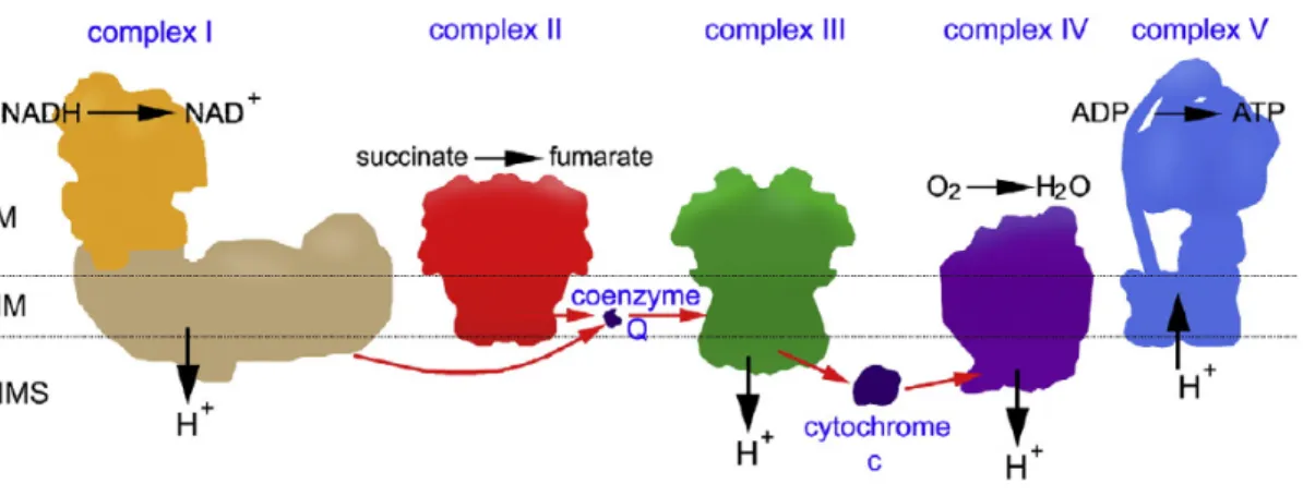
As stated earlier, one of the major functions of mitochondria is to generate energy in the form of ATP through a process called oxidative phosphorylation. The mitochondrial respiratory chain consists of five protein complexes (Complex I-V) that are embedded
into the lipid bilayer of the inner mitochondrial membrane (Figure 1.2). Out of the 92 identified structural OXPHOS subunit genes, 79 are encoded by nuclear genome and 13 are encoded by mtDNA genome which are the building blocks for the formation of the five OXPHOS complexes 12. The subunits of Complex I and Complex III-V are encoded by both nuclear DNA and mitochondrial DNA (mtDNA) whereas subunits of Complex II is exclusively encoded by nuclear genome. ATP is generated in a two-step process. In the first step electrons from NADH (reduced nicotinamide adenine dinucleotide) and FADH2
(flavin adenine dinucleotide), produced by the oxidation of nutrients such as glucose and fatty acids, are passed along a series of carrier molecules called the electron transport chain to molecular oxygen to form water (Figure 1.2) 16. This generates an electrochemical gradient that allows Complex I, III and IV to pump protons across the inner membrane. This creates a proton gradient across the membrane, which is used by Complex V to produce ATP in the second step.
Complex I or NADH dehrdrogenase (NADH: ubiquinone oxidoreductase) is the largest complex of the respiratory chain consisting of 44 subunits (14 core subunits - 7 from mtDNA and 7 from nDNA) and another 30 nDNA accessory subunits including a FMN- containing flavoprotein and 6 iron-sulfur centers proposed to maintain the stability of the complex 10,12. Complex I is L-shaped comprising of a long and short arm. The long arm forms the hydrophobic integral membrane protein and the hydrophilic short arm contains the flavin mononucleotide (FMN) and the NADH active center extending into the matrix
10. In this complex, oxidation of NADH allows the transfer of two electrons from NADH to FMN thereby reducing it to FMNH2. The electrons are further transferred to ubiquinone (Q) via iron sulfur cluster of Complex I. This electron transport is coupled with the transfer of four protons from mitochondrial matrix into the intermembrane space thereby creating the proton gradient (Figure 1.2).
Complex II or succinate dehydrogenase (succinate: ubiquinone oxidoreductase) is the only complex consisting of 4 subunits that are encoded entirely by nDNA 12. This is another point where electrons can enter the electron transport chain besides Complex I.
Complex II catalyzes the oxidation of succinate via flavin adenine dinucleotide (FAD) to form fumarate during which electrons travel from FADH2 through iron sulphur clusters to ubiquinone (Figure 1.2) 16.
Ubiquinone is a lipid soluble benzoquinone that diffuses in the phospholipid bilayer of the inner membrane thereby assisting in shutting electrons between membrane proteins
(Figure 1.2) 10.
Complex III or cytochrome bc1 (ubiquinol: cytochrome c oxidoreductase) is a complex comprising of 11 subunits; out of which 1 (cytochrome b) is encoded by mtDNA and 10 are encoded by nDNA 12. This complex catalyzes the transfer of electrons from ubiquinols to cytochrome c that resulted in the translocation of four protons across the inner membrane (Figure 1.2) 16.
Cytochrome c is a peripheral protein facing the intermembrane space and transfers electrons from complex III to complex IV (Figure 1.2) 10.
Complex IV (cytochrome c oxidase) is the last enzyme of the electron transport chain consisting of 3 subunits encoded by mtDNA and 11 subunits encoded by nDNA 12. This complex transfers four electrons from reduced cytochrome c to O2 thereby producing two molecules of water. This electron transfer further resulted in the translocation of four protons across the inner membrane (Figure 1.2) 16.
Complex V (F0F1-ATP synthase) consists of 19 subunits where 2 subunits are encoded by mtDNA and the remaining 17 subunits are encoded by nDNA 12. The electrochemical gradient is utilized by Complex V to drive the synthesis of ATP from ADP and Pi.
Powered by the translocation of three protons into the mitochondrial matrix, Complex V synthesizes one molecule of ATP, which is the energy currency of cell that is finally transported outside mitochondria by adenine nucleotide translocase (ANT1) (Figure 1.2)
16.
Figure 1.2 OXPHOS system.
Schematic representation of the OXPHOS system showing its individual components. The position of the matrix (M), the intermembrane space (IMS) and cristae or inner membrane (IM) has been indicated 17.
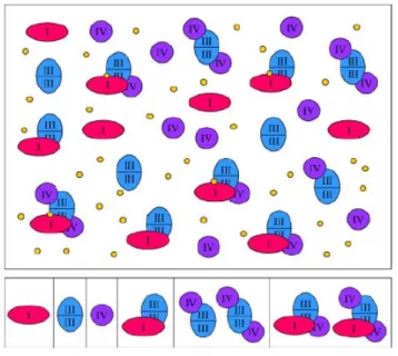
Two models have been proposed describing the supramolecular organization of these five complexes that together form the OXPHOS system. (i) The “fluid state” model postulates that the respiratory chain complexes diffuse freely in inner mitochondrial membrane and the electron transfer is based on random collisions of the single complexes 17,18. This is supported by the fact that all five complexes of the OXPHOS system can be purified to homogeneity in an enzymatically active form using isolated mitochondrial membranes
17,19
Figure 1.3 Model of the supramolecular structure of the OXPHOS system.
Single complexes co-exist with supramolecular assemblies. Complex I (red) can associate with complex III2 (blue). Complex III2 can associate with one or two copies of complex IV (purple).
The largest assemblies include complex I, dimeric complex III, and one or several copies of complex IV. (Yellow circles, ubiquinol, which either freely diffuses within the inner mitochondrial membrane or might form part of the I+III2 supercomplex. For simplicity, complex II was omitted from the figure because it is not known to form part of OXPHOS supercomplexes.
Furthermore, cytochrome c, alternative oxidoreductases, and the ATP synthase complex are omitted from the figure) 19.
.
(ii) The “solid-state” model proposes stable interactions between the OXPHOS complexes within entities named supercomplexes or respirasomes 17. Several evidences has been reported in support of this model:(a) Supercomplexes including more than one type of OXPHOS complex are resolved by blue native polyacrylamide gel electrophoresis (BN-PAGE) and are shown to be active by in-gel activity experiments
20,21; (b) Single particle electron microscopy studies revealed defined associations of OXPHOS complexes within the isolated respiratory supercomplexes 22,24; (c) Studies showed that point mutations in genes encoding one of the subunits of one OXPHOS
complex affect the stability of other OXPHOS complexes 25; (d) Flux control experiments indicate that respiratory chain operates as one functional unit 26,27; (e) Various reconstitution experiments showed that different OXPHOS complexes when present at defined stoichiometries generated highest electron transfer activities 19;28; (f) Cardiolipin seems to assists the formation of some supercomplexes 29,30.
Complex I, III, IV has been shown to form supercomplexes with defined stoichiometric composition (Figure 1.3). Electron microscopy studies have revealed interaction between complexes I and III, among complexes I, III and IV and in a dimeric form of complex V, between two ATP synthase monomers. The I+III2+IV1–2 supercomplex is known as respirasome that can autonomously carry out respiration in the presence of ubiquinone and cytochrome c (Figure 1.3) 17. Complex II in general, seems to maintain its singular state due to its direct involvement in the citric acid cycle and does not take part in formation of the respiratory chain supercomplexes 17. However one study reported Complex II containing supercomplexes 31.
Although the debate on the membrane state of OXPHOS system is still ongoing, it is clear that many experimental observations cannot be explained only with “fluid state”
model whereas other observations do not indicate only towards a “solid state” model.
Hence this arise a possibility where the membrane state of OXPHOS system is of dynamic nature where respiratory supercomplexes may co-exist within the inner mitochondrial membrane with single OXPHOS complexes (Figure 1.3) 17,19.
1.4. Replication of mtDNA
mtDNA replication occurs randomly throughout the cell cycle and is independent of nuclear DNA replication. mtDNA replication machinery and proteins for maintaining its integrity are encoded by nuclear genome and subsequently imported into mitochondria.
In eukaryotes, mtDNA replication takes place in a ‘replisome’ by mtDNA polymerase g (Pol g) which is a heterotrimeric protein consisting of a catalytic subunit (Pol g A) and a dimeric accessory subunit (Pol g B) (Figure 1.4). This heterotrimeric protein has three activities: DNA polymerase activity, 3’-5’ exonucleolytic proofreading activity and a 5’dRP lyase activity that is required for enzymatic DNA repair 12. The additional components included in the replisome are mitochondrial single stranded binding protein (mtSSB) and TWINKLE which is a 5’-3’DNA helicase (Figure 1.4). Moreover mitochondrial transcription factor A (TFAM), RNA polymerase (POLRMT), RNA
processing enzymes (RNaseH1) and topoisomerase (mtTOP1) are required for the mtDNA replication. During mtDNA replication the TWINKLE helicase forms a hexamer that unwinds mtDNA in the 5’-3’direction facilitating mtDNA synthesis. The resulting single stranded region of mtDNA at replication fork is stabilized by mtSSB, which further stimulates the TWINKLE dependent mtDNA unwinding and enhancement of polymerase gamma activity (Figure 1.4) 12,13. POLRMT act as a primase to provide primers required for the initiation of lagging-strand DNA synthesis 15. RNaseH1 is involved in removing the short RNA primers required for the initiation of H and L strand DNA synthesis. mtTOP1 is proposed to have a role in relaxing negative supercoils and removing positive supercoils at the replication fork generated by DNA helicase 13. Finally TFAM plays a role in the maintenance of mtDNA integrity by actively participating in bending and packaging of mtDNA into nucleiods 32.
Figure 1.4 The mtDNA replication machinery.
The TWINKLE helicase has 5’to 3’directionality and unwinds the duplex DNA template. The mtSSB proteinstabilizes the unwound conformation and stimulates DNA synthesis by the POLγ holoenzyme 13.
1.5. Mitochondrial transcription
mtDNA transcription generates the RNA primers for initiation of mtDNA replication at OH along with mtDNA expression. The principal mitochondrial transcription machinery consists of the mitochondrial RNA polymerase (POLRMT), mitochondrial transcription factor B2 (TFB2M), and mitochondrial transcription factor A (TFAM) 33. Recent studies have shown that TFB1M, which is a paralog of TFB2M, has no direct role in mtDNA transcription. However, TFB1M functions as a 12S rRNA methyltransferase that stabilizes the small subunit of the mitochondrial ribosome 34. Mitochondrial transcription is a bidirectional process that is initiated in the D-loop region where light-strand transcription starts from the light-strand promoter and heavy-strand transcription initiates
from two heavy strand promoters: HSP1 and HSP2 (Figure 1.1). TFAM is important for transcription initiation, as it preferentially binds the mtDNA upstream of the promoters thereby regulating RNA transcription. mtDNA transcription that is initiated from HSP1 promoter terminates at the tRNALeu (UUR), thereby transcribing tRNAVal, tRNAPhe and 2 ribosomal RNA (12S and 16S) whereas transcription initiated from HSP2 promoter transcribes the full-length mtDNA 35. Moreover, mtDNA transcription that is initiated from LSP promoter can either proceed through entire mtDNA or can terminate prematurely to prime mtDNA replication. There are additional factors that modulate the activity of basal mitochondrial transcription machinery thereby affecting both transcription initiation and termination. POLRMT plays a role in elongation of transcription, which is also enhanced by mitochondrial transcription elongation factor (TEFM) 12. Termination of both H and L strand transcription has been proposed to be carried out by the mitochondrial transcription termination factor 1 (MTERF1) that binds downstream of the rRNA genes 12,15,36. In mammals, there is a family of four MTERF proteins (MTERF1–4) 15,37, where MTERF1-3 has been proposed to bind to the promoter region thereby modulating mtDNA transcription 35. Recent studies have shown different roles of the MTERF family proteins: MTERF2 has been shown to regulate oxidative phosphorylation by modulating mtDNA transcription 38. MTERF3 is shown to be a negative regulator of mtDNA transcription 39 and also plays a role in regulating mitochondrial ribosome biogenesis 40. MTERF4 is reported to regulate mitochondrial translation and ribosomal biogenesis by targeting RNA methyltransferase NSUN4 to the large ribosomal subunit 41.
1.6. Mitochondrial translation
Mitochondrial protein synthesis is an important process for all mammals since it produces thirteen polypeptides that are key components of the OXPHOS complexes. Despite of a 50-year period of research where many factors critical for mitochondrial translation have been identified, the molecular details underlying the mitochondrial translation process remains incomplete. One of the major reasons behind this is the absence of an in vitro reconstituted system established from mammalian mitochondria that is capable of correct initiation and synthesis of a mitochondrial encoded protein. However studies in the past years have provided valuable information on specific features of mammalian mitochondrial protein synthesis where a number of the individual steps of protein
synthesis have been successfully carried out in vitro. Mitochondrial ribosome or mitoribosome is the key component of the mitochondrial protein synthesis process.
Studies have shown that mammals possess a distinct set of ribosomes which sediment as 55S particles and comprise 2 subunits, a 28S small subunit (mt-SSU) and 39S large subunit (mt-LSU) 42,43. Only two rRNA species has been identified in each subunit of mammalian mitoribosomes, 12S rRNA in small subunits and 16S rRNA in large subunit.
In addition, ribosomes may also carry a 5S rRNA has been reported 44. Mammalian mitoribosomes have lower sedimentation coefficient (55S) compared to prokaryotes (70S) and cytosolic (80S) counterparts. This is due to the change in protein to RNA ratio.
For prokaryotes/eukaryotic cytosolic ribosomes the protein:RNA ratio is 1:2 whereas for mammalian mitoribosome it is 2:1 suggesting a relatively low RNA content thereby compensated by a large number of mitoribosomal proteins, reviewed in 43. The small subunit comprises of 29 proteins out of which 14 are homologues of prokaryotic ribosomes whereas the large subunits is composed of 48 proteins and 28 of them are homologues of bacterial ribosomal proteins 45. There are four steps (phases) for the protein synthesis in mammalian mitochondria: Initiation, elongation, termination and ribosomal recycling.
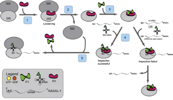
(i) Translation initiation of mammalian mitochondria:
The translation initiation process of mammalian mitochondria is different from that in prokaryotes and eukaryotic cytoplasm. It is still unclear how the mitoribosomes are directed to the initiation codon since mitochondrial mRNAs are not capped and lack any upstream leader sequences 46. Till date only two mitochondrial initiation factors have been identified, mitochondrial initiation factor 2 (IF2mt) and mitochondrial initiation factor 3 (IF3mt). According to a recent study it was shown in vitro that with IF2mt and IF3mt it was possible to assemble an initiation complex on 55S mitoribosomes with fMet- tRNA correctly positioned at the start codon of a mitochondrial mRNA 42. Based on various studies a current working model has been proposed for the initiation of translation (Figure 1.5). In the first step, IF3mt interacts with 55S ribosome thereby loosening the interaction between the two subunits, releasing the 39S subunit and forming a transient 28S:IF3mt complex in the second step (Figure 1.5). This is followed by the binding of IF2mt: GTP to the small subunit in the third step. In the fourth step, it has been shown that the mRNA feeds into the 28S subunit via an mRNA entrance gate (Figure 1.5) 47.
Figure 1.5 Model for the initiation phase of mitochondrial translation.
In the current model for the initiation of protein synthesis, mitochondrial initiation factor 3 (IF3mt) actively dissociates 55S ribosomes, forming a transient [IF3mt:55S] complex (Step 1) and leading to the formation of an IF3mt:28S complex (Step 2). Mitochondrial initiation factor 2 (IF2mt) bound to GTP binds to the small subunit (Step 3), followed by the fMet-tRNA and mRNA (Step 4), although the exact order of binding is not clear. Once the 28S initiation complex has formed, the large subunit joins, and along with the hydrolysis of GTP to GDP, the initiation factors exit (Step 5) leaving a 55S:fMet-tRNA:mRNA complex that is ready for the elongation phase of protein synthesis 42.
After the first 17 nucleotides of mRNA enters the ribosome the 5’end of mRNA pauses at the P-site of the ribosome. During this time, inspection is carried out by 28S subunit at the 5’ end of mRNA for the start codon. At this time, IF2mt: GTP may promote the binding of fMet-tRNA to the ribosome. If correct start codon in present in the P-site of mitoribosomes, a stable 28S initiation complex is formed via the codon:anticodon interactions between the fMet-tRNA and the 5’AUG start codon (Figure 1.5). However, if the inspection fails, when fMet-tRNA binds without mRNA or mRNA lacks a proper 5’ start codon, the mRNA slides through the small subunit and finally dissociates (Figure 1.5). But once the inspection succeeds and 28S initiation complex is formed, the large subunit joins the complex followed by hydrolysis of GTP to GDP by IF2mt in the fifth step. The initiation factors are released and 55S:fMet-tRNA:mRNA complex is formed that is ready for the elongation phase of protein synthesis (Figure 1.5) 42.
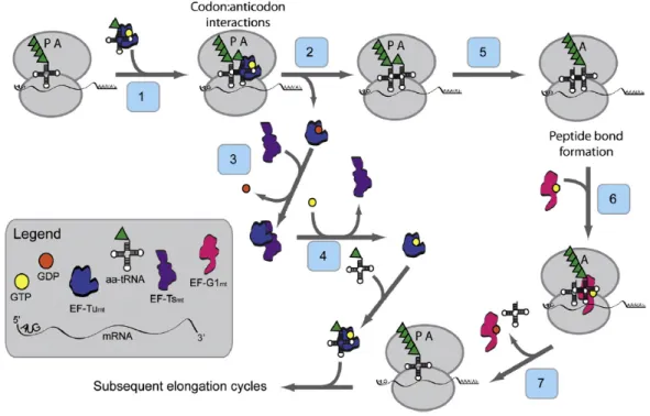
(ii) Translation elongation of mammalian mitochondria:
Three mitochondrial elongation factors, EF-Gmt, EF-Tsmt and EF-Tumt are involved in the process of polypeptide chain elongation. This elongation phase in mammalian mitochondria has many similarities to the process in prokaryotes 48, as compared to initiation and termination phases. The tRNA containing the growing polypeptide chain is located in the P-site of the mitoribosome. In the first step, GTP bound elongation factor Tu (EF-Tumt) that is the active form, binds aminoacyl-tRNA (aa-tRNA) to form a ternary complex (EF-Tumt-GTP-aa-tRNA) and enters the A-site of mitoribosome (Figure 1.6) 42. In the second step, once the codon:anticodon interactions take place, the ternary complex is selected triggering the hydrolysis of GTP to GDP by EF-Tumt, thereby releasing EF- Tumt-GDP from the ribosome.
Figure 1.6 Model for the elongation phase of mitochondrial translation.
The tRNA containing the growing polypeptide chain is located in the P-site of the ribosome. EF- Tumt brings the aa-tRNA to the A-site of the ribosome (Step 1). In concert with the hydrolysis of GTP to GDP, EF-Tumt leaves the ribosome (Step 2). EF-Tsmt binds to EF-Tumt, displacing the GDP molecule and forming an EF-Tumt·EF-Tsmt complex (Step 3). A GTP molecule displaces EF-Tsmt, and an EF-Tumt:GTP complex is formed (Step 4) which can then bind another aa-tRNA reforming the ternary complex. The large ribosomal subunit catalyzes peptide bond formation and the growing polypeptide chain is transferred to the tRNA in the A-site of the ribosome (Step 5).
EF-G1mt:GTP binds to the ribosome at the A-site (Step 6) and catalyzes translocation of the ribosome, moving the deacylated tRNA out of the P-site and the peptidyl-tRNA from the A-site to the P-site (Step 7). A new cycle of elongation can then begin 42.
.
The third step follows the binding of Elongation factor Ts (EF-Tsmt) to EF-Tumt thus displacing GDP. Subsequently GTP molecule binds to EF-Tumt displacing EF-Tsmt and forming EF-Tumt:GTP complex in the fourth step. This can now bind to another aa-tRNA to form a ternary complex for the next round (Figure 1.6) 42. In Step 5, the large ribosomal subunit catalyzing the peptide bond formation and the growing polypeptide chain is transferred to the tRNA in the A-site of the mitoribosome thus leaving a deacylated tRNA in the P-site. In the next step (Step 6), another mitochondrial elongation factor G1 (EF-G1mt) binds to GTP, forming EF-G1mt:GTP that binds to A-site of ribosome. It has been reported that this binding catalyzes the translocation of mitoribosome in the final step (Step 7) thereby removing the deacylated tRNA from the P-site and moving the peptidyl-tRNA from A-site to P-site (Figure 1.6) 42. Cryo-EM studies of the mitochondrial ribosome suggested that mitoribosomes lacks E-site 47. (iii) Termination of translation and ribosome recycling of mammalian mitochondria:
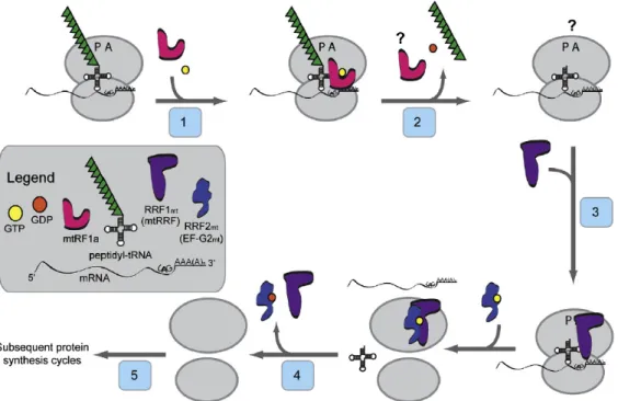
It has been shown that UAA and UAG serve as stop codons but these stop codons are not recognized by any so far identified release factors. According to a recent study, it has been suggested that they promote a −1 frameshift thereby moving a classical UAG codon into the A-site for termination 49. In the first step, when the termination codon UAA or UAG enters the A-site of mitoribsomes, it is recognized by mitochondrial release factor mtRF1a (Figure 1.7). In presence of GTP, mtRF1a binds to the A- site in the second step, thereby promoting the hydrolysis of the peptidyl-tRNA bond (by the peptidyl transferase center on the 39S subunit) and subsequent release of the completed polypeptide (Figure 1.7) 42. However, the manner in which mtRF1a is released from the mitoribosome after the release of polypeptide is still elusive. In the next step (step 3) mitochondrial ribosome recycling factor (RRF1mt)binds to the A-site of the ribosome, which is then accompanied by binding of RRF2mt (EF-G2mt). This binding enhances the dissociation of ribosomal subunits and release of deacylated tRNA and mRNA in the fourth step (Figure 1.7). The dissociation of ribosomal subunits and its release is dependent on the combined action of RRF1mt and RRF2mt (EF-G2mt) 29,50,51. Following the release of RRF1mt and RRF2mt from the ribosomse in the final step the ribosome begins another round of protein synthesis (Figure1.7) 42.
Figure 1.7 Model for the termination and ribosome recycling phases of mitochondrial protein synthesis.
As the termination codon (UAG here) enters the A-site of the ribosome, mtRF1a and GTP bind to the A-site (Step 1) and promote GTP-dependent hydrolysis and release of the polypeptide chain (Step 2). How mtRF1a is released from the ribosome is not known. RRF1mt binds to the A-site of the ribosome (Step 3) and is joined by RRF2mt (also termed EF-G2mt). These factors promote the dissociation of the ribosomal subunits and release of the deacylated tRNA and the mRNA (Step 4). Following release of RRF1mt and RRF2mt (Step 5), the ribosome begins another round of protein synthesis 42.
1.7. Mitochondrial quality control (MQC)
Mitochondria are dynamic organelles possessing various essential biological roles in cellular physiology; hence to maintain a healthy mitochondrial population, several interdependent mechanisms exist, starting from the molecular, organellar to the cellular level. These conserved mechanisms known as mitochondrial quality control (MQC) maintains the mitochondrial homeostasis. There are several issues that impose significant challenges to proper mitochondrial function:
(a) The first challenge for maintaining homeostasis is the generation of reactive oxygen species (ROS), a byproduct of oxidative phosphorylation. Accumulation of ROS can cause damage to mtDNA, oxidative modification to the mitochondrial proteins, leading to misfolding and aggregation thereby disrupting their native functions (Figure 1.8A).
(b) The second problem arises from the bigenomic nature of the mitochondrial proteome (Figure 1.8B). Mitochondrial proteome comprises of almost 1500 proteins and majority
of proteins are encoded by nuclear genome, synthesized in the cytosol and imported into mitochondria through specific translocation machineries. According to recent studies,
~500 proteins reside in the matrix 52, ~60 proteins in inter membrane space (IMS) 53,54 and ~100 polypeptides located in the outer membrane (OM). But the inner membrane (IM) proteome is the most enriched consisting ~840 proteins that comprises the electron transport chain and F1 FO ATPase complexes (OXPHOS system) 55. Out of the 1500 proteins, only 13 polypeptides that are subunits of the respiratory chain are encoded by the mitochondrial genome and needs to be assembled together with the nuclear encoded ones. This complex biogenesis pattern requires a coordinated expression and communication between the two genomes and proper sorting-folding and assembly of these polypeptides into right stoichiometric complexes within the IM. If there is a failure of coordination or perturbation in homeostasis especially under stress condition these newly synthesized proteins are at a risk to misfolding and aggregation. Therefore mitochondria have its own quality control team consisting of chaperones and proteolytic machineries that deal with such problems and thereby maintain integrity.
Figure 1.8 Biochemical stresses that challenge normal mitochondrial function.
(A) Stalling the high-energy electrons at respiratory complexes I and III leads to generation of superoxide anion which—either directly or via subsequent ROS radicals—can damage biological molecules like mtDNA and propel additional damage. The biogenesis of OXPHOS complexes requires tight coordination between synthesis and assembly of the mitochondrial- and nuclear- coded proteins. (B) Polypeptides derived from the nuclear genome are translated on cytosolic ribosomes and imported in an unfolded state into the mitochondrion via presequence translocases of the outer (TOM) and inner (TIM) membranes. Imported polypeptides are inserted into the IM where they are joined with mitochondria-synthesized subunits. Mismatches in subunit stoichiometry can lead to accumulation of unfolded or unassembled proteins that can affect functional integrity of mitochondria. In addition, the electron transport chain units of OXPHOS contain redox-active cofactors poised for rapid electron exchange reactions. (C) When improperly assembled, these prosthetic groups can act as pro-oxidants through their inherent ability to generate ROS via Fenton-like reactions 55.
(c) The final threat comes from the multiple redox cofactors of electron transport chain (ETC) (Figure 1.8C). If these cofactors are not assembled properly, they may act as pro oxidants through their inherent ability to generate ROS via Fenton-like reactions, thereby further adding to the challenging biochemical environment 56,57. If these challenges are not taken care of properly, they may further distort protein homeostasis leading to progressive failure of the organelle 55.
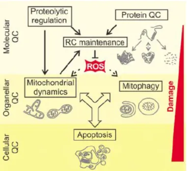
In order to cope up with the above-mentioned challenges, cells have evolved elaborate quality control mechanisms that engage at several levels depending on the extent of damage (Figure 1.9). The molecular level of MQC is the first line of defense that comprises of highly conserved molecular chaperones and energy dependent proteases distributed across the mitochondrial compartments and also cytosolic proteolytic systems like the ubiquitin–proteasome system (UPS), which associates with the OMM 55. The primary role of the chaperones is to ensure proper folding and assembly of mitochondrial proteins and that of proteases is to remove damaged proteins from the organelle 58. The second line of defense is provided by the fusion and fission events that mediate organellar dynamics and regulate even redistribution of mtDNA and proteome throughout the mitochondrial network (Figure 1.9). Fusion with neighboring healthy mitochondria helps in restoring the function of damaged mitcohondria. However if the mitochondria are severely damaged, then fusion is impaired resulting in fragmentation of mitochondria that are selectively removed by mitochondria-specific type of autophagy known as mitophagy. Occurrence of mitophagy inhibits the release of pro-apoptotic proteins from damaged mitochondria thereby suppressing apoptosis, which is the final line of defense at cellular level (Figure 1.9) 58.
In last years, mitochondrial derived vesicles (MDV) carrying selected oxidized cargo and delivering to lysosomes, has been identified as a new pathway to MQC. This process has been shown to be independent of mitochondrial dynamics and mitophagy. One of the proposed roles is to remove segments of the mitochondrial membranes containing damaged, hard to dissociate protein complexes and/or reactive prosthetic groups reviewed in 55.
Figure 1.9 Quality control (QC) surveillance of mitochondria.
Intraorganellar proteases exert QC and regulatory functions to maintain respiratory chain (RC) activity. The functionality of damaged mitochondria can be restored by fusion and content mixing within the mitochondrial network. Severely damaged mitochondria fragment and are removed by mitophagy or induce apoptosis by the release of pro-apoptotic proteins 58.
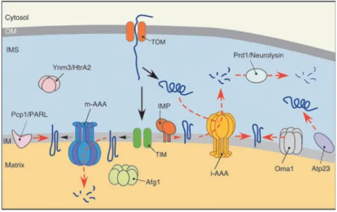
1.7.1 MQC in the matrix:
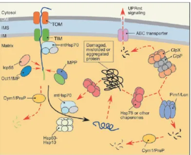
Protein regulation and quality control is tightly regulated in the mitochondrial matrix, which is the most protein dense region since it harbors the enzymes of TCA cycle and other metabolic enzymes along with translational machinery 59. Most of the proteins are synthesized in the cytosol and imported as unfolded polypeptides into mitochondria through the outer and inner membrane translocators (TOM and TIM23, respectively) in a membrane potential (ΔΨm) dependent manner (Figure 1.10). A team of chaperones and proteases are involved to ensure proper translocation of these pre-proteins followed by removal of the N-terminal MTS and folding into their native state. In the first stage, mtHsp70 and J-type co-chaperones participate in import of precursor proteins that later with help of Hsp60-Hsp10 chaperone system promote folding of the imported polypeptides (Figure 1.10) 55. mtHsp70 harbors diverse roles in mitochondrial matrix.
mtHsp70 is recruited to TIM23 translocase and at the channel it functions in the multi- subunit PAM (Presequence Translocase-Associated Motor) thereby interacting with the translocating polypeptides to drive their import through the channel into the matrix 60. At