Cellular maturation of mitochondrial molybdoenzymes
Inaugural-Dissertation zur
Erlangung des Doktorgrades
der Mathematisch-Naturwissenschaftlichen Fakultät der Universität zu Köln
vorgelegt von
Julian Klein
aus Köln
Berichterstatter: Prof. Dr. G. Schwarz (Gutachter) Prof. Dr. T. Langer
Tag der mündlichen Prüfung: 6. November 2012
published: Klein JM, and Schwarz G. Cofactor-dependent maturation of mammalian sulfite oxidase links two mitochondrial import pathways.
Journal of Cell Science (2012)
submitted: Klein JM, Busch JD, Potting C, Baker MJ,
Langer T, and Schwarz G. The mitochondrial amidoxime-reducing component (mARC1) is a novel signal-anchored protein of the outer mitochondrial membrane.
Journal of Biological Chemistry (2012)
Conference contributions
2012: Klein JM, and Schwarz G. Mitochondrial
maturation of sulfite oxidase. Gordon Research Conference “Protein Transport Across Cell
Membranes”; Galveston, USA.
(Poster presentation)
2011: Klein JM, and Schwarz G. Assembly and
maturation of sulfite oxidase. “Molybdenum and Tungsten Enzyme Conference”;
Edmonton, Canada (Oral presentation)
2010: Klein JM, and Schwarz G. Assembly and
maturation of sulfite oxidase. Gordon Research Conference “Mitochondria and Chloroplasts”;
Barga, Italy.
(Poster presentation)
Table of contents
1 Introduction 1
1.1 Molybdenum and Moco 1
1.2 Moco synthesis 2
1.3 Moco dependent enzymes 4
1.3.1 Mammalian cytosolic Moco enzymes 4
1.3.2 Mammalian mitochondrial Moco enzymes 5
1.3.2.1 Sulfite oxidase 5
1.3.2.2 Sulfite oxidase- and Moco-deficiency 8
1.3.2.3 Mitochondrial amidoxime reducing components 10
1.4 Mitochondrial architecture and function 12
1.5 Mitochondrial protein import 13
1.5.1 Import of proteins containing cleavable pre-sequences 14 1.5.2 Import of proteins lacking cleavable pre-sequences 16
1.5.2.1 Insertion of proteins into the inner membrane 16
1.5.2.2 Import of small IMS proteins 17
1.5.2.3 Integration of β-barrel proteins into the outer membrane 17 1.5.2.4 Integration of α-helical proteins into the outer membrane 18
1.6 Cofactors and metabolites in mitochondria 20
1.6.1 Heme synthesis and transport 21
1.7 Aims of the current study 23
2 Results 24
2.1 Assembly and maturation of mammalian SO 24
2.1.1 Amplification, purification and characterization of mouse SO 24
2.1.2 Moco dependent mitochondrial localization of SO 27
2.1.3 Mitochondrial import of SO 32
2.1.4 Retrograde translocation of SO to the cytosol in absence of Moco 35 2.1.5 The role of heme in the mitochondrial maturation of SO 40 2.1.6 The impact of homodimerization on the mitochondrial maturation of SO 43 2.1.7 SO-independent population of Moco in mitochondria 46
2.2 Mitochondrial maturation of mARC1 48
2.2.1 Sub-cellular localization of mARC1 48
2.2.2 Localization of mARC1 within the mitochondrial compartments 50
2.2.3 Mitochondrial targeting of mARC1 53
2.2.4 Mechanims of mitochondrial mARC1 import 57
3 Discussion 61
3.1 Maturation of mammalian SO 61
3.1.1 Moco dependent mitochondrial localization of SO 61
3.1.2 The SO import vs. other proteins with bipartite targeting signals 64 3.1.3 The role of heme and dimerization in the mitochondrial maturation of SO 66
3.1.4 Processing of SO in the IMS 68
3.1.5 Moco stabilization 69
3.1.6 Assembly and maturation of SO 70
3.2 Subcellular localization and sorting of human mARC1 71 3.2.1 Localization of mARC1 to the outer mitochondrial membrane 71 3.2.2 Targeting of mARC1 to the outer mitochondrial membrane 73
3.2.3 Outer membrane targeting of mARC1 vs inner membrane sorting of SO 76
3.2.4 In vitro import of mARC1 79
3.2.5 Assembly and maturation of mARC1 80
4 Material and methods 82
4.1 Material 82
4.1.1 Organisms 82
4.1.2 Plasmids 83
4.1.3 Enzymes and chemicals 83
4.1.4 Antibodies 84
4.2 Methods 85
4.2.1 Biochemical methods 85
4.2.1.1 Expression of recombinant SO variants in E. coli 85
4.2.1.2 E. coli cell disruption 85
4.2.1.3 Purification of recombinantly expressed SO variants 85
4.2.1.4 Concentration of purified proteins 86
4.2.1.5 Analytical size exclusion chromatography 86
4.2.1.6 Buffer exchange 86
4.2.1.7 Determination of protein concentration 86
4.2.1.8 SDS-PAGE 87
4.2.1.9 Western blot 87
4.2.1.10 Blue Native-PAGE 87
4.2.1.11 Sulfite:Cytochrome c assay 88
4.2.1.12 HPLC Form A analysis 88
4.2.1.13 Nit-1 assay 89
4.2.1.14 TCA precipitation 90
4.2.2 Molecular biological methods 90
4.2.2.1 Cloning 90
4.2.2.2 Plasmid amplification and purification 90
4.2.2.3 Site directed mutagenesis 90
4.2.2.4 Fusion PCR 90
4.2.3 Cell biological methods 91
4.2.3.1 Cultivation of mammalian cells 91
4.2.3.2 Transfection of mammalian cells 91
4.2.3.3 shRNA mediated knockdown of gene products 91
4.2.3.4 Harvesting and disruption of mammalian cultured cells 92
4.2.3.5 Microscopic preparations 92
4.2.3.6 Staining of mitochondria 92
4.2.3.7 Confocal laser microscopy 92
4.2.3.8 Determination of the Pearson Correlation Coefficient 93 4.2.4 Methods to study mitochondrial localization and import of proteins 93
4.2.4.1 Enrichment of mitochondria 93
4.2.4.2 Purification of mitochondria 93
4.2.4.3 Na2CO3 extraction of mitochondrial proteins 94
4.2.4.4 Protease treatment of mitochondria 94
4.2.4.5 Hypotonic swelling of mitochondria 94
4.2.4.6 In vitro translation 95
4.2.4.7 Mitochondrial in vitro import studies 95
5 Appendix 97
5.1 Primers 97
5.2 Constructs 98
5.3 Sequences 99
5.3.1 Mouse SO 99
5.3.2 Human mARC1 100
6 References 101
Figures and tables
Figures
Figure 1.1 Structures of two eukaryotic types of Moco 2
Figure 1.2 Human Moco biosynthesis 3
Figure 1.3 Reaction mechanism and crystal structure of animal SO 6 Figure 1.4 Severe neurological symptoms of SO deficiency 9 Figure 1.5 Composition and electron transfer in the N-reductive system 11 Figure 1.6 Mitochondrial import of pre-sequence containing precursors 15 Figure 1.7 Possible import pathways of signal-anchored proteins 20
Figure 1.8 Mammalian heme biosynthesis 21
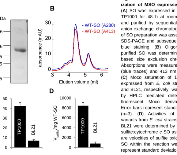
Figure 2.1 Multiple sequence alignment of mammalian SO variants 25 Figure 2.2 Purification and characterization of MSO expressed in E. coli 26
Figure 2.3 Moco dependent localization of SO 28
Figure 2.4 Characterization of a Moco-deficient mutant variant of SO 29
Figure 2.5 Cellular distribution of Moco-deficient SO 30
Figure 2.6 Western blot analysis of Moco dependent distribution of SO 32 Figure 2.7 SO contains an N-terminal mitochondrial targeting signal 33 Figure 2.8 Moco independent mitochondrial processing of SO 34
Figure 2.9 Processing of SO by the IMP complex 35
Figure 2.10 Design of an unprocessed SO-TIM50 chimera 36
Figure 2.11 Similar enzymatic activities of WT-SO and SO-TIM50 37 Figure 2.12 Moco independent mitochondrial localization of SO-TIM50 38 Figure 2.13 Moco “pulls” SO across the outer mitochondrial membrane 39 Figure 2.14 Characterization of a heme deficient mutant variant of SO 41 Figure 2.15 Heme independent mitochondrial localization of SO 42 Figure 2.16 Heme mediated trapping of the SO heme domain 43 Figure 2.17 Characterization of a monomeric mutant variant of SO 44 Figure 2.18 Dimerization independent mitochondrial localization of SO 45 Figure 2.19 Mitochondria contain an SO independent population of Moco 47
Figure 2.20 Alignment of human mARC1 and mARC2 48
Figure 2.21 Analysis and prediction of mARC1 sequence and structural motifs 49
Figure 2.22 Mitochondrial localization of mARC1-GFP 50
Figure 2.23 Association of mARC1 with mitochondrial membranes 51 Figure 2.24 mARC1 localizes to the outer mitochondrial membrane 52
Figure 2.25 Mitochondrial targeting motifs of mARC1 55
Figure 2.26 Moco independent mitochondrial targeting of mARC1 56
Figure 2.27 In vitro translation of mARC1 57
Figure 2.28 mARC1 is not processed 58
Figure 2.29 Mitochondrial in vitro import of mARC1 59
Figure 3.1 Assembly and maturation of mammalian SO 71
Figure 3.2 Assembly and maturation of human mARC1 80
Tables
Table 3.1 Comparison of IMS proteins with bipartite N-terminal targeting signals 65 Table 3.2 Analysis of matrix-targeting capability of different protein N-termini 74 Table 3.3 TM domain comparison between signal-anchored proteins and inner
membrane/IMS proteins with bipartite targeting signals 76
Table 4.1 E. coli cells used in this study 82
Table 4.2 Mammalian cells used in this study 82
Table 4.3 N. crassa cells used in this study 83
Table 4.4 Plasmids used in this study 83
Table 4.5 Primary antibodies used in this study 84
Table 4.6 Secondary antibodies used in this study 84
Tab. 5.1 Primers used in this study 97
Tab. 5.2 Constructs used in this study 98
Abbreviations
A absorption aa amino acid
ADP adenosine diphosphate AMP adenosine monophosphate AO aldehyde oxidase ATP adenosine triphosphate AU absorption units
BN-PAGE Blue Native-polyacrylamide gel electrophoresis BSA bovine serum albumin cDNA complementary DNA CDS coding sequence CMV cytomegalovirus cPMP cyclic pyranopterin monophosphate Da Dalton DMSO dimethyl sulfoxide DNA deoxyribonucleic acid DTT dithiothreitol ECL enhanced chemiluminescence EDTA ethylenediaminetetraacetic acid ELISA enzyme-linked immunosorbent assay ER endoplasmic reticulum ERMES ER-mitochondria encounter structure FAD flavin adenine dinucleotide FPLC fast performance liquid chromatography g, mg, µg, ng gram, milligram, microgram, nanogram GFP green fluorescent protein GSH glutathione GTP guanosine triphosphate h hours
Hepes 2-[4-(2-hydroxyethyl) piperazin-1-yl] ethanesulfonic acid His-tag histidine-tag
HPLC high performance liquid chromatography HRP horseradish peroxidase
IBM inner boundary membrane IM mitochondrial inner membrane IMP inner membrane peptidase IMS mitochondrial intermembrane space
IPTG isopropyl-β-D-1-thiogalactopyranoside
kbar kilo bar kDa kilo Dalton l, ml, µl liter, milliliter, microliter LB lysogeny broth M, mM, µM molar, millimolar, micromolar mARC mitochondrial amidoxime reducing component min minutes
MINOS mitochondrial inner membrane organizing system
mm, µm, nm millimeter, micrometer, nanometer Mo molybdenum MoCD molybdenum cofactor deficiency Moco molybdenum cofactor mol, mmol, µmol, pmol mole, millimole, micromole, picomole MPP matrix processing peptidase MPT molybdopterin MPT-AMP molybdopterin-adenosine monophosphate MRI magnetic resonance imaging mRNA messenger RNA MSO mouse sulfite oxidase mt mitochondrial MTS mitochondrial targeting signal NAD/NADH nicotinamide adenine dinucleotide NADPH nicotinamide adenine dinucleotide phosphate NED N-(1-Naphtyl)-ethylendiamine-dihydrochloride Ni-NTA nickel-nitrilotriacetic acid NO nitric oxide NOHA N-hydroxy-arginine NR nitrate reductase OM mitochondrial outer membrane P pellet PAGE polyacrylamide gel electrophoresis PAM pre-sequence associated motor complex PBS phosphate buffered saline
PCC Pearson Correlation Coefficient PCR polymerase chain reaction PEI polyethylenimine PK proteinase k PMSF phenylmethanesulfonylfluoride PSO plant sulfite oxidase PVDF polyvinylidenfluorid RNA ribonucleic acid ROS reactive oxygen species rpm revolutions per minute RT room temperature SA sulfanilamide
SAM sorting and assembly machinery SDS sodium dodecyl sulfate sec second shRNA small hairpin ribonucleic acid SN supernatant SO sulfite oxidase TBS tris buffered saline TBST tris buffered saline tween TCA trichloroacetic acid TIM translocase of the inner membrane TOM translocase of the outer membrane Tris tris(hydroxymethyl)aminomethane
VDAC voltage dependent anion channel WT wildtype
XDH xanthine dehydrogenase XO xanthine oxidase
XOR xanthine oxidoreductase
Abbreviations of species
E. coli Escherichia Coli M. musculus Mus musculus N. crassa Neurospora crassa
S. cerevisiae Saccharomyces cerevisiae
Abstract
The molybdenum cofactor (Moco) is an essential component present in nearly all domains of life. In mammals, Moco is part of four currently known enzymes and constitutes a crucial redox-active center involved in a number of fundamental cellular reactions. Moco-dependent enzymes are present in the cytosol but also in or at mitochondria, where Moco is integrated into sulfite oxidase (SO) and the mitochondrial amidoxime-reducing component (mARC), respectively. The family of mitochondrial Moco-enzymes is of particular interest considering the cytosolic synthesis of enzymes and cofactor, which requires a coordinated mitochondrial transport and assembly process. In the current study, the mitochondrial maturations of SO and mARC1 were thus analyzed to obtain a mechanistic understanding of the processes starting with the cytosolic syntheses of apo-proteins all the way to the formation of the mature mitochondrial enzymes.
The first part of this work uncovered the cellular assembly of SO, a soluble protein of the mitochondrial intermembrane space, and revealed a Moco-dependent mitochondrial targeting mechanism. In spite of its functional bipartite N-terminal targeting signal, about 70%
of SO mislocalized to the cytosol if Moco was not present. Following the identification of SO processing by the inner membrane peptidase (IMP) complex, prevention of this cleavage and thus anchoring of SO in the inner mitochondrial membrane resulted in an efficient mitochondrial targeting even in absence of Moco. SO was thereby identified to undergo a reverse translocation to the cytosol in absence of Moco, which is required to trap SO in the intermembrane space and to constitute in addition a vectorial driving force for completion of SO translocation across the TOM complex. The integration of Moco is not only essential for correct sub-mitochondrial localization, but also a prerequisite for in vivo heme integration and homodimerization of SO. In conclusion, the identified molecular hierarchy of SO maturation represents a novel link between the canonical pre-sequence pathway and folding-trap mechanisms of mitochondrial import.
The other mitochondrial Moco-enzyme mARC1 was recently discovered and its sub- mitochondrial localization had remained unclear. In the second part of this study, mARC1 was shown to be localized to the outer mitochondrial membrane. As a result of the translocation process, the C-terminal catalytic core of the protein remains exposed to the cytosol and confers an N(in)-C(out) membrane orientation of mARC1. This localization is mediated by the N-terminal domain of the enzyme, being composed of a classical but weak N-terminal targeting signal and a downstream transmembrane domain. Thereby, the transmembrane domain of mARC1 is sufficient for mitochondrial targeting, while the N- terminal targeting signal seems to function as a supportive receptor for the outer mitochondrial membrane. According to its localization and targeting mechanism, mARC1 is classified as a novel signal-anchored protein. Considering the membrane integration of mARC1, an SO-similar demand of Moco for mitochondrial retention of mARC1 is not required and its N-terminal targeting motifs are sufficient for adequate mitochondrial localization.
During mitochondrial import, mARC1 is not processed and membrane integration proceeds membrane potential independently but requires external ATP, which finally results in the assembly of mARC1 into high-oligomeric protein complexes.
Zusammenfassung
Der Molybdän-Cofaktor (Moco) ist ein essentieller Bestandteil in allen Organismenreichen. In Säugetieren bildet Moco ein wichtiges redox-aktives Zentrum von bisher vier bekannten Enzymen und ist dadurch an einer Vielzahl fundamentaler, zellulärer Reaktionen beteiligt.
Moco-abhängige Enzyme liegen sowohl im Zytosol als auch in Mitochondrien vor, wobei Moco hier in die Sulfitoxidase (SO) und in die mitochondriale Amidoxim-reduzierende Komponente (mARC) eingebaut ist. Unter Berücksichtigung der zytosolischen Synthese von Moco, SO und mARC ist die Familie der mitochondrialen Moco-Enzyme dabei von besonderem Interesse, da diese einen koordinierten mitochondrialen Transport und einen entsprechend regulierten Reifungsprozess der beiden Enzyme verlangt. In dieser Arbeit wurden die mitochondrialen Reifungsprozesse von SO und mARC analysiert, um ein mechanistisches Verständnis dieser Prozesse zu erlangen.
Im ersten Teil wurden dabei hierarchische Stufen zur zellulären Reifung der SO aufgedeckt.
SO ist ein lösliches Protein des mitochondrialen Intermembranraums und zeigte dabei eine Moco-abhängige mitochondriale Lokalisierung. Ungeachtet der zweigeteilten N-terminalen mitochondrialen Zielsequenz wurden in Abwesenheit von Moco etwa 70% des Enzyms im Zytosol detektiert. Nachdem die Innere-Membran-Peptidase (IMP) als SO-prozessierende Protease identifiziert wurde, konnte eine über Mutationen verhinderte Prozessierung und damit eine Verankerung der SO in der inneren Membran eine vollständige mitochondriale Lokalisation auch in Abwesenheit von Moco erreichen. Dieses Experiment belegte, dass die SO einer reversen Translokation in Richtung Zytosol unterliegt, wenn Moco nicht eingebaut werden kann. Moco ist dabei für die Initiierung der SO-Faltung verantwortlich und verhindert dadurch zum einen den Rücktransport ins Zytoplasma und greift dadurch zum anderen auch aktiv in die Translokation der SO ein, indem die Faltung eine zusätzliche vektoriell getriebene Kraft für die vollständige Translokation in den Intermembranraum darstellt. Der Einbau des Moco ist nicht nur für die Lokalisation der SO essentiell, sondern auch eine Voraussetzung für den Einbau des Häm-Cofaktors und die Homodimerisierung der SO. Insgesamt stellt die dargestellte molekulare Hierarchie der SO-Reifung eine neue Verbindung zwischen dem kanonischen Prä-Sequenz Importweg und faltungsabhängigen Importmechanismen dar.
Das mitochondriale Moco-Enzym mARC1 wurde erst kürzlich entdeckt, wobei seine sub- mitochondriale Lokalisation unklar blieb. Im zweiten Teil dieser Arbeit konnte nun die Assoziation von mARC1 mit der mitochondrialen Außenmembran demonstriert werden. Als Resultat des Translokationsprozesses bleibt die C-terminale katalytische Domäne dem Zytosol exponiert und verleiht mARC1 eine N(innen)-C(außen) Membrankonformation. Diese Lokalisation wird über die N-terminale Domäne des Enzyms gesteuert, welche aus einem klassischen aber schwachen N-terminalen Ziel-Signal und einer folgenden Transmembrandomäne besteht. Die Transmembrandomäne ist dabei hinreichend für die Lokalisation, wobei das Ziel-Signal als unterstützender Rezeptor für die mitochondriale Außenmembran zu dienen scheint. Sowohl die Lokalisation als auch der Transportmechanismus klassifizieren mARC1 dabei als ein neues Signal-verankertes Protein. Aufgrund der Membranverankerung von mARC1 ist der Einbau des Moco kein essentieller Bestandteil des Translokationsprozesses, welcher ausschließlich von den beiden N-terminalen Motiven gesteuert wird. Während des Importprozesses wird mARC1 nicht prozessiert und der Membraneinbau erfolgt unabhängig vom Membranpotential, erfordert jedoch die externe Zufuhr von ATP. Dies führt final zur Integration von mARC1 in hoch- oligomere Proteinkomplexe in der Außenmembran.
1
1 Introduction
1.1 Molybdenum and Moco
Molybdenum (Mo) belongs to the group of transition metals and constitutes an essential trace element for animals, plants and microorganisms. In nature, Mo occurs in different chemical compounds and exhibits a rich coordination and redox chemistry, as illustrated by its extended spectrum of oxidation states ranging from –ll to +Vl. Therefore, Mo depicts a potent catalyst of a large variety of redox reactions in biological systems. Although Mo is of very low abundance in the cell, its uptake requires an adequate and efficient mechanism to guarantee a constant supply of Mo. Amongst different forms and oxidation states of Mo occurring in nature, the molybdate anion (MoO4-2
) constitutes the only Mo compound that organisms can acquire from their environment (Llamas et al., 2011). While bacteria mediate uptake of molybdate by means of a high-affinity ATP-binding-cassette transporter (Maupin- Furlow et al., 1995), a homologous system has not been found in eukaryotes, yet. Instead, plants contain two different molybdate transporters referred to as MOT1 and MOT2, showing distant relations to sulfate transporters of the SULTR family (Tejada-Jimenez et al., 2007).
Animals lack MOT1, while recently, MOT2 was identified as the first molybdate transporter in animals (Tejada-Jimenez et al., 2011).
Upon its successful uptake from the environment, Mo is complexed by a pterin compound to build the biologically active molybdenum cofactor (Moco). Moco is composed of a tricyclic pterin coordinating Mo via a dithiol group within the third pyrano ring. With the exception of bacterial nitrogenase, in which a unique iron-molybdenum cofactor confers catalytic activity, all other Mo-dependent enzymes contain a pterin type cofactor (Hille, 1996).
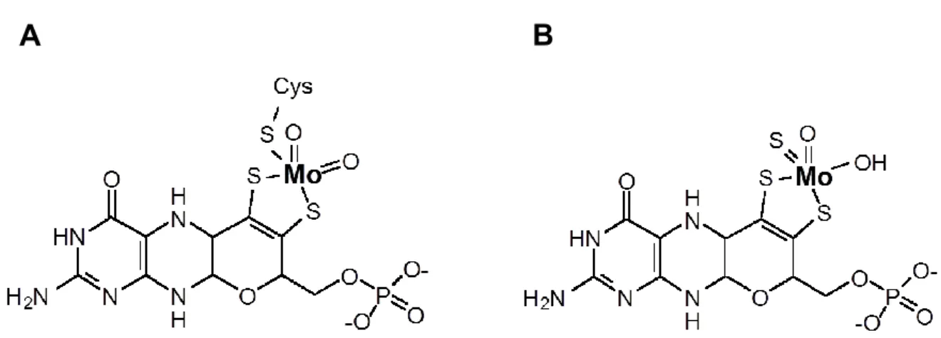
Eukaryotic Moco occurs in two different forms that share a common backbone but differ in the coordination of Mo. In one variant, Mo is covalently bound to a conserved cysteine residue of the Moco binding domain (Figure 1.1 A), while in the other variant instead a third terminal sulfur ligand is bound to Mo with the cofactor remaining non-covalently bound to the respective protein (Figure 1.1 B) (Schwarz and Mendel, 2006). Integration of Mo into both types of Moco permits the positioning of Mo within the protein active site, thus controlling its redox behavior on the one hand and aligning the pterin ring system for electron transfer from or to Mo on the other hand (Mendel, 2007). In respect to its involvement in electron transfer, the pterin moiety is fully reduced and thus believed to be prone to oxidation. In addition, the isolated coordination of Mo by means of the dithiol group is fragile, finally resulting in destabilization of Moco in protein free environments (Rajagopalan and Johnson, 1992).
Consequently, Moco was assumed not to occur free in the cell but rather to be associated rapidly with its respective target-enzymes.
2
1.2 Moco synthesis
The biosynthesis of Moco is highly conserved from bacteria to eukaryotes and is composed of four enzymatic steps (Figure 1.2), which are catalyzed by six gene products in eukaryotes.
The first step of the Moco synthesis cascade starts out from GTP, which is converted to cyclic pteranopterin monophosphate (cPMP) by two proteins (MOCS1A and MOCS1B in humans) in a complex reaction mechanism. MOCS1A contains two [4Fe-4S] clusters and belongs to the group of S-adenosyl-methionine dependent radical enzymes (Hanzelmann et al., 2004). Although the overall mechanism of cPMP synthesis is not fully understood, MOCS1A and MOCS1B are believed to build a complex with S-adenosyl-methionine and GTP (Hanzelmann et al., 2002) to generate cPMP as a fully reduced tetrahydro- pteranopterin backbone (Santamaria-Araujo et al., 2012). The subcellular localization of both MOCS proteins have remained uncharacterized in mammals, their N-terminal mitochondrial targeting signals however strongly suggest the synthesis of cPMP to take place in mitochondria. Moreover, the plant homologues Cnx2 and Cnx3 have been exclusively detected in mitochondria (Teschner et al., 2010).
The second step of Moco synthesis occurs in the cytosol, suggesting that cPMP is exported from mitochondria to become converted to the next intermediate. In plants, the inner mitochondrial membrane transporter Atm3 has been shown to be involved in the export of cPMP to the cytosol (Teschner et al., 2010). Following its mitochondrial export, two sulfur atoms are transferred to cPMP to build molybdopterin (MPT) dithiolate. This reaction is
A B
Figure 1.1 Structures of two eukaryotic types of Moco. Within eukaryotic enzymes, Mo is either (A) covalently connected to a conserved cysteine or (B) non-covalently bound to the protein and instead exposing a third terminal sulfur ligand. Figure modified from (Mendel and Bittner, 2006).
3
catalyzed by MPT-synthase, a hetero-tetrameric complex consisting of two large (MOCS2A in humans) and two small (MOCS2B in humans) subunits. While the large subunits mediate oligomerization, the small subunits each carry a single sulfur atom as thiocarboxylates being sequentially transferred to cPMP (Gutzke et al., 2001). Following the completion of a reaction cycle, the small subunits dissociate from the MPT-synthase and transiently bind to the MOCS3 protein, where they are re-sulfurated in an ATP-dependent reaction (Matthies et al., 2004).
The third and fourth steps of the Moco synthesis pathway are catalyzed by gephyrin in humans. Gephyrin is composed of an N-terminal G-domain and a C-terminal E-domain, which are both involved in separate reactions during the last steps of Moco synthesis (Schwarz and Mendel, 2006). First, the G-domain binds and adenylates MPT in an Mg2+ and ATP dependent manner, yielding MPT-AMP as the last intermediate of the Moco synthesis pathway. The reaction mechanism was uncovered in plants and revealed the binding of MPT to the homologous Cnx1G protein and the subsequent transfer of AMP to the terminal phosphate of MPT (Llamas et al., 2004). In the last step of Moco synthesis, Mo is attached to the dithiolate of the MPT backbone by the gephyrin E-domain. As again first discovered for the plant protein, MPT-AMP is hydrolyzed by the homologous Cnx1E protein and Mo is simultaneously transferred to the MPT dithiolate, finally resulting in the formation of Moco (Llamas et al., 2006).
Figure 1.2 Human Moco biosynthesis.
Structures of all intermediates are given and names are depicted in red. Catalyzing proteins are colored green. Step 1 occurs in mitochondria and is schematized accordingly.
The in vivo sulfur source for MOCS3 (X-S) is not known. SAM, S-adenosyl-methionine.
GEPH, gephyrin. See text for details.
4
1.3 Moco dependent enzymes
Following the completion of its synthesis, Moco becomes incorporated into a multitude of different Mo-enzymes to fulfill its biological function. More than 50 Moco-dependent enzymes were described so far, most of them catalyzing redox reactions that are important for the global cycles of nitrogen, carbon and sulfur (Schwarz et al., 2009). The majority of these proteins exclusively occur in prokaryotes, while to date five Moco-dependent enzymes are known in eukaryotes.
One of these enzymes, nitrate reductase (NR), is solely present in plants and fungi and plays a key role during nitrogen assimilation by catalyzing the reduction of nitrate to nitrite. Moco is covalently bound by a conserved cysteine as depicted in figure 1.1 A. Apart from Moco, NR requires the integration of a cytochrome b5 type heme and a FAD-cofactor as well as homodimerization to achieve catalytic activity. Thereby, electrons are transferred from NAD(P)H to FAD and via heme to the Moco domain, which harbors the active site where nitrate is reduced.
Animals do not have a NR, while they comprise a subset of four other Moco enzymes, which can be classified into two groups according to their active site structure as well as sub- cellular distributions. The first group is composed of the cytosolic proteins aldehyde oxidase (AO) and xanthine oxidoreductase (XOR), both sharing the terminal sulfide group, while mitochondria constitute the second site of Moco-activity and harbor sulfite oxidase (SO) as well as the mitochondrial amidoxime reducing component (mARC 1+2), each having a cysteine linked Moco in their active site (Chamizo-Ampudia et al., 2011, Rajapakshe et al., 2011).
1.3.1 Mammalian cytosolic Moco enzymes
AO and XOR can be grouped according to their cytosolic localizations, but they also share significant structural and functional similarities. Both proteins contain Moco within their C- terminal domains, which also mediate homodimerization in each case. In contrast to NR and mitochondrial molybdoenzymes, Moco is not covalently bound by a protein derived conserved cysteine, but instead contains a third terminal sulfido group as illustrated in figure 1.1 B. This sulfur atom is added to the cofactor by the enzyme Moco sulfurase in a final maturation step (Hille et al., 2011). As additional redox active domains, AO and XOR contain N-terminal [2Fe-2S] clusters and a central FAD domain to build the conserved tripartite structure of the AO/XOR enzyme family.
5
AO acts on a large array of substrates, but the general types of reactions imply hydroxylation of heterocycles and oxidation of aldehydes to the corresponding carboxylic acids (Garattini et al., 2008). Mechanistically, substrates are oxidized in the substrate binding pocket at the Mo-center. Electrons are transferred from Moco via the [2Fe-2S] clusters to FAD. During electron transfer, the [2Fe-2S] clusters act as electron sinks to store reducing equivalents during catalysis (Hille, 2002). While the human genome harbors a single AO gene, other vertebrates contain several different AO isoforms. Physiological functions of AO are poorly understood, an AO knockout mouse however revealed a function in the biosynthesis of retinoic acid (Terao et al., 2009). Furthermore, mammalian AOs represent an import drug-metabolizing system in the cytoplasm of hepatic cells. Thereby, AOs are proposed to act in concert with the microsomal cytochrome P450 system and to activate or inactivate various types of drugs and toxic compounds (Garattini et al., 2008).
XOR resembles AO in respect to structure and reaction mechanism, but catalyzes the hydroxylation of a different subset of substrates, which is much better defined as for AO.
XOR mediates the oxidation of hypoxanthine to xanthine and the downstream reaction of xanthine to uric acid (Hille and Nishino, 1995). XOR exists in two forms, as xanthine dehydrogenase (XDH), constituting the primary gene product, and as xanthine oxidase (XO), arising from XDH by formation of internal disulfide bonds. While XD favors NAD+ as a primary electron acceptor, XO does not bind NAD+ and instead transfers electrons to O2. Therefore, numerous reactive oxygen species (ROS) are formed, that are proposed to function in the innate immune response (Vorbach et al., 2003) and together with the antioxidant uric acid act in the regulation of the cellular redox potential (Droge, 2002). While no isolated forms of AO-deficiency are known, inactive XOR, either occurring in response to mutations in the XOR gene (Ichida et al., 1997) or secondary caused by the loss of Moco- sulfurase (Ichida et al., 2001), results in xanthinuria. The symptoms of the disease are not lethal, but affected patients suffer from xanthine stones due to elevated levels of xanthine.
1.3.2 Mammalian mitochondrial Moco enzymes 1.3.2.1 Sulfite oxidase
Sulfite oxidase (SO) is generally referred to as the most important eukaryotic Moco enzyme, as its depletion results in a severe neurodegenerative phenotype (see chapter 1.3.2.2 for details). SO catalyzes the essential oxidation of toxic sulfite to nonhazardous sulfate and thereby mediates the final step in the oxidative degradation of the sulfur-containing amino acids methionine and cysteine (Figure 1.3 A).
6
In contrast to AO and XOR, SO covalently binds Moco by a conserved cysteine in the central domain of the protein. Furthermore, Moco is deeply buried within the protein, forming numerous stabilizing hydrogen bonds to several residues of the central Moco-binding domain, as illustrated by the chicken SO crystal structure (Figure 1.3 B) (Kisker et al., 1997).
To become catalytically active, SO requires the integration of a cytochrome b5 type heme as a second metal containing cofactor. Although not covalently attached to the protein, heme is stably integrated into the N-terminal domain of SO with two conserved histidines symmetrically coordinating the heme iron (Kisker et al., 1997). In analogy to structurally related plant NR and also both cytosolic Moco enzymes, SO undergoes homodimerization mediated by a large interface of the C-terminal domain. Upon the integration of both cofactors and homodimerization, SO is catalytically active and ready for sulfite oxidation.
Figure 1.3 Reaction mechanism and crystal structure of animal SO. (A) Sulfite is oxidized at the Mo-active site of SO (depicted in blue with all three domains) and sulfate is released. The two electrons are sequentially transferred to the b5 heme and cytochrome c as the final electron acceptor.
Monomeric SO is depicted for simplicity. (B) Crystal structure of mature chicken liver SO. The N- terminal heme domain is depicted in red, the central Moco domain in yellow and the C-terminal dimerization domain in green. Moco and heme are shown in ball-stick representations. The gray dotted lines connect the metal centers of the cofactors, the red dotted lines indicate a loop region, which is weakly defined in the electron density. Figure modified from (Kisker et al., 1997).
A
B
7
Sulfite oxidation takes place in the mitochondrial intermembrane space (IMS) in mammals, where cytochrome c is reduced as the physiological and final electron acceptor.
The catalytic mechanism of mammalian SO starts with the oxidation of sulfate at the Mo- active site to generate a two-electron reduced Mo(IV) state (Hille, 1994). In a one-electron transfer reaction, b5 heme is reduced to generate the EPR visible Mo(V)/Fe(II) intermediate of SO (Astashkin et al., 2002). This part is referred to as the reductive half reaction, resulting in the release of sulfate and the formation of fully reduced SO. In the oxidative half reaction, the electron is passed to cytochrome c to build a one electron reduced Mo(V)/Fe(III) form of the enzyme. In analogy to the first electron transfer reaction from the Mo center to cytochrome c, the second electron is transferred to a second equivalent of cytochrome c to regenerate the fully oxidized Mo(VI)/Fe(III) form of SO (Feng et al., 2007). As a multi-redox center enzyme, SO performs rapid electron transfer between Mo- and heme-domain to achieve an efficient oxidation of sulfite (Pacheco et al., 1999). However, the crystal structure of chicken SO revealed a comparatively large distance of ~32Å between both domains (Kisker et al., 1997). This conflict has led to the proposal of domain movements prior to electron transfer in order to bring Mo- and heme-domain in closer proximity. Consistently, the electron transfer rate of SO was shown to be dependent on solution viscosity and dropped in the presence of increasing concentrations of sucrose or polyethylene-glycol (Feng et al., 2002).
SO is mainly present in the liver as the predominant site of methionine and cysteine catabolism, but it is also abundant heart, kidney and to a lesser extent in brain to ensure comprehensive protection from sulfite accumulation (Moriwaki et al., 1997). Using cytochrome c as the final electron acceptor of the sulfite oxidation, SO is localized in the IMS of mitochondria as a soluble enzyme (Ito, 1971). As most mitochondrial proteins, SO is synthesized in the cytosol and imported into mitochondria. The mechanism of mitochondrial translocation of SO was investigated by pioneer work of Ono and Ito in the early 1980’s.
While the presence of ribosomes on the surface of mitochondria suggested a co-translational import of some precursors (Kellems et al., 1975, Ades and Butow, 1980), the translation of SO was demonstrated to take place on free ribosomes in the cytosol, indicating a post- translational import of SO (Ono et al., 1982). Following its translation, SO was shown to enter mitochondria ATP- and membrane potential-dependently and the inner mitochondrial membrane was found to be involved in the translocation process (Ono and Ito, 1982b).
Import of SO is accompanied by processing, as illustrated by a truncated mature enzyme compared to the precursor during in vitro mitochondrial import experiments (Ono and Ito, 1984). Upon completion of import, the half-life of SO was measured to last between three and four days (Ono and Ito, 1982a).
8
The significance of sulfite oxidation is not restricted to animals, but also an important reaction in plants and bacteria. In plants, a protein homologous to animal SO was discovered (Eilers et al., 2001). Its primary and crystal structure however revealed a number of differences to animal SO, as plant SO (PSO) lacks a heme cofactor and Moco thus constitutes the only redox center of the enzyme (Schrader et al., 2003). Accordingly, PSO does not use cytochrome c but instead molecular oxygen as a physiological electron acceptor with the corresponding reaction taking place in peroxisomes. Bacteria oxidize sulfite by means of a sulfite dehydrogenase, a heterodimer of a large Mo-binding subunit and a small heme c-containing subunit (Kappler et al., 2000). While the sulfite dehydrogenase sequence is not related to mammalian SO, the heme c containing subunit revealed structural similarities to the b5 heme domain of mammalian SO (Kappler and Bailey, 2005).
1.3.2.2 Sulfite oxidase- and Moco-deficiency
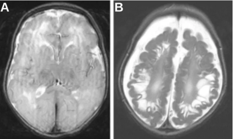
The physiological relevance of SO is displayed by a severe neurodegenerative disorder termed isolated SO-deficiency, rapidly evolving in absence of functional SO. Affected patients suffer from neurological abnormalities such as microcephaly, mental retardation and seizures, usually accompanied by death in early infancy (Figure 1.4). The disease follows an autosomal recessive trait and is very rare, with less than 30 described cases in the literature (Johnson et al., 2002, Tan et al., 2005). SO-deficiency either occurs in response to mutations in the SO gene or based on a secondary loss of activity caused by mutations in one of the four genes involved in Moco biosynthesis. The latter causes a simultaneous loss of all five mammalian Moco enzymes with the corresponding disease termed Moco-deficiency (Schwarz, 2005). The clinical symptoms of Moco-deficiency are however hardly distinguishable from those of isolated SO-deficiency, demonstrating the loss of SO to be the predominant cause of the pathophysiology in Moco-deficiency and SO to be the most important Moco enzyme in humans.
In absence of functional SO, sulfite initially accumulates in the liver as the main site of methionine and cysteine catabolism and subsequently spreads out the entire body via the blood circulation and finally reaches the brain. The pathogenesis of SO- and Moco-deficiency is not completely understood and may derive from sulfite toxicity, a lack of sulfate or the accumulation of sulfur-containing compounds that are formed in response to excessive sulfite accumulation. Sulfite is a strong nucleophile breaking disulfide bridges and thereby affecting numerous proteins and cellular functions. In addition, sulfite exposure to mouse neuronal cells was shown to increase reactive oxygen species (ROS) and to simultaneously decrease intracellular ATP production. The concomitant inhibition of glutamate dehydrogenase by sulfite led to the proposal of a general neuronal energy deficit during SO
9
deficiency, resulting in neuronal ischemia followed by brain lesions, as seen in most patients (Zhang et al., 2004).
Accumulation of sulfite within the cells also leads to the excess formation of sulfur compounds like s-sulfocysteine, a structural analog of glutamate potentially contributing to neuronal death by hyperactivating NMDA-receptors (Olney et al., 1975, Salman et al., 2002).
Finally, inactive SO causes a cellular deficit of sulfate, which is required for the synthesis of myelin stabilizing sulfatides in the brain. A lack of sulfate was thus proposed to result in myelin destabilization and the following neurologic dysfunctions observed upon SO- deficiency. Characterizations of neuropathological changes in isolated SO-deficiency however revealed normal sulfatide levels, contradicting a deficiency of sulfate to be the primary cause of the SO-deficiency symptoms (Rosenblum, 1968).
Efficient therapeutic treatments of isolated SO-deficiency are not available so far, given that a cellular enzyme replacement therapy as the most obvious and promising solution would fail due to inefficient cellular uptake of externally applied SO. The mitochondrial localization of SO increases this problem and would interfere with successful sub-cellular sorting of cofactor loaded and folded SO upon a hypothetical cellular uptake.
Therefore, mostly unsuccessful attempts to attenuate the symptoms of isolated SO- deficiency have been reported. Low protein diets aiming in a decrease of sulfite production (Touati et al., 2000) or inhibition of NMDA-receptor channels to circumvent their potential hyperactivation during disease progression (Kurlemann et al., 1996) did however not sustainably improve the symptoms.
In contrast to isolated SO-deficiency, an efficient therapy is available for a group of patients suffering from Moco-deficiency. Treatment of patients with externally applied Moco could theoretically cure all types of Moco-deficiency, but is less likely due to the high
A B Figure 1.4 Severe neurological
symptoms of SO deficiency. Axial magnetic resonance imaging (MRI) scan of an isolated SO deficiency patient brain. (A) MRI scan 11 days after birth revealed a diffuse loss of gray-white distinction and (B) severe encephalomalacia and increasing neurodegeneration after 3.5 months.
Figure modified from (Tan et al., 2005).
10
instability of protein-free cofactor (Deistung and Bray, 1989). However, cPMP as the first and most stable precursor of the Moco synthesis pathway turned out to be able to significantly improve the phenotypes of patients suffering from mutations affecting the first step of Moco synthesis (Veldman et al., 2010).
1.3.2.3 Mitochondrial amidoxime reducing components
Numerous drugs and drug candidates contain strongly basic functional groups like amidines, which interfere with efficient absorption by the gastrointestinal tract in response to their protonation under physiological conditions. Therefore, a so-called prodrug principle was developed to enhance oral bioavailability of such molecules (Ettmayer et al., 2004). By N- hydroxylation of certain functional groups, the latter become less basic and unprotonated under physiological conditions, thus increasing intestinal uptake by diffusion. These N- hydroxylated prodrugs must then be converted to the active drug upon cellular assimilation by reduction and reformation of the basic functional groups. The functionality of this principle and its application on a wide range of different drugs implied the presence of a cellular N- reductive system catalyzing the reduction and activation of N-hydroxlated prodrugs (Clement, 2002).
In mammals, outer mitochondrial membrane proteins cytochrome b5 and its reductase as well as a third unidentified component were shown to be involved in these prodrug activating reductions (Kadlubar and Ziegler, 1974, Clement et al., 1997). In a screen for the missing third component of the N-reductive system, Havemeyer et al. (2006) identified a novel mitochondrial protein annotated as MOSC2 (Moco-sulfurase C-terminal domain) according to its similarities to the C-terminal domain of the Moco-sulfurase, which sulfurates Moco in XO and AO. Due to its involvement in the reductive activation of N-hydroxylated prodrugs, the enzyme was termed the mitochondrial amidoxime reducing component 2 (mARC2). The human chromosome 1 harbors a second gene in tandem orientation to MOSC2, revealing striking sequence similarities and being annotated as MOSC1. The corresponding protein was shown to have similar functions in the reduction of N-hydroxylated prodrugs and was hence termed mARC1 (Gruenewald et al., 2008).
Considering the structural analogies of both mARC proteins to Moco sulfurase, their potential Moco incorporation was tested and thereby indeed revealed mARC1 and mARC2 to be novel mammalian Moco enzymes. In contrast to cytosolic animal Moco enzymes, Moco of both mARC proteins was shown not to contain a third terminal sulfido group (Wahl et al., 2010). Instead, pulsed EPR spectroscopy suggested a protein derived equatorial Moco ligand and both mARC proteins to join the SO/NR family covalently binding Moco by a conserved cysteine (Rajapakshe et al., 2011). This was confirmed and extended by
11
characterizations of the Chlamydomonas reinhardtii homologue crARC, which revealed cysteine 252 to be essential for Moco binding and catalysis (Chamizo-Ampudia et al., 2011).
The mARC proteins confer a number of unique traits that distinguish it from all other eukaryotic Moco enzymes. First, purification and oligomerization analyses revealed both mARC proteins to be monomeric (Wahl et al., 2010), while most other eukaryotic Moco enzymes require homodimerization for catalytic activity. Moreover, mARC proteins contain Moco as a single redox active center and do not contain any further cofactors, qualifying mARC as the simplest animal Moco enzyme. Instead, mARC is integrated into a three component enzyme system with the overall cofactor composition mirroring eukaryotic NR proteins (FAD, heme, Moco). Interestingly, the electrons pass from NADH to FAD containing cytochrome b5 reductase and via heme containing cytochrome b5 to Moco within the mARC subunit, which harbors the active site for substrate reduction (Figure 1.5). Thereby, not only cofactor composition, but also cofactor arrangement and electron flow of the N-reductive system are similar to NR.
While many N-hydroxylated compounds were identified as substrates for native and recombinant mARC proteins, the physiological functions of mARC remain poorly understood.
So far, the only known physiological role of both mARC proteins accounts for their involvement in the regulation of nitric oxide (NO) synthesis (Kotthaus et al., 2011). NO synthases catalyze the oxidation of arginine to citrulline and NO via the intermediate N- hydroxy-arginine (NOHA). NO is an essential cellular signaling molecule with versatile functions in vascular homeostasis and innate immune response. However, overproduction of NO can cause severe diseases like ischemia or septic shocks, thus requiring a balanced regulation of NO synthesis (Moncada et al., 1991). Both mARC proteins were shown to be involved in one of those regulative mechanisms and to catalyze the reduction of NOHA to arginine in cooperation with cytochrome b5 and its reductase (Kotthaus et al., 2011).
Figure 1.5 Composition and electron transfer in the N-reductive system. NADH is oxidized by FAD containing NADH cytochrome b5 reductase (NADH Cytb5R) and electrons are passed to heme integrating cytochrome b5 (Cytb5). Moco binding mARC receives electrons from Cytb5
and reduces the substrate within the active site.
12
1.4 Mitochondrial architecture and function
Mitochondria harbor SO and mARC and thereby exert crucial cellular functions in sulfite detoxification and NO synthesis regulation. These reactions however only represent a small fraction of the multitude of essential cellular processes and functions mitochondria are involved in. In addition to their central role in ATP production by oxidative phosphorylation, they play key roles in the metabolism of amino acids and lipids as well as in iron-sulfur cluster biogenesis (Lill, 2009, Osman et al., 2011). Further, mitochondria are fundamental for the regulation of programmed cell death and mitochondrial dysfunction is hallmark of many neurodegenerative diseases (Zeviani, 2004, Wang and Youle, 2009).
The cellular functions of mitochondria are tightly linked to its architecture, which is characterized by two membranes of distinct structure. The outer mitochondrial membrane builds the border to the cytosol and harbors voltage dependent anion channels (VDAC), which permit passive exchange of small molecules and metabolites between the cytosol and the mitochondrial IMS. The outer membrane also contains the translocase of the outer membrane (TOM) complex as the main entry gate for proteins, the sorting and assembly machinery (SAM) as well as mitofusins being involved in fusion of mitochondria.
The inner mitochondrial membrane constitutes a significantly larger surface area than the outer membrane and can be divided into two main regions. The inner boundary membrane (IBM) is juxtaposed to the outer membrane with the diameter of the IMS not exceeding 2-3 nm (Neupert, 2012). The IBM is rich in proteins involved in transport of other proteins as well as metabolites and harbors the translocase of the inner membrane (TIM) complex. Invaginations of the IBM, termed cristae, form a multitude of shapes from tubular to lamellar structures and comprise the second compartment of the inner mitochondrial membrane. Cristae membranes mainly contain components of the respiratory chain and the F1F0-ATP-synthase. IBM and cristae membranes are connected by narrow tubular openings called cristae junctions, which are believed to limit diffusion between both membranes and between intracristae space and the remainder of the IMS (van der Laan et al., 2012). The mechanisms controlling the ultrastructural organization of mitochondria had remained largely unknown, until the mitochondrial inner-membrane organizing system (MINOS) was recently discovered in Saccharomyces cerevisiae (S. cerevisiae) as the first protein scaffold regulating mitochondrial architecture (Harner et al., 2011, Hoppins et al., 2011, von der Malsburg et al., 2011). This large complex is composed of at least six subunits and is involved in the maintenance of inner membrane organization by regulation of cristae morphogenesis and controlling the lateral diffusion of membrane components between IBM and cristae. The MINOS complex further interacts with the TOM and SAM complex of the