Kamp et al.: IC AM-1 on alveolar macrophage surface 455
Eur. J. Clin. Chem. Clin. Biochem.
Vol. 32, 1994, pp. 455-460
© 1994 Walter de Gruyter & Co.
Berlin · New York
A Rapid Semiautomatical Enzyme Linked Immtmoassay Identifying Intercellular Adhesion Molecule-1 (ICAM-1)
on the Alveolar Macrophage Surface
By Sabine Kamp, B. Kreft, J. Braun and K. Dalhoff
Klinik f r Innere Medizin (Direktor: Prof. Dr. H. L. Fehm), Medizinische Universit t zu L beck, Germany(Received June 8, 1993/February 11, 1994)
Summary: Intercellular adhesion molecule-1 (ICAM-1), a raember of the immunoglobulin gene superfamily, is a
cytokine-inducible adhesion molecule, which plays a central role in leukocyte migration into sites of acute or chronic inflammation. In this article we describe a sandwich immunoenzymometric method which allows rapid, semiquantitative (in "enzyme immunoassay units", EU) identification of ICAM-1 on the surface of alveolar macro- phages. We evaluated this method in two groups of patients with pulmonary sarcoidosis (n = 12) or bacterial pneumonia (n = 11) and a group of healthy volunteers (n = 6), comparing the results with those obtained by immunocytochemical staining. ICAM-1 expression on the sarcoid alveolar macrophages surface was significantly elevated, s compared with control alveolar macrophages (0.76 EU ± 0.27 vs. 0.44 EU ± 0.12, p < 0.01). ICAM- 1 expression on the surface of alveolar macrophages from patients with pneumonia was not elevated (0.48 EU
± 0.35). Stimulation with tumour necrosis factor-α (TNF-a) or interferon-γ (100 kU/1) led to a significant induction of ICAM-1 on the surface of control alveolar macrophages (0.76 EU ± 0.18, p < 0.005 for TNF-a, 0.64 EU ± 0.10, p < 0.005 for interferon-γ), whereas alveolar macrophages from both patient groups did not respond to cytokines even at high dosages. ICAM-1 expression on the surface of alveolar macrophages from patients with sarcoidosis correlated with the spontanepus release of TNF-α by macrophages (R = 0.77, p < 0.05). To summarize, the evalu- ation of ICAM-1 levels on the surface of cells harvested from the pulmonary c mpartment is a useful tool for interpreting the mechanisms leading to leukocyte cc mulation and activation in inflarnmatory disorders of the lung.
Introduction (5, 6). Structurally, ICAM-1 is a member of the
T
^
1t, „ . . t , /T^AAVT ix · ^ immunoglobulin supergene family with 5 immunoglob- Intercellular adhesion molecule-1 (ICAM- 1) is a cyto- , . . . , , . ,* ~ ., . , , , , . . , ".-.. ,, .
Λ. , ., ulm-hke domains (7). Considermg current knowledge on kme-inducible adhesion molecule expressed on a wide · ' .
ΤΛ
ΑΛ^ Λ Λ - · ^
variety of cells at sites of inflammation (i, 2). It is the
the^
nctloun of IC^
M-
du""g ^zmmMon, stud.es principal receptor for the integrin lymphocyte fonction
K^dm^ theP
osslble roleof ICAM-1 express,on on the associated antigen-1 (3) aod mediales a n mfcer of ad-
surface of alveolarmacrophages in the pathogenesis of hesion events among leukocytes and'between leukocytes inflammatory disorders of the lung are remarkably rare and other cells types (l, 4). ICAM-1
1) plays a major role (
8~
10)·
Wetherefore developed a rapid and reliable me- in controlling leukocyte trafficking into inflamed organs
thod foridentification of ICAM-1 levels on the surface of alveolar macrophages harvested by bronchoalveolar -- lavage. Based on cell-physiological ELISA methods i ') Abbreviations: ICAM-1 (Intercejlular Adhesion Molecule-i, (Π), we describe in this report an immunoenzymometric
· which a " ows *
gamma (interferon-γ). quantitative and reproducible detection of ICAM-1 on
Eur. J, Clin. Chem. Clin. Biochem. / Vol. 32, 1994 / No. 6
Ihe surface of alveolar macrophages. We evaluated this method in two groups of patients with pulmonary sar- coidosis or bacterial pneumonia, comparing the results with those obtained from alveolar macrophages of heal- tliy volunteers. We examined both basal and stimulated ICAM-1 expression following 16 hours of incubation with interferon-γ or TNF-α. ICAM-1 expression in the sarcoidosis group was compared with TNF-α concen- tration in the supernatants of alveolar macrophages s an established characteristic of macrophage activity (10).
Patients suffering from pulmonary sarcoidosis were chosen because this disease provides an example of a strongly compartmentalized inflammatory disorder with accumulation of highly activated macrophages and lym- phocytes in the lung (13—20). Patients with bacterial pneumonia were chosen because this disease, in con- trast, is characterized by neutrophil accumulation in the pulmonary compartment.
Tab. l Age, sex and lavage cell characteristics of patients and healthy volunteers.
Age (a)
Pneumonia n = 11 49 ±23
Sarcoidosis n = 12 36.7 ± 14
Control n = 6 27 ± 4 Sex- male
- female Bronchoalveolar lavage
Total cell count
(io
6)
Alveolar macro- phages (%) Lymphocytes (%) Granulocytes (%) Eosinophils (%) CD4+ (%) CD4+/CD8 +
83
27 77 164 1
± 23
± 15
± 4± 10
± 1
48
24 ±20 65 ± 19 32 ± 19 1 ± 1 1 ± 1 82 ± 7 8.8 ± 4.6
60
11 ± 6 90 ±7 8 ± 5 1 ± 1
Materials and Methods Reagents
Human recombinant tumour necrosis factor-α and human recombi- nant interferon-γ were obtained from Serva, Heidelberg, Germany.
Stock Solutions of cytokines were stored aliquoted at —80 °C. The freeze-dried monoclonal mouse antibody against human ICAM-1 (clone 84H10) was purchased from Dianova, Hamburg, Germany.
The antibody was reconstituted with phosphate-buffered saline (0. l mol/1, pH 7.2) containing l g/I bovine serum albumin (Serva, Hei- delberg, Germany) and sodium azide (l g/l) to a final antibody concentration of 0.1 g/l. This stock solution could be stored at 4 °C for up to three months. Monoclonal antibodies against CD3, CD4, and CD8 were purchased from Dianova, Hamburg, Germany and Ortho Diagnostics, Beerse, Belgium, respectively. Peroxidase con- jugated goat anti-mouse antibody was obtained from Sigma, Dei- senhofen, Germany. Appropriate stock Solutions of these antibodies in l g/l bovine serum albumin were stored at 4 °C and dissolved in phosphate-buffered saline (0.1 mol/1, pH 7.2) for use in the experiments. The neutralizing anti-TNF-α antibody was obtained through H. Biermann, Bad Nauheim, Germany from British Bio- technology and was kept aliquoted at -20 °C. 2,2'-Azino-bis(3- ethylbenzthiazoline-6-sulphonic acid) diammonium salt (ABTS) was purchased from Sigma. Reagents for immunocytochemical staining were from Dako, Hamburg, Germany. All cell culture me- dia and Supplements were obtained from Gibco, Eggenstein, Ger- many.
Study population
The study population consisted of 12 consecutive, untreated patients with active biopsy-proven sarcoidosis of the lung (radio- logical type I: n = 9, type II: n = 3) and 11 untreated patients with clinical and radiological signs of bacterial pneumonia. As controls, six healthy volunteers were studied. The three groups were com- parable with respect to Smoking habits, which have been shown lo influence expression of adhesion molecules (21).
Ethical committee approval and individual informed consent were obtained.
Clinical data and lavage cell characteristics are summarized in tablel.
Bronchoalveolar lavage and cell Isolation
Bronchoalveolar lavage was performed using a flexible fiberscope in the right middle lobe under Standard conditions (22). Lavage volume was 200 ml with a ine n recovery of 84%, and was not statistically diiferent in patients and controls. The fifst 20 ml ali- quot of the lavage, which is known to be contaminated with bron^
chial secretions (23), was discarded and remaining portions were pooled. After evaluation of total cell count on a haemocytometer (Neubauer), lavage cell differentials were determined by Wright- Giemsa staining of cytocentrifuge preparations (Cytospin II, Shan- don). Cell viability was assessed nsirig trypan blue dye exclusion (88.7% ± 6.6 for all cases). For microscopical differentiation of lymphocyte subsets (CD3, CD4, CD8), fluorescein-conjug ted monoclonal antibodies were used. Lavage cells were washed twice using phosphate-buffered saline and resuspended at a viable al- veolar macrophages concentration of 109/1 in M199 supplemented with foetal calf serum (50 ml/l, Gibco), Z,-glutamine (2 mmol/1, Gibco) and penicillin/streptomycin (l g/l, Gibco).
ICAM-l expression on the surface of alveolar macrophages
ICAM-1 expression on the surface of alveolar macrophages was determined using an immunoenzymometric assay teohnique.
Mononuclear lavage cells containing viable alveolap macroph' ges at a density of 109/1 were seeded into 96-well flat bottom microtitre plates (100 μΐ/well) for three hours (37 °C, 5% CO2) to allow the macrophages to adhere. We chose this cell concentrations from the linear portion of the cell number/absorbance curve. Non-adherent cells were removed by gently washing three times with warm cell culture medium. Serum-supplemented M199 (100 μΐ/well) with or without 100 kU/1 interferon-γ or TNF-α was added. Incubation was continued for 16 hours at 37 °C in a totaHyJhumidified atmosphere containing 5% CO2. At the end of the inc bation time cells were washed twice and fixed with 10 g/l paraformaldehyde (l h, 21 °C).
After washing the fixed cells t ree times with phosphate-buffered saline (0.1 mol/1, pH 7.2) free birtding sites were blocked by adding a 20 g/l solution of bovine serum albumin dil ted in phosphate- buffered saline, 0. l mol/1 for l -houtit 37 °C. The blocking solution was removed by Inversion of the microtitre plate and gently tap-
Kamp et al.: ICAM-1 on alveolar macrophage surface 457
- l i
ping it on a soft paper tissue. A total of 100 μΐ of the monoclonal mousc anti-ICAM-1 antibody in an antigen-saturating concen- tration (l : 1000, containing l g/l bovine serum albumin) was ad- ded to each well for two hours at 37 °C. At the end of the two hours, plates wcre washed three times and 100 μΐ of the developing antibody was added (peroxidase conjugated goat anti-mouse anti- body, l : 1000, containing l g/l bovine serum albumin). After incu- bating the plates for l hour at 37 °C, the enzyme conjugate was removed and plates were washed four times with phosphate-buff- ered saline. Finally ABTS (l g/l) and hydrogen peroxide (2.8 μΐ 300 g/kg H2O2 per 10 ml) in 0.1 mol/1 sodium acetate, (pH 4.2) was added. The colour was allowed to develop for 120 minutes and plates were read with a microplate reader (Behring EL 311) at 405 nm. Appropriate controls were included in each assay. These include omission of the primary antibody and/or the developing antibody and/or the Substrate or the cells. Non-specific binding of the primary monoclonal mouse antibody to alveolar macrophages was quantified using mouse IgGi instead of the anti-ICAM-1 anti- body. Results are expressed s enzymeimmunoassay units (EU) from quadruplicate wells calculated s the difference of ab- sorbances (ΔΑ405 nm) measured in the wells with added anti-ICAM- 1 antibody and in the control wells with added mouse IgGj. One EU is defined s A^snm = A405nm anti-ICAM-1 antibody — A405nm mouse IgG^ intraassav Variation 9.7%. The number of ad- herent cells at the end of each experiment was not different in patient samples and controls s quantified by crystal violet staining.
Cell viability after 16 hours of Stimulation was determined ran- domly in separate experiments using a colorimetric assay according to Mosmann (24).
linmunocytochemical staining
For immunocytochemical staining of ICAM-1 cytocentrifuge prep- arations of the lavage cells were dried at room temperature, wrapped in aluminium foil and stored at —20 °C until use.
Immunocytochemical staining was performed using the alkaline phosphatase monoclonal mouse anti-alkaline phosphatase complex s described (25). Briefly, frozen slides were warmed to room tem- perature before unwrapping, After fixing the cells for 2 min with pure acetone and washing with Tris^buffered saline (pH 7.6), slides were incubated for 30 min at 21 °C with 200 μΐ of the monoclonal anti-ICAM-1 antibody (l : 25) in a humidified chamber.
The primary antibody was removed and slides were washed twice before adding 200 μί of a rabbit anti-mouse antibody for 30 min.
After two washings with Tris-buffered saline slides were incubated with 200 μΐ of a l : 50 dilution of the alkaline phosphatase mono- clonal mouse anti-alkaline phosphatase complex.
The alkaline phosphatase product was visualized using a naphthol AS-MX phosphate fast red solution (Sigma, Deisenhofen, Ger- many), containing l mol/1 levamjsole to inhibit endogenous alka- line phosphatase. Finally, slides were coutterstained with haem- alaun (20 min) and blued under running tap water (10 min).
Tumour necrosis factor-α bioassay
For determination of TNF-α, alveolar macrophages from patients with sarcoidosis and healthy volunteers were cultured in 96-well microtitre plates s described above. After removing non-adherent ceils 100 μΐ fresh Ml99 supplemented with 50 ml/l foetal calf seruni was added to each well. Supernatants were removed from at least 6 wells after 60 min (37 °C). Samples from triplicate wells were pooled into one tube and immediately frozen at -20 °C.
TNF-α was measured using a L929 fibroblast lytic assay s prc- viously described (26). Briefly, L929 cells (60000/well) were cul- tured in 96-well flat'bottom microtitre plates (Nunc) containing threefoid serial dilutions of conditioned supernatant of sarcoid al- veolar macrophages, in the presence of actinomycin D (l mg/J, to- tal assay v lume: 200 μΐ/well). After 20 hours of incubation, the renoaining L929 cells were stained with crystal! violet. The TNF-a
concentration of the triplicate samples was quantified by compar- ing the results with the linear portion of a Standard curve, pbtained with human recombinant TNF-a (6.6 X l O6 kU/g). The specificity of this bioassay was tested by neutralizing peak samples with a goat anti-human-TNF-α antibody (0.1-100 mg/1).
Statistics
Non-parametric statistics were used throughout the study. The Wil- coxon signed rank test was used for paired samples and the Mann- Whitney Uztest for independent samples. Correlations were madc with the Spearman rank correlation. The median was used s a marker of central tendency. Differences were considered statisti- cally significant per p < 0.05 (27).
Results
Total and differential cell count
Table l shows the lavage cell characteristics. As ex- pected, the total number of recovered bronchoalveolar lavage cells was increased in both patient groups. The percentage of lymphocytes in the sarcoidosis group was significantly increased, compared with healthy controls (p < 0.005). In patients with pneumonia, neutrophils but not lymphocytes were significantly increased in bron- choalveolar lavage.
Expression of ICAM-1 on alveolar macrophages
Using the immunoenzymometric assay, ICAM-1 ex- pression on the surface of alveolar macrophages was de-
Control Pneumonia Sarcoidosis
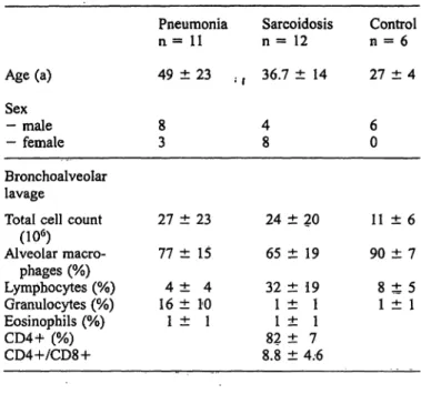
Fig. l Bas l (dark columns) and interferon-γ induced (100 kU/1, white columns) ICAM-1 expression on the surface of alveolar ma- crophages from patients and controls. Mean ± SD.
* = p < 0.01 versus basal expression on the surface of control alveolar macrophages.
** = p < 0.05 versus basal expression on the surface of control alveolar raacrophages.
Control (n = 6);
Pneumonia (n = 11), Sarcoidosis (n = 12).
Eur. J. Clin. Chem. Clin. Biochem. / Vol. 32, 1994 / No. 6
tected in all cases (flg. 1). Specific antibody binding was determined for each patient/control by subtracting non- specific binding of mouse IgG, to alveolar macrophages (fig. 2). In all cases absorbance due to non-specific bind- ing did not exceed 10% of maximum absorbance ob- tained after addition of the specific anti-IC AM-1 anti- body. We found significantly elevated ICAM-1 ex- pression on the surface of sarcoid alveolar macrophages, compared with those of healthy controls (0.76 EU
±0.27 vs. 0.44 EU ±0.12, p < 0.01). In contrast, ICAM-1 expression on the surface of alveolar macro- phages from patients with pneumonia was not elevated (0.48 EU ± 0.35, p = not significant).
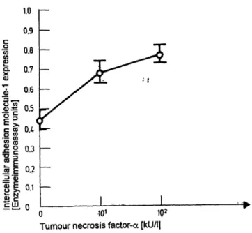
ICAM-1 expression on the surface of alveolar macro- phages from healthy volunteers was augmented after Stimulation with interferon-γ or TNF-α (fig. 1). In three controls, a dose dependent increase of ICAM-1 ex- pression was demonstrated (fig. 3). However patients' alveolar macrophages did not respond to cytokines even at high doses (500 kU/1 interferon-γ or 500 kU/1 TNF- a, data not shown).
ίο h-
0.9 0.8 0.7
ε °·
6 8 0.5 0.4 0.3 0.2 0.1 Ο1.0 Γ-
Γ
5! Pl
1 3
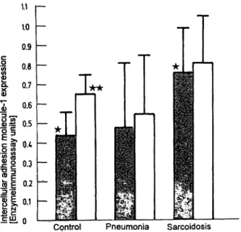
Fig. 2 Assay results given s A40s nm for the expression of ICAM- 1 on the surface of alveolar macrophages.
ELISA tests were performed s follows:
No.
21 34
Specific Mouse monoclonal IgGj pnmary-
anti-ICAM-1 antibody
—+ —
— +
+
Enzyme- linked second antibody
-f
—_|-
+
strateSub-
+ +4.
-1-
0 101 102
Tumpur necrosis f ctor^a [kU/l]
Fig. 3 TNF-a ihduced ICAM-1 expression pn the surface of alve- olar macrophages from healthy volunteers. Mean ± SD from 3 cases.
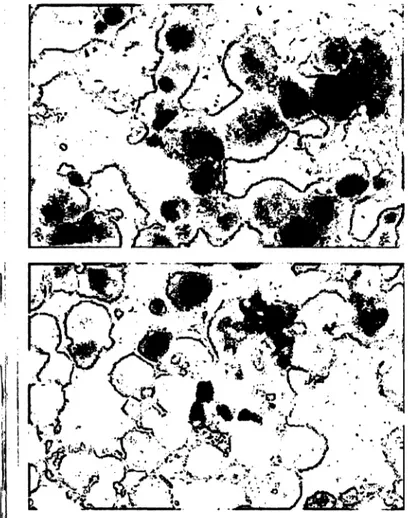
Irnmunocytochemical staining on cytocentrifuge prep- arations correlated with the results obtained with the im- m noenzymometric assay method. In pulmon ry s r- coidosis the number of visibly stained cells was signifi- cantly elevated when compared with healthy volunteers (78% ± 11 vs. 40% ± 8 positive cells, p < 0.05). Fur^
thermore, we observed that staining of patients' alveolar macrophages resulted in more intense colouring of the cells than immunocytochemical staining of control al- veolar macrophages (fig. 4).
Tumour necrosis factor-α bioassay
The black part of column 4 corresponds to the expression of the ICAM-1 on the cell surface.
was determined in the supernatants of alveolar macrophages. All sarcoid alveolar macrophages spon- taneously released TNF-α, whereas only low TNF-a concentration could be detected in the supernatants of controls. TNF-α concentrations in patients ranged from 5-1375 kU/1 reflecting different stages of activity of the disease. TNF-α concentration in the supernatants of sarcoid alveolar macrophages correlated with the·
ICAM-1 surface expression of these cells (r s? 0.-77, p < 0.05; 10). TNF-α was not determined in bacterial pneumonia.
Discussion
We desoribe a method for the rapid, semi-a t m tic
identification of IC M-1 on the surface of alveolar mac-
rophages. Our results correlate with those obtained by
immunocytochemical staining, a technique which has
been used previously to esttoate surface expression of
adhesion molecules on lveot r macroplaages (8, 9). We
Kamp et al.: ICAM-1 on alveolar macrophage surface 459
Fig. 4 Immunocytochemical staining of cytospin preparations of sarcoid alveolar macrophages (upper panel) and control alveolar macrophages (lower panel) using alkaline phosphatase monoclonal mouse anti-alkaline phosphatase complex (magnification: 400 X).
did not directly compare our ELISA with flow eytomet- ric methods, which can also be used for detection of cell surface markers. Flow cytometry is widely established for lymphocyte typing and provides the advantage of testing more than one antibody on identical cells. How- ever alveolar macrophages show high background fluor- escence, which interferes with the specific signal, es^
pecially when low receptor expression is expected (28).
In this fespect, we fouüd the immunoenzymometric as- say to be very precise, allowing the quantification of small differences in ICAM^l expression that are difficult to assess by microscopic aiiälysis. The use of a micro- plate reader for measuring the indicator enzyme activity at the end of the experiment is not only convenient but leads to more objective quantitative results than those obtained by microscopic evaluation of cell stains. In ad- dition, large numbers of cells can be analysed in one experiment (e. g. investigation of 10 wells of a microtitre plate equals the analysis of 10 X l O
5cells), which leads to highly reproducible results.
Furthermore, differences in the colour intensity of the immunocytochemically stained cells are not quantifiable
by microscopic evaluation. We observed marked Vari- ation in this respect between patient samples and con- trols, which may reflect up-regulation of the number of binding sites expressed per cell (1).
Finally, the immunoenzymometric assay provides a set- ting suitable for studying inodulatory effects of cyto- kines or other agents on the expression of ICAM-1 on the surface of alveolar macrophages in vitro. In sum- mary we see this method äs an approach to a better semi-quantitative identification of adhesion molecules on the surface of alveolar raacrophages, compared with conventional immunocytochemical staining.
The main finding of the clinical part of this study was that ICAM-1 expression on the surface of alveolar mac- rophages is differently regulated in patients with sar- coidosis (mononuclear cell alveolitis) and bacterial pneumonia (neutrophil alveolitis). Sarcoid alveolar mac- rophages expressed significantly higher amounts of ICAM-1 than macrophages of healthy volunteers. Our data are consistent with recent work demonstrating in- creased expression of ICAM-1 on the surface of alveolar macrophages from patients with pulmonary sarcoidosis using immunocytochemical staining (8, 9). We fiirther demonstrated that ICAM-1 expression of control al- veolar macrophages can be significantly increased by Stimulation with either TNF- or interferon- . It has been demonstrated previously that ICAM-1 expression can be induced by cytokines on various cell types (1), but no data were hitherto available on the in vitro induc- tion of ICAM-1 on the surface of alveolar macrophages.
In contrast, ICAM-1 expression on the surface of al- veolar macrophages from patients with bacterial pneu- monia was not elevated, proving that upregulated ICAM-1 expression is not an unspecific finding in pul- monary inflammation. In the light of present knowledge, ICAM-1 expression on the alveolar macrophages sur- face seems to be crucial in the pathogenesis of lympho- cyte accumulation and activation in the pulmonary com- partment (10, 29). In neutrophil alveolitis different mechanisms of bronchoalveolar cell activation are likely to be involved.
Since alveolar macrophages can be harvested in large numbers from sites of disease activity by bronchoalveo- lar lavage, rapid and reliable identification of adhesion molecules such äs ICAM-1 on the surface of cells from the pulmonary compartment might be a useful tool for further elucidation of the pathogenesis of these diseases.
Acknowledgement
The authors acknowledge the expert technical assi Karlberg and Monika Losch.
Eur. J. Clin. Chem. Clin. Biochem. / Vol. 32,1994 / No. 6
References
1. Dustin, M. L., Rothlein, R., Bhan, A.K., Dinarello, C. A. &
Springer, T. A. (1986) Induction by IM and interferon- : Tis- sue distribution, biochemistry, and function of a natural adher- cnce molecule (ICAM-l). J. Immunol. 137, 245-254.
2. Springer, T. A. (1990) Adhesion receptors of the immune Sys- tem. Nature 346, 425-434.
3. Mariin, S. D. & Springer, T. A. (1987) Purified intercellular adhesion molecule-1 (lCAM-1) is a ligand for lymphocyte function associated antigen (LFA-1). Cell 57, 813-819.
4. Wawryk, S. O., Novotny, J. R., Wicks, 1. R, Wilkinson, D., Mäher, D., Salvaris, E., Welch, K., Fecondo, J. & Boyd, A. W.
(1989) The role of the LFA-l/ICAM-1 interaction in human leukocyte homing and adhesion. Immunol. Rev. 108, 135- 5. Barton, R. W., Rothlein, R., Ksiazek, J. & Kennedy, C. (1989)161.
The effect of anti-intercellular adhesion rnolecule-1 on phor- bol-ester-induced rabbit lung inflammation. J. Immunol. 143,
1278-1282.
6. Isobe, M., Yagita, H., Okumura, K. & Diara, A. (1992) Specific acceptance of cardial allograft after treatment with antibodies to ICAM-l and LFA-1. Science 255, 1125-1128.
7. Staunton, D. E., Mariin, S. D., Dustin, M. L. & Springer, T. A.
(1988) Primary structure of ICAM-l demonstrates interaction between members of the immunoglobulin and integrin super- family. Gell 52, 925-933.
8. Melis, M., Gjomarkaj, M., Pace, E., Malizia, G. & Spatafora, M. (1991) Increased expression of leukocyte function associ- ated antigen-1 (LFA-1) and intercellular adhesion molecule-1 (ICAM-l) by alveolar macrophages of patients with pulmon- ary sarcoidosis. Chest 100, 910-916.
9. Striz, L, Wang, Y.-M., Kalaycioglu, O. & Costabel, U. (1992) Expression of alveolar macrophage adhesion molecules in pul- monary sarcoidosis. Chest 102, 882—886.
10. Dalhoff, K., Bohnet, S., Braun, J., Kreft, B. & Wießmann, K.
J. (1993) Intercellular adhesion molecule l (ICAM-l) in the pathogenesis of mononuclear cell alveolitis in pulmonary sar- coidosis. Thorax 48, 1140-1144.
11. Rothlein, R., Czajkowski, M., O'Neill, M. M., Mariin, S. D., Mainolfi, E. & Merluzzi, V. J. (1988) Induction of intercellular adhesion molecule l on primary and continuous cell lines by pro-inflammatory cytokines. J. Immunol. 141 (1665—1669.
12. Müller-Quernheim, J., Pfeifer, S., Männel, D., Strausz, J. &
Ferlinz, R. (1992) Lung-restricted activation of the alveolar macrophage/monocyte System in pulmonary sarcoidosis. Am.
Rev. Respir. Dis. 145, 187-192.
13. Robinson, B. W. S., McLemore, T. L. & Crystal, R. G. (1981) Gamma interferon is spontaneously released by alveolar mac- rophages and lung T-lymphocytes in patients with pulmonary sarcoidosis. J. Clin. Invest. 75, 1488-1495.
14. Hunninghake, G. W. (1984) Release of interleukin-1 by al- veolar macrophages of patients with active pulmonary sarcoid- osis. Am. Rev. Respir. Dis. 129, 569-572.
15. Bachwich, P. R., Lynch III, J. R, Larrick, J. W., Spengler, M.
& Kunkel, S. L. (1986) Tumor necrosis factor production by human sarcoid alveolar macrophages. Am. J, Pathol. 725, 421-426.
16. Sernenzato, G. (1986) The immunology of sarcoidosis. Semin.
Respir. Med. 8, 17-29.
17. Aerts, C., Wallaert, B. & Grobois Jr, C. (1986) Release of Superoxide anion by alveolar macrophages in pulmonary sar- coidosis. Arm. N. Y. Acad. Sei. 4 , 192-200.
18. Dalhoff, K., Braun, J., Ldpp, R., Schnabel, A. & Wießmann, K.
J. (1992) Sauerstofrradikalbildüng bei pulmonaler Sarkoidose.
Dtsch. Med. Wochenschr. 117, 887-892,
19. Keogh, A., Hunninghake, G. W., Line, B; R. & Crystal, R. G.
(1983) The alveolitis of pulmonary sarcoidosis. Am. Rev. Re- spir. Dis. 128, 256-265.
20. Crystal, R. G., Bitterman, P. B., Rennard, S. L, Hatice, A. J.
& Keogh, B. A. (1984) Interstitial lung disease of unknown cause: disorders characterized by chronic inflammation of the lower respiratory tract. N. Engl. J. Med. 310, 154-166 and 235-244.
21. Hoogsteden, H. C., van Hai, P. T. W., Wijkhuijs, J. M., Hop, W., Verkaik, A. P. K. & Hivering, C. (1991) Expression of the CD 11/CD 18 cell surface adhesion glycoprotein family on alveolar macrophages in smokers and nonsmokers. Chest 100, 1567-1571.
22. Crystal, R. G., Reynolds, H, Y. & Kalaca, A. R. (1986) Bron- choalveolar lavage. Chest 90, 120-133-
23. Kelly, C. A., Ward, C., Stenton, S. C., Heridriok, D. J. & Wal- ters, E. H. (1988) Assessment of pulmonary macrophage and neutrophil function in sequential bronchoalveolar lavage aspir- ates in sarcoidosis. Thorax 43, 787-791.
24. Mosmann, T. (1983) Rapid colorimetric assay for cellular growth and survival: Application to proliferation and cytotox- icity assays. J. Immunol. Methods 65, 55—63.
25. Erber, W. N., Pinching, A. J. & Mason, D. Y. (1984) Immuno- cytochemical detection of T and B cell populations in routine blood smears. Lancet /, 1042-1046.
26. Feist, W., Ulmer, A. J.^ Musehold, J., Brade, H., Kusumoto, S-
& Flad, H.^D. (1989) Induction of tumor necrosis factor-alpha release by lipopolysaecharide and defmed lipopolysaccharide partial structures. Immunbiology 179, 293-307.
27. Sachs, L. (1984) Angewandte Statistik. Springer Verlag, Berlin, pp. 230-238.
28. Lehnert, B. E., Valdez, Y. E., Filak, D. A., Steinkamp, J. A.
& Stewart, C. C. (1986) Flow cytometric characterization of alveolar macrophages. J. Leukocyte Biol. 39, 285—298.
29. Bohnet, S., Braun, J. & Dalhoff, K. (1994) Intercellular ad- hesion molecule l is upregulated on alveolar macrophages from AIDS-patients. Eur. Respir. J. (in press).
30. Sibille, Y. & Marchandise, F. X. (1993) Pulmonary immune cells in health and disease: Polymorphonuclear neütrophils.
Eur. Respir. J. 6, 1529-1543.
Dr. med. K. Dalhoff Klinik für Innere Medizin
Medizinische Universität zu Lübeck Ratzeburger Allee 160
D-23538 Lübeck Germany