Dictyostelium discoideum
INAUGURAL-DISSERTATION
Zur Erlangung des Doktorgrades der mathematisch-naturwissenschaftlichen
Fakultät der Universität zu Köln
Vorgelegt von
Huajiang Xiong
aus Chang Chun, V.R. China
Köln, Februar 2007
This research work was carried out under the supervision of Prof. Dr.
Angelika A. Noegel from April 2004 to December 2006, in the Institute for Biochemistry I, Medical Faculty, University of Cologne, Cologne, Germany.
Diese Arbeit wurde unter der Anleitung von Frau Prof. Dr. Angelika A.
Noegel vom April 2004 bis Dezenber 2006 im Institut für Biochemie I der Medizinischen Fakultät der Universität zu Köln, angefertigt.
Referees/Berichterstatter: Prof. Dr. Angelika A. Noegel Prof. Dr. Siegfried Roth
Oral examination expected/
Mündliche Prüfung
vorraussichtlich: 24. April 2007
Ich möchte mich ganz herzlichst bei Frau Prof. Dr. Noegel bedanken, daß sie mich in ihre Gruppe aufgenommen und mir die wunderbare Gelegenheit gegeben hat, an diesem spannenden Projekt zu arbeiten. Dank ihrem herausragenden Wissen und ihrer Unterstützung auf allen Gebieten wurde diese Arbeit so vielfältig gestaltet.
Ich danke den Herren Prof. Dr. Siegfried Roth und Prof. Dr. Jens Brüning für die freundliche Übernahme der jeweiligen Koreferate, außerdem der „International Graduate School for Genetics and Functional Genomics“ für die finazielle Unterstützung und wissenschaftliche Förderungskurse.
Besonderer Dank gilt Dr. Francisco Rivero für die Hilfe bei allen Mikroskopieproblemen, die Bereitstellung der interaptin-Stämme, Scholade, Süßigkeiten und Knaller in jeglichen Versteckmöglichkeiten…Du bist der Knaller.
Herzlichen Dank an das Labor 14, das Labor mit den Golden Girls, Anne (Tanz mir den Dicty!), Mary (Sushi?), Monika, Rosi (Falcon-Sekt hat Stil und Du solltest Klaus öfters mitbringen!), Julia, Tanja (schön mit dir die Bench zu teilen), Surayya und the best Indian dancer/dressman/white collar Subhanjan and the karaoke master, and Vivek (do more of the Oooouuhs, good to have you guys in the lab, you are the balance for all the estrogens!).
Ich danke alle Mitarbeiter der Biochemie I, Wenshu (my favourite batch mate), Georgia (meine „Nachbarin“, ich drücke Dich so gern), Jianbo, Rui, Ludwig, Yvonne, Verena, Sascha, Akis, Ria, Martina, Jessica, Kristina, Eva- Maria, Rolf, Charles, Christoph, Christian, Maria, Dörte, Budi, Gudrun, Berthold, Claudia, Bärbel, Brigitte, Sonya, Oliver und Chrisitina, sowie Ursula Euteneuer für die herrlichen EM Aufnahmen und Annette Vogel für ihre Hilfe
Ein liebevoller Dank gilt meinen Eltern und Großeltern, die mich immer
gefördert und unterstützt haben. Ich hoffe, die Arbeit gefällt Euch!
Für meine Eltern
Table of contents
1 Introduction ...1
1.1 Organization of the nuclear envelope ... 1 1.2 Nuclear positioning ... 2 1.2.1 KASH domain proteins in the ONM________________________________ 2 1.2.2 SUN domain proteins in the INM__________________________________ 7 1.3 The model system Dictyostelium discoideum... 12 1.3.1 Aim of the study _____________________________________________ 13
2 Material and Methods...14
2.1 Materials... 14 2.1.1 Kits _______________________________________________________ 14 2.1.2 Enzymes, antibodies and antibiotics ______________________________ 14 2.2 Media and buffers ... 16 2.2.1 Media and buffers for Dictyostelium culture ________________________ 16 2.2.2 Media for Escherichia coli culture ________________________________ 16 2.2.3 Buffers_____________________________________________________ 16 2.2.4 Biological materials ___________________________________________ 17 2.2.5 Plasmids and constructs _______________________________________ 17 2.2.6 Oligonucleotides _____________________________________________ 18 2.3 Methods ... 19 2.3.1 Generation of cDNA __________________________________________ 19 2.3.2 Plasmid DNA and total RNA isolation _____________________________ 19 2.3.3 Constructs __________________________________________________ 19 2.4 Biochemical methods... 20 2.4.1 Preparation of total lysates and intact nuclei________________________ 20 2.4.2 Gel electrophoresis and immunoblotting ___________________________ 21 2.4.3 Proteinase K protection assay __________________________________ 21 2.4.4 Chromatin immunoprecipitation (ChIP) ____________________________ 22 2.4.5 Preparation of GST fusion proteins _______________________________ 22 2.4.6 GST pull-down assay _________________________________________ 23 2.4.7 In vitro cross-linking experiment _________________________________ 23 2.4.8 Indirect immunofluorescence microscopy __________________________ 23 2.4.9 Electron microscopy __________________________________________ 24 2.4.10 Metaphase arrest of cell division ______________________________ 24 2.5 Growth and development of D. discoideum... 25
3 Results ...26
3.1 SUN domain proteins in Dictyostelium... 26 3.1.1 Sun-1 and Sun-2 in Dictyostelium________________________________ 26 3.1.2 Domain architecture of Sun-1 and Sun-2 __________________________ 26 3.1.3 Multiple alignment of the SUN domains ___________________________ 29 3.2 Generation of Sun-1 monoclonal antibodies... 30 3.3 Biochemical properties of Sun-1 ... 34 3.3.1 Sun-1 is restricted to the nucleus ________________________________ 34 3.3.2 Sun-1 is an integral membrane protein ____________________________ 36 3.3.3 Sun-1 oligomerizes via the coiled-coil domains _____________________ 37 3.3.4 Sun-1 may form dimers and trimers in vivo_________________________ 40 3.3.5 Sun-1 is a type II transmembrane protein __________________________ 42 3.3.6 Sun-1 associates with chromatin ________________________________ 44 3.4 Sun-1 and the KASH domain protein interaptin... 46 3.4.1 The KASH domain protein homolog interaptin ______________________ 46 3.4.2 Expression profile of Sun-1 and interaptin _________________________ 49 3.4.3 Localization of Sun-1 and interaptin ______________________________ 50 3.4.4 The KASH motif of interaptin is required for nuclear membrane targeting _ 53 3.4.5 The KASH motif of interaptin competes with Sun-1 for the nuclear membrane localization _________________________________________________ 55 3.5 N-terminal truncation of Sun-1 ... 57 3.5.1 Expression of GFP-∆NSun-1 deforms the nucleus ___________________ 57
Table of contents
3.5.4 GFP-∆NSun-1 cells formed protrusion to the centrosome _____________ 65 3.5.5 GFP-∆NSun-1 disconnected the nucleus from centrosomes ___________ 67 3.5.6 GFP-∆NSun-1 deformed the nucleus during intracellular movement _____ 72
4 Disussion ...75
4.1 SUN domain proteins in D. discoideum... 75
4.2 Subcellular localization of Sun-1... 77
4.3 Sun-1 forms dimers and trimers in vitro... 78
4.4 Sun-1 is a type II nuclear envelope protein and associated with chromatin... 80
4.5 Sun-1 and interaptin compete for the nuclear membrane localization ... 82
4.6 The N-terminus of Sun-1 confers INM localization ... 85
4.6.1 GFP-∆NSun-1 affects the nuclear shape __________________________ 86 4.6.2 GFP-∆NSun-1 causes centrosome amplification ____________________ 88 4.7 Proposed model for the function of Sun-1 in D. discoideum... 91
5 Summary...93
6 Zusammenfassung ...94
7 References...95
8 Table of figures ...100 9 Appendix...I
General abbreviations________________________________________________I Eidesstattliche Erklärung ____________________________________________III Curriculum vitae __________________________________________________ IV Lebenslauf _______________________________________________________ V
1 Introduction
1.1 Organization of the nuclear envelope
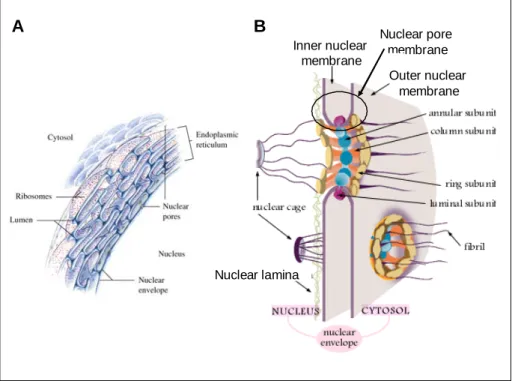
In eukaryotic cells, the interphase nucleus is surrounded by the nuclear envelope (NE) that functions as a barrier separating the nuclear and the cytoplasmic compartment. The NE is a highly specialized membrane system composed of the morphologically distinct outer and inner nuclear membrane (ONM, INM), the nuclear pore complexes (NPCs) and the nuclear lamina (Figure 1A). The ONM is contiguous with the rough endoplasmic reticulum (rER) that is decorated with ribosomes (Franke et al., 1981; Gerace and Burke, 1988; Newport and Forbes, 1987; Watson, 1955) and joins the INM at the sites of the nuclear pore membrane within the NPCs (Figure 1B). Further, the INM is supported by the nuclear lamina, a dense network consisting of the intermediate filaments lamins and lamin-associated proteins which confers mechanical stability to the nucleus and provides an anchor for integral proteins of the INM (Gruenbaum et al., 2005; Holmer and Worman, 2001).
A B
Nuclear poremembrane Inner nuclear
membrane
Outer nuclear membrane
Nuclear lamina
Figure 1: Composition of the nuclear envelope in eukaryotic cells. A. The nuclear envelope is a system of nuclear membranes continuous with the rough endoplasmic reticulum. B.
1 Introduction
The inner nuclear envelope is supported by the nuclear lamina and joins the outer nuclear membrane at the sites of the nuclear pore complex (Horton et al., 2002).
1.2 Nuclear positioning
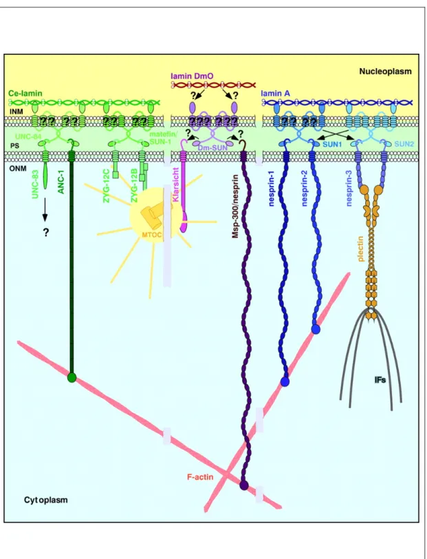
The well-defined position of the nucleus within a cell is essential for many processes such as mitosis, meiosis, fertilization as well as cell migration, differentiation and polarization which represent the repertoire of all eukaryotic cells (Morris, 2000). However, nuclear positioning and migration requires a concerted action of the cytoskeleton that includes the actin filaments (F-actin), microtubules (MTs) and intermediate filaments (IFs), to which the nucleus must be connected. In recent years, two conserved nuclear envelope protein families, the KASH domain proteins and the SUN domain proteins have been described as molecular linkers connecting the nuclear envelope and the cytoskeleton.
1.2.1 KASH domain proteins in the ONM
Although the ONM is contiguous with the tubular system of the rER
and allows integral proteins to diffuse to both membranes (Ellenberg et al.,
1997; Lippincott-Schwartz et al., 2000), they possess distinct protein
compositions. A new protein family termed the KASH domain proteins is mainly
localized in the ONM and has been proposed to connect the nucleus to the
different cytoskeletal elements. These proteins can be divided into three
groups based on their interaction with (1) F-actin: Anc-1 of Caenorhabditis
elegans (Hedgecock and Thomson, 1982; Starr and Han, 2002), Msp-
300/Nesprin in Drosophila melanogaster (Rosenberg-Hasson et al., 1996; Volk,
1992; Zhang et al., 2002), as well as Nesprin-1 and 2 (synonym Syne, Myne,
Enaptin and NUANCE) in mammals (Apel et al., 2000; Padmakumar et al.,
2005; Zhang et al., 2001; Zhen et al., 2002); (2) IFs: Nesprin-3 in mammals
(Wilhelmsen et al., 2005); (3) centrosome and/or MTs: Zyg-12 (Fridkin et al.,
2004; Malone et al., 2003) and UNC-83 in worms (Malone et al., 1999; Reinsch and Gonczy, 1998) and Klarsicht in flies (Fischer-Vize and Mosley, 1994;
Patterson et al., 2004).
All KASH domain proteins reported so far possess a highly conserved C-terminal region referred to as the Klarsicht/Anc-1/Syne-1 homology (KASH) domain harboring a single transmembrane domain and a short tail sequence of approximately 35 amino acids (Starr and Han, 2005; Wilhelmsen et al., 2006).
Regardless of the KASH domain, the three groups of KASH domain proteins
differ in their N-terminal domain organization that is specialized for the
connection to the different cytoskeletal components (Figure 2).
1 Introduction
Figure 2: Proposed model for nuclear positioning and migration. SUN domain proteins, e.g.
UNC-84, reside in the inner nuclear membrane (INM) by interaction with the nuclear lamina or lamin-binding proteins. The SUN domain of UNC-84 binds directly to the KASH domain proteins (UNC-83- or Nesprin-related protein), which are retained specifically in the outer nuclear membrane (ONM). UNC-83-related proteins connect the nucleus to microtubules, whereas Nesprin-related proteins link the nucleus to actin filaments (Wilhelmsen et al., 2006).
1.2.1.1 Nuclear positioning on F-actin and IFs
The F-actin binding KASH domain proteins contain two N-terminally located calponin-homology (CH) domains followed by a central stretch of spectrin-like repeats and the KASH domain, giving rise to the giant molecules of up to 1 MDa. Anc-1 is unique as it harbors six interspersed coiled-coil repeats in its elongated domain. Worms, carrying mutated anc-1 (nuclear anchorage defective, expressed as a 950 kDa protein) failed to position the nuclei and mitochondria correctly and the nuclei floated freely in the cytoplasm (Hedgecock and Thomson, 1982; Starr and Han, 2002).
In agreement, flies generated with the germ-line specific msp-300
SZ-75allele for a mutated 300 kDa muscle-specific protein were observed with defective nuclear positioning during oogenesis (Yu et al., 2006). Moreover, binding of Msp-300/Nesprin to F-actin in vivo is essential for myogenesis in Drosophila embryos, as the lethal msp-300
SZ-75mutation caused defects in myotube migration, attachment and contraction (Rosenberg-Hasson et al., 1996; Volk, 1992).
In mammals, Nesprin-1 and 2 occur as diverse isoforms generated by alternative splicing, transcription initiation and termination of the genes syne-1 and syne-2 (Warren et al., 2005), whereas the giant Nesprin-1 and Nesprin-2 isoforms Enaptin and NUANCE (976 and 764 kDa) encompass completely the CH domains and the KASH domain and can potentially reach out 500 nm from the nuclear envelope into the cytoplasm (Starr and Han, 2005). Similar to Msp- 300/Nesprin in flies, Nesprin-1 (Myne-1) was first found to position the postsynaptic nuclei at neuromuscular junctions and within myocytes, but is also expressed in other cell types (Apel et al., 2000; Mislow et al., 2002;
Padmakumar et al., 2005; Zhang et al., 2001; Zhen et al., 2002). Nevertheless, Nesprin-1 may have other functions despite of nuclear positioning, e.g. when overexpressed in epithelial cells it acted as a dominant-negative inhibitor for the morphology of the Golgi apparatus that collapsed into a condensed structure near the centrosome (Gough et al., 2003).
Additionally, the mammalian Nesprin-3, a protein of 110 kDa, lacking a
calponin homolgy domain interacts with plectin a protein that in turn is capable
1 Introduction
to cross-link cytoplasmic intermediate filaments and F-actin as it also harbors an F-actin binding domain. This indicates that the small Nesprin isoform can also contribute to nuclear positioning (Wilhelmsen et al., 2005).
1.2.1.2 Nuclear positioning on centrosome and microtubules Evidently, positioning and migration using F-actin and IFs may be assisted by the additional connection of the nucleus to the centrosomes and microtubules, a property attributed to the second group of KASH domain proteins including Zyg-12 and UNC-83 in C. elegans and Klarsicht in D.
melanogaster.
In detail, worm embryos with the zygote defective (zyg-12) genetic background die owing to chromosome segregation defects caused by the nucleus-centrosome detachment (Malone et al., 2003). Notably, Zyg-12 binds to dynein and is a member of the Hook protein family that has been suggested to link membrane compartments and microtubules (Walenta et al., 2001).
However, of the three Zyg-12 isoforms (A, B and C) derived from alternative splicing of a single gene the isoforms B and C are localized at the ONM, whereas A lacking a KASH domain is associated with the centrosome. In the proposed two-step model, all Zyg-12 isoforms migrate on microtubules in a dynein-dependent manner, but Zyg-12B and C are localized at the nuclear envelope and Zyg-12A is attached to the centrosome. Once the nucleus and centrosome are located in close proximity the connection is provided by heterodimerization of Zyg-12B and/or C with Zyg-12A (Malone et al., 2003).
Comparable to Zyg-12, Klarsicht of D. melanogaster participates in
nuclear migration from the basal compartment to the apex of the imaginal disc
during the compound eye development by attachment of the nucleus to the
centrosome and microtubules (Fischer et al., 2004; Fischer-Vize and Mosley,
1994; Patterson et al., 2004). Distinctly different from Zyg-12, Drosophila
embryos bearing a null mutation in the gene klarsicht are viable and fertile
regardless of the misshapen photoreceptors.
UNC-83 is essential for the nuclear migration in certain tissues during embryonic development of the worm, such as the hypodermal cells and the intestine. Interestingly, though mutations in the unc-83 gene abrogated nuclear migration, the nucleus-centrosome connection was not disrupted hinting at a putative function of UNC-83 in nuclear migration along microtubules, but not essential for the connection to centrosomes yet to be uncovered (Lee et al., 2002; Starr et al., 2001; Sulston and Horvitz, 1981). In summary, KASH domain proteins of the ONM engage in interactions with different cytoskeletal elements to allow the processes nuclear positioning and migration and are thus essential both for the embryonic development and the organization of the cytoplasm.
1.2.2 SUN domain proteins in the INM
As the integral proteins can diffuse laterally throughout the rER and the ONM, specific mechanisms are required to retain distinct proteins at the ONM. Studies on the KASH domain proteins revealed that they are recruited to the ONM by the SUN domain proteins, a new INM protein family.
The first SUN domain protein UNC-84 was identified in C. elegans.
Mutants exhibiting an uncoordinated movement due to the defects in nuclear positioning and migration resulted from alterations in the gene unc-84 (Horvitz and Sulston, 1980; Malone et al., 1999; Sulston and Horvitz, 1981).
Interestingly, the phenotype of the unc-84 mutation resembles that of the unc-
83 worms indicating that both proteins are involved in a common mechanism
(Starr et al., 2001). The N-terminal domain of UNC-84 containing several
putative transmembrane domains does not show sequence or domain
similarities with any known proteins whereas a C-terminal region of
approximately 120 amino acids shares a significant homology with the C-
terminus of the spindle pole body protein Sad1 in Schizosaccharomyces
pombe (Hagan and Yanagida, 1995). Therefore it was designated as the
Sad1/UNC-84 (SUN) domain (Malone et al., 1999; McGee et al., 2006). As
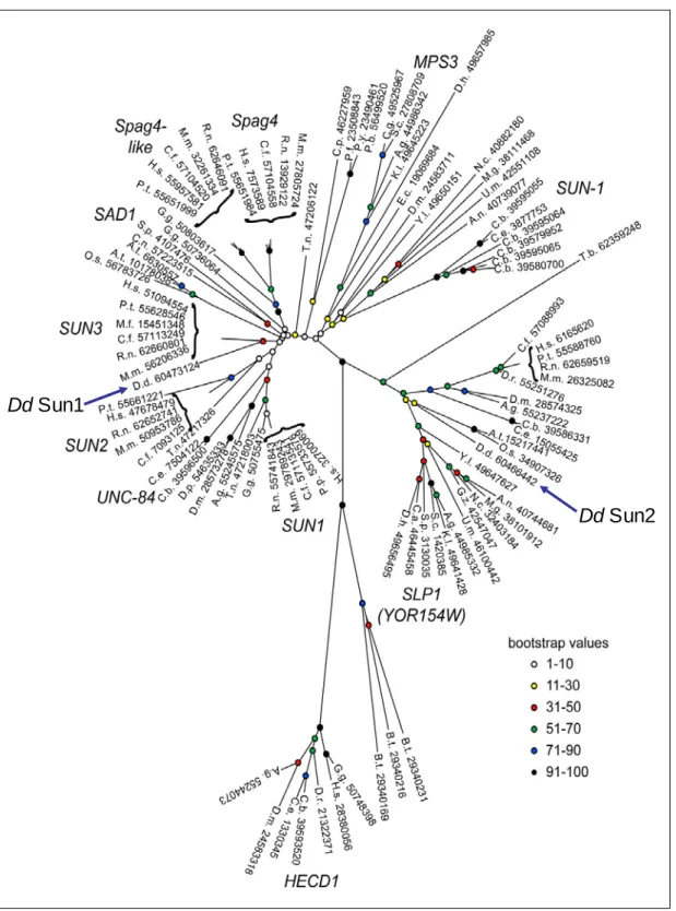
illustrated in Figure 3, further database searches discovered two SUN domain
1 Introduction
proteins in D. melanogaster and D. discoideum (termed Dd Sun-1 and Dd Sun- 2) and four in mammals (Crisp et al., 2006; Dreger et al., 2001; Hasan et al., 2006; Jaspersen et al., 2006; Malone et al., 1999; Padmakumar et al., 2005).
The presence of SUN domain proteins across many species and their
propensities to interact with KASH domain proteins indicate a conserved
mechanism for nuclear positioning and migration.
Dd Sun2 Dd Sun1
Figure 3: Phylogeny of SUN domain proteins (Jaspersen et al., 2006). SUN domain proteins are present throughout various species. Based upon the sequence homology in the SUN domains, SUN domain proteins are grouped into subfamilies. D. discoideum Sun- 1 is grouped into the SUN3 subfamily, whereas D. discoideum Sun-2 is grouped into the subfamily of the SUN-like proteins (SLP).
1 Introduction
1.2.2.1 Domain architecture of SUN domain proteins
In metazoa, SUN domain proteins interact with the nuclear lamina, whereas the conserved C-terminal SUN domain is responsible for the binding to the KASH domain proteins. Despite of these characteristics, they exhibit great variabilities in their N-terminal domain architecture that appear to contain functionally related domains. In general, most of these proteins possess at least one transmembrane domain, e.g Sad1 and human Sun-3 (Crisp et al., 2006; Hagan and Yanagida, 1995), whereas some may have multiple transmembrane domains as nine hydrophobic domains were pedicted for UNC-84 and three for human Sun-1 (Malone et al., 1999; McGee et al., 2006;
Padmakumar et al., 2005). Extraordinarily, none of the two uncharacterized SUN domain proteins in D. melanogaster possess a transmembrane domain. A further feature of the SUN domain proteins is the presence of at least one coiled-coil domain that may promote dimerization or oligomerization (Crisp et al., 2006).
1.2.2.2 INM targeting of SUN domain proteins
In general, integral proteins of the INM are translated on the rER and
diffuse along the ONM to the INM by passing the highly curved nuclear pore
membrane of the NPCs which occurs in an ATP-dependent and temperature-
sensitive manner (Ohba et al., 2004; Soullam and Worman, 1993; Soullam and
Worman, 1995). According to the “Diffusion-Retention” model implying that
INM proteins may be trapped in there by interaction with lamins or chromatin-
binding proteins or both (Ellenberg et al., 1997; Ohba et al., 2004; Ostlund et
al., 1999; Smith and Blobel, 1993; Worman and Courvalin, 2000), SUN domain
proteins may be targeted to the INM and retained there by interaction with
lamins, as C. elegans UNC-84 and matefin/Sun-1, mammalian Sun-1 and Sun-
2 colocalize with lamins (Crisp et al., 2006; Fridkin et al., 2004; Hodzic et al.,
2004; Lee et al., 2002; Padmakumar et al., 2005). Interestingly, the N-termini
of matefin/Sun-1 and mammalian Sun-1 and Sun-2 bind to lamins in vitro, but
this interaction is not essential in vivo since they localized to the INM in the
absence of lamins (Crisp et al., 2006; Fridkin et al., 2004; Haque et al., 2006;
Hasan et al., 2006; Padmakumar et al., 2005). On the contrary, though UNC- 84 does not bind to lamin, it is localized to the INM in a lamin-dependent fashion (Lee et al., 2002) suggesting that an alternate mechanism for INM retention may exist.
1.2.2.3 Interaction of the SUN with the KASH domain proteins In the nuclear envelope, the SUN and the KASH domain proteins interact with each other by projection of their C-termini into the perinuclear space, connecting the nucleoskeleton with the cytoskeleton (Figure 2). The combination of the SUN domain proteins with various KASH domain proteins gives rise to a great repertoire linking the nucleus silmultaneously to different cytoskeletal elements: (1) combinations of UNC-84 with Anc-1 or UNC-83 provide nuclear positioning on F-actin or migration along MTs (Lee et al., 2002;
Starr and Han, 2002; Starr et al., 2001); (2) interaction of mammalian Sun-1 and Sun-2 with Nesprin-1/Nesprin-2 or Nesprin-3 contribute to the nuclear positioning on F-actin or Ifs (Crisp et al., 2006; Haque et al., 2006;
Padmakumar et al., 2005; Wilhelmsen et al., 2005); (3) the complex formed by matefin/Sun-1 and Zyg-12 is capable to transport the nucleus on MTs towards the centrosome and maintain their proximity (Fridkin et al., 2004; Malone et al., 2003). Likewise, interaction of Sad1 with Kms1, which is a KASH domain protein in S. pombe, is required for the localization of Sad1 at the spindle pole body, the centrosome equivalent of yeast (Niwa et al., 2000).
Based on their location and their interactions SUN domain proteins as
linker proteins in the INM may transduce mechanical signals originating in the
cytoplasm to the nucleus that responds with biochemical processes. Despite of
the important role of the SUN domain proteins in nuclear positioning and
migration, non-mechanical roles such as influencing the fat metabolism,
germline cell proliferation/maintenance and telomere clustering has become
evident (Fridkin et al., 2004; Greer and Brunet, 2005; Niwa et al., 2000).
1 Introduction
1.3 The model system Dictyostelium discoideum
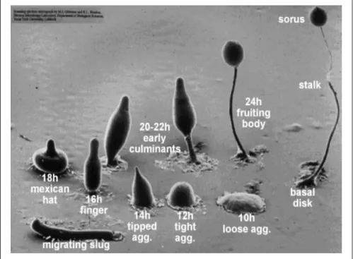
Dictyostelium discoideum is a haploid eukaryote with a simple but fast and well-defined life-cycle which facilitates genetic, biochemical and cell biological studies. The social amebae feed on bacteria, and live as single cells that develop into multicellular organisms in response to starvation. During the development, cells migrate chemotactically following a central cAMP source to form aggregates (slugs) that induce the expression of cell-cell adhesion molecules establishing the mound structures. The mounds differentiate into the terminal fruiting bodies composed of a head filled with spores supported by a stalk of vacuolated cells (Figure 4).
D. discoideum shares several features with mammalian cells, thus it is a well-accepted model to study many processes such as chemotaxis, cell-cell adhesion and morphogenesis. As nuclear positioning and migration is important for the development of various tissues in worm, fly and mammals, we set out to examine the new field of nuclear envelope proteins in D. discoideum.
Figure 4: Development of D. discoideum from single-cell ameba into the multicellular fruiting bodies (courtesy of M. Grimson, R. Blanton, Texas Tech University).
1.3.1 Aim of the study
To date, the organization and function of the nuclear envelope is not understood in D. discoideum, this challenged us to shed light onto these aspects. The first nuclear envelope protein described so far, interaptin (Rivero et al., 1998), may function as a KASH domain protein as suggested by the findings in higher eukaryotes.
The prediction of two SUN domain proteins (Sun-1 and Sun-2) in the
D. discoideum proteom prompted us to investigate the function of the SUN
domain proteins using biochemical and cell biological approaches. In this study,
we focus on the proteins Sun-1 and interaptin. Particularly, we are interested in
(1) whether Sun-1 is targeted to the inner nuclear membrane in the absence of
lamins, as reported for other SUN domain proteins; (2) whether Sun-1 interacts
with interaptin in a manner similar to mammalian SUN domain proteins and the
Nesprins to connect the nucleus to the elements of the cytoskeleton; (3)
whether these proteins have evolved other functions in the ameba.
2 Material and methods
2 Material and Methods
2.1 Materials
Standard laboratory reagents and materials were obtained from local suppliers, fine chemicals from Sigma if not otherwise indicated and instruments were supplied by the Departmental facility.
2.1.1 Kits
Nucleobond AX 100 Macherey Nagel
NucleoSpin Extraction Kit Macherey Nagel Qiagen RNeasy Mini Kit Qiagen
pGEM-T easy Cloning Kit Promega
2.1.2 Enzymes, antibodies and antibiotics
2.1.2.1 Enzymes for molecular biology purposes Calf intestinal alkaline phosphatase (CIP) Roche
Klenow fragment Roche
Lysozyme Sigma M-MLV reverse transcriptase Promega
Proteinase K Merck
Restriction endonucleases New England Biolabs Ribonuclease A (RNase A) Sigma
Ribonuclease H (RNase H) Boehringer
T4 DNA ligase Invitrogen
Taq polymerase Amersham Pharmacia
2.1.2.2 Primary antibodies Mouse monoclonal anti-actin antibody (Act1-7)
(Simpson et al., 1984) Mouse monoclonal anti-GFP antibody
(K3-184-2)
(Noegel et al., 2004)
Rabbit polyclonal anti-GFP antibody Gift from M. Schleicher
Rabbit polyclonal anti-GST antibody Unpublished
Mouse monoclonal anti-interaptin antibody (260-60-10)
(Rivero et al., 1998) Mouse monoclonal anti-PDI-1 antibody
(221-135-1)
(Monnat et al., 1997) Mouse monoclonal anti-Sun-1 antibody
(K55-432-2), western blot analysis
This study Mouse monoclonal anti- Sun-1 antibody
(K55-460-1 and K55-450-1), immunofluorescence,
(chromatin) immunoprecipitation
This study
Mouse monoclonal anti-Spermatozopsis αβ- tubulin antibody (D. discoideum centrosome) (K29-359-31)
Gift from K. Herkner
Mouse monoclonal anti-α-tubulin antibody (6-11B1)
(Piperno and Fuller, 1985) (Generous gift from Michael Koonce)
Rat monoclonal anti-α-tubulin antibody (YL1/2)
Kilmartin et al., 1982
2.1.2.3 Secondary antibodies
Goat anti-mouse IgG, peroxidase conjugated Sigma Goat anti-rabbit IgG, peroxidase conjugated Sigma Goat anti-mouse IgG, Cy3 conjugated Sigma Goat anti-mouse IgG, Cy5 conjugated Sigma
Goat anti-mouse IgG, Alexa 568 conjugated Molecular probes Goat anti-rat IgG, Alexa 568 conjugated Molecular probes
2.1.2.4 Antibiotics
Ampicillin Gruenenthal Blasticidin S ICN Biomedicals
G418 Sigma Dihydrostreptomycinsulfate Sigma
Tetracyclin Sigma
2 Material and methods
2.2 Media and buffers
All media and buffers were prepared using deionized water, filtered through an ion-exchange unit (Membra Pure). All media and buffers were sterilized by autoclaving at 120°C; the antibiotics were added to the media after cooling to approx. 50°C. Agar plates were prepared using a semi- automatic plate-pouring machine (Technomat).
2.2.1 Media and buffers for Dictyostelium culture
AX2 medium, pH 6.7 (Claviez et al., 1982)
7.15 g yeast extract 14.3 g peptone (proteose) 18.0 g maltose
0.486 g KH
2PO
40.616 g Na
2HPO
4x2H
2O ad 1liter H
2O
Soerensen phosphate buffer, pH 6.0 (Malchow et al., 1972)
2 mM Na
2HPO
4,
14.6 mM KH
2PO
4Phosphate agar plates, pH 6.0 9 g agar
ad 1 liter Soerensen phosphate buffer
SM agar plates,
pH 6.5 (Sussman, 1951)
9 g agar 10 g peptone 10 g glucose 1 g yeast extract 1 g MgSO
4x7H
2O 2.2 g KH
2PO
41 g K
2HPO
4ad 1 liter H
2O
2.2.2 Media for Escherichia coli culture
LB medium, pH 7.4 (Sambrook et al., 1989) SOC medium, pH 7.0 (Sambrook et al., 1989)
2.2.3 Buffers
10x MOPS (pH 7.0 or pH 8.0) 41.9 g MOPS
7 ml 3 M sodium acetate
20 ml 0.5 M EDTA
ad 1 liter H
2O
10x NCP (pH 8.0) 12.1 g Tris/HCl, pH 8.0 87.0 NaCl
5 ml Tween 20 ad 1 liter H
2O 1x PBS (pH 7.4) 8.0 g NaCl
0.2 g KH
2PO
41.15 g Na
2HPO
40.2 g KCl
ad 1 liter H
2O 20x SSC (pH 7.0) 3 M NaCl
0.3 M sodium citrate TE buffer (pH 8.0) 10 mM Tris/HCl, pH 8.0
1 mM EDTA 10x TAE buffer (pH 8.3) 27.22 g Tris
13.6 g sodium acetate 3.72 g EDTA
ad 1 liter H
2O
2.2.4 Biological materials
Bacterial strains
E. coli DH5α (Hanahan, 1983)
E. coli XL1 Blue Bullock et al., 1987
Klebsiella aerogenes (Williams and Newell, 1976)
D. discoideum strains
AX2-214, an axenically growing derivative of wild strain NC-4, commonly referred to as AX2
(Wallraff et al., 1986)
abpD- (AX2 strain carrying a disrupted locus of interaptin)
(Rivero et al., 1998) abpD+ (AX2 strain overexpressing interaptin) (Rivero et al., 1998) MAD-GFP (AX2 overexpressing the C-
terminal region of interaptin, referred to as GFP-IntCT in this study)
(Rivero et al., 1998)
2.2.5 Plasmids and constructs
pGEM-T easy Promega
pGEX-4T2
pGEX-4T3 Clontech
pDEX-GFP79
pGEX-4T2-SunCT1 Amino acid 411 to 572, overlapping the
coiled-coil domains located between the
2 Material and methods
transmembrane domain and the SUN domain (cloned into pGEX4T2 using XmaI/XhoI)
pGEX-4T2-SunCT2 Amino acid 700 to 905, overlapping the SUN domain
(cloned into pGEX4T2 using XmaI/XhoI) pDEX-GFP79-∆NSun-1 Amino acid 283 to 905 overlapping the
transmembrane domain, coiled-coil domains and SUN domain (cloned into pDEX-GFP-79 using ClaI)
2.2.6 Oligonucleotides
SunCT1
Xma-SunCT1-For 5'-CCCGGGGCATCATCAAACATTTTACACAATAGAT TTAGTAATAGTA
Xho-SunCT1-Rev 5'-CTCGAGACTACCATAATAAAAGTTTTTCCATGGG TCTC
SunCT2
Xma-SunCT2-For 5'-CCCGGGGCTACAAAT TGGATATTCCCACAACCA AAA
SunCT2-Xho-Rev 5’-CTCGAGCTCTTGAATAATTTGTATTTGTTCTTGTT CTGGAT
GFP-∆NSun-1
ClaI-∆NSun-1-For 5’-ATCGATAAAGTTAATTTTAAACAAGCTATTTGGAT TTTC
∆NSun-1-ClaI-Rev 5’-ATCGATATCGATTTATAATTCATCAGATTGTTGTT
G
2.3 Methods
Standard molecular biology techniques were performed as described in “A Laboratory Manual. Cold Spring Harbor Laboratory Press, NY, Vol 1-3 (Sambrook et al., 1989).
2.3.1 Generation of cDNA
For the generation of cDNA, 1-5µg of total RNA was incubated with the M-MLV reverse transcriptase in each reaction as recommended in the manufacturer’s protocols.
2.3.2 Plasmid DNA and total RNA isolation
Plasmid DNA from bacteria and total RNA from vegetative AX2 cells (shaking culture) were extracted using Nucleobond AX 100 and Qiagen RNeasy, repectively, following the manufacturer’s instructions. The plasmid DNA was used for transfection of AX2 cells; RNA samples were used in for reverse transcription and subsequent cDNA generation.
2.3.3 Constructs
2.3.3.1 GST-SunCT1 fusion vector
The two coiled-coil domains amino acid 412 to 571 is encoded in the
cDNA position 1223 to 1710 bp. A PCR fragment extending from DNA
position 1220 to 1713 (overlapping amino acid 411 to 572) was generated
with a 5’-XmaI and a 3’-XhoI, the fragment was cloned as into the expression
vector pGEX4T2 using XmaI/XhoI. The resulting vector GST-SunCT1 was
expressed in the E. coli strain XL1 Blue.
2 Material and methods
2.3.3.2 GST-SunCT2 fusion vector
The SUN domain spans the amino acid 712 to 859 that corresponds to the position 2133 to 2877 in the cDNA. We have cloned the downstream conding region from position 1885 to 2718 bp including both the SUN domain and the ER retention signal (amino acid 608 to 905) XmaI/XhoI into the expression vector pGEX4T2. Expression of the recombinant fusion protein GST-SunCT2 was carried out in the E. coli strain XL1 Blue.
2.3.3.3 GFP-∆NSun-1 fusion construct
A DNA fragment from position 853 to 2718 bp of the encoding region, corresponding to the amino acid position 283 to 905, was cloned into the vector pDEX-GFP79 using ClaI. After tranfection of the construct lacking the N-terminus (GFP-∆NSun-1) into AX2 cells, positive clones were obtained by addition of G418 (0.4µg/ml) as a selection drug.
2.4 Biochemical methods
2.4.1 Preparation of total lysates and intact nuclei
Total cell lysates were prepared from vegetative cells or at the indicated time points. After washing twice with Soerenen buffer, cells were resuspended in TMS buffer containing:
50 mM Tris/HCl pH 7.4
100 mM NaCl
5 mM MgCl
2 250 mM Sucrose
1 mM EDTA
1 mM EGTA
1 mM DTT
1 mM Benzamidine
1 mM PMSF
Total cell lysates were obtained by passing through Nuclepore membrane (5µm diameter, Whatman) and used in further experiments. Intact nuclei were isolated from vegetative cells. Nuclei were collected after lysis through Nuclepore membrane by spinning for 5 min at 4000 g.
2.4.2 Gel electrophoresis and immunoblotting
Cells were resupended and lysed in TMS buffer in a concentration of 2x10
6/ml. Protein samples were separated on 10% polyacrylamide gels (SDS-PAGE), then either stained with Coomassie Brillant Blue, or transferred onto nitrocellulose membranes (Schleicher and Schuell) semi-dry and wet blotting transfer systems, respectively. After electro-transfer, the membranes were blocked with 5% (w/v) milk in 1x NCP prior to the appropriate antibody detections. The primary antibodies were detected using the according peroxidase-conjugated secondary antibodies and visualized by ECL (enzyme chemiluminescence) reactions. ECL reactions on the nitrocellulose membranes were documented on X-ray films.
2.4.3 Proteinase K protection assay
Intact nuclei were isolated from AX2 cells and incubated in
proteinase K protection assay buffer (10mM Tris/HCl pH7.4, 250mM sucrose,
1µg/ml proteinase K) without and as a positive control for the enzyme activity
with 0.5(v/v)% Triton X-100 on ice. Samples were collected from both
reactions after 5, 10, 30, 45 min and the activity of proteinase K was
immediately stopped by adding PMSF to a final concentration of 1mM and
boiling (95°C, 5 min) in SDS sample buffer. The samples were further
analyzed by SDS-PAGE and western blot.
2 Material and methods
2.4.4 Chromatin immunoprecipitation (ChIP)
Vegetative AX2 cells were harvested and lysed in TMS buffer in a density of 5x10
7cells per ChIP reaction. To reduce the viscosity of genomic DNA and disrupt the nuclear envelope, total cell lysates were sonified (10 impulses, each of the twice repeated procedure was paused for 10 sec). The appropriate mAb was coupled to protein A-beads and PBS was used as a negative control (1hr, 4°C).
The beads were washed three times with PBS then incubated with sonified total cell lysate (2hrs on a vertical rotator, 4°C). Unspecific protein and DNA bindings were removed by five times washing with PBS and once with TE buffer and divided into two aliquotes.
One aliquote of the each ChIP reaction was used for elution of DNA by adding 100 µl of TE buffer containing 1% SDS. After Phenol/Chloroform and ethanol precipitation DNA was resuspended in 50µl aqua bidest, 5µl of this DNA was subsequently used for PCR amplification to detect the presence or absence of coprecipitated DNA. To the second aliquote 20 µl SDS sample buffer was added and subjected to SDS-PAGE and western blotting.
2.4.5 Preparation of GST fusion proteins
Recombinant GST-u84CT1 expression was induced in E. coli strain XL1-Blue by 0.3 mM IPTG for 5h at room temperature. Cells were harvested by centrifugation at 5000 g, 4°C for 15 min (Beckman) and resuspended in TM buffer (TMS without sucrose). After sonification the soluble fraction of the bacterial lysate was obtained by centrifugation at 13000 g, 4°C for 30 min.
The GST-u84CT1 was isolated from the supernatant by incubating with
glutathione agarose beads (4°C, 16 h). Glutathione agarose beads coupled
with GST-u84CT1 was washed five times with PBS (500 g, 4°C, 10 min)
before performing thrombin cleavage (4°C, 16 h) or incubating with AX2 total
cell lysate.
2.4.6 GST pull-down assay
Total cell lysate of AX2 was prepared as described above.
Glutathione agarose beads coupled with GST-SunCT1 (see above) were incubated with total cell lysate at 4°C for 5 h. The beads were washed three times with PBS (500 g, 4°C, 1min) and boiled in SDS sample buffer (95°C, 5 min). Samples were analyzed using 10% SDS polyacrylamide gels and stained with Coomassie Brilliant Blue. Protein bands of interest, when indicated, were excised from gels and subjected to peptide sequencing by MALDI.
2.4.7 In vitro cross-linking experiment
To address protein-protein interaction, the GST tag of the recombinant fusion proteins was removed by thrombin cleavage. 10 µg of the protein of interest was incubated in the phosphate potassium buffer (pH7.4) containing 0.001(v/v)% glutaraldehyde at room temperature, samples were taken at the time points 5, 10 and 20 min. The reaction was stopped by quenching the cross-linker glutaraldehyde with glycin (added to a final concentration of 0.1M). All samples were subsequently investigated by SDS- PAGE and western blotting.
2.4.8 Indirect immunofluorescence microscopy
Approximately 1x10
6cells were transferred onto each coverslip (10mm diameter) and allowed to adhere to the surface for 20 min at room temperature.
Pre-incubation buffer: 1x PBS containing 25mM glycine
Blocking solution: 1x PBS containing 0.5 (w/v)% BSA and 0.1 (v/v)% fish gelatine
If not mentioned otherwise, standard immunofluorescence stainings were carried out using ice-cold methanol as fixative (5 min, -20 °C) prior to incubation with the pre-incubation buffer (three times, 5 min, room temperature) and blocking solution (two times, 15 min, room temperature).
The appropriate antibodies were diluted in the blocking solution to the
2 Material and methods
working concentration and applied for 1hr at room temperature; the excess of antibodies was removed by washing with the blocking solution prior to the 1hr of incubation with the according secondary antibodies. Nuclear DNA was stained with 4’-6-diamidino-2-phenylindole (DAPI) or ToPro-3 (Invitrogen).
For sequential digitonin/Triton X-100 permeabilization experiments, cells adhered on the coverslips were fixed by application of 4% (w/v) paraformaldehyde for 20 min. After three times washing with PBS, cells were first permeabilized by short incubation with prechilled 10µg/ml digitonin solution in PBS (5 min, on ice) and washed five times with PBS. Blocking reaction and incubation with the primary antibodies was performed as described above. After removal of excess of the primary antibodies (six washes with PBS), cells were permeabilized by application of 0.2% Triton X- 100 in PBS (10 min, room temperature). The permeabilized nuclear envelope was blocked before probing with the second epitope-specific antibodies. The two specific primary antibodies were finally visualized by simultaneous incubation of the according secondary antibodies.
Confocal images were acquired with an inverted Leica LSM 410 laser-scanning microscope using 40x and 100x Neofluar oil immersion objectives.
2.4.9 Electron microscopy
Intact nuclei were isolated GFP-∆NSun-1 cells following the procedure described above and fixed with 4% (w/v) formaldehyde prior to blocking and immunogold labeling using polyclonal rabbit anti-GFP antibodies for subsequent electron microscopy studies.
2.4.10 Metaphase arrest of cell division
Cells were placed on a culture dish covered with converslips and
allowed to attach to that for 2hrs before adding nocodazol to a final
concentration of 33µm in the media. The incubation with nocodazole was
maintained for 4hrs at room temperature that increased cells arrested in the
metaphase of mitotsis. Chromosome structures were then fixed on the
coverslips in ethanol/acetic acid (volume ratio 3/1) for 1hr on ice, followed by a change of fresh chromosome fixative for an additional 10 min on ice, before proceeding with DAPI staining.
2.5 Growth and development of D. discoideum
The strain AX2 was either grown in liquid media at 22°C and shaking at 160 rpm or on SM agar plates along with Klebsiella aerogenes. To allow development, AX2 cells grown to the density of 2x to 3x 10
6/ml and washed twice with Soerensen phosphate buffer (500 g, 4°C, 2 min). 1x 10
8cells were spread on one phosphate agar plate and incubated at 22°C during development.
3 Results
3 Results
3.1 SUN domain proteins in Dictyostelium
3.1.1 Sun-1 and Sun-2 in Dictyostelium
In D. discoideum, two SUN domain proteins, Sun-1 (dictybase accession code DDB0219949) and Sun-2 (DDB0186751), are predicted in the database, each encoded by a single-copy gene in the genome.
The gene sun-1 is located on chromosome 2 at the coordinates 2044015 to 2046802 overlapping 2788 bp of the Crick strand. The genomic locus of sun-1 consists of two exons, the first spans 1075 bp which is intercalated by an intron of 69 bp from the second exon initiating from position 1146 encoding for a mRNA of 2718 bp, accordingly a protein of 905 amino acids.
The gene sun-2 is located on chromosome 4 spanning the genomic coordinates 3835569 to 3839405 on the Watson strand and does not contain introns, resulting in a coding region of 3837 bp and a protein of 1278 amino acids.
3.1.2 Domain architecture of Sun-1 and Sun-2
In D. discoideum, Sun-1 comprises 905 amino acids giving rise to a protein of 105 kDa. Analysis of the domain structure using the program SMART revealed an N-terminal coiled-coil domain (amino acids 170 to 221), a single transmembrane domain (amino acids 291 to 313), followed by a pair of coiled-coil domains (amino acids 412 to 457 and 507 to 571).
Additional analysis of the primary protein sequence with the program
InterProScan revealed a C-terminal SUN domain (amino acid 712 to 859),
which terminates with a putative ER retention signal SDEL (Figure 5A). The
presence of an ER retention signal in Dictyostelium Sun-1 is unique for a
SUN domain protein, since ER retention signals were not found in SUN domain proteins from other species. Sun-1 resembles features found in different SUN domain protein homologues: (1) it shares the single transmembrane domain with the C. elegans UNC-84 and the S. pombe Sad1. (2) Like the mammalian SUN domain proteins, Sun-1 possesses coiled-coil domains, that are absent in Ce UNC-84 and Sp Sad1 (Hagan and Yanagida, 1995; Malone et al., 1999).
Sun-2 represents the second SUN domain protein in D. discoideum
containing 1278 amino acids with a molecular weight of 146 kDa. The
program SMART predicted an N-terminal transmembrane domain (amino
acids 7 to 25), a long coiled-coil domain (amino acids 177 to 360) and a C-
terminal histone deacetylase (HDAC) interaction domain at the amino acid
position 842 to 914 of Sun-2 (Figure 5B). Further, the program InterProScan
identified a centrally located SUN domain (amino acids 534 to 657). The
domain architecture of Sun-2 is atypical compared to the common domain
architecture of SUN domain proteins from other subfamilies. Thus proteins
with a central SUN domain are termed as the SUN-like proteins (SLP),
which are also present in the proteom of yeast and flies. Due to the central
SUN domain Sun-2 represents rather a putative SUN-like protein than an
orthologue of the mammalian Sun-2. The outstanding domain architecture of
the SUN-like proteins demonstrates that this subfamily diverged from other
SUN domain proteins early during evolution. However, SUN domain
proteins, reported to be involved in nuclear positioning and migration,
possess a C-terminal SUN domain. Hence, SUN-like proteins may function
in other cellular mechanisms yet to be identified. One considerable
functional aspect of Sun-2 in D. discoideum may be the regulation of gene
expression or the modification of chromatin due to a putative HDAC
interaction domain.
3 Results
A
B
Figure 5: Primary structure of Sun-1 and Sun-2 in D. discoideum. The domain predictions were obtained using the programs SMART and InterProScan. The putative domains are highlighted: coiled-coil domains (blue), SUN domain (pink box) and transmembrane domain (grey box). A. Sun1 possesses an additional ER retention signal at the C-terminus (green box). Monoclonal antibodies were raised against the pair of coiled-coil domains (underlined). B. Sun-2 encompasses an N-terminal transmembrane domain, a putative coiled-coil domain, a centrally located SUN domain and a histone deacetylase (HDAC) interaction domain (yellow box).
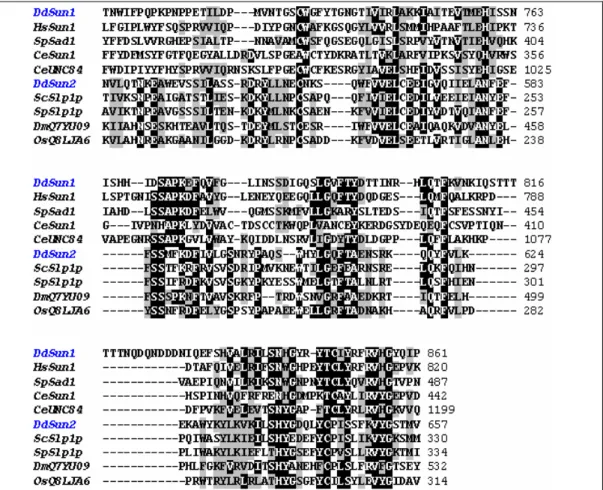
3.1.3 Multiple alignment of the SUN domains
In C. elegans, D. melanogaster and mammals SUN domain proteins bind directly or indirectly to the KASH domain proteins forming a molecular bridge between the INM and the ONM. The SUN domain is of high biological relevance for the binding to KASH domain proteins. Thus, we analyzed the SUN domain of Sun-1 and Sun-2 in detail. The SUN domain of Sun-1 shares 28% homology with the SUN domain of Ce UNC-84 (Figure 6).
Although D. discoideum is a lower eukaryote and diverged in the evolutionary tree before the fungi (Eichinger et al., 2005), the SUN domain of Sun-1 is closer related to human SUN proteins (39% identity to SUN-1, 38% identity to SUN-2) than to SUN proteins in yeast (27% identity) or worm (28% identity). In the evolution, D. discoideum represents an intermediate organism between yeast and mammals therefore proteins from D.
discoideum show higher homology to mammalian proteins. With respect of the overall structure and sequence homology Sun-1 belongs to the subfamily of SUN3 proteins along with mouse and human Sun-3. Due to the domain architecture, Sun-1 may be involved in the organization of the nuclear envelope in D. discoideum.
The centrally located SUN domain of Sun-2 shared sequence
homology with the SUN domains of SUN-like proteins found in S. cerevisiae,
S. pombe, and D. melanogaster, demonstrating that Sun-2 belongs to the
subfamily of the SUN-like proteins (Figure 6). However, the SUN domain
sequences of SUN-like proteins are distinct from those of other SUN domain
proteins, suggesting that the SUN-like proteins are specific for other
conserved cellular processes than organization of the nuclear envelope
3 Results
Figure 6: Sequence homology of SUN domains of D. discoideum Sun-1 (DDB0219949), human Sun-1 (NP 079430), S. pombe Sad1 (Q09825), C. elegans Sun-1 (Q20924) and UNC-84 (Q20745), D. discoideum Sun-2 (DDB0186751), S. cerevisiae Slp1p (Q12232), S. pombe Slp1p (O59729), D. melanogaster Q7YU09 and O. sativa Q8LJA6.
3.2 Generation of Sun-1 monoclonal antibodies
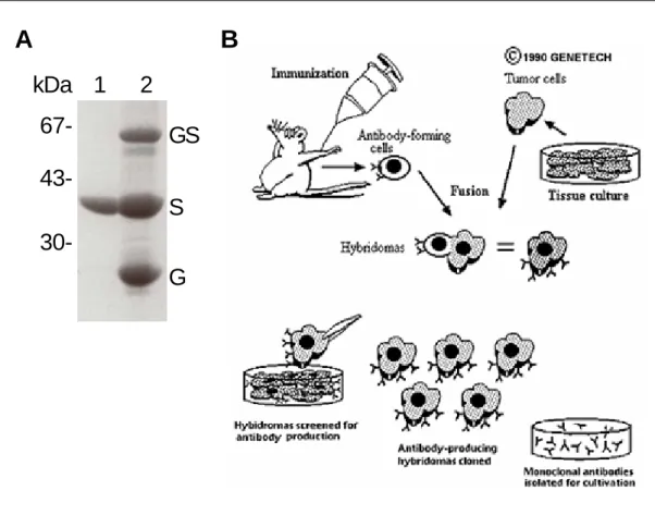
To address the biochemical and the functional aspects of Sun-1, we
generated monoclonal antibodies against the amino acids 337 to 671
spanning the two coiled-coil domains (SunCT1) that are located between
the transmembrane domain and the SUN domain. We targeted the coiled-
coil domains of Sun-1 due to (1) the uniqueness of the protein sequence in
the D. discoideum proteom and (2) the higher solubility during heterologous
protein expression. The DNA sequence encoding the two coiled-coil
domains (SunCT1) was cloned into the bacterial expression vector pGEX-
4T2. The 65 kDa fusion protein GST-SunCT1 was purified from the
Escherichia coli strain XL1-Blue and the GST tag was subsequently removed by thrombin cleaveage. After thrombin digestion the SunCT1 had an apparent molecular weight of 40 kDa (Figure 7A). Two naïve mice were immunized with 10 µg of the purified SunCT1. After three weeks the mice were boosted twice with 10 µg SunCT1 and the mouse with higher antiserum titer was sacrificed for isolation of spleen cells, which were fused to the myeloma cell lines AG8 and PAI, respectively (Figure 6B).
A kDa
67- 43- 30-
1 2 GS S G
B
Figure 7: Generation of monoclonal antibodies against Sun-1. A. The fusion protein GST- SunCT1 (GS) was purified from bacterial cell lysate. The 40 kDa SunCT1 (S, lane 1) was cleaved from the GST moiety (G) prior to immunization of the mice. B. Scheme illustrating the process of monoclonal antibody generation (Genetech, 1999). Mice were immunized with purified SunCT1. Spleen cells from the mouse with the highest serum titer were isolated and fused to myeloma cells AG8 and PAI, respectively, giving rise to hybridoma clones. The immortalized hybridoma clones were screened for specificity to Sun-1 by ELISA and western blots.
3 Results
Immunoreactivity of the hybridoma clones for SunCT1 was screened by ELISA assay and on western blot stripes. Promising clones were subjected to single-cell subcloning. In the screening experiments, three single-cell subclones were identified to be specific for biochemical or cell biological studies, i.e. K55-432-2, K55-450-1 and K55-460-1. Each of the single-cell clones recognized specifically the endogenous Sun-1 in western blot screening experiments. Further screenings of these clones by immunofluorescence and immunoprecipitation assays revealed that the clones K55-450-1 and K55-460-1 were suitable for immunolocalization and immunoprecipitation studies of Sun-1.
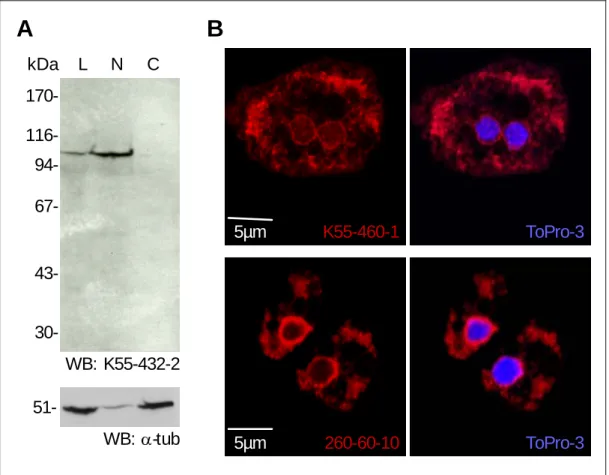
The monoclonal antibody (mAb) K55-432-2 detected Sun-1 in the AX2 total cell lysate with the expected molecular mass of 105 kDa. In a crude fractionation experiment, K55-432-2 localized the endogenous Sun-1 specifically to the nuclear fraction but not to the cytoplasmic fraction (Figure 8A), indicating that Sun-1 is a protein associated with the nuclear compartment. To monitor the efficiency of the fractionation procedure, the samples were probed with monoclonal antibodies against α-tubulin (mAb 6- 11B1, Piperno and Fuller, 1985, a kind gift from Michael Koonce). The 51 kDa protein α-tubulin was found both in the total cell lysate and in the cytoplasmic fraction (Figure 8A). However, low amount of α-tubulin was also detected in the nuclear fraction, which may be derived from the microtubule (MT) tips associated with the microtubule organization center (MTOC) at the centrosome. Since the mAb K55-432-2 displayed the highest immunoreactivity in western blot analysis, this clone was used for all biochemical studies.
In cell biological screening, the mAb K55-432-2 failed to stain Sun-1
by indirect immunofluorescence. The inability of mAb K55-432-2 to detect
Sun-1 in AX2 cells may be due to the specificity of mAb K55-432-2 for
denatured Sun-1 protein. In further immunofluorescence screenings, a
second mAb clone, K55-460-1, localized the endogenous Sun-1 to the
nuclear envelope and some cytoplasmic compartments in AX2 cells (Figure
8B). To confirm the nuclear envelope localization, nuclei of AX2 cells were
stained with ToPro-3 to visualize the DNA. In a merged image, Sun-1
surrounded the nucleus in a rim-like pattern, demonstrating that mAb K55- 460-1 recognizes specifically the native epitope of Sun-1 in vivo.
Subsequently, mAb K55-460-1 was used for all cell biological analyses.
Interaptin has been reported to be a nuclear envelope protein in D.
discoideum (Rivero, et al., 1998). Thus, the localization pattern of the endogenous Sun-1 was compared to that of interaptin. The mAb 260-60-10 localized interaptin predominantly to the nuclear envelope but also to further cytoplasmic compartments, most likely the peripheral ER. However, the cytoplasmic staining of Sun-1 in vivo may be derived from unspecific antibody binding, since endogenous Sun-1 was not distributed to the cytoplasmic fraction as shown in the crude AX2 cell lysate fractionation experiment (Figure 8A).
B A
kDa 170- 116- 94- 67-
43-
30-
L N C
WB: K55-432-2 51-
WB: α-tub
K55-460-1 ToPro-3
5µm 260-60-10 ToPro-3
5µm
Figure 8: Screenings of monoclonal antibodies against Sun-1. A. The mAb K55-432-2 detected endogenous Sun-1 in the AX2 total cell lysate (L) and in the nuclear fraction (N) but not in the cytoplasmic fraction (C). The mAb 6-11B1 (Piperno and Fuller, 1985), a kindly gift from Michael Koonce, monitored α-tubulin in the AX2 total cell lysate (L). In the crude fractionation experiment, α-tublin was associated with the nuclear fraction to some extent (N), but enriched in the cytoplasmic fraction
3 Results
envelope and some cytoplasmic compartments. The mAb 260-60-10 stained interaptin at the nuclear envelope, as well as some cytoplasmic compartments.
Indirect immunofluoresence (Cy3 conjugated anti mouse IgG, red), DNA was visualized by ToPro-3 (blue).
3.3 Biochemical properties of Sun-1
3.3.1 Sun-1 is restricted to the nucleus
To examine the subcellular localization of endogenous Sun-1 in more detail, cellular components were isolated by ultracentrifugation on discontinuous sucrose gradients. In brief, 5x10
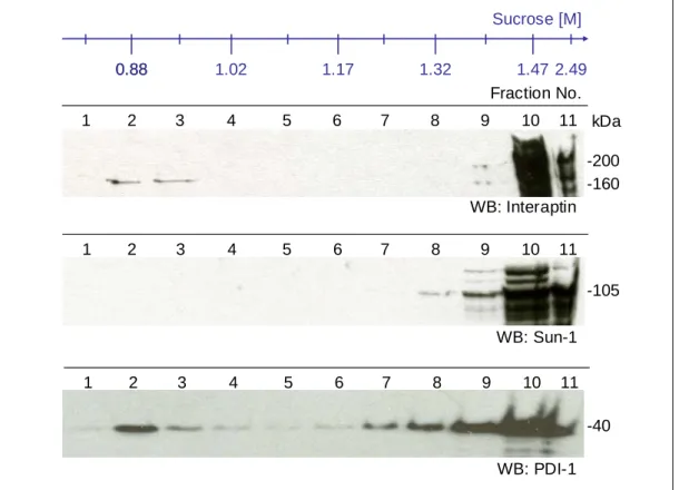
8AX2 cells were lysed in 5ml TMKS buffer by passing through a Nucleopore filter (5µm pore diameter, Whatman). To reduce the loading volume from 5ml to 1ml, the total cell lysate was spun for 30 min at 15000 g. After centrifugation, the pellet containing all cellular organells was resuspended in 1ml of TMKS buffer and was separated on a discontinuous sucrose gradient ranging from 0.85M to 2.49M of sucrose. Samples were analyzed on western blots.
To test the accuracy of the fractionation procedure, the samples
from all gradients were probed with antibodies against interaptin (mAb 260-
60-10) and protein disulfide isomerase 1 (PDI-1, mAb 221-135-1),
respectively. To some extent, interaptin was localized with the ER marker,
PDI-1, to the low-density sucrose gradient fractions two and three
confirming that interaptin is partially targeted to the peripheral ER (Figure 9,
fractions 2 and 3). As a nuclear envelope protein, the majority of
endogenous interaptin was predominantly localized to the high-density
sucrose fractions corresponding to the nuclear fractions (Figure 9, fractions,
9 to 11). However, PDI-1 was also found in high-density fractions,
correlating to the rough ER, which is contiguous with the nuclear envelope
(Figure 9, fractions 7 to 11). Comparable to the nuclear envelope marker
interaptin and the ER marker PDI-1, Sun-1 was recovered in the high-
density sucrose fractions representing the nuclear fractions
(Figure 9,
fractions 8 to 11). Distinct from the localization pattern of interaptin and PDI-
1, the endogenous Sun-1 was exclusively found in the nuclear fractions and was absent from the peripheral ER. These data indicate that the distribution of Sun-1 is limited to the nuclear envelope. Thus we conclude that the cytoplasmic staining observed in the immunfluorescence experiment may be derived from non-specific interaction of mAb K55-460-1.
11 10 9 8 7 6 5 4 3 2 1
WB: Interaptin
WB: Sun-1
WB: PDI-1 Fraction No.
11 10 9 8 7 6 5 4 3 2 1
11 10 9 8 7 6 5 4 3 2 1
kDa -200 -160
-105
-40 0.88
Sucrose [M]
0.88 1.02 1.17 1.32 1.47 2.49
Figure 9: Localization of Sun-1 to the nuclear compartment. Total cell lysate of AX2 was separated on a discontinuous sucrose gradient. Interaptin is localized to peripheral ER (Fraction 2 and 3) and to the nuclear compartment (Fraction 9 to 11) overlapping with the distribution pattern of PDI (mAb 221-135-1). Colocalizing with the nuclear envelope marker interaptin, Sun-1 is limited to the high-density fractions representing the nuclear fractions (Fraction 8 to11).
3 Results
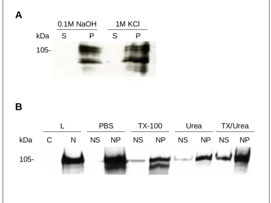
3.3.2 Sun-1 is an integral membrane protein
Analysis of the Sun-1 primary protein sequence using the program SMART revealed one putative transmembrane domain. To examine, whether Sun-1 exhibits properties of a transmembrane protein, extraction experiments were carried out. Application of alkali (0.1M NaOH) or high salt (1M KCl) extracts peripheral membrane proteins from cytoplasmic and nuclear membranes. Wildtype AX2 cell lysates were obtained by filtration of cells through Nucleopore membranes (5µm pore diameter, Whatman). Intact nuclei were isolated from the cell lysate by 10 min spinning at 3000 g.
Addition of alkali (0.1M NaOH) or high salt (1M KCl) to the nuclei led to some degradation of Sun-1. However, Sun-1 was not extracted from the nuclear membrane fraction by alkali or high salt indicating that Sun-1 is not peripherally anchored on the surface of nuclear membranes. Moreover, Sun-1 was only partially solubilized from the nuclear membranes in the presence of 1% Triton X-100 or 8M Urea or a combination of both reagents, demonstrating that Sun-1 is highly hydrophobic (Figure 10B).
Hydrophobicity and nuclear membrane association of Sun-1 agreed with the
properties observed for interaptin (Rivero et al., 1998), thus Sun-1 is a
putative integral protein of the nuclear membranes in D. discoideum.
A
B
105-
kDa S P S P 0.1M NaOH 1M KCl
105-
kDa C N NS NP NS NP NS NP NS NP
L PBS TX-100 Urea TX/Urea
Figure 10: Sun-1 is a putative integral nuclear membrane protein. AX2 cells were lysed by filtration through Nucleopore membrane (5µm pore diameter, Whatman). Intact nuclei were isolated from the cell lysate by centrifugation for 10 min at 3000 g. A.
Sun-1 was resistant to extraction with alkali (0.1M NaOH) or high salt (1M KCl) and remained membrane-associated. The endogenous Sun-1 was partially degraded due to alkali or high salt extraction. B. The intact nuclei (N), obtained after filtration through Nucleopore membrane (5µm pore diameter, Whatman), were used for further extractions. Sun-1 was partially solublilized from nuclear membranes by addition of 1% Triton X-100 (NS and NP), 8M Urea (NS and NP) or a combination of both reagents (NS and NP). Western blots were probed with mAb K55-432-2.
3.3.3 Sun-1 oligomerizes via the coiled-coil domains
As mentioned above, two putative coiled-coil domains were
predicted between the transmembrane domain and the SUN domain of Sun-
1 by the program SMART. The first coiled-coil domain spans the amino
acids 412 to 457, whereas the second coiled-coil domain extends from
amino acid 507 to 571 (Figure 11A). As coiled-coil domains represent
oligomerization motives, we tested whether the coiled-coil domains of Sun-1
are capable to oligomerize. For this purpose, recombinant SunCT1
overlapping the amino acid 419 to 578 bearing the two coiled-coil domains
was expressed as GST fusion protein and purified from E. coli using GST-
Sepharose beads (Amersham). To eliminate the chance of unspecific
3 Results
protein dimerization via the GST moiety, GST was removed from SunCT1 by thrombin cleavage.
The native protein, SunCT1, occurs as a monomer of approximately 40 kDa. Notably, low amount of dimeric (80 kDa) and trimeric (120 kDa) SunCT1 was not dissociated into monomers under denaturating and reducing conditions (Figure 11A). The presence of dimeric and trimeric SunCT1 after addition of 1% SDS and 0.1% β-Mercaptoethanol to the sample buffer indicate a strong affinity within the dimers and trimers. The strong affinity within the dimers and trimers may result from covalent bonds given by intermolecular disulfide bridges, thus we sought for cysteine residues within the protein sequence of the coiled-coil domains. Unlike mouse Sun-1 (unpublished data from our lab), Dictyostelium Sun-1 does not harbor any cysteine residue in these coiled-coil domains. Thus, the dimers and trimers were not stabilized by disulfide bonds, which can be reduced in presence of β-Mercaptoethanol. The affinity between the subunits of a dimer or a trimer may be a consequence of strong association of intermolecular hydrogen bonds, salt bridges, aromatic and/or hydrophobic interactions inducing conformational changes within the coiled-coil domains that were not taken apart by denaturation, i.e. boiling in 1% SDS containing sample buffer.
The amount of dimers and trimers observed in SDS polyacrylamide gels were enriched by incubation of 5µg purified SunCT1 with the cross- linker glutaraldehyde that stabilizes oligomers by creating covalent bonds between the interacting subunits. In a time-dependent fashion, an increase of SunCT1 dimers, trimers, tetramer and pentamer was observed 20 min after addition of 0.001% glutaraldehyde to the native SunCT1 (Figure 11B).
The stabilized SunCT1 dimers and trimers resembled with respect to their
migration behaviour those detected in the absence of glutaraldehyde hinting
to a specific cross-linking of SunCT1 dimers and trimers (Figure 11B).
A
C B
413 572
GST
1 CC T CC CC SUN 905
170
221 291
313 412
457 507
571
712
859 CC CC
kDa 94-
67-
43-
Fract. No. Fract. No.
Native X-linked
Tri
Di
Mo Input
N X 9 10 11 9 10 11 kDa
94- 67-
43-
- 5‘ 10‘ 20‘
Glutaraldehyde
Tri Di
Mo Tet Pen
Figure 11: Recombinant SunCT1 forms oligomers in vitro. A. The recombinant GST- SunCT1 harbors the two predicted coiled-coil domains (amino acids 419 to 578). B.
The native SunCT1 occurs as a 40 kDa monomer (Mo), whereas low amounts of dimers (80 kDa, Di) and trimers (120 kDa, Tri) were detected under denaturating and reducing conditions. 5µg of SunCT1 was incubated with 0.001%
glutaraldehyde. At the indicated time points, the cross-linking reaction was stopped by addition of 1M glycine to a final concentration of 0.1M. The amount of dimers, trimers, tetramers (160 kDa, Tet) and pentamers (200 kDa, Pen) were increased 20 min after addition of glutaraldehyde. C. Gel filtration of native SunCT1 (N) and cross-linked SunCT1 (X). 10µg of native or cross-linked SunCT1 were analyzed by size-exclusion chromatography using a Sephadex G75 column. The native SunCT1 trimers (Fraction 9), dimers (Fraction 10) and monomers (Fraction 11) were eluted from the column. Cross-linked SunCT1 trimers and dimers were recovered in fraction 10, whereas the monomeric SunCT1 was found in fraction 11. Western blots were probed with mAb K55-432-2.