Characterisation of PhdB, a pleckstrin homology domain containing protein in Dictyostelium discoideum
INAUGURAL-DISSERTATION zur
Erlangung des Doktorgrades
der Mathematisch-Naturwissenschaftlichen Fakultät der Universität zü Köln
vorgelegt von
Dhamodharan Neelamegan aus
Bangalore, Indien
Köln, 2004
Referees/Berichterstatter: Prof. Dr. Angelika A. Noegel Prof. Maria Leptin
Date of oral examination: 07.07.2004 Tag der mündlichen Prüfung
The present research work was carried out under the supervision of
Prof. Dr. Angelika A. Noegel, in the Institute of Biochemistry I, Medical Faculty, University of Cologne, Cologne, Germany. From June 2001 to July 2004.
Diese Arbeit wurde von Juni 2001 bis Juli 2004 am Biochemischen Institut I der Medizinischen Fakultät der Universität zu Köln unter der Leitung von
Prof. Dr. Angelika A. Noegel durchgeführt.
Acknowledgement
I wish to express my deep sense of gratitude to Prof. Dr. Angelika A. Noegel, esteemed advisor of my promotion studies, for encouraging and supporting me through out my study and for critically reviewing the thesis.
I am extremely grateful to Dr. Francisco Rivero, Dr. Akis Karakesisoglou and Dr.Ludwig Eichinger for their keen interest, advice, and encouragement through out my course of my study.
I am thankful to Rosi, Maria and Berthold for their help and cooperation during my stay here in the lab. Thanks to Bettina Lauss, for her continuous help with official works during the period of my stay in Köln. My due thanks to Dr. Budi Tunggal for his help in sorting out the computer problems encountered during my work and thesis writing.
I take this opportunity to express my humble and sincere thanks to my parents for all they have done to bring me to this level.
I am thankful to all my previous and present lab colleagues for their direct and indirect help during my work. I am also thankful to all my friends and well wishers who helped me directly or indirectly for the successful completion of my research work. I also thank Dr. Subramanya Hegde for all his help during my stay in Köln.
The financial assistance received by me from the Graduate School ‘International graduate school in genetics and functional genomics’ University of Cologne Germany, in the form of Stipend is highly acknowledged.
Also the financial assistance received by me from the DFG is highly acknowledged.
04 May 2004 Dhamodharan. N
Cologne, Germany.
1. INTRODUCTION... 4
1.1 Dictyostelium life cycle and molecular techniques ... 4
1.1.2 Chemotaxis... 5
1.1.3 Dictyostelium chemotaxis... 9
1.1.4 Cell-cell adhesion during Dictyostelium development... 10
1.2 AIM OF THE STUDY ... 12
2. MATERIALS AND METHODS ... 13
2.1 MATERIALS ... 13
2.1.1 Laboratory materials... 13
2.1.2 Instruments and equipments... 13
2.1.3 Kits ... 14
2.1.4 Enzymes, antibodies, substrates, inhibitors and antibiotics ... 15
2.1.5 Chemicals and reagents... 16
2.1.6 Media and buffers... 17
2.1.7 Media and buffers for Dictyostelium culture... 18
2.1.8 Media for E. coli culture... 18
2.1.9 Media for hybridoma cells ... 19
2.1.10 Buffers and other solutions... 20
2.1.11 Biological materials... 21
2.1.12 Plasmids ... 21
2.1.13 Primers ... 21
2.2 METHODS... 22
2.2.1 General methods... 22
2.2.2 Genomic and cDNA database screening... 22
2.2.3 CELL BIOLOGICAL METHODS ... 22
2.2.4 Growth in liquid nutrient medium... 22
2.2.5 Growth on SM agar plates... 22
2.2.6 Development of Dictyostelium... 23
2.2.7 Aggregation competent cells... 23
2.2.8 Aggregation analysis ... 23
2.2.9 Chemotaxis analysis... 23
2.2.10 Development on phosphate-buffered agar plates or water agar plates... 24
2.2.11 Preservation of Dictyostelium cells ... 24
2.2.12 Preservation of Dictyostelium spores ... 25
2.2.13 Transformation of Dictyostelium cells by electroporation ... 25
2.2.14 MOLECULAR BIOLOGICAL METHODS ... 26
2.2.15 Purification of plasmid DNA ... 26
2.2.16 Isolation of Dictyostelium genomic DNA ... 26
2.2.17 DNA agarose gel electrophoresis ... 27
2.2.18 Southern blotting ... 28
2.2.19 Isolation of total RNA from Dictyostelium cells... 28
2.2.20 RNA formaldehyde-agarose gel electrophoresis... 29
2.2.21 Northern blotting ... 30
2.2.22 Hybridisation of Southern or northern blots with a radiolabelled DNA probe ... 30
2.2.23 PCR-mediated screening of Dictyostelium transformants... 31
2.2.24 Construction of vectors ... 32
2.2.25 Construction of vector for expressing different functional domains of PhdB as GFP
fusion proteins ... 32
2.2.26 Construction of a vector for expression of the C-terminal peptide (ArfGAP/PH) as a GST-fusion protein... 33
2.2.27 Construction of a vector for expression of the C-terminal peptide (ArfGAP/PH) without any additional tag. ... 33
2.2.28 Construction of a PhdB gene replacement vector ... 34
2.2.29 Lambda ZAP II cDNA library screening ... 34
2.2.30 Preparation of plating bacteria ... 34
2.2.31 Plating a lambda ZAP bacteriophage cDNA library... 34
2.2.32 Screening bacteriophage plaques by hybridisation ... 35
2.2.33 Phage purification ... 35
2.2.34 Excision of plasmid containing the cDNA from bacteriophages ... 36
2.2.35 Plating excised phagemid... 36
2.2.36 Transformation of E. coli... 37
2.2.37 Preparation of competent E. coli cells by the CaCl2 method... 37
2.2.38 Transformation of CaCl2 competent E. coli cells... 37
2.2.39 BIOCHEMICAL METHODS... 37
2.2.40 Preparation of total protein from Dictyostelium... 37
2.2.41 SDS-polyacrylamide gel electrophoresis ... 37
2.2.42 Western blotting using the semi-dry method ... 38
2.2.43 Immunodetection of membrane-bound proteins ... 39
2.2.44 Enzymatic chemi-luminescence (ECL) detection system ... 39
2.2.45 BCIP/NBT colour development substrate reaction... 40
2.2.46 Purification of GST-CTPhdB fusion protein by sarkosyl solubilisation... 40
2.2.47 Purification of the C-terminal peptide of PhdB fusion protein expressed without additional tag. ... 41
2.2.48 Quantification of protein ... 41
2.2.49 Subcellular fractionation ... 42
2.2.50 Triton X-100 extraction of Dictyostelium cells... 42
2.2.51 IMMUNOLOGICAL TECHNIQUES ... 42
2.2.52 Generation of monoclonal antibodies ... 42
2.2.53 Preparation of mouse feeder cells for fusion and cloning... 43
2.2.54 Fusion ... 43
2.2.55 Screening of hybridoma clones ... 45
2.2.56 Indirect ELISA for screening of hybridoma clones ... 45
2.2.57 Stripe test for screening of hybridoma clones... 46
2.2.58 Cloning of hybridoma cells ... 46
2.2.59 Freezing and recovery of hybridoma cell lines ... 47
2.2.60 Recovery of frozen cell lines... 48
2.2.61 Generation of polyclonal antibodies against PhdB ... 48
2.2.62 Preparation of Dictyostelium cells for immunofluorescence analysis... 49
2.2.63 Methanol fixation ... 49
2.2.64 Picric acid-paraformaldehyde fixation ... 49
2.2.65 Immunolabelling of fixed cells ... 50
2.2.66 Mounting of cover slips... 50
2.2.67 DAPI and phalloidin staining of fixed cells ... 51
2.2.68 MICROSCOPY... 51
2.2.69 Computer analysis ... 52
3. RESULTS... 53
3.1 Screening of genomic and cDNA data bases sequences of Dictyostelium... 53
3.1.1 Generation of monoclonal and polyclonal antibodies against PhdB... 55
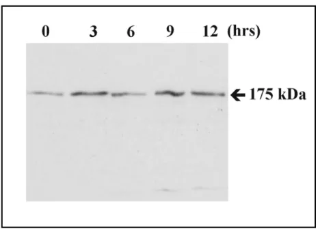
3.1.2 PhdB is expressed throughout the development of Dictyostelium... 57
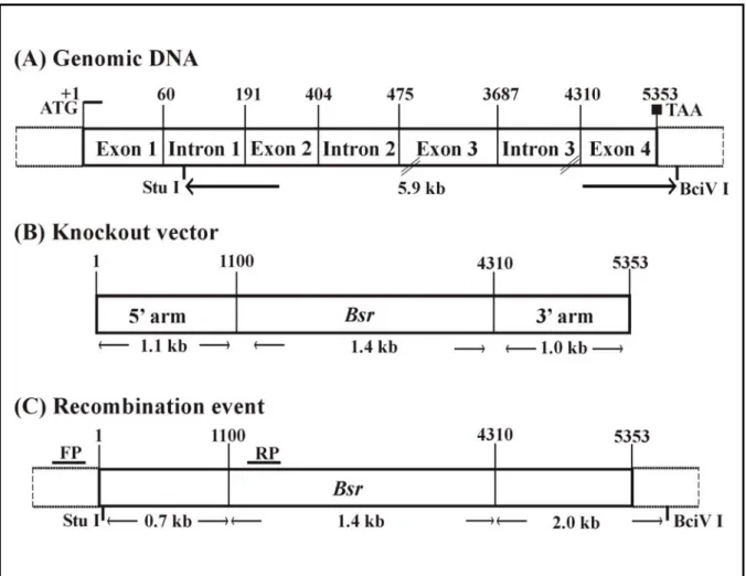
3.1.3 PhdB- mutant generation by homologous recombination ... 58
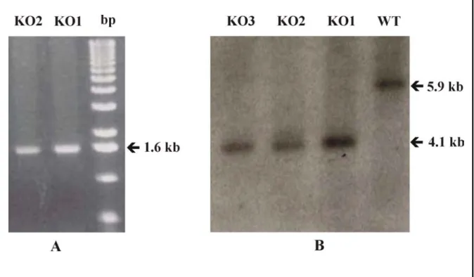
3.1.4 PCR analysis ... 60
3.1.5 Southern blot analysis ... 60
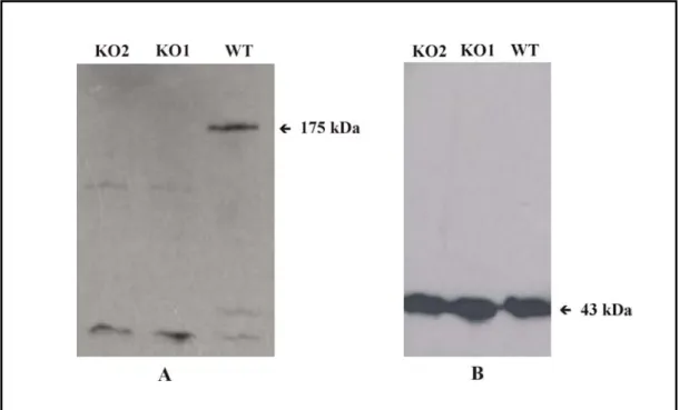
3.1.6 Western blot analysis... 61
3.1.7 PhdB- cells exhibit an aggregation defect ... 63
3.1.8 PhdB- cells exhibit a defect in streaming during Dictyostelium chemotaxis ... 65
3.1.9 PhdB- cells leading edge is equally sensitive to cAMP as the one of wild type cells. 68 3.1.10 Analysis of cell motility ... 69
3.1.11 PhdB- cells exhibit a defect in chemoattractant mediated actin polymerisation ... 70
3.1.12 PhdB- cells express normal levels of csA... 71
3.1.13 PhdB- cells exhibit normal growth in axenic medium ... 78
3.1.14 PhdB- cells growth on agar plates with bacteria as food source ... 79
3.1.15 PhdB- cells exhibit normal pinocytosis ... 80
3.1.16 PhdB- cells exhibit normal phagocytosis ... 81
3.1.17 Developmental analysis... 82
3.1.18 Localisation of actin, CAP and filamin in PhdB- cells... 84
3.1.19 PhdB is associated with the Dictyostelium Triton X-100 insoluble cytoskeleton... 85
3.1.20 PhdB is associated with membrane fractions in Dictyostelium... 86
3.1.21 The C- terminal peptide of PhdB binds to the F-actin cytoskeleton ... 86
3.1.22 The C- terminal peptide of PhdB interacts with phosphoinositides... 88
4. DISCUSSION... 89
4.1 Identification of PhdB, a 175 kDa protein ... 89
4.1.1 PhdB- null cells exhibit abnormal aggregation... 89
4.1.2 PhdB- cells exhibit normal chemotaxis ... 90
4.1.3 PhdB- cells aggregation is not mediated by csA ... 91
4.1.4 PhdB- cells exhibit a streaming defect during chemotaxis... 94
4.1.5 PhdB- cells exhibit normal pinocytosis, phagocytosis and development... 96
4.1.6 The PhdB protein may have a function at the membrane in association with the cytoskeleton... 96
5. SUMMARY / ZUSAMMENFASSUNG... 99
6. BIBLIOGRAPHY ... 101
Abbreviations:
AP Alkaline phosphatase APS Ammonium persulphate Bsr Blasticidin resistance cassette BSA Bovine serum albumin
CH Calponin homology
CRAC Cytosolic regulator of adenylyl cyclase cAMP Adenosine- 3', 5'- cyclic monophosphate cAR cAMP receptor
DEPC Diethylpyrocarbonate DMSO Dimethylsulfoxide
dNTP Deoxyribonucleotide triphosphate DTT 1,4-dithiothreitol
EDTA Ethylenediaminetetraacetic acid
EGTA Ethyleneglycol-bis (2-amino-ethylene) N,N,N,N-tetraacetic acid GEF Guanine-nucleotide exchange factor
GPCR G-protein coupled receptor GFP Green Fluorescence Protein GAP GTPase activating protein GST Glutathione S-transferase FITC Fluorescein-5-isothiocyanate HRP Horse radish peroxidase
HEPES N- (2-hydroxyethyl) piperazine-N-2-ethanesulphonic acid IPTG Iso-propylthio-galactopyranoside
IgG Immunoglobulin G Kb Kilobase pairs
kDa KiloDalton
MES Morpholinoethansulphonic acid MOPS Morpholinopropanesulphonic acid mRNA messenger ribonucleic acid
mAb Monoclonal Antibody
NP-40 Nonylphenylpolyethyleneglycol OD Optical density
PIPES Piperazine-N,N.-bis(2-ethanesulphonic acid) PMSF Phenylmethylsulphonylfluoride
PKB/Akt Protein Kinase B
PI3K Phosphatidylinositol 3 kinase PAGE Polyacrylamide gel electrophoresis PH Pleckstrin homology
rpm Rotations per minute SDS Sodium dodecyl sulphate
TRITC Tetramethylrhodamine β isothiocyanate TAE Tris borate EDTA
WT Wild type
X-gal 5-bromo-4-chloro-3-indolyl-D-galactopyranoside
1. INTRODUCTION
1.1 Dictyostelium life cycle and molecular techniques
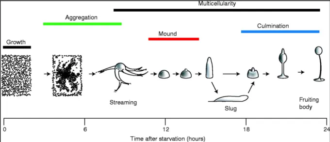
Dictyostelium discoideum is a non-metazoen eukaryote, which lives as a unicellular amoeba in the soil and feeds on bacteria. When the food source is exhausted the amoebae aggregate into a mass of up to 105 cells, which differentiate and develop into a mushroom shaped fruiting body (Kessin, 2000).
In the laboratory Dictyostelium are grown on a lawn of bacteria or in a liquid medium. To begin development in the laboratory we remove the source of nutrition and put the cells on a moist solid substratum. The substratum can be agar or filter paper which is sufficiently moist.
As no nutrients are provided the amoebae aggregate and make a fruiting body entirely using the metabolic reserves accumulated during the trophic phase. The fruiting body consists of a ball of spores supported by a stalk. The striking feature of Dictyostelium during starvation is that they undergo a switch in behaviour by inducing the expression of additional genes to form a fruiting body (Figure I).
Dictyostelium is an excellent organism for the study of the molecular mechanisms of cell motility, signal transduction, cell-type differentiation and developmental processes. It is haploid and therefore mutants can be easily generated. The molecular genetic techniques available include gene inactivation by homologous recombination, gene replacement, antisense strategies, restriction enzyme-mediated integration (REMI), library complementation and expression of green fluorescent protein (GFP) fusion proteins. The hereditary information is carried on 6 chromosomes with sizes ranging from 4 to 8 Mb (Cox et al., 1990; Kuspa and Loomis, 1996) resulting in a total of about 34 Mb of DNA; a multicopy 90 kb extrachromosomal element that harbours the rRNA genes, and the 55 kb mitochondrial genome.
The genome sequencing is completed and the estimated number of genes in the genome is
~12,000. Many of the known genes show a high degree of sequence similarity to homologues in vertebrate species (Eichinger and Noegel, 2003). Furthermore Dictyostelium cells resemble human leukocytes in their motility characteristics.
Figure I. Dictyostelium development. During growth phase Dictyostelium exists as single cell amoeba. Upon starvation Dictyostelium undergoes chemotaxis towards a pulsatile cAMP source secreted by the cells at the centre. During aggregation cells coalesce into adherent cells and form streams. The streams eventually come together to form the mound. The mound is the first stage in the multicellular development. It develops a tip, which coordinates further development. Next a finger like structure emerges which either immediately forms a fruiting body or a motile slug that migrates to a favourable environment for culmination into the fruiting body. The scale shows the relative timing of development (The picture was taken from Coates and Harwood, 2001).
1.1.2 Chemotaxis
Chemotaxis is a process by which cells respond to an extracellular chemical signal and guide their movement towards the chemical signal. Chemotaxis has a role in various functions of a cell, prokaryotes tracing their food, protozoa like Dictyostelium forming multicellular structures, axon guidance and embroygenesis is in part due to chemotaxis.
Eukaryotic cells respond to chemoattractant gradients by adapting a polarised morphology of their cytoskeleton towards the origin of the signal (Chen et al., 1997). The actin cytoskeleton is an important player in this event, and allows protrusion of an actin-rich lamellipod (pseudopod). In chemotaxing cells, including Dictyostelium and leukocytes, the forward movement is driven by extending the leading edge through localised polymerisation of F- actin. This change is coupled to an actin-myosin mediated contraction of uropod (posterior region of a cell), which allows the uropod to be released and retracted towards the direction of movement. Small GTPases like Rac and Cdc42 have been shown to regulate the process of F- actin assembly at the leading edge of the cell (Hall et al., 1989). Dominant negative Rac
prevents F-actin assembly, whereas constitutively active Rac results in excessive F-actin assembly and membrane ruffling (Rivero et al., 2001). It has been shown that the Rho family members Cdc42 is required for proper orientation of the cytoskeleton towards the direction of the chemoattractant signal. Macrophages expressing dominant negative Cdc42 move in response to directional signals but the movement lacks directionality (Allen et al., 1998).
These observations indicate that Cdc42 may act in leading the cell movement towards a particular direction. Both Dictyostelium and leukocytes use G-protein coupled serpentine receptors to transduce the extracellular signal. In the mammalian system binding of chemoattractant to the receptor elicits a transient increase in phosphoinositides (PIs), cAMP, cGMP, Ca2+ and rearrangement of F-actin. In both leukocytes and Dictyostelium the receptor molecules remain uniformly distributed across the cell surface during chemotaxis (Xiao et al., 1997; Postma et al., 2002). Thus despite uniform distribution of chemoattractant receptors on the cell surface the cells extend their leading edge towards the source of chemoattractant. It was concluded therefore that the cell adopts an internal asymmetric signaling relative to the external gradient. In addition to the small GTPases as important players at the leading edge of the cell the phosphatidylinositol 3 kinase (PI3K) signalling pathway is another essential component. The main feature of the PI3K pathway is the recruitment of PI3K at the leading edge, which results in localised production of PI (3,4,5) P3. PIP3 serves as docking site for many pleckstrin homology (PH) domain containing proteins. Studies on this pathway have led to the identification of several PH domain containing proteins as effectors of PI3K. They include Akt/PKB, CRAC and PhdA.
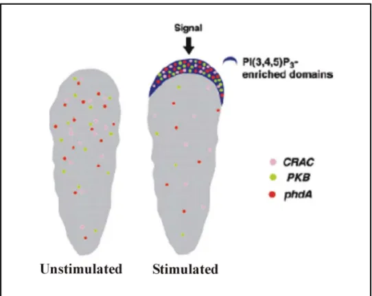
Studies with Akt/PKB, CRAC (required for receptor activation of the adenylyl cylcase; Insall et al., 1994) and PhdA (Funamoto et al., 2001), a newly identified PH domain containing protein in Dictyostelium, provide evidence for localised activation of responses leading to directional cell movement. These proteins transiently localize to the plasma membrane in response to cAMP. When Dictyostelium cells are bathed in cAMP the GFP fusions of CRAC and Akt/PKB are uniformly distributed along the plasma membrane, indicating that the chemoattractant activates the receptors uniformly distributed on the cell surface. Further insights were obtained when the response was examined in chemotaxing cells. Under these conditions the GFP-PH domain fusion proteins of both CRAC and Akt/PKB were found to localise to the leading edge of the cells. This observation strongly indicates that receptor mediated responses are locally activated at the leading edge (Figure II; Firtel and Chung, 2000).
Figure II. Asymmetric accumulation of PIP(3) and PH domain containing proteins at the leading edge of the cell. In an unstimulated cell the PH domain containing proteins CRAC, Akt/PKB and PhdA are cytosolic. In chemotaxing cell, these proteins become localised to the leading edge.
In neutorophils it has been shown that PI3K, Rac and F-actin pathways are integrated and function through a positive feed back loop, by which localised signals are amplified to allow formation of the leading edge (Leevers et al., 1999; Weiner et al, 2002).
In mammals four isoforms of PI3Ks are known, α, β, γ and δ. But only leukocyte specific PI3Kγ has been tentatively implicated in translocating signals from G protein linked receptors into specialized responses such as chemotaxis (Janetopoulos et al., 2001). Studies with knock out mice lacking the catalytic subunit of PI3Kγ, the p110γ subunit, indicate the possibility of PI3K signalling pathway (Zhou et al., 1995) involved in directional movement. The leukocytes lacking PI3Kγ failed to move directionally in response to chemotactic stimulation.
This is also in accordance with the response of cells expressing dominant negative Cdc42.
This observation also suggests a co-operative relationship between these two processes (Allen et al., 1998).
In addition to the GTPases and PI3K role at the leading edge of a cell, recently in leukocytes it has been shown that PAK1 and PIXα are recruited to the leading edge which are down stream of the heterotrimeric G protein coupled receptor. The recruitment and activation of
PAK1 is mediated by direct binding of PAK1 to Gβγ. The interaction of Gβγ and PAK1 also brings PIXα into the complex. As a result Cdc42 is activated, leading to the activation of PAK1 (Daniels et al., 1999). Studies with PIXα knockout mice reported that the mice were normal, however neutrophils isolated from these mice exhibit severe defect in chemotaxis and chemotactic signaling (Li et al., 2003) .
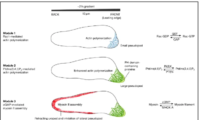
Based on the recent findings chemotaxis can be broadly divided into three modules as shown in figure III. Module 1 represents the event at the leading edge when the chemoattractant receptor is activated, small GTPases along with GEF/GAP factors regulate modulation of F- actin polymerization at the leading edge. Module 2 represents the activation of PI3K signalling that contributes to the localised accumulation of phosphoinositides, which mediates actin polymerization by recruiting PH domain containing proteins. Module 3 represents the myosin based retraction of the uropod which is regulated by cGMP signalling at the posterior end of the cell.
Figure III. Module 1, activation of the chemoattractat receptor leads to formation of active small G proteins of the Rho/Rac group, leading to actin polymerisation and a small pseudopod is extended at the front of the cell. It is proposed that specific activators of GTPases, guanine exchange factors (GEFs) and inhibitors GTPase-activating proteins (GAPs) are preferentially recruited at the higher chemoattractant concentrations. The polarity generated by the first module is used by the second module to trigger a patch of phosphatidylinositol-3,4,5 trisphosphate (PtdIns (3,4,5) P3) at the leading edge, which enhances actin polymerisation and extension of the pseuodopod. The third module inhibits the formation of pseudopodia at the back and sides of the cells by cementing the uropod with myosin II assembly. Increase in cGMP concentration leads to myosin filament formation at the uropod (Graphic from Postma et al., 2004).
1.1.3 Dictyostelium chemotaxis
cAMP plays an important role in the development of Dictyostelium. Early in the developmental cycle, when cells are starved some cells begin to secrete pulses of cAMP. The surrounding cells respond by undergoing chemotactic migration towards these cells. When extracellular cAMP binds to the cell surface receptor, signals are transduced and this results in the formation of intracellular cAMP, which is then secreted into the medium eliciting cAMP responses in other cells further away from the aggregation centre.
The cAMP signalling is mediated through a cell surface receptor, which activates G-protein dependent signalling, which finally activates adenylyl cyclase (Van Haastert et al., 1991).
The signalling pathway leading to the activation of the adenylyl cyclase ACA during aggregation are well studied. In addition to the Gβγ subunit, which appear to be the direct activator of ACA, proteins like, Pianissimo, CRAC (Cytosolic Regulator of Adenylyl Cyclase) and MAP kinase ERK2 play important roles in controlling the production of cAMP, but do not affect chemotaxis (Wu et al., 1995; Chen et al., 1997; Firtel, 1996). Many of the components of aggregation stage signalling pathways are induced by nanomolar pulses of cAMP via an autoregulatory loop, by ACA. By this chemotactic mediated mechanism, cells are able to form mounds of ~ 105 cells. Also cAMP is an important regulator of gene expression. At the early aggregation stage of development, pulses of low concentration of cAMP control the expression of a number of genes. At later stages higher concentrations of cAMP are required for gene expression.
The chemoattractant signal is perceived by G-protein coupled receptor (GPCR) cAR1. Using GFP tagged cAR1 it was demonstrated that the receptor remains evenly distributed along the plasma membrane in highly polarised chemotaxing cells (Servent et al., 1999). This observation indicated different activation of signalling pathways in the front and back of the cells, which is not dependent on differential distribution of the receptor. The G-protein β subunit was shown to exhibit a shallow anterior-posterior gradient similar to the extracellular chemoattractant. This reflects a shallow receptor occupancy gradient. Studies with GFP fusions of PH domain containing proteins including CRAC and the PI3K effector Akt/PKB demonstrated that proteins carrying PH domains that bind to the PI3K products PtdIns(3,4,5)P3 / PtdIns(3,4)P2 rapidly translocate to the plasma membrane in response to a uniform stimulation with the chemoattractant cAMP (Parent et al., 1998). Analysis of Dictyostelium PI3K1 and PI3K2 localisation revealed that in vivo these two proteins rapidly and transiently translocate to the plasma membrane in response to stimulation by cAMP. Both PI3Ks localise to the leading edge during chemotaxis. The localisation and activation of PI3K
at the leading edge could thus mediate generation of a steep PtdIns(3,4,5)P3 gradient (Zhou et al., 1998). In cells lacking the Gα2 or the Gβ subunit (Lilly and Devreotes, 1995) the translocation of PI3Ks to the leading edge does not occur. This observation shows that the localisation of PI3K is a downstream event of the heterotrimeric G protein. The interacting partners of PI3Ks at the membrane are presently not known. The persistence of the steep PtdIns(3,4,5)P3 gradients indicates that a mechanism must restrict the PtdIns(3,4,5)P3 at the leading edge. One possibility is that PtdIns(3,4,5)P3 is degraded along the lateral side of the cell. A candidate enzyme is the tumour suppressor PTEN a phosphatase, that dephosphorylates PtdIns(3,4,5)P3 and PtdIns(3,4)P2 to PtdIns(4,5)P2 / PtdIns(4)P, respectively (Lee et al., 1999). Dictyostelium has provided more evidence for a direct role of PTEN in regulating chemotaxis (Funamoto et al., 2002). These studies revealed that in resting cells PTEN is localised to the plasma membrane and uniformly distributed around the cell. In response to global stimulation the phosphatase PTEN transiently delocalises from the plasma membrane. In chemotaxing cells, PTEN is excluded from the leading edge but is present along the sides and the back of the cell. Thus PI3K and PTEN exhibit opposite patterns of spatial localisation during Dictyostelium chemotaxis.
The defects of PI3K1/PI3K2 null cells are more severe than those of Akt/PKB null cells. PI3 kinase presumably has downstream effectors in addition to Akt/PKB that are required for chemotaxis. Towards this, PH domain containing proteins have gained importance, as they may have an essential role in the PI3Kinase activated localised signalling.
1.1.4 Cell-cell adhesion during Dictyostelium development.
Two mechanisms of cell-cell adhesion, characterised as EDTA sensitive and EDTA resistant aggregation have been shown to operate at different stages of development in Dictyostelium.
Upon starvation the cells undergo a developmental programme where cells migrate chemotactically towards regions of higher concentration of cAMP and form multicellular aggregates. Within 24 h morphogenesis takes place and the cells eventually form a fruiting body consisting of two major types of cells, spore cells and stalk cells. During the first few hours of starvation, the cells produce and accumulate a glycoprotein of 24 kDa (gp24).
Expression of gp24 confers cells to acquire EDTA sensitive binding sites (Knecht et al., 1987) that are disrupted by low concentration (1 mM) of EDTA. The cells acquire EDTA resistant cell binding sites a few hours later, at the early aggregation of development (Gerisch et al., 1985). The adhesion sites of these cells are stable in the presence of 10 mM EDTA. The
adhesion in these cells are mediated by the homophilic interaction of an 80 kDa glycoprotein (gp80) also known as contact site A (csA; Muller and Gerisch, 1978; Siu and Kamboj, 1990).
The first phase of csA expression is in the very early stage of aggregation and results in a low basal level of expression. The mechanism involved in the induction of this low level expression of csA is not known. The low level of expression of csA does not seem to be dependent on cAMP (Ma and Siu, 1990). The second phase requires cAMP and results in high level of csA expression. As the chemotaxing Dictyostelium cells respond by secreting cAMP into the medium, the secreted cAMP augments the expression of csA. In this cAMP signal pathway, cAMP binds to the cell surface cAMP receptor (Klein et al., 1988) and this binding leads to the G- protein coupled activation of adenylyl cyclase. The activated adenylyl cyclase now produces intracellular cAMP (Snaar-Jagalska et al., 1988). The cAMP is then secreted into the medium and binds to the cAMP receptor on adjacent cells. Since the cAMP receptor is desensitised soon after the ligand (cAMP) binding, cAMP is produced and secreted in a pulsatile manner. This pulsatile cAMP production and release leads to the differential activation of certain aggregation stage specific genes, including the regulation of csA expression. The expression of csA begins by transient activation of the cell surface cAMP receptor. The receptor which is coupled to the G-protein, Gα2 subunit transduces the down stream signalling (Newell et al., 1988; Pupillo et al., 1989). Also it was shown that adenylyl cyclase which is the down stream effector of cAMP receptor activation, is not involved in this pathway, and that the intracellular cAMP is not required for csA expression (Ma and Siu, 1990).
In the present study we have identified a novel PH domain containing protein from the cDNA and genomic DNA databases of Dictyostelium the protein is referred as PhdB (Pleckstrin homology domain containing Protein B). We also generated Dictyostelium mutants, in which the PhdB gene was inactivated by homologous recombination, and analyse its role in Dictyostelium aggregation.
1.2 AIM OF THE STUDY
This study was initiated since the Dictyostelium genome sequencing was in progress and information on potential novel genes was available in the databases.
Dictyostelium is haploid and simple, and most of the signalling pathways as well as chemotactic behaviors like those seen in white blood cells are highly conserved throughout evolution. Hence Dictyostelium genome offers a great value to understand and determine the functions of novel genes as well as homologous genes from other species.
The aims set for the study were to characterise novel actin binding proteins, by developing tools like monoclonal and polyclonal antibodies against the new protein, and to generate Dictyostelium mutants which lack the expression of the gene of interest by homologous recombination to understand the exact function of the new gene.
2. MATERIALS AND METHODS
2.1 MATERIALS
2.1.1 Laboratory materials
Cellophane sheet, Dry ease Novex
Centrifuge tubes, 15 ml, 50 ml Greiner
Coverslips (glass), ∅ 12 mm, ∅ 18 mm, ∅ 55 mm Assistent
Corex tube, 15 ml, 50 ml Corex
Cryo tube, 1 ml Nunc
Electroporation cuvette, 2 mm electrode gap Bio-Rad Microcentrifuge tube, 1.5 ml, 2.2 ml Sarstedt Micropipette, 1-10 µl, 10-200 µl, 100-1,000 µl Gilson
Needles (sterile), 18G–27G Terumo, Microlance Nitrocellulose membrane, BA85 Schleicher and Schuell Nitrocellulose-round filter, BA85, ∅ 82 mm Schleicher and Schuell
Nylon membrane, Biodyne B Pall
Parafilm American National Can
Pasteur pipette, 145 mm, 230 mm Brand, Volac
PCR soft tubes, 0.2 ml Biozym
Petri dish (35 mm, 60 mm, 100 mm) Falcon
Petri dish (90 mm) Greiner
Plastic cuvette, semi-micro Greiner
Plastic pipettes (sterile), 1 ml, 2 ml, 5 ml, 10 ml, 25 ml Greiner
Quartz cuvette, Infrasil Hellma
Quartz cuvette, semi-micro Perkin Elmer
Saran wrap Dow
Slides, 76 x 26 mm Menzel
Syringes (sterile), 1 ml, 5 ml, 10 ml, 20 ml Amefa, Omnifix Syringe filters (Acrodisc), 0.2 µm, 0.45 µm Gelman Sciences Tissue culture flasks, 25 cm2, 75 cm2, 175 cm2 Nunc Tissue culture dishes, 6 wells, 24 wells, 96 wells Nunc
Whatman 3MM filter paper Whatman
X-ray film, X-omat AR-5, 18 x 24 mm, 535 x 43 mm Kodak
2.1.2 Instruments and equipments
Centrifuges (microcentrifuges):
Centrifuge 5417 C Eppendorf
Centrifuge Sigma B. Braun Biotech Instruments
Cold centrifuge Biofuge fresco Heraeus Instruments Centrifuges (table-top, cooling, low speed):
Centrifuge CS-6R Beckman
Centrifuge RT7 Sorvall
Centrifuge Allegra 21R Beckman
Centrifuges (cooling, high speed):
Beckman Avanti J25 Beckman
Sorvall RC 5C plus Sorvall
Centrifuge-rotors:
JA-10 Beckman
JA-25.50 Beckman
SLA-1500 Sorvall
SLA-3000 Sorvall
SS-34 Sorvall
Dounce homogeniser, 10 ml B. Braun
Electroporation unit, Gene-Pulser Bio-Rad
ELISA reader Lab Systems
Fluorescence spectrophotometer Photon Technology International
Freezer (-80°C) Nunc
Freezer (-20°C) Siemens, Liebherr
Gel-documentation unit MWG-Biotech
Heating block, DIGI-Block JR neoLab
Hybridising oven Hybaid
Incubators:
CO2-incubator, BBD 6220, BB 6220 Heraeus
Incubator, microbiological Heraeus
Incubator with shaker, Lab-Therm Kuehner Laminar flow, Hera Safe (HS 12) Heraeus
Model 422 Electro-Eluter Bio-Rad
Microscopes:
Light microscope, CH30 Olympus
Light microscope, DMIL Leica
Light microscope, CK2 Olympus
Fluorescence microscope, DMR Leica
Fluorescence microscope, 1X70 Olympus
Confocal laser scan microscope, DM/IRBE Leica
Stereomicroscope, SZ4045TR Olympus
Oven, conventional Heraeus
PCR machine, PCR-DNA Engine PTC-2000 MJ Research
pH-Meter Knick
Refrigerator Liebherr
Semi-dry blot apparatus, Trans-Blot SD Bio-Rad
Sonicator, Ultra turrax T25 basic IKA Labortechnik Speed-vac concentrator, DNA 110 Savant
Spectrophotometer, Ultraspec 2000, UV/visible Pharmacia Biotech Ultracentrifuges:
Optima TLX Beckman
Optima L-70K Beckman
Ultracentrifuge-rotors:
TLA 45 Beckman
TLA 100.3 Beckman
SW 41 Beckman
UV-crosslinker, UVC 500 Hoefer
UV- transilluminator, TFS-35 M Faust
Vortex, REAX top Heidolph
Waterbath GFL
2.1.3 Kits
Advantage cDNA PCR kit Clontech
Nucleobond AX Macherey-Nagel
NucleoSpin Extract 2 in 1 Macherey-Nagel
NucleoSpin Plus Macherey-Nagel
Original TA Cloning Invitrogen
Qiagen Midi- and Maxi-prep Qiagen
Stratagene PrimeIt II Stratagene
2.1.4 Enzymes, antibodies, substrates, inhibitors and antibiotics
Enzymes used in the molecularbiology experiments:Calf Intestinal Alkaline Phosphatase (CIAP) Boehringer
Deoxyribonuclease I (DNase I) Boehringer
Klenow fragment Boehringer
Lysozyme Sigma
Proteinase K Sigma
Restriction endonucleases Amersham, Life technologies, New England Biolabs
Reverse transcriptase, Superscript II Life technologies
Ribonuclease H (RNase H) Boehringer
Ribonuclease A (RNase A) Sigma
S1-nuclease Amersham
T4 DNA ligase Boehringer
Taq-polymerase Life technologies/Boehringer
Primary antibodies:
Goat anti-GST antibody Pharmacia
Mouse anti-Actin monoclonal antibody, Act 1 Simpson et al., 1984 Mouse anti-CAP monoclonal antibody, 231-18-8 Gottwald et al., 1996 Mouse anti-csA monoclonal antibody, 33-294 Berthold et al., 1985 Mouse anti-Filamin monoclonal antibody, 82-382-8 Brink et al., 1985 Secondary antibodies:
Mouse anti-goat IgG, alkaline phosphatase conjugated Sigma Goat anti-mouse IgG, peroxidase conjugated Sigma Goat anti-mouse IgG, alkaline phosphatase conjugated Sigma Goat anti-rabbit IgG, peroxidase conjugated Sigma Mouse anti-goat IgG, peroxidase conjugated Sigma Sheep anti-mouse IgG, Cy3 conjugated Sigma Substrates:
BCIP/NBT Promega
pNPP, Sigma Fast tablet sets, 5ml, 20 ml Sigma Inhibitors:
Diethylpyrocarbonate (DEPC) Sigma
Leupeptin Sigma
Pepstatin Sigma
Phenylmethylsulphonylfluoride (PMSF) Sigma Antibiotics:
Ampicillin Gruenenthal
Blasticidin S ICN Biomedicals
Chloramphenicol Sigma
Dihydrostreptomycinsulphate Sigma
Geneticin (G-418) Life technologies
Kanamycin Sigma, Biochrom
Tetracyclin Sigma
2.1.5 Chemicals and reagents
Acetic acid (98-100%) Riedel-de-Haen
Acrylamide (Protogel: 30:0,8 AA/Bis-AA) National Diagnostics
Adenosine triphosphate (ATP) Sigma
Agar-Agar (BRC-RG) Biomatic
Agarose (Electrophoresis Grade) Life technologies
3-Aminophthalhydrazide Fluka
Bacto-Agar Difco
Bacto-Peptone Difco
Bacto-Tryptone Difco
Boric acid Merck
Bovine serum albumin (BSA) Roth
5-Brom-4-chlor-3-indolyl-β-D-galactopyranoside (X-Gal) Roth
Bromophenol blue (Na-Salt) Serva
Caesium chloride Biomol
Calcium chloride-dihydrate Merck
Chloroform Riedel-de-Haen
Coomassie Brilliant Blue R 250 Serva
p-Coumaric acid Fluka
cyclic Adenosine monophosphate Sigma
Cyclohexamide Sigma Deoxyribonucleotide triphosphates (dNTP) Sigma, Roche
Dimethylformamide Riedel-de-Haen
Dimethylsulfoxide (DMSO) Merck
Dithiothreitol (DTT) Sigma
Ethanol Riedel-de-Haen
Ethidium bromide Sigma
Ethylene diamine tetraacetate (EDTA), disodium salt Merck Ethylene glycol-bis [2-aminoethylether]-
-N,N,N’,N’-tetraacetate (EGTA) Sigma
Formamide Merck
Gelatin (Teleostean), cold-water fish skin Sigma
Glucose Merck
Glycerine Riedel-de-Haen
Glycine Riedel-de-Haen
Hydrochloric acid, 32% Fluka
Hydrogen peroxide (H2O2), 30% Merck
Imidazole Merck
Isopropanol Merck
Isopropypl-β-D-thiogalactopyranoside (IPTG) Loewe Biochemica
Magnesium acetate-tetrahydrate Merck
Magnesium sulphate-heptahydrate Merck
Maltose Merck
β-Mercaptoethanol Sigma
Methanol Riedel-de-Haen Morpholino ethane sulphonic acid (MES) Merck
Morpholino propane sulphonic acid (MOPS) Gerbu N- [2-Hydroxyethyl] piperazine-N’-2-
-ethanesulfonic acid (HEPES) Biomol Nonylphenyl-polyethyleneglycol (NP-40) Fluka Peptone Oxoid
Phenol Roth
Phosphoric acid Merck
Piperazine-N, N’-bis [2-ethane sulphonic acid] (PIPES) Sigma Polyethyleneglycol 4000 (PEG 4000), solution Sigma Polyoxyethylene-sorbitan monolaurate (Tween 20) Roth Polyvinyl alcohol, Av. MW 30,000-70,000 Sigma
Polyvinylglycol MW 10,000 Sigma
Ponceau S Concentrate Sigma
Potassium acetate Fluka
Potassium chloride Fluka
Potassium dihydrogen phosphate Fluka
di-Potassium hydrogen phosphate Merck
Sodium acetate Fluka, Merck
Sodium azide Merck
Sodium chloride Fluka
Sodium citrate Fluka, Merck
Sodium dihydrogen phosphate-dihydrate Merck
Sodium dodecyl sulphate (SDS) Serva
di-Sodium hydrogen phosphate Merck
Sodium hydroxide Riedel-de-Haen
Sodium lauryl sarcosinate Sigma
Sorbitol Fluka
Sucrose Fluka
N,N,N’,N’-Tetramethylethylenediamine (TEMED) Merck
Trichloroacetic acid Merck
Tris [hydroxymethyl] amino methane Fluka, Riedel-de-Haen
Triton X-100 Merck
Trypan blue Merck
Xylene cyanol Fluka
Yeast extract Oxoid
Radiolabelled nucleotide:
α-32P-deoxyadenosine triphosphate, (10 mCi/ml) Amersham
2.1.6 Media and buffers
All media and buffers were prepared with deionised water, filtered through an ion-exchange unit (Membra Pure). The media and buffers were sterilized by autoclaving at 120ºC and antibiotics were added to the media after cooling to approx. 50ºC. For making agar plates, a semi-automatic plate-pouring machine (Technomat) was used.
2.1.7 Media and buffers for Dictyostelium culture
AX2-medium, pH 6.7:(Claviez et al., 1982) 7.15 g yeast extract
14.3 g peptone (proteose)
18.0 g maltose
0.486 g KH2PO4
0.616 g Na2HPO4.2H2O
add H2O to make 1 liter Phosphate agar plates, pH 6.0:
9 g agar
add Soerensen phosphate buffer, pH 6.0 to make 1 liter Salt solution:
(Bonner et al., 1947) 10 mM NaCl 10 mM KCl 2.7 mM CaCl2
Starvation buffer, pH 6.5:
(Shaulsky et al., 1998) 10 mM MES, pH 6.5 10 mM NaCl
10 mM KCl 1 mM CaCl2
1 mM MgSO4
SM agar plates, pH 6.5:
(Sussman, 1951) 9 g agar
10 g peptone
10 g glucose
1 g yeast extract 1 g MgSO4.7H2O
2.2 g KH2PO4
1 g K2HPO4
add H2O to make 1 liter Soerensen phosphate buffer, pH 6.0:
(Malchow et al., 1972) 2 mM Na2HPO4
14.6 mM KH2PO4
2.1.8 Media for E. coli culture
LB medium, pH 7.4:(Sambrook et al., 1989) 10 g bacto-tryptone 5 g yeast extract 10 g NaCl
adjust to pH 7.4 with 1 N NaOH add H2O to make 1 liter
For LB agar plates, 0.9% (w/v) agar was added to the LB medium and the medium was then autoclaved. For antibiotic selection of E. coli transformants, 50 mg/l ampicillin, kanamycin or chloramphenicol was added to the autoclaved medium after cooling it to approx. 50ºC. For blue/white selection of E. coli transformants, 10 µl 0.1 M IPTG and 30 µl X-gal solution (2%
in dimethylformamide) was plated per 90 mm plate and the plate was incubated at 37ºC for at least 30 min before using.
SOC medium, pH 7.0:
(Sambrook et al., 1989) 20 g bacto-tryptone 5 g yeast extract 10 mM NaCl 2.5 mM KCl
dissolve in 900 ml deionised H2O adjust to pH 7.0 with 1 N NaOH
The medium was autoclaved, cooled to approx. 50ºC and then the following solutions, which were separately sterilized by filtration (glucose) or autoclaving, were added:
10 mM MgCl2.6H2O 10 mM MgSO4.7H2O 20 mM Glucose
add H2O to make 1 liter
2.1.9 Media for hybridoma cells
Freezing medium: 80 ml Normal media
10 ml foetal calf serum, heat inactivated (Roche) 10 ml DMSO, hybri-max (Sigma)
3x HAT medium: 572 ml Normal media
36 ml 50x HAT supplement (Biochrom) 1x HAT medium: 572 ml Normal media
12 ml 50x HAT supplement (Biochrom)
1x HT medium: 572 ml Normal media
12 ml 50x HT supplement (Biochrom) Normal media (NM): 500 ml RPMI 1640 (Biochrom)
55 ml Foetal calf serum, heat-inactivated (Roche) 11 ml Kanamycin, 5 mg/ml
6 ml 1mM β-mercaptoethanol, freshly made
RPMI 1640: Readymade RPMI 1640 medium (1x) containing 25 mM HEPES, 0.532 g/l L-glutamine, 5.5 g/l NaCl, 5 mg/l phenol-red and 2.0 g/l NaHCO3 was obtained from the company Biochrom.
RPMI 1640 (w/o HEPES, Same as RPMI 1640 except that it is without HEPES w/o glutamine): and L-glutamine. It was obtained readymade (1x) from
the company Biochrom.
2.1.10 Buffers and other solutions
The buffers and solutions that were commonly used during the course of this study are mentioned below-
Hepes-phenol:
1 kg phenol was melted at 60ºC in a water-bath and equilibrated with 1 vol. of 1 M Hepes, pH 7.5. The equilibrated phenol was aliquoted in 50 ml Falcon tubes and stored at –20ºC.
10x MOPS (pH 7.0/ pH 8.0):
41.9 g MOPS
7 ml 3 M sodium acetate
20 ml 0.5 M EDTA, add H2O to make 1 liter 10x NCP-Puffer (pH 8.0):
12.1 g Tris/HCl, pH 8.0 87.0 g NaCl
5.0 ml Tween 20
2.0 g sodium azide add H2O to make 1 liter PBG (pH 7.4):
0.5 % bovine serum albumin
0.1 % gelatin (cold-water fish skin) in 1x PBS, pH 7.4 1x PBS (pH 7.4):
8.0 g NaCl 0.2 g KH2PO4 1.15 g Na2HPO4
0.2 g KCl dissolve in 900 ml deionised H2 adjust to pH 7.4 add H2O to make 1 liter, autoclave 1.2 M Phosphate buffer (pH 6.8):
1.2 M Na2HPO4, pH 9.1 was mixed with 1.2 M NaH2PO4, pH 4.02 in the ratio of 2:1.
20x SSC (pH 7.0):
3 M NaCl
0.3 M sodium citrate TE buffer (pH 8.0):
10 mM Tris/HCl, pH 8.0 1 mM EDTA, pH 8.0 Tris-phenol:
1 kg phenol was melted at 60ºC in a water-bath and equilibrated with 1 vol. of 1 M Tris/HCl, pH 8.0. The equilibrated phenol was aliquoted in 50 ml Falcon tubes and stored at –20ºC.
10x TAE buffer (pH 8.3):
27.22 g Tris
13.6 g sodium acetate 3.72 g EDTA
add H2O to make 1 liter
2.1.11 Biological materials
Bacterial strains:E. coli BL21 (DE) Studier and Moffat,1986 E. coli DH12S Life technologies
E. coli DH5α Hanahan, 1983
E. coli JM 83 Vieira and Messing,1982 E. coli MC1061 Wertman et al., 1986 E. coli XL1 blue Bullock et al., 1987
Klebsiella aeorgenes Williams and Newell, 1976 Dictyostelium discoideum strain:
AX2-214 An axenically growing derivative of wild strain, NC-4 (Raper et al., 1935)
2.1.12 Plasmids
pBluescript II SK(+/-) Stratagene
pBsrGFP Unpublished
pGEX-43T Pharmacia Biotech
pIMS Simon et al., 1988
2.1.13 Primers
dh6 5’ATGCAACCCAAAGATTATATG 3’
dh7 5’TTGTTGTTCTTGTAAATGATC 3’
dh8 5’TTAATGAGTTGTATGAGAAGA 3’
dh16 5’TGTGCAGAATGTGGAGCATCA 3’
dh29 5’TCTTGGAGATCAAAGAAATTC 3’
dh30 5’TCCAATTTCTTTTTAATTGAT 3’
Gefrp 5’ACTATTGTAACGATGGATGAT 3’
kobsrrp 5’TCTACTAATTCTAGATCTTG T 3’
kofp 5’AACAACAATAACAACACAGAT 3’
2.2 METHODS
2.2.1 General methods
2.2.2 Genomic and cDNA database screening
Screening of genomic and cDNA database was performed by T-BLASTN searches from the data base www.sdsc.edu/mpr/dicty which contain the cDNA and genomic DNA sequence data information of Dictyostelium. The well characterised actin binding domain of filamin was used as bait in TBLASTN searches to identify the new proteins from the database that contain similar sequences as the bait. PhdB was identified as a new protein, which contained an actin binding domain similar to filamin. The PhdB actin binding domain has 40% identity with the first CH domain of filamin (Figure A)
Filamin_CH MAAAPSGKTWIDVQKKTFTGWANNYLKERILKIEDLATSLEDGVLLINLLEIISSKKILK PhdB_CH ---QIDSFTSWINQHLSERGLSVKDLSVDFQDGVLLLNLLEILSGKKIAR Filamin_CH YNKAPKIRMQKIENNNMAVNFIKSE-GLKLVGIGAEDIVDSQLKLILGLIWTLILRYQIQ PhdB_CH YVRSPKFLQHKIDNIMIAFNFMEKAFDIKVFGCNAKDIVDGNLKQTMGVIFLLIQK----
Figure A. Amino acids in red are identical, green and blue colors exhibit similarity.
2.2.3 CELL BIOLOGICAL METHODS
Growth and development of Dictyostelium 2.2.4 Growth in liquid nutrient medium
The procedure was adopted from Claviez et al., (1982). Dictyostelium discoideum AX2 and the derived transformants were grown in liquid AX2 medium containing dihydrostreptomycin (40 µg/ml) and other appropriate selective antibiotics (depending upon the mutant) at 21°C either in a shaking-suspension in Erlenmeyer flasks with shaking at 160 rpm or the cells were grown on petri dishes. For all the cell biological work, the culture was harvested at a density of 3-5 x 10⁶ cells/ml.
2.2.5 Growth on SM agar plates
In general, Dictyostelium cells were plated onto SM agar plates overlaid with Klebsiella aerogenes and incubated at 21ºC for 3-4 days until Dictyostelium plaques appeared on the
bacterial lawns. To obtain single clones of Dictyostelium, 50-200 cells were suspended in 100 µl Soerensen phosphate buffer and plated onto Klebsiella-overlaid SM agar plates. Single plaques obtained after incubation at 21ºC for 3-4 days were picked with sterile tooth-picks, transferred either to new Klebsiella-overlaid SM agar plates or to separate petri dishes in AX2 medium supplemented with dihydrostreptomycin (40µg/ml) and ampicillin (50 µg/ml) to get rid of the bacteria and any other appropriate selective antibiotic (depending upon the mutant).
2.2.6 Development of Dictyostelium
Development in Dictyostelium is induced by starvation. For analysis of development in suspension culture and on phosphate agar, cells grown to a density of 2-3 x 10⁶ cells/ml were pelleted by centrifugation at 2,000 rpm (Sorvall RT7 centrifuge) for 2 min at 4ºC and were washed two times in an equal volume of cold Soerensen phosphate buffer in order to remove all the nutrients present in the AX2 media. After washing twice in Soerensen phosphate buffer, the cells were resuspended in Soerensen phosphate buffer at a density of 1 x 10⁷ cells/ml and were shaken at 160 rpm at 21ºC for the desired time periods.
2.2.7 Aggregation competent cells
Vegetative cells grown axenically to a density of 3x10⁶ cells /ml were harvested and washed twice in ice cold Soerensen buffer. The cells were again reconstituted at a density of 1x10⁷ cells/ml in Soerensen buffer and incubated for 6 h at 21°C in shaking culture (160 rpm). The cells obtained by this method are referred as t6 cells or aggregation competent cells.
If the cells are harvested after 8 h of incubation in Soerensen buffer it is referred as t8 cells.
2.2.8 Aggregation analysis
Vegetative cells grown axenically to a density of 3x10⁶ cells/ml were harvested and washed twice in ice cold Soerensen buffer. The cells were again reconstituted at a density of 5x10⁷ cells/ml in Soerensen buffer and plated as monolayers on a 6 well plates (5x10⁷cells/ml/well).
At different time points during aggregation images was captured using an Olympus IX70 inverse microscope.
2.2.9 Chemotaxis analysis
Aggregation competent cells were washed and resuspended in Soerensen buffer. A small volume of cells was platedon a glass-bottomed chamber and allowedto adhere to the surface for ~20 min. A micropipette filledwith cAMP (100 µM) was positioned to stimulate cells by
using a micromanipulator (Eppendorf-Netheler-Hinz GmbH), andthe response and movement of cells was recorded by using a time-lapsevideo recorder and NIH Image software (1 image every 30 s). Themovement of cells and changes of cell shape were analyzed withthe DIAS program (Dynamic Image Analysis System; Wessels et al., 1998).
2.2.10 Development on phosphate-buffered agar plates or water agar plates
Cells grown to a density of 2-3 x 10⁶ cells/ml were washed twice with equal volumes of Soerensen phosphate buffer. 5 x 10⁷ cells were then resuspended in 1 ml Soerensen phosphate buffer and evenly distributed onto phosphate-buffered agar plates (90 mm) or water agar plates (90 mm). The plates were allowed to air dry and any excess liquid was carefully aspirated without disturbing the cell layer. The plates were then incubated at 21°C. Different stages of development were observed and the images were captured at indicated time points.
2.2.11 Preservation of Dictyostelium cells
To ensure proper maintenance of the Dictyostelium AX2 strain, mutants and the transformants, adequate stocks of frozen cultures and spores were prepared.
Dictyostelium cells were allowed to grow densely in AX2 medium to a concentration of 4-5 x 10⁶ cells/ml. In a 15 ml Falcon tube, 9 ml of the densely grown culture was collected at 4°C and supplemented with 1 ml Horse serum and 1 ml DMSO on ice. The contents were mixed by gentle pipetting, followed by preparing aliquots of 1 ml in cryotubes (1 ml, Nunc). The aliquots were incubated on ice for 60 min, followed by incubation at –20ºC for at least 2 h.
Finally; the aliquots were transferred to –80ºC for long term storage.
For reviving the frozen Dictyostelium cells, the aliquot was taken out from –80ºC and thawed immediately at 37ºC in a waterbath. In order to remove DMSO, the cells were transferred to a falcon tube containing 30 ml AX2 medium and centrifuged at 2,000 rpm (Sorvall RT7 centrifuge) for 2 min at 4ºC. The cell pellet was resuspended in 10 ml of AX2 medium and 200 µl of the cell suspension was plated onto SM agar plates overlaid with Klebsiella, while the remaining cell suspension was transferred into a 100-mm petri dish (Falcon) and appropriate antibiotics were added. Cells in the petri dish were allowed to recover overnight at 21ºC and the medium was changed the next day to remove the dead cells and the traces of DMSO, whereas, the SM agar plates coated with cell suspension and bacteria were incubated at 21ºC until plaques of Dictyostelium cells started to appear.
2.2.12 Preservation of Dictyostelium spores
Dictyostelium cells were harvested and plated onto 90 mm phosphate-buffered agar plates as described above (materials and methods, 2.2.6). The plates were incubated at 21°C till the mature fruiting bodies appeared. The spores were collected from the fruiting bodies, resuspended in Soerensen phosphate buffer to a density of 1 x 10⁷ to 1 x 10⁸ spores/ml and aliquoted 1 ml each in cryotubes (1.5 ml, Nunc). After immediately freezing the spores in liquid nitrogen, the aliquots were transferred to –80°C for long term storage.
For germination of the frozen spores, one of the aliquots was taken out of –80°C and thawed quickly to room temperature. The spore suspension was resuspended in 30 ml of AX2 medium in an Erlenmeyer flask and incubated at 21°C and 160 rpm.
2.2.13 Transformation of Dictyostelium cells by electroporation
The electroporation method for transformation of Dictyostelium cells described by de Hostos et al. (1993) was followed with little modifications. Dictyostelium discoideum AX2 or mutant cells were grown axenically in suspension culture to a density of 2-3 x 10⁶ cells/ml. Cell suspension was incubated on ice for 20 min and centrifuged at 2,000 rpm (Sorvall RT7 centrifuge) for 2 min at 4ºC to collect the cells. The cells were then washed with an equal volume of ice-cold Soerensen phosphate buffer followed by an equal volume of ice-cold Electroporation-buffer. After washings, the cells were resuspended in Electroporation-buffer at a density of 1 x 10⁸ cells/ml. For electroporation, 20-25 µg of the plasmid DNA was added to 500 µl (5 x 10⁷ cells) of the above cell suspension and the cell-DNA mixture was transferred to a pre-chilled electroporation cuvette (2 mm electrode gap, Bio-Rad).Electroporation was performed with an electroporation unit (Gene Pulser, Bio-Rad) set at 0.9 kV and 3 µF without the pulse controller. After electroporation, the cells were immediately spread onto a 100-mm petri dish (Falcon) and were allowed to sit for 10 min at 21ºC.
Thereafter, 1 ml of Healing-solution was added dropwise onto the cells and the petri dish was incubated at 21ºC on a shaking platform at 50 rpm for 15 min. After 15 min, 10 ml of AX2 medium was added into the petri dish and the cells were allowed to recover overnight. On the next day, the medium was changed to the selection medium containing appropriate antibiotic.
To select stable transformants, selection medium was replaced after every 24 - 48 h until the control petri dish (containing cells electroporated without any DNA) was clear of live cells.
Electroporation-buffer:
100 ml 0.1 M potassium phosphate buffer 17.12 g sucrose
add distilled H2O to make 1 litre and autoclave.
0.1 M Potassium phosphate buffer:
170 ml 0.1 M KH2PO4
30 ml 0.1 M K2HPO4
adjust to pH 6.1 Healing-solution:
150 µl 0.1 M CaCl2
150 µl 0.1 M MgCl2
10 ml electroporation-buffer
2.2.14 MOLECULAR BIOLOGICAL METHODS 2.2.15 Purification of plasmid DNA
Pure plasmid preparations in small and large scale were done by kits provided either by Macherey-Nagel (Nucleo-Spin kit for small scale plasmid preparations) or by Qiagen (Qiagen Midi- and Maxi-Prep kit for large scale plasmid preparations). These kits were used when the pure plasmid DNA was required for sequencing, PCR or transformation. Overnight culture of bacteria containing the plasmid is pelleted and the cells are lysed by alkaline lysis. The freed plasmid DNA is then adsorbed on a silica matrix, washed with ethanol, and then eluted into TE, pH 8.0.
This method avoids the requirement of caesium chloride or phenol-chloroform steps during purification.
2.2.16 Isolation of Dictyostelium genomic DNA
Genomic DNA from Dictyostelium was prepared according to the method described by Nellen et al. (1987), with slight modifications. Dictyostelium cells were allowed to grow on Klebsiella- covered SM plates at 21ºC. After 2-3 days, when the plates were covered with densely grown Dictyostelium, cells were collected in 15 ml ice-cold water, pelleted and washed twice with ice- cold water to get rid of Klebsiella. Alternatively, the pellet of 1 x 10⁸ Dictyostelium cells grown in shaking suspension was washed twice with ice-cold Soerensen phosphate buffer. The pellet of Dictyostelium cells was finally resuspended in 5 ml cold Nucleolysis buffer. The nuclei fraction was obtained by centrifugation at 3,000 rpm (Sorvall RT7 centrifuge) for 10 min. The nuclear pellet obtained was carefully resuspended in 1 ml TE, pH 8.0, with 0.5% SDS and 0.1 mg/ml proteinase K and incubated at 37ºC for 3-5 h. The genomic DNA was extracted twice with phenol/chloroform (1:1 v/v), precipitated by adding 2.5 vol. 96% ethanol and 1/10 vol. 3 M sodium acetate, pH 5.2. The DNA precipitate was carefully spooled with a Pasteur pipette, washed with 96% ethanol, air-dried and dissolved in the desired volume of TE, pH 8.0.
Nucleolysis buffer:
10 mM magnesium acetate 10 mM NaCl
30 mM HEPES, pH 7.5 10% sucrose
2% Nonidet P40
Estimation of DNA concentration:
1 O.D at 260 nm = 50 µg DNA
2.2.17 DNA agarose gel electrophoresis
Agarose gel electrophoresis was performed according to the method described by Sambrook et al. (1989) to resolve and purify the DNA fragments.
Agarose gel preparation:
Horizontal submarine gel electrophoresis was performed with 0.7% (w/v) agarose in 1x TAE buffer. Only for resolving fragments less than 1,000 bp, 1% (w/v) agarose gels in 1x TAE buffer were used. Briefly, the agarose was solubilized in 1x TAE buffer by boiling, cooled to
~60ºC, ethidium bromide was added to a final concentration of 0.5 µg/ml and mixed thoroughly. The agarose solution was then poured into a sealed gel-casting chamber of the desired size and the well-forming-comb was placed. After the gel was completely set, it was submerged in 1x TAE buffer in an electrophoresis tank, DNA samples were loaded and electrophoresis was performed at 1-5 V/cm. DNA-size marker (Life technologies) was always loaded along with the DNA samples in order to estimate the size of the resolved DNA fragments in the samples. The gel was run until the bromophenol blue dye present in the DNA-loading buffer had migrated the appropriate distance through the gel. The gel was examined under UV light at 302 nm and was photographed using a gel-documentation system (MWG-Biotech)
Sample preparation for electrophoresis:
To the DNA solution, 0.2 vol. of the DNA-loading buffer (10 mM Tris/HCl, pH 8.0, 0.1 mM EDTA, 40% sucrose, 0.5% SDS, 0.25% bromophenol blue) was added and the mixture was loaded into the gel.
2.2.18 Southern blotting
Southern blotting (Southern, 1975) is a technique used to transfer DNA from its position in an agarose gel to a nitrocellulose/nylon membrane. After transfer, the membrane can be hybridised with a radiolabelled probe to identify specific fragments.
The ethidium bromide stained agarose gel was photographed using a scale under UV light to document migration of DNA fragments with respect to the DNA-size marker. DNA was depurinated by incubating the gel in 2 vol. of 0.25 M HCl for 20 min at room temperature with gentle shaking. The gel was rinsed in deionised H2O to remove excess HCl and was then incubated in 2 vol. of denaturation solution (0.5 M NaOH, 1.5 M NaCl) for 30 min in order to denature the DNA. Now the transfer was performed by capillary transfer technique. Briefly, the gel was transferred directly from the denaturation solution to a buffer reservoir containing a supporting wick (made up of Whatman 3MM paper) and 20x SSC. A dry nylon membrane (Biodyne B membrane, Pall) of the same size as the gel was then directly placed on the alkaline gel. Three pieces of Whatman 3MM paper followed by blotting pads, all cut to the same size as the gel, were placed on top of the nylon membrane. A glass plate supporting approximately 500 g weight was finally kept on top of the stack and transfer of DNA to the membrane was allowed to proceed for overnight. Next day, position of the wells as well as the orientation of the membrane was marked before removing the membrane from the gel surface.
The transferred DNA was then immobilized onto the membrane by baking at 80ºC for 1 h.
Thereafter, the membrane was used for hybridisation with a desired radiolabelled probe.
2.2.19 Isolation of total RNA from Dictyostelium cells
The pellet of 1 x 10⁸ cells (harvested at growth or different stages of development) was washed with ice-cold DEPC-H2O (0.1% DEPC, mixed by stirring for 5-6 h, autoclaved) and resuspended in 10 ml 50 mM Hepes buffer, pH 7.5. To the cell suspension, 100 µl DEPC and 1 ml 10% SDS was added, mixed briefly, followed by immediately adding 1 vol. of phenol saturated with Hepes buffer, pH 7.5. The sample was then vortexed strongly and centrifuged at 3,000 rpm (Sorvall RT7 centrifuge) for 20 min at 4ºC. The upper aqueous phase was collected carefully and was extracted with an equal volume of phenol/chloroform (1:1 v/v), till no interphase was visible. This was followed by an extraction with an equal volume of chloroform and finally the RNA present in the upper aqueous phase was precipitated by adding 2 vol. ethanol and 1/10 vol. 2 M sodium acetate, pH 5.2 and incubating the samples overnight at –20ºC. Next day, the RNA was pelleted, washed with 70% ethanol, air-dried and dissolved in the desired volume of DEPC-H2O. The concentration of RNA was determined by measuring the O.D260 of the solution using a spectrophotometer. The RNA samples were stored at –80ºC.
DEPC-H2O:
0.1% DEPC in H20
mixed by stirring for 5-6 h autoclaved
Estimation of RNA concentration 1 O.D at 260 nm = 40 µg RNA
2.2.20 RNA formaldehyde-agarose gel electrophoresis
The formaldehyde-agarose denaturing electrophoresis (Lehrach et al., 1977) is used for separation and resolution of single stranded RNA.
Sample preparation for electrophoresis:
In general, 30 µg of purified total RNA was mixed with an equal volume of RNA-sample buffer and denatured by heating at 65ºC for 10 min. After denaturation, the sample was immediately transferred to ice and 1/10 vol. of RNA-loading buffer was added. Thereafter, the RNA samples were loaded onto a denaturing formaldehyde-agarose gel.
Formaldehyde-agarose gel preparation:
For a total gel volume of 150 ml, 1.8 g agarose (final concentration 1.2%) was initially boiled with 111 ml DEPC-H2O in an Erlenmeyer flask, cooled to 60ºC and then 15 ml of RNA-gel- casting buffer, pH 8.0 and 24 ml of 36% formaldehyde solution were added. The agarose solution was mixed by swirling and poured into a sealed gel-casting chamber of the desired size. After the gel was completely set, denatured RNA samples were loaded and the gel was run in 1x RNA-gel-running buffer, pH 7.0, at 100 V until the bromophenol blue dye had migrated the appropriate distance through the gel.
A test gel was sometimes run with 5 µg of total RNA to check the quality of the RNA samples. In such a case, 10 µg/ml ethidium bromide was added to the RNA-sample buffer during sample preparation and after electrophoresis the gel was examined under UV light at 302 nm and was photographed using the gel-documentation system.
10x RNA-gel-casting buffer (pH 8.0): 10x RNA-gel-running buffer (pH 7.0):
200 mM MOPS 200 mM MOPS
50 mM sodium acetate 50 mM sodium acetate
10 mM EDTA 10 mM EDTA
adjust pH 8.0 with NaOH adjust pH 7.0 with NaOH
autoclaved autoclaved
RNA-sample buffer: RNA-loading buffer:
50% formamide 50% sucrose, RNase free
6% formaldehyde 0.25% bromophenol blue
in 1x RNA-gel-casting buffer, pH 8.0 in DEPC-H2O Internal RNA-size standard:
26S rRNA (4.1 kb) 17S rRNA (1.9 kb) 5S rRNA (0.1kb)
2.2.21 Northern blotting
After electrophoresis, the RNA formaldehyde agarose gel was rinsed in sufficient amount of deionised H2O for 5 min and then equilibrated in 10x SSC for 25 min. The resolved RNA was then transferred from the gel to the nylon membrane (Biodyne B membrane, Pall) using the transfer setup as described for Southern blotting (see materials and methods 2.2.18). After overnight transfer with 20x SSC, the transferred RNA was immobilised by baking the membrane in an oven at 80ºC for 1 h.
2.2.22 Hybridisation of Southern or northern blots with a radiolabelled DNA probe
Southern- or northern-blots were rinsed briefly with 2x SSC and incubated in a heat-sealable hybridisation-bag (Life technologies) in 15-20 ml of pre-hybridisation buffer for 1h at 37ºC on a shaking platform. After pre-hybridisation, the denatured radiolabelled DNA probe was added directly to the pre-hybridisation-buffer in the hybridisation-bag and the hybridisation was performed by incubating the blot overnight at 37ºC. After hybridisation, the blot was washed twice with 2x SSC/0.1% SDS for 10 min each at room temperature with gentle shaking followed by two washings with wash buffer for 30 min each at 37ºC with gentle shaking. The blot was then wrapped in a plastic wrap and autoradiography was performed by exposing the blot to X-ray film at –70ºC for the desired time.
Pre-hybridisation/Hybridisation buffer:
50% formamide
1% sodium lauryl sarcosinate 0.2% SDS
2 mM EDTA, pH 7.2, adjusted with NaOH 0.12 M phosphate buffer, pH 6.8:
2x SSC
4x Denhardt’s reagent 2% bovine serum albumin Wash buffer:
Same contents as Pre-hybridisation / hybridisation buffer without 4x Denhardt’s reagent 100x Denhardt’s reagent
2% ficoll 400
2% polyvinylpyrolidone
2.2.23 PCR-mediated screening of Dictyostelium transformants
A PCR approach was used for screening of the PhdB- mutant. AX2 cells were transformed with the PhdB gene replacement vector (see materials and methods, 2.2.28) and the transformants were selected for resistance to blasticidin (5 µg/ml). Single cell transformants were then obtained by spreader dilution of the whole pool of transformants onto SM plates overlaid with Klebsiella. Thereafter, the single transformants were picked up and grown in separate wells in a 24 well tissue culture plate. The selection medium contained streptomycin (40 µg/ml) and ampicillin (50 µg/ml) to get rid of the bacteria.
Preparation of DNA for PCR reaction:
After the cells had grown to confluency in the wells, cells were suspended in the medium present in the wells and transferred to a 1.5 ml microcentrifuge tube. The cells were then pelleted by centrifugation in a microcentrifuge at maximum speed for 15 s. The cells were washed twice with 1 ml of ice-cold H2O and resuspended in 100 µl of lysis buffer. The cells were then incubated at 56ºC for 45 min followed by incubation at 95ºC for 10 min to liberate the genomic DNA.
PCR conditions:
For PCR, 15 µl of the processed cell suspension containing the liberated genomic DNA was used as a template. Reaction programme and composition of the reaction-mix are indicated below:
Lysis buffer: Reaction-mix (50µl final volume):
0.5% Nonidet P-40 15 µl template
0.05 mg/ml proteinase K 2.5 µl kofp 5’ primer (2 pmol/µl)
in 1x PCR buffer 2.5 µl kobsrrp 3’ primer (2 pmol/µl)
1.0 µl dNTP-mix (10 mM each) 5.0 µl 10x PCR buffer
1.0 µl Taq polymerase (1 U/µl) 23 µl H2O