Non-tuberculous mycobacterial ocular infection masquerading as choroidal tumour – a diagnostic conundrum
Abstract
Purpose:To report a rare case of non-tuberculous mycobacterial (NTM) choroiditis masquerading as choroidal tumour, where the initial diagnos- is was masked by keratitis.
Khy Ching Yeap
1Premala Devi Sivagurunathan
1Case description:A 57-year-old heroin chaser with a pre-existing left
eye blindness due to past blunt trauma presented with diffuse bacterial
Puspha Raman
1keratitis on the same side. Systemic examination revealed multiple non-
Khairul Husnaini Mohd Khalid
1tender cervical lymphadenopathies. B-scan ultrasonography showed a hyperechoic choroidal mass with surrounding exudative retinal detach- ment, resembling a choroidal tumour. However, computed tomography
(CT) and magnetic resonance imaging (MRI) scan findings were sugges- 1 Department of
Ophthalmology, Hospital tive of inflammatory choroidal changes. Inflammatory markers were
Tuanku Ampuan Najihah Kuala Pilah, Malaysia significantly raised and infective screening was positive for HIV and
Hepatitis C. Tuberculosis workup was normal. In view of intractable pain, evisceration was done and his vitreous humour was sent for polymerase chain reaction (PCR). It was reported to be positive for My- cobacterium Fortuitum.
Conclusion:NTM ocular infections are rare, challenging to diagnose, and potentially sight threatening. Early recognition and prompt treatment is life and vision saving.
Keywords:non-tuberculous mycobacteria, atypical, Mycobacterium Fortuitum, choroidal tumour
Introduction
Non-tuberculous mycobacteria (NTM) are ubiquitous or- ganisms most commonly found in soil, water, air, and food. Although NTM ocular infections are rare, there has been a steady increase of such cases in the past two decades [1]. Risk factors for ocular NTM infections are trauma, corneal surgery or previous infection, corticoste- roid use, and systemic immunosuppression [2]. The commonly isolated NTM species in ocular infections are the rapid growing types including Mycobacterium Chelonae, Mycobacterium Fortuitum and Mycobacterium Abscessus [3].
M. Fortuitum is usually associated with indolent keratitis and low grade ocular inflammation. The first case was reported in 1965 where M. Fortuitum caused chronic keratitis following corneal foreign body removal [4]. After that more cases of M. Fortuitum keratitis were reported in laser refractive surgery or keratoplasty patients [2], [5]. There are few reported cases of M. Fortuitum endoph- thalmitis after vitrectomy and glaucoma surgeries [6].
These cases are characteristically caused by an initial corneal trauma or surgery and followed by a chronic course of disease progression. We present a rare case of M. Fortuitum causing rapidly destructive keratitis asso-
ciated with choroidal granuloma, leading to an unfavour- able outcome in a HIV patient.
Case description
A 57-year-old man, who has been blind in the left eye secondary to blunt trauma for the past ten years, presented with a three-day history of left eye pain associ- ated with purulent discharge after a foreign body entry.
The patient had no known medical illness and he was a heroin chaser. On examination, he had no light perception in the left eye with positive relative afferent pupillary light defect. There was a melting central corneal ulcer, associ- ated with proptosis, periorbital swelling, diffuse conjunctiv- al chemosis, and injection (Figure 1, Figure 2). His in- traocular pressure, measured gently with a tonopen, was elevated to 24 mmHg. The iris and fundus details were not visible due to the opaque cornea. B-scan ultrasono- graphy showed a hyperechoic mass with surrounding ex- udative retinal detachment, resembling a choroidal tu- mour (Figure 3). Systemic examination showed multiple non-tender cervical lymphadenopathies with rubbery consistency, measuring 0.5 cm by 0.5 cm. Otherwise, his cardiovascular, respiratory, and abdominal examinations were normal with no evidence of malignancy or systemic
1/4 GMS Ophthalmology Cases 2019, Vol. 9, ISSN 2193-1496
Case Report
OPEN ACCESS
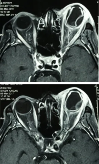
infection. The patient was initially diagnosed with left bacterial keratitis secondary to a foreign body entry with possibly an underlying choroidal tumour. Our differential diagnosis was left bacterial keratitis resulting from a ruptured bullous keratopathy secondary to chronically raised IOP due to the tumour. However, the MRI imaging showed left choroidal detachment with lens displacement, surrounding inflammatory changes and no evidence of tumour. There was no increase in vascularity within the mass as well (Figure 4). Blood investigations showed a markedly elevated erythrocyte sedimentation rate (ESR) of 105 mm/hour (reference range: 1–10 mm/hour), raised C-reactive protein of 69.6 mg/L (reference range:
<5 mg/dL) with positive HIV and Hepatitis C status. His corneal scraping grew Pseudomonas Aeruginosa.
Due to the patient’s immunocompromised status, markedly elevated ESR, lymphadenopathy, and inflam- matory changes of the mass, a possible diagnosis of in- traocular tuberculosis (TB) was made. However, TB work- up, which included chest X-ray, Mantoux test and sputum to look for acid-fast bacilli were normal. The patient was started on Ceftazidime 5% and fortified Gentamicin 0.9%
eye drops which was sensitive for the Pseudomonas Aeruginosa species and two IOP lowering agents. Despite the initial targeted treatment, the patient’s condition worsened. His cornea perforated within two days of presentation and the patient was in intractable pain, not responding to analgesics. After an elaborated discussion and counselling, we proceeded with evisceration of the affected eye. The corneal tissue and vitreous humour was sent for TB polymerase chain reaction (PCR). The PCR result came back positive for M. Fortuitum. The pa- tient was not started on any systemic antibiotics as there was no evidence of systemic dissemination of the infec- tion.
Figure 1: External photograph showing swollen periorbital tissue with proptosis
Figure 2: External photograph showing melting central corneal ulcer with hypopyon
Figure 3: B-scan showing hyperechoic mass with surrounding exudative retinal detachment
Figure 4: MRI orbit showing left choroidal detachment with lens displacement
2/4 GMS Ophthalmology Cases 2019, Vol. 9, ISSN 2193-1496
Yeap et al.: Non-tuberculous mycobacterial ocular infection masquerading ...
Discussion
This case illustrates that NTM keratitis and choroiditis are rare opportunistic infections that can pose both diagnostic and therapeutic challenges. In the literature, the average duration from onset of symptoms to diagnosis is 10 weeks [3]. In this case, it took approximately 6 weeks from the onset of symptoms for the diagnosis of NTM choroiditis to be confirmed. From the systematic reviews and the reported cases, it is deducible that NTM infections are great masquerades. The clinical features and indolent nature of NTM keratitis can mimic those of infectious keratitis by other pathogens, especially fungus, herpes simplex virus, and Acanthamoeba [1]. An indolent inflammatory process and recalcitrance to traditional antibacterial therapy should increase the level of suspi- cion of NTM infections. Contrary to the reported cases, this patient presented with progressive keratitis that resulted in perforation despite the intensive medical treatment. The presence of a dual infection of Pseudomo- nas Aeruginosa and M. Fortuitum may explain the altered course of the disease.
The B-mode ultrasonography features of this patient mimicked a choroidal tumour with collar stud appearance with low internal reflectivity. Similar to our case, Lai et al.
from Japan reported a case of NTM choroiditis in a HIV patient which was mistaken for ocular lymphoma [7]. All the other reported NTM choroiditis cases also experienced delay in diagnosis due to misdiagnosis, misidentification of the organism, and delay in taking cultures [1].
The three commonest NTM species, M. Chelonae, M. Fortuitum and M. Abscessus are part of Group IV NTM, which are also known as rapidly growing NTM. These or- ganisms take 7–10 days to culture, unlike other groups of NTM, which take up to 2–4 weeks [2], [8]. Based on the literature, NTM are best diagnosed firstly using a Löwenstein-Jensen medium to culture for acid-fast bacilli.
However, it was reported that the initial NTM isolates identified via conventional biochemical techniques could possibly belong to different NTM subtypes, hence PCR is employed to reduce misidentification of the species [9].
This is important as different NTM species have different antimicrobial susceptibilities.
Intraocular NTM infections were reported to have poorer response to medical treatment as compared periocular, adnexal, and ocular surface NTM infections. Studies have shown that Amikacin and Clarithromycin were more effi- cacious, while fluoroquinolones in general were less ef- fective [3], [9]. Combination therapy using amikacin, a fluoroquinolone, and a macrolide is preferred over mono- therapy to reduce resistance and to increase efficacy by increasing penetration through the corneal epithelium.
Presence of a foreign body or implant and a non-respond- ing intraocular NTM infection may warrant surgical inter- vention such as removal of the foreign body or implant itself, pars plana vitrectomy, evisceration, or enucleation.
In this patient, evisceration was necessary to remove the infected ocular tissue.
Conclusion
NTM ocular infections are rare and pose diagnostic challenges. A delay in diagnosis leads to severe morbidity and poor visual outcome. Hence, high index of suspicion, prompt investigation and treatment could be sight saving.
Therapeutic surgical intervention may be necessary to control the infection.
Notes
Competing interests
The authors declare that they have no competing in- terests.
Patient consent
Written consent to publish the case report was obtained.
Acknowledgments
• Radiology Department of Hospital Tuanku Ampuan Najihah, Kuala
• Pilah for interpretation of relevant imaging
References
1. Moorthy RS, Valluri S, Rao NA. Nontuberculous mycobacterial ocular and adnexal infections. Surv Ophthalmol. 2012 May- Jun;57(3):202-35. DOI: 10.1016/j.survophthal.2011.10.006 2. Kheir WJ, Sheheitli H, Abdul Fattah M, Hamam RN.
Nontuberculous Mycobacterial Ocular Infections: A Systematic Review of the Literature. Biomed Res Int. 2015;2015:164989.
DOI: 10.1155/2015/164989
3. Girgis DO, Karp CL, Miller D. Ocular infections caused by non- tuberculous mycobacteria: update on epidemiology and management. Clin Experiment Ophthalmol. 2012 Jul;40(5):467- 75. DOI: 10.1111/j.1442-9071.2011.02679.x
4. Turner L, Stinson I. Mycobacterium fortuitum; as a cause of corneal ulcer. Am J Ophthalmol. 1965;60(2):329–31. DOI:
10.1016/0002-9394(65)90934-7
5. Dugel PU, Holland GN, Brown HH, Pettit TH, Hofbauer JD, Simons KB, Ullman H, Bath PE, Foos RY. Mycobacterium fortuitum keratitis. Am J Ophthalmol. 1988 Jun 15;105(6):661-9. DOI:
10.1016/0002-9394(88)90061-X
6. Shah M, Relhan N, Kuriyan AE, Davis JL, Albini TA, Pathengay A, Miller D, Flynn HW Jr. Endophthalmitis Caused by Nontuberculous Mycobacterium: Clinical Features, Antimicrobial Susceptibilities, and Treatment Outcomes. Am J Ophthalmol. 2016 Aug;168:150- 6. DOI: 10.1016/j.ajo.2016.03.035
7. Lai LJ, Chen SN, Kuo YH, Ho JD, Ho CL. Presumed choroidal atypical tuberculosis superinfected with cytomegalovirus retinitis in an acquired immunodeficiency syndrome patient: a case report. Jpn J Ophthalmol. 2002 Jul-Aug;46(4):463-8. DOI:
10.1016/S0021-5155(02)00500-2
3/4 GMS Ophthalmology Cases 2019, Vol. 9, ISSN 2193-1496
Yeap et al.: Non-tuberculous mycobacterial ocular infection masquerading ...
8. Sharma K, Gautam N, Sharma M, Dogra M, Bajgai P, Tigari B, Sharma A, Gupta V, Sharma SP, Singh R. Ocular mycobacteriosis- dual infection of M. tuberculosis complex with M. fortuitum and M. bovis. J Ophthalmic Inflamm Infect. 2017 Dec;7(1):2. DOI:
10.1186/s12348-016-0121-0
9. Paulose RM, Joseph J, Narayanan R, Sharma S. Clinical and microbiological profile of non-tuberculous mycobacterial endophthalmitis-experience in a tertiary eye care centre in Southern India. J Ophthalmic Inflamm Infect. 2016 Dec;6(1):27.
DOI: 10.1186/s12348-016-0096-x
Corresponding author:
Dr. Puspha Raman
Department of Ophthalmology, Hospital Tuanku Ampuan Najihah, Jalan Melang, Kampung Gemelang, 72000 Kuala Pilah, Negeri Sembilan, Malaysia, Phone: +60123728048 puspha@gmail.com
Please cite as
Yeap KC, Sivagurunathan PD, Raman P, Khalid KHM. Non-tuberculous mycobacterial ocular infection masquerading as choroidal tumour – a diagnostic conundrum. GMS Ophthalmol Cases. 2019;9:Doc25.
DOI: 10.3205/oc000114, URN: urn:nbn:de:0183-oc0001147
This article is freely available from
http://www.egms.de/en/journals/oc/2019-9/oc000114.shtml Published:2019-07-05
Copyright
©2019 Yeap et al. This is an Open Access article distributed under the terms of the Creative Commons Attribution 4.0 License. See license information at http://creativecommons.org/licenses/by/4.0/.
4/4 GMS Ophthalmology Cases 2019, Vol. 9, ISSN 2193-1496
Yeap et al.: Non-tuberculous mycobacterial ocular infection masquerading ...