Non-Hodgkin lymphoma with relapses in the lacrimal glands
Abstract
Objective:To report an unusual case of systemic non-Hodgkin lymphoma (NHL) with repeated relapse in the lacrimal glands, in spite of complete remission for several years after treatment.
Rita Couceiro
1Helena Proença
1Filomena Pinto
1Methods:A 78-year-old male with small lymphocytic B cell NHL, stage
IV disease (lung invasion), was submitted to surgery and chemotherapy
Ana Fonseca
1in 2001, with complete remission of the disease. In 2003 he developed
Manuel Monteiro-Grillo
1a nodular lesion in the right lacrimal fossa. Pathology results revealed a local relapse of NHL. Radiation and chemotherapy were initiated and
complete remission was again achieved. In 2012 the patient developed 1 Hospital de Santa Maria, Department of
a new nodular lesion located in the left lacrimal fossa, resulting in dip-
Ophthalmology, Lisbon, Portugal
lopia, ptosis and proptosis of the left eye. Orbital computerized tomog- raphy (CT), ocular ultrasound and incisional biopsy were performed.
Results:Orbital CT revealed a lesion infiltrating the left lacrimal gland and encircling the globe. Biopsy results confirmed a local relapse of B cell NHL. The patient was submitted to local radiation therapy with progressive resolution of ptosis, proptosis and diplopia. Response to treatment was monitored with ocular ultrasound.
Conclusions: Patients with NHL diagnosis should be immediately investigated if ophthalmic or orbital symptoms develop. NHL extension to the orbit and adnexa is infrequent (5% of NHL cases) but may occur at any stage of the disease, including as a relapse site. In such cases, radiation and chemotherapy achieve good results, inducing long periods of remission.
Keywords:lacrimal gland, non-Hodgkin lymphoma relapse, ocular ultrasound, orbit
Introduction
Systemic non-Hodgkin’s lymphoma (NHL) is reported to have secondary extension to the orbit and adnexa in about 5% of cases [1] and an orbital presentation in 2.4%
of all NHL cases [2]. However, given that clinical manifest- ations of orbital lymphoma are sometimes subtle, some authors suggest that this prevalence might be higher [1].
Patients with NHL diagnosis should therefore be suspect- ed of having orbital involvement if an orbital mass, ptosis, proptosis or lid edema develop [1]. On the other hand, patients with primary orbital and ocular adnexal lymph- oma evidence extraorbital involvement in more than 50%
of cases, which determines the need for extensive sys- temic workup at diagnosis [3].
The typical presentation of adnexal lymphoproliferative disease includes a painless mass, swelling or proptosis [4]. Involvement of adnexal structures in systemic NHL may appear at any time during the course of the disease, including as a relapse site [1], [5].
Treatment with systemic chemotherapy or systemic im- munotherapy and combination with local radiation treat- ment is beneficial in secondary orbital lymphomas, induc- ing prolonged remission [1], [6].
With this report we aim to describe a rare case of systemic non-Hodgkin lymphoma (NHL), with repeated relapses in the lacrimal glands, in spite of complete and sustained remission after treatment.
Case description
A 78-year-old caucasian male, without any prior relevant medical history, was diagnosed with small lymphocytic B cell NHL in 2000, presenting with stage IV disease with lung invasion. He was submitted to surgery and chemo- therapy (cyclophosphamide, vincristine, prednisone;
8 cycles), with complete regression of the disease.
In 2003 the patient developed intermittent diplopia in right gaze and a nodular lesion was noted at the lateral portion of his right orbit. CT imaging revealed a lesion in the right lacrimal fossa, pressing the globe down and forward without invading it. An incisional biopsy was per- formed and pathology results revealed local recurrence of NHL, but no immunohistochemistry analysis was con- ducted due to a small sample size with artifactual changes. The patient underwent chemotherapy (cyclo- phosphamide, hydroxydaunorubicin, oncovin, prednisone;
1/3 GMS Ophthalmology Cases 2015, Vol. 5, ISSN 2193-1496
Case Report
OPEN ACCESS
4 cycles) and radiation therapy (12 cycles), with complete and sustained response to treatment.
In 2012 the patient developed progressive ptosis of the left eye (OS) and intermittent diplopia, with no other visual complaints. Concurrently, a new nodular lesion was noted in the lateral portion of the left orbit.
On observation the patient presented best corrected visual acuities (BCVA) of 20/25 in both eyes. Inferior dystopia and proptosis in OS were noted; Hertel examin- ation (106 mm) results were 15 mm in the right eye and 20 mm in OS. There was slight restriction of movements in OS in up and left gaze, resulting in diplopia. A clear enlargement of the left lacrimal gland was evident on examination, with discrete associated conjunctival chemosis. The remaining anterior segment features and fundoscopy were unremarkable.
Orbital CT imaging revealed an infiltrative hyperdense lesion in the left lacrimal gland, encircling the globe, the muscle cone and the lateral and proximal portions of the optic nerve, without invading any of these structures, nor the extraocular muscles or the orbital walls (Figure 1).
On ocular ultrasound the lacrimal gland lesion exhibited well defined borders, internal homogeneous structure and medium/low reflectivity.
Figure 1: Orbital CT, axial view shows an infiltrative lesion, located in the left lacrimal gland.
Incisional biopsy of the left lacrimal gland was performed and pathology results revealed B cell NHL relapse. Im- munohistochemistry results were positive for CD20 and bcl-2 and negative for CD5, CD10, CD23, CD43 and cyclin D1.
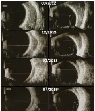
Local radiation therapy (total dosing: 8 Gy/2 fractions) was undertaken and during the following 6 months there was a gradual resolution of diplopia, ptosis and proptosis in OS. Progressive reduction in lesion size was monitored with ultrasound until its complete regression (Figure 2).
At last follow-up BCVA remained 20/25 bilaterally.
Figure 2: Consecutive ultrasounds of the left eye – superior transversal view (left column) and lateral transversal view (right column) – documenting progressive resolution of the lacrimal gland lesion after radiation therapy treatment was performed
in November 2012.
Discussion
The differential diagnosis of an orbital mass lesion is vast [7] and although the orbit is rarely a secondary site of lymphoma dissemination, patients with NHL diagnosis should be immediately investigated if ophthalmic or orbit- al symptoms develop [1].
The reported case is unusual for the repeated NHL re- lapses in the lacrimal glands, separated by several years of complete remission. Orbit and adnexa NHL have indeed been documented in relapses of previously diagnosed lymphomas [5].
In the reported case, the patient was initially diagnosed with small lymphocytic B cell NHL, which is considered an indolent lymphoma with a variable clinical course [8].
This might include relapses at the original site or at new locations and even a change in lymphoma histology can occur [9]. Immunohistochemistry analysis was not per- formed in the first relapse, but in 2012 results suggest the existence of a MALT subtype NHL (CD20+, CD5–, CD10–, CD23–), confirming a change in NHL histology.
However, this does not represent a change into a more aggressive histology and therefore, we believe, should not influence the patient’s survival.
2/3 GMS Ophthalmology Cases 2015, Vol. 5, ISSN 2193-1496
Couceiro et al.: Non-Hodgkin lymphoma with relapses in the lacrimal ...
Radiation and chemotherapy have proved to achieve good results in secondary orbital NHL [1], [6] and were effective in treating both relapses in this case. Sustained remission after relapse of indolent lymphomas can often be obtained as long as the disease histology remains low- grade, although relapse will usually ensue [9], determining the need for prolonged follow-up.
Conclusions
Patients with NHL diagnosis should be immediately investigated if ophthalmic or orbital symptoms develop.
NHL extension to the orbit and adnexa is infrequent (5%
of NHL cases) but may occur at any stage of the disease, including as a relapse site. In such cases, radiation and chemotherapy achieve good results, inducing long periods of remission.
Notes
Competing interests
The authors declare that they have no competing in- terests. No financial support was received for this submis- sion.
Informed consent
This case report was performed with informed consent regarding the consultation of human medical records.
Acknowledgments
Dr. Ana Cristina Ferreira, Dr. Dolores López-Presa – De- partment of Pathology, Hospital de Santa Maria, Lisbon, Portugal.
References
1. Bairey O, Kremer I, Rakowsky E, Hadar H, Shaklai M. Orbital and adnexal involvement in systemic non-Hodgkin's lymphoma.
Cancer. 1994 May 1; 73(9):2395-9. DOI: 10.1002/1097- 0142(19940501)73:9<2395::AID-CNCR2820730924>3.0.CO;2- V
2. Lazzarino M, Morra E, Brusamolino E, Pagnucco G, Bernasconi C, Rosso R, Castello A, Ghisolfi A, Tafi A, Zennaro G.
Clinicopathologic and immunologic characteristics of non- Hodgkin's lymphomas presenting in the orbit. A report of eight cases. Cancer. 1985 May 1; 55(9):1907-12. DOI: 10.1002/1097- 0142(19850501)55:9<1907::AID-CNCR2820550913>3.0.CO;2- J
3. Hatef E, Roberts D, McLaughlin P, Pro B, Esmaeli B. Prevalence and nature of systemic involvement and stage at initial examination in patients with orbital and ocular adnexal lymphoma. Arch Ophthalmol. 2007 Dec;125(12):1663-7. DOI:
10.1001/archopht.125.12.1663
4. Sullivan TJ, Whitehead K, Williamson R, Grimes D, Schlect D, Brown I, Dickie G. Lymphoproliferative disease of the ocular adnexa: a clinical and pathologic study with statistical analysis of 69 patients. Ophthal Plast Reconstr Surg. 2005
May;21(3):177-88. DOI:
10.1097/01.IOP.0000159173.42243.AD
5. Ferry JA, Fung CY, Zukerberg L, Lucarelli MJ, Hasserjian RP, Preffer FI, Harris NL. Lymphoma of the ocular adnexa: A study of 353 cases. Am J Surg Pathol. 2007 Feb;31(2):170-84. DOI:
10.1097/01.pas.0000213350.49767.46
6. Esmaeli B, Ahmadi MA, Manning J, McLaughlin PW, Ginsberg L.
Clinical presentation and treatment of secondary orbital lymphoma. Ophthal Plast Reconstr Surg. 2002 Jul;18(4):247- 53. DOI: 10.1097/00002341-200207000-00003
7. Saunders TS, Anis S, Doych Y, Moran A, Hou JS, Chen X, Yanoff M. Systemic non-Hodgkin's lymphoma involving the orbit and leptomeninges. Dig J of Ophthal. 2010;16(3):9-12. DOI:
10.5693/djo.02.2010.06.001
8. Rizzo K, Nassiri M. Diagnostic Workup of Small B Cell
Lymphomas: A Laboratory Perspective. Lymphoma. 2012;2012:1- 15. DOI: 10.1155/2012/346084
9. National Cancer Institute. Adult Non-Hodgkin Lymphoma Treatment (PDQ®). General Information About Adult Non-Hodgkin Lymphoma. Bethesda, MD: NCI; 2014. Available from: http://
www.cancer.gov/cancertopics/pdq/treatment/adult-non- hodgkins/Patient [last accessed 2014 Nov 15]
Corresponding author:
Rita Couceiro, MD
Avenida Professor Egas Moniz, Hospital Santa Maria - Secretariado do Serviço de Oftalmologia 1649-035 Lisboa, Portugal, Phone: 00351 913988901, Fax: +351 217 805 653
anaritacouceiro@gmail.com
Please cite as
Couceiro R, Proença H, Pinto F, Fonseca A, Monteiro-Grillo M.
Non-Hodgkin lymphoma with relapses in the lacrimal glands. GMS Ophthalmol Cases. 2015;5:Doc04.
DOI: 10.3205/oc000026, URN: urn:nbn:de:0183-oc0000260
This article is freely available from
http://www.egms.de/en/journals/oc/2015-5/oc000026.shtml Published:2015-06-03
Copyright
©2015 Couceiro et al. This is an Open Access article distributed under the terms of the Creative Commons Attribution 4.0 License. See license information at http://creativecommons.org/licenses/by/4.0/.
3/3 GMS Ophthalmology Cases 2015, Vol. 5, ISSN 2193-1496
Couceiro et al.: Non-Hodgkin lymphoma with relapses in the lacrimal ...