Method:A 42-year-old female patient with past history of hysterectomy
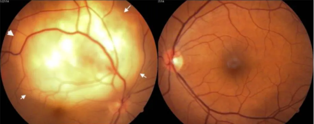
presented with diminution of vision in the right eye. Fundus examination 1 Sarojini Devi Eye Hospital / Osmania Medical College, Hyderabad. India in the right eye showed a yellowish white choroidal mass with associated
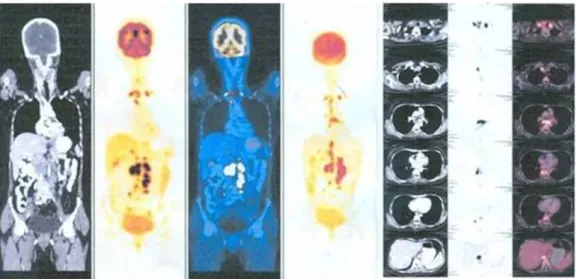
bullous retinal detachment superotemporal to fovea. Left eye fundus was normal. Fundus flourescein angiography showed early and late hyperflourescence with late pooling in serous detachments. Complete systemic evaluation did not yield a clue to diagnosis. Positron emission tomography scan (PET scan) showed enlarged lymph nodes in cervical, mediastinal and peritoneal regions. Lymph node biopsy showed case- ating granulomas.
Results:The granuloma subsided and a scar formed 5 months after starting anti-tuberculous treatment with improvement in vision. Six months later, the vision deteriorated again with the development of a choroidal neovascular membrane (CNVM) at the margin of the scar.
The CNVM resolved and all the signs of activity subsided after giving intravitreal antivascular endothelial growth factor (anti-VEGF) injections.
Conclusions:Making a diagnosis of tuberculous granuloma in a case of choroidal mass lesion is a challenge. PET scan helps in identifying metabolically active lymph nodes appropriate for biopsy. Healed scars of tuberculous choroid lesions should be followed closely to detect the development of CNVM.
Keywords:tuberculosis, choroidal granuloma, exudative retinal detachment, CNVM, bevacizumab
Introduction
Tuberculosis (TB) is a common infectious disease, nearly one-third of world population is infected with it. The esti- mated figure for prevalence of tuberculosis in India is given as 2.5 million [1]. It is estimated that 40% of the population in India is infected with tuberculous bacteria and that the majority of them has latent rather than active tuberculosis [2]. Tuberculosis primarily affects the lungs, but may also affect extrapulmonary organs including the eyes [3]. Tuberculosis can affect the anterior and posteri- or segments of the eye as well as the adnexa with various presentations. These pleomorphisms of lesions, as well as the lack of uniformity in diagnostic criteria, make a diagnosis of intraocular tuberculosis a challenge.
Choroidal neovascular membrane (CNVM) can arise as a complication of any pathologic process that involves retinal pigment epithelium and damages Bruch’s mem- brane [4]. The association between choroidal neovascular membrane (CNVM) and age-related macular degeneration or myopia is well known. CNVM may also develop as a complication in posterior uveitis; the reported incidence
is 2% [5]. The different uveitic entities associated with CNVM include toxoplasmosis, punctuate inner choroido- pathy (PIC) (17–40%), idiopathic multifocal choroiditis (MC) (33%), and serpiginous choroiditis [6]. It is also re- ported in ten eyes (9%) of seven patients in a follow-up of 58 patients (116 eyes) with Vogt-Koyanagi-Harada syndrome [7].
We report an interesting case of isolated choroidal granuloma without systemic tuberculosis in an immuno- competent young female that responded very well to anti- tuberculous treatment with good visual recovery. CNVM developed at the margin of the lesion that had regressed after tuberculostatic therapy. After four anti-VEGF injec- tions, control of the CNVM was achieved.
Figure 1: RE showing subretinal mass lesion. White arrows demarcating the outline of retinal detachment.
Figure 2: FFA shows early hyperflourescence with late pooling in serous detachment.
Case description
A 42-year-old female patient presented with a history of diminution of vision in the right eye for fifteen days. She had no other systemic complaints. She had undergone hysterectomy for an indication of dysfunctional uterine bleeding a year and a half ago. On examination, she had best corrected visual acuity of 20/400 in the right eye and 20/20 in the left eye. Both eyes anterior segment examination was within normal limits. Fundus examina- tion of the right eye showed a yellowish white subretinal mass lesion six to eight disc diameters in size, located at the superotemporal arcade, just reaching the fovea with an overlying exudative detachment (Figure 1). Left eye fundus was normal. The differential diagnosis included choroidal metastasis, choroidal tuberculoma or amelan- otic melanoma. Because of hysterectomy in the recent past, the possibility of choroidal metastases was strongly considered.
Fundus flourescein angiography showed an early hyper- flourescence in the region of the lesion with progressive increase in hyperflourescence and pooling of the dye outlining the serous detachment by late phases (Figure 2).
Ultrasonography showed a mass lesion of uniform echo- genecity with low internal reflectivity (Figure 3).
Figure 3: Ultrasonography shows a mass lesion of uniform echogenecity and low internal reflectivity.
Figure 4: PET scan shows enlarged lymph nodes in cervical, mediastinal, and retroperitoneal regions.
The patient was referred to the physician for systemic evaluation. The patient underwent chest radiography, Mantoux test, complete blood picture, erythrocyte sedi- mentation rate (ESR) and HIV test. Positron emission tomography (PET) scan was advised by the physician.
Chest x-ray was normal, Mantoux test (with 5 IU of purified protein derivatives) showed 15 mm induration. Erythro- cyte sedimentation rate (ESR) was more than 20 mm.
PET scan (Figure 4) reporting was presence of multiple
18F-fluorodeoxyglucose (FDG) avid nodes (up to level IV) in cervical, mediastinal and retroperitoneal region – suggestive of an inflammatory pathology (tuberculosis) or metastases. Fine needle aspiration biopsy (FNAC) of cervical lymph node was inconclusive. Cervical lymph node biopsy showed caseous necrotic inflammation probably of tuberculous etiology. There was no evidence of neoplasia (Figure 5). AFB culture of lymph nodes did not show any growth of acid fast bacilli.
Figure 5: Showing granuloma with Langhan’s giant cells and caseation necrosis
The patient was started on four drug anti-tuberculosis treatment (isoniazid, rifampicin, etambutol, pyrazinamide with pyridoxine) with oral prednisolone (1 mg/kg body weight). Oral corticosteroids were tapered after the first four weeks and were stopped within eight weeks. The
lesion started showing healing from the edges within two weeks after starting anti-tuberculous treatment (ATT) (Figure 6 and Figure 7).
Figure 6: Healing lesion two months after starting anti-tuberculous treatment
Figure 7: Scarred granuloma seven months after starting anti-tuberculous treatment. Fovea is spared.
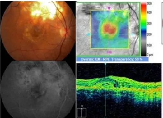
At ten-month follow-up, the scarred granuloma was stable with a visual acuity of 6/12p. A month later, still on ATT, the patient reported diminution of vision in the right eye, with best corrected visual acuity (BCVA) falling to 6/36p in RE. RE fundus examination showed macular edema with subretinal blood and exudates adjacent to the scar of the previous choroidal granuloma (Figure 8). Fundus
Figure 8: Juxtafoveal yellowish white lesion adjacent to healed scar with retinal edema, subretinal blood, exudates and subretinal fluid. FFA and SD-OCT showed signs of active choroidal neovascular membrane at the margin of the scar.
Figure 9: At one-year follow-up, fundus picture showed healed, pigmented scarred lesion at fovea. OCT showed reduced thickness, scarred lesion with partial restoration of foveal contour.
fluorescein angiography (FFA) showed patchy hypofloures- cence due to subretinal blood and exudates and mild diffuse late leakage due to juxtafoveal CNVM. Spectral domain optical coherence tomography (SD-OCT) showed an increased foveal thickness, a dome-shaped hyperre- flectivity at the level of the retinal pigment epithelium
(RPE) and adjacent intraretinal cystic changes indicative of an active CNVM.
After informed consent, intravitreal anti-vascular endotheli- al growth factor (anti-VEGF) (Bevacizumab) was given.
The CNVM resolved after four intravitreal Bevacizumab (Avastin) injections, leaving a scar with BCVA of 6/36 (Figure 9).
choroiditis, retinal vasculitis, sub retinal abscess, and endophthalmitis [10].
In this case, a history of hysterectomy with a subretinal mass lesion prompted a PET scan to look for any focus of malignancy. This identified metabolically active lymph nodes at several sites, including cervical nodes, which on biopsy identified caseating granulomas. This confirma- tory histopathology allowed timely and appropriate treat- ment and presumably resulted in a better visual and anatomical outcome. PET/CT scans may suggest the sites of safe high-yield biopsies, thus yielding confirmatory histopathology, cultures, or PCR samples. Doycheva et al. found that 18F-FDG-PET/CT is useful in identifying metabolically active nodes appropriate for a biopsy that helps to establish the diagnosis and start appropriate treatment in presumed intraocular tuberculosis [11].
However, in the present case the development of choroidal neovascular membrane at fovea at the margin of the scar of regressed granuloma jeopardized the good visual outcome. CNVM, though uncommon, is a sight threatening complication of posterior uveitis. It results either from an inflammation induced angiogegenic drive or a degenerative disruption of RPE-choroid complex. The prime inducer of neovascularisation is the vascular en- dothelial growth factor [12]. The reported incidence of CNVM developing as a complication of uveitis is 2% [5].
Bansal et al. report 3.5% (5 eyes) developing CNVM in a study of 105 patients (141 eyes) of tubercular serpiginous like choroiditis with a minimum follow-up of 9 months [13]. Inflammatory CNVM develops close to the healed chorioretinal scar or choroidal granuloma – may be foveal, juxtafoveal and extrafoveal [14]. In this case, a juxtafoveal inflammatory CNVM has developed eleven months after the appearance of choroidal granuloma. Therapies available for inflammatory CNVM include photodynamic therapy (PDT), intravitreal bevacizumab and intravitreal ranibizumab [13]. In our case, the patient was continuing with ATT and the CNVM resolved and all the signs of activity subsided after four injections of intravitreal beva- cizumab. The vision was stabilized at 6/36 and no recur- rence occurred during a follow-up of one year. The median number of intravitreal anti-VEGF injections required to achieve an inactive CNVM in different studies is reported as 2 (range 1–5) [14]. In a study on “Risk of choroidal neovascularization among the uveitides” [5], Baxter et al. have concluded that CNVM is rare among cases of anterior and intermediate uveitis and found much more frequently (both at initial presentation as well at follow-
Therefore, clinicians should educate the high-risk cases of posterior uveitis with potential symptoms of CNVM and provide them with screening tests like Amsler grid for an early detection and treatment.
Notes
Competing interests
The authors declare that they have no competing in- terests.
References
1. World Health Organization. Global Tuberculosis Control 2015.
Geneva: WHO; 2015.
2. Global Health Education (GHE). TB Facts. TB Statistics for India.
National and state statistics. Available from: http://
www.tbfacts.org/tb-statistics-india/
3. Golden MP, Vikram HR. Extrapulmonary tuberculosis: an overview.
Am Fam Physician. 2005 Nov;72(9):1761-8.
4. Campa C, Costagliola C, Incorvaia C, Sheridan C, Semeraro F, De Nadai K, Sebastiani A, Parmeggiani F. Inflammatory mediators and angiogenic factors in choroidal neovascularization:
pathogenetic interactions and therapeutic implications. Mediators Inflamm. 2010;2010. pii: 546826. DOI: 10.1155/2010/546826 5. Baxter SL, Pistilli M, Pujari SS, Liesegang TL, Suhler EB, Thorne
JE, Foster CS, Jabs DA, Levy-Clarke GA, Nussenblatt RB, Rosenbaum JT, Kempen JH. Risk of choroidal neovascularization among the uveitides. Am J Ophthalmol. 2013 Sep;156(3):468- 77.e2. DOI: 10.1016/j.ajo.2013.04.040
6. Kuo IC, Cunningham ET Jr. Ocular neovascularization in patients with uveitis. Int Ophthalmol Clin. 2000 Spring;40(2):111-26.
DOI: 10.1097/00004397-200004000-00009
7. Moorthy RS, Chong LP, Smith RE, Rao NA. Subretinal neovascular membranes in Vogt-Koyanagi-Harada syndrome. Am J Ophthalmol. 1993 Aug 15;116(2):164-70. DOI: 10.1016/S0002- 9394(14)71280-2
8. Bramante CT, Talbot EA, Rathinam SR, Stevens R, Zegans ME.
Diagnosis of ocular tuberculosis: a role for new testing modalities? Int Ophthalmol Clin. 2007;47(3):45-62. DOI:
10.1097/IIO.0b013e318074de79
9. Demirci H, Shields CL, Shields JA, Eagle RC Jr. Ocular tuberculosis masquerading as ocular tumors. Surv Ophthalmol. 2004 Jan- Feb;49(1):78-89. DOI: 10.1016/j.survophthal.2003.10.009 10. Sharma A, Thapa B, Lavaju P. Ocular tuberculosis: an update.
Nepal J Ophthalmol. 2011 Jan-Jun;3(1):52-67. DOI:
10.3126/nepjoph.v3i1.4280
11. Doycheva D, Deuter C, Hetzel J, Frick JS, Aschoff P, Schuelen E, Zierhut M, Pfannenberg C. The use of positron emission tomography/CT in the diagnosis of tuberculosis-associated uveitis. Br J Ophthalmol. 2011 Sep;95(9):1290-4. DOI:
10.1136/bjo.2010.182659
12. Grossniklaus HE, Ling JX, Wallace TM, Dithmar S, Lawson DH, Cohen C, Elner VM, Elner SG, Sternberg P Jr. Macrophage and retinal pigment epithelium expression of angiogenic cytokines in choroidal neovascularization. Mol Vis. 2002 Apr;8:119-26.
13. Bansal R, Gupta A, Gupta V, Dogra MR, Sharma A, Bambery P.
Tubercular serpiginous-like choroiditis presenting as multifocal serpiginoid choroiditis. Ophthalmology. 2012 Nov;119(11):2334- 42. DOI: 10.1016/j.ophtha.2012.05.034
14. D’Ambrosio E, Tortorella P, Iannetti L. Management of uveitis- related choroidal neovascularization: from the pathogenesis to the therapy. J Ophthalmol. 2014;2014:450428. DOI:
10.1155/2014/450428
Corresponding author:
Sikander A. K. Lodhi
Sarojini Devi Eye Hospital, “HILL VIEW”, 10-3-300/3, Humayun Nagar, Masab Tank, Hyderabad 500028, Telangana State, India, Phone: +91 9848020497, +91 40 23533497
sikanderlodhi@gmail.com
Please cite as
Lodhi SA, Saifuddin K, Devulapally S. Inflammatory choroidal neovascular membrane after healed tuberculous choroidal granuloma. GMS Ophthalmol Cases. 2017;7:Doc06.
DOI: 10.3205/oc000057, URN: urn:nbn:de:0183-oc0000571
This article is freely available from
http://www.egms.de/en/journals/oc/2017-7/oc000057.shtml Published:2017-03-03
Copyright
©2017 Lodhi et al. This is an Open Access article distributed under the terms of the Creative Commons Attribution 4.0 License. See license information at http://creativecommons.org/licenses/by/4.0/.