Model membrane systems, membrane- active peptides and application
possibilities
Inaugural-Dissertation
zur
Erlangung des Doktorgrades
der Mathematisch-Naturwissenschaftlichen Fakultät der Universität zu Köln
vorgelegt von
Mareike Horn aus Friedrichroda
Köln
2017
Berichterstatter: Prof. Dr. Ines Neundorf
Prof. Dr. Ulrich Baumann
Tag der mündlichen Prüfung: 19. Dezember 2017
Die im Rahmen der vorliegenden Arbeit durchgeführten Experimente und Untersuchungen
wurden im Zeitraum von April 2014 bis August 2017 am Institut für Biochemie der
Universität zu Köln unter der Anleitung von Frau Prof. Dr. Ines Neundorf durchgeführt.
The ability of cell-penetrating peptides (CPPs) to translocate a variety of covalently or non- covalently linked cargoes, particularly therapeutics and imaging agents, across the plasma membrane of living cells renders them of broad interest in the medical field as well as in biotechnological development. Thus, the use of CPPs, e.g. in therapeutics, increases the necessity of understanding their mechanism of action, a subject still under debate.
For the first step of peptide internalization, the interaction between the positively charged amino acid side chains within the peptide sequence and the anionic constituents of the membrane bilayer is assumed. However, owing to the complexity of biological plasma membranes, studying the mode of action of CPPs on a molecular level is very difficult. With this in mind, model membrane systems, especially large and giant unilamellar vesicles and giant plasma membranes vesicles derived from live cells plasma membranes, were prepared and several techniques were established in order to monitor the initial step of cell entry – binding to and translocation through the cell membrane as well as identification of key components of the plasma membrane required for efficient uptake.
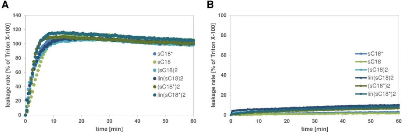
The first chapter focuses on the membrane interactions and perturbation mechanism of two structural modified variants of the well-described CPP sC18, a cyclized and a dimeric branched version, with artificial model membranes. Fluorescence analysis demonstrated the importance of negatively charged lipids for an efficient uptake of Arg-rich CPPs, supporting the assumption that electrostatic interactions are required for the initial stage of cell entry. In case of dimeric variants, the strong affinity was further accompanied by intense membrane destabilizing effects, presumably responsible for their lytic behavior as anticancer peptide. Moreover, the cyclization of the peptide backbone turned out to be a promising strategy as well to improve their cell-penetrating capability. Using CD spectroscopy, structural arrangements when in contact with neutral and anionic vesicles further revealed that a certain flexibility and thus the static arrangement of the guanidinium groups within the cycle seem to positively affect peptide-membrane interaction. In summary, these evidences suggest that the internalization of the peptide variants depends highly on the membrane composition of the target cell.
In the second chapter, the focus lay on the design and characterization of novel peptide
conjugates, composing of a wound healing promoting peptide and sC18 as cell penetrating
peptide, with the final goal to further enhance the wound healing activity. Besides, the new
conjugate Tylotoin-sC18* exhibited further promising antimicrobial activity and moreover a
certain selectivity towards cancer cells, suggesting that Tylotoin-sC18* is a good candidate
for further studies and the possible development as a therapeutic agent for infected wounds.
Zusammenfassung
Die Fähigkeit zellpenetrierender Peptide (cell-penetrating peptides, CPPs) die Plasmamembran lebender Zellen passieren zu können und dabei kovalent und nicht- kovalent gebundene Moleküle, insbesondere therapeutisch aktive Wirkstoffe und Farbstoffe, zu transportieren, macht sie sowohl für die Medizin als auch für den Biotechnologiesektor besonders interessant. Für eine erfolgreiche therapeutische Anwendung zellpenetrierender Peptide ist die Erforschung derer genauen Wirkmechanismen essentiell. Als erster Schritt der Internalisierung wird dabei weithingehend die Interaktion der positiv geladenen Aminosäuren innerhalb der Peptidsequenz mit den negativ geladenen Komponenten der Membrandoppelschicht angenommen. Eine besondere Herausforderung der Forschung stellt jedoch die Komplexität biologischer Plasmamembranen dar, weswegen sich die Aufklärung der Wirkmechanismen zellpenetrierender Peptide auf molekularer Ebene als besonders schwierig erweist.
Vor diesem Hintergrund wurde sich für den Einsatz artifizieller Modell-Membran-Systeme, insbesondere unilamellare Liposomen (large and giant unilamellar vesicles, LUVs and GUVs) und Membranvesikel (ginat plasma membrane vesicles, GPMVs), die von lebenden Zellmembranen abstammen, entschieden. Darauf basierend wurden in dieser Arbeit verschiedene Techniken etabliert, welche die Untersuchung des ersten Schrittes der Internalisierung in die Zelle – die Anbindung, sowie die Translokation durch die Zellmembran – ermöglichen sollten. Weiterhin sollten die Schlüsselkomponenten, welche zur effizienten Aufnahme beitragen, identifiziert werden.
Das erste Kapitel widmet sich den Interaktions- und Destabilisierungsprozessen zweier
strukturell modifizierten Varianten des bereits gut untersuchten zellpenetrierenden Peptides
sC18, einer zyklisierten und einer verzweigten Version, unter Verwendung von artifiziellen
Modellmembranen. Dabei stellten Fluoreszenzanalysen die Wichtigkeit negativ geladener
Lipide für die effiziente Aufnahme Arg-reicher Peptide heraus. Dies unterstützt die
Vermutung, dass elektrostatische Interaktionen in der ersten Phase der Zellpenetration eine
zentrale Rolle spielen. Im Fall der verzweigten Varianten zeigte sich, dass deren starke
Affinität von intensiven Destabilisierungseffekten der Membran begleitet wurde. Diese
könnten für deren lytisches Verhalten als Zytostatika-Peptid verantwortlich sein. Weiterhin
erwies sich die Zyklisierung des Peptid-Rückgrats als vielversprechende Strategie zur
Verbesserung der Zellinternalisierung. CD-spektroskopische Analysen mit neutral und
negativ geladenen Vesikeln zeigten, dass eine gewisse Flexibilität und damit die Anordnung
der Guanidiniumgruppen innerhalb des Zyklus die Peptid-Membran-Interaktion positiv
beeinflussen können. Zusammengefasst lassen die Ergebnisse darauf schließen, dass die
Internalisierung der Peptid-Varianten stark von der Membranzusammensetzung der Zielzelle abhängig ist, wodurch eine gewisse Selektivität vermutet wird.
Im zweiten Kapitel lag der Fokus auf dem Design und der Charakterisierung eines neuen
Peptid-Konjugats, welches sich aus dem wundheilungsfördernden Peptid Tylotoin und dem
zellpenetrierenden Peptid sC18 zusammensetzt, mit dem Ziel den Wundheilungsprozess
noch weiter zu beschleunigen. Das neue Konjugat Tylotoin-sC18* zeigte darüber hinaus
eine vielversprechende antimikrobielle Wirkung und eine gewisse Selektivität gegenüber
einer getesteten Krebszelllinie. Diese Eigenschaften machen Tylotoin-sC18* zu einem
aussichtsreichen Kandidaten für weitere Studien, um eine neue Klasse von therapeutischen
Wirkstoffen zur Behandlung infizierter Wunden zu entwickeln.
Table of contents
Abstract ... I Zusammenfassung ... III Table of contents ... V
1. Introduction ... 1
1.1 Biological and model membranes ... 1
1.1.1 Eukaryotic and bacterial membranes ... 1
1.1.2 Vesicles as artificial model membrane system ... 3
1.2 Cell-penetrating peptides ... 5
1.2.1 Classification and uptake mechanisms ... 6
1.2.2 Interactions of CPPs with membranes ... 9
1.3 Skin and wound healing ... 11
1.3.1 Skin as barrier ... 11
1.3.2 Wound healing and peptides ... 12
1.4 Peptides used within the thesis ... 14
1.4.1 The cationic cell-penetrating peptide sC18 ... 14
1.4.2 The naturally derived peptide Tylotoin ... 15
2. Aims of the thesis ... 16
2.1 Establishment of diverse bilayer model systems and their applications in peptide-lipid interactions... 16
2.2 CPPs and wound healing ... 16
3. Materials and methods ... 17
3.1 Peptides and lipids ... 19
3.1.1 Peptides ... 19
3.1.2 Lipids ... 20
3.2 Solid phase peptide synthesis (SPPS) ... 20
3.2.1 Automated solid phase peptide synthesis ... 20
3.2.2 5(6)-carboxyfluorescein (CF) – labeling on resin ... 22
3.2.3 Kaiser test [88] ... 22
3.2.5 Full cleavage of peptide ... 23
3.3 Peptide analysis ... 23
3.3.1 Analytical HPLC-MS ... 23
3.3.2 Preparative RP-HPLC ... 23
3.3.3 Circular dichroism (CD) spectroscopy ... 24
3.4 Peptide-lipid interactions ... 24
3.4.1 Preparation of large unilamellar vesicles (LUVs) ... 24
3.4.2 Preparation of giant unilamellar vesicles (GUVs) ... 25
3.4.3 Preparation of giant plasma membrane vesicles (GPMVs) ... 26
3.4.4 Secondary structure via CD spectroscopy ... 27
3.4.5 Peptide-induced CF-release ... 27
3.4.6 Flow cytometric binding studies ... 27
3.4.7 Confocal laser scanning microscopy ... 27
3.5 In vitro studies ... 28
3.5.1 Microorganisms and media ... 28
3.5.2 Antimicrobial activity ... 28
3.5.3 Cell lines and culture conditions ... 29
3.5.4 Maintenance and seeding cells ... 29
3.5.5 Freezing and thawing cells ... 30
3.5.6 Cell viability assays ... 30
3.5.7 Internalization studies ... 31
3.5.8 In vitro scratch assay ... 32
3.5.9 Western blot analysis ... 32
4. Results and discussion ... 34
4.1 Investigating CPP-lipid membrane interactions ... 34
4.1.1 Experimental approaches on CPP-lipid membrane interactions ... 34
4.1.2 Impact of branched and cyclic CPPs on model membranes ... 40
4.2 Cell-penetrating peptide conjugate with wound healing promoting activity ... 61
4.2.1 Design of peptides and peptide synthesis ... 61
4.2.2 Cytotoxicity against keratinocytes and cancer cells ... 65
4.2.3 Internalization into cells... 67
4.2.4 Peptide-lipid membrane interaction ... 77
4.2.5 Efficacy of Tylotoin-sC18* on keratinocyte migration in a scratch-wound closure assay ... 80
4.2.6 Mechanism of peptide-induced cell migration ... 82
4.2.7 Antimicrobial activity ... 83
5. Conclusion and outlook... 86
6. References ... 92
7. Attachment ... 110
7.1 List of abbreviations ... 110
7.2 List of figures ... 114
7.3 List of tables ... 117
7.4 Declaration ... 118
1. Introduction
1.1 Biological and model membranes
1.1.1 Eukaryotic and bacterial membranes
Biological membranes play a crucial role in the cellular protection against the surrounding environment as well as in the control and the transport of nutrients. Particularly, cellular membranes are only permeable for compounds within a narrow range of size, net charge and polarity. Thereby, the membrane provides an efficient way for a cell to prevent uncontrolled influx or efflux of solutes and compounds, which could otherwise be harmful.
[5]
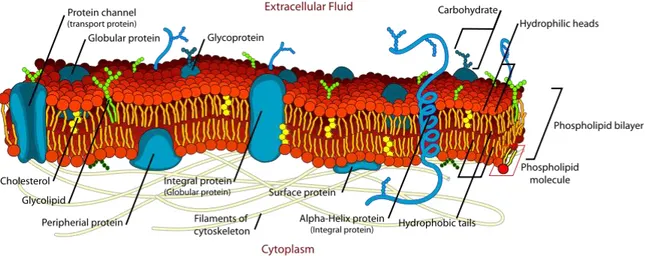
In 1925, Gorter et al. first suggested the concept that biological membranes are composed of two opposite layers of lipids [65]. Almost 50 years later, Singer et al. proposed the advanced fluid mosaic model, in which the biological membrane is considered as a two- dimensional liquid where lipids and proteins diffuse more or less unhindered within the plane of the membrane [170].
Since then, many developments were brought to this model in terms of the membrane composition and molecular organization, e.g. the discovery of lateral microdomain structures [167], revealing the complex nature of the cell membrane structure (Figure 1) [137].
Figure 1. A schematic representation of the plasma membrane. (from Mariana Ruiz, 2007)
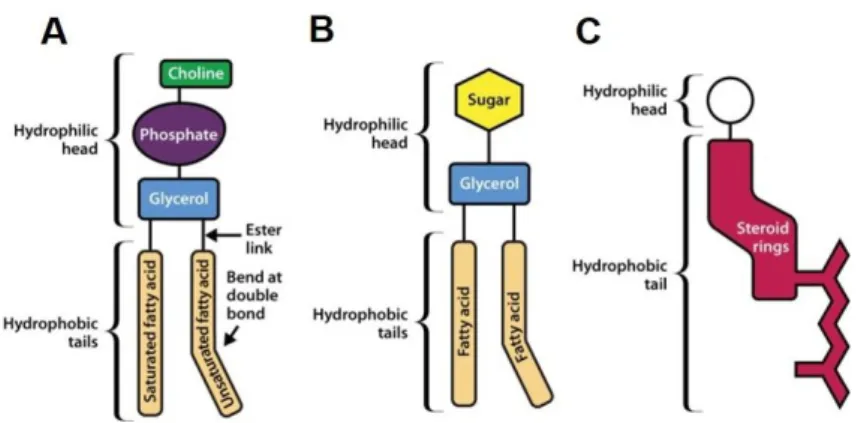
In general, the plasma membrane is composed of three categories of membrane lipids
according to their chemical structure, namely glycerol-based lipids, ceramide-based
sphingolipids and cholesterol (Figure 2) [137].
Figure 2. Schematic illustration of the three main membrane lipids. Phospholipids (A) Glycolipid (B) and Cholesterol (C).
(adapted from Watson, H., 2015 [197])
The glycerol-based lipids or phospholipids are the most abundant membrane lipids consisting of two fatty acid chains linked to glycerol and a phosphate group [197]. They are further classified into various subgroups based on their hydrophilic head groups which are then attached to the phosphate group [137]. The four major glycerol phospholipids are phosphatidylcholine (DOPC), phosphatidylethanolamine (DOPE), phosphatidylserine (DOPS) and phosphatidylinositol (PI) [204].
Sphingolipids, another important group of membrane lipids, are composed of a sphingosine, instead of the glycerol backbone, which is attached to a long unsaturated hydrocarbon chain [5]. Moreover, sphingomyelin, which is the major sphingolipid in mammalian cells, is believed to be involved in the formation of lateral microdomains, so called lipid rafts [26]. In addition, cholesterol is also enriched in these microdomains of tightly packed domains [49]
as result of its favorable interaction with the glycosphingolipids [159]. Subsequently, cholesterols hydrophilic carboxyl group interacts with the hydrophilic head groups of glycosphingolipids and the bulky hydrocarbon rings interact with the hydrophobic acyl chains of membrane lipids [137].
The lipid compositions can be very different in various types of membranes, organisms and
also between the two leaflets of the lipid bilayer [5, 60, 107]. Thus, several lipid species are
distributed differentially in the two monolayers of biological membranes, resulting in
asymmetric lipid distribution [107]. In mammalian cells, the outer leaflet of the plasma
membrane contains predominantly phosphatidylcholine and sphingomyelin, whereas lipids
with neutral or negatively charged head groups (e.g., phosphatidylethanolamine,
phosphatidylserine, and phosphatidylinositol) are preferentially located in the inner leaflet
[197]. However, cholesterol is present in both layers [203]. Furthermore, several studies
revealed that the cell membrane composition changes in cancer cells, which is indicated by
the overexpression of certain types of proteoglycans and the enrichment of negatively
charged lipids, more precisely phosphatidylserine, in the outer leaflet [85, 120, 193]. Thus,
phosphatidylserine is no longer restricted to the cytosolic leaflet of the plasma membrane
and is translocated to the outer leaflet by the action of an enzyme called scramblase [118, 197]. Therefore, the membrane of cancer cells become more anionic than those of healthy cells.
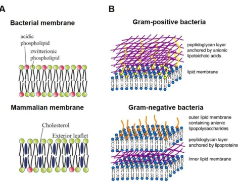
Moreover, the cell membranes of bacteria (gram-positive and gram-negative) differ in their composition significantly from mammalian cell membranes [60]. In particular, one of the major differences between bacterial and eukaryotic cell membranes is the presence of a large amount of cholesterol in eukaryotic cell membranes, while in bacterial cells, cholesterol is completely missing (Figure 3A) [23]. In addition, the membranes of bacteria are rich in negatively charged lipids, like phosphatidylglycerol and cardiolipin.
Phosphatidylethanolamine is the most common neutral phospholipid in bacterial cell membranes (Figure 3A) [60, 63].
Figure 3. Bacterial versus mammalian membranes. Schematic representation of the key features of the bacterial and mammalian cell membrane compositions (A). The membrane structure of gram-positive and gram-negative bacteria (B). ((A) adapted and modified from Brender et al., 2012 [23]; (B) adapted and modified from Chen and Bothun, 2014 [32])
Moreover, the outer membrane of gram-negative bacteria additionally comprises lipopolysaccharides as anionic components, whereas lipoteichoic acids are found in gram- positive bacteria (Figure 3B) [60].
1.1.2 Vesicles as artificial model membrane system
In general, model membrane systems are defined as artificial lipid bilayers in which the lipid compositions mimic the arrangement of those in biological cell membranes [137]. The interior of the spherical-shaped vesicles is characterized by aqueous solutions [164, 194].
Such liposomes are usually categorized, according to their size and lamellarity, into
LUVs and GUVs) [4]. Since MLVs are composed of multiple lipid bilayers, they are therefore not suitable and only rarely used as model membrane system [37].
Large and giant unilamellar vesicles
Large and giant unilamellar vesicles are the most studied and predominantly used model systems not only because of the more homogenous lipid distribution but also because of their suitable size. LUVs are characterized by diameters varying from 10 to 100 nm, while giant unilamellar vesicles have diameters above 10 µm and are therefore close to actual cell size [137].
Furthermore, they can be relatively easy prepared by several methods. The most common method for the preparation of large unilamellar vesicles is illustrated in Figure 4.
Figure 4. Formation of large unilamellar vesicles. Thin dried lipid films were hydrated and stacks of liquid crystalline bilayers become fluid and swell. Subsequently, hydrated lipid sheets self-close to form large, multilamellar vesicles (MLVs).
To get LUVs, the size of the formed MLVs must be reduced by mechanical energy (extrusion). (modified after Lasic, A. D., 1988 [103])
Classically, LUVs are prepared by dissolving lipids of interest in organic solvents like chloroform, followed by in vacuo evaporation to form a thin dried lipid film [137].
Subsequently, the dried lipid film is hydrated in aqueous buffer and MLVs will be formed. In order to obtain large unilamellar vesicles, the MLV suspension is then disrupted by several freeze-thaw cycles, which results in homogenous size distribution in the final suspension [137]. Finally, this suspension is subjected to extrusion through polycarbonate filters to produce large unilamellar vesicles with diameters up to 200 nm, depending on the pore size of the polycarbonate filter.
GUVs are mostly prepared by electroformation [10], but this technique requires
considerable technical effort. Whereas, the gentle hydration method, provided by Horger et
al., allows the preparation of giant unilamellar vesicles in a fast manner without the need of specialized equipment [76]. Going into detail, Horger and coworkers described the formation of giant unilamellar vesicles by hydration from a hybrid film of partially dried agarose and lipids [76]. In brief, a thin film of agarose will be prepared on a clean glass slide. Next, the lipid mixture, dissolved in chloroform, is applied on the dried agarose film resulting in the formation of a hybrid film of agarose and lipids. Finally, the last stage comprises the hydration of the hybrid film leading to the formation of GUVs within minutes.
Giant plasma membrane vesicles obtained from live cells
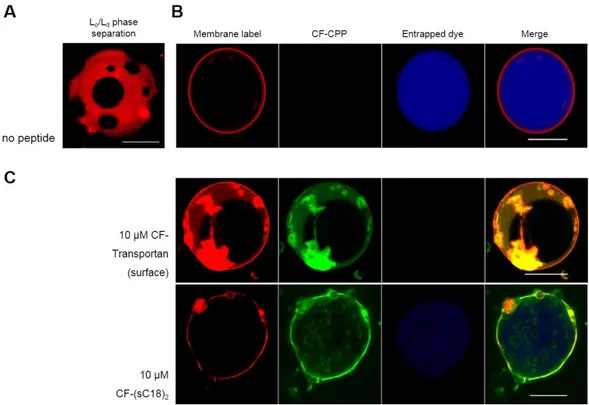
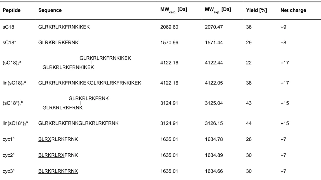
Giant plasma membrane vesicles are cell-derived vesicles and therefore provide a more natural system than large and giant unilamellar vesicles [162]. The attractiveness of GPMVs originates mostly from their complexity, since they maintain the lipid and protein diversity of cellular membranes, which they derived from [165]. GPMVs are released from cells upon chemical induction by either dithiothreitol (DTT) and formaldehyde (PFA) (Figure 5) or by N-ethylmaleimide (NEM) to avoid cross-linking-derived artifacts of lipids [14, 134, 165].
Figure 5. Principle of giant plasma membrane vesicle formation. (after Säälik et al., 2011 [156])
Moreover, GPMVs phase-segregate easily into liquid-ordered (L
o) and -disordered (L
d) lipid domains [156] They showed spherical shapes and varied in size up to 15 µm and are therefore distinctly smaller than GUVs.
1.2 Cell-penetrating peptides
The cellular plasma membrane constitutes an effective semi-permeable barrier that primarily protects living cells form the surrounding environment [111, 206]. At the same time, the cell membrane represents a barrier to the intracellular delivery of large hydrophilic molecules, e.g. for therapeutic applications [69]. Thus, the successful transport of such macromolecules to target specific cells and tissues is a major hurdle for administering bioactive molecules [52], which results in the necessity to find strategies allowing the efficient delivery of therapeutics across the membrane [145].
In this regard, the ability of certain peptides, so called cell-penetrating peptides (CPPs), to
cross the cellular membrane was simultaneously reported by Frankel and Pabo [57], and
the field of AIDS revealed that the TAT peptide, derived from the human immunodeficiency virus (HIV), passes very efficiently through cell membranes of HeLa cells and promotes viral gene expression [57, 66].
In general, cell-penetrating peptides are relatively short peptides containing less than 40 amino acids, which are accompanied by an amphipathic or highly cationic character, usually rich in basic amino acids [36, 196]. Furthermore, CPPs are able to mediate the uptake of bioactive molecules into the cell, and thus transporting therapeutically active cargoes, including small molecules, plasmid DNA, siRNA, therapeutic proteins and imaging reagents [144, 145].
Thus, the discovery of CPPs almost 30 years ago has paved the way for the development of cell delivery approaches and so, CPPs have gained considerable interest in clinical applications.
1.2.1 Classification and uptake mechanisms
Cell-penetrating peptides can be classified into different subgroups according either to their origin or to their physical-chemical characteristics [111, 210]. Based on their origin, three main classes of CPPs can be distinguished, including protein-derived peptides, chimeric peptides that are formed by the combination of two natural sequences, and finally synthetic peptides, which are rational designed sequences [16, 111]. Classical examples of each class are depicted in the following table.
Table 1. Sequences and origins of well-studied CPPs.
Peptide Origin Sequence Ref.
Protein-derived peptides
Tat(48-60) Protein Tat HIV-1 GRKKRRQRRRPPQ [192]
Penetratin Antennapedia homeodomain
RQIKIWFQNRRMKWKK [40]
Chimeric peptides
Transportan Galanin/
mastoparan
GWTLNSAGYLLGKINLKALAALAKKIL [139]
MAP de novo KLALKLALKALKAALKLA [132]
Synthetic peptides
Oligoarginines Model peptide R
n[59,
125]
Pep-1 reverse
transcriptase/T antigen
KETWWETWWTEWSQPKKKRKV [42]
According to their structure, CPPs can be categorized as cationic CPPs with a high positive net charge, hydrophobic CPPs consisting mostly of nonpolar amino acid residues and as amphipathic peptides comprising both polar and nonpolar regions [121, 122, 210]. The class of amphipathic CPPs can be further subdivided into primary and secondary amphipathic CPPs [210].
The first class, the cationic or nonamphipathic CPPs, are defined as rather short peptides, like Tat(48-60) or nona-arginine (R9), having a high content of positively charged amino acids (Arg or Lys) [151]. Further, it was shown that cationic peptides bind electrostatically to negatively charged components of the plasma membrane of cells, leading to efficient translocation [97]. Primary amphipathic CPPs, such as Transportan, typically consist of more than 20 amino acid residues and their primary structure includes hydrophilic and hydrophobic domains [155]. The uptake mechanism of these peptides is proposed to occur by the insertion into the surface of the cellular membrane followed by pore forming events (direct translocation, Figure 6) [210, 211]. The last group comprises the secondary amphipathic peptides, e.g. penetratin, containing normally less than 20 amino acids in their sequence [111]. They reveal their amphipathic properties only through a change in their secondary structure upon the interaction with the lipid membrane [111, 151, 210]. The membrane binding affinity of these peptides increases with the negatively charged lipid content of the membrane [210].
Although, the exact mechanism used by CPPs to translocate across the cellular plasma membrane is not fully resolved, it is widely believed that the uptake mechanism varies for different classes of CPPs, and moreover, several mechanisms might even coexist depending on experimental conditions, such as the applied concentration, temperature, incubation time, the cell line used or the attached cargo [70, 111, 145, 151].
However, the two proposed major translocation mechanisms of cell-penetrating peptides
include the energy-dependent endocytotic pathway and the direct translocation via energy-
independent pathways [111, 206].
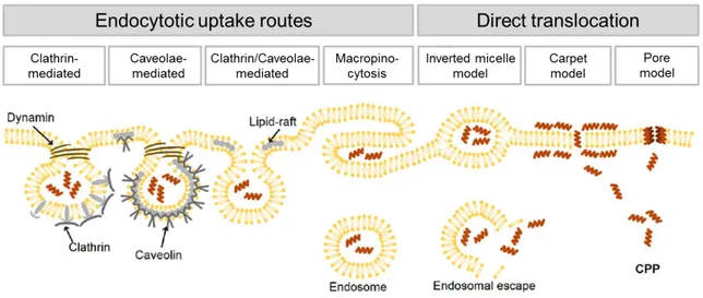
Figure 6. Scheme of different suggested uptake pathways for cell-penetrating peptides. (modified after Mickan et al., 2014 [121])
Figure 6 summarizes the different uptake routes by which CPPs might penetrate the cell membrane. The endocytosis-mediated translocation mechanism, including clathrin- mediated endocytosis and caveolae/lipid raft-mediated endocytosis as well as macropinocytosis, which provides the main pathway utilized by most CPPs, especially when peptides are attached to large cargo molecules [145, 206]. These four morphologically distinct pinocytotic pathways differ in the size of the coat (if any) and in the size of the detached vesicles [99]. However, endocytosis generally consists of two steps: the endocytotic entry which is followed by the endosomal release, whereby the latter step is crucial in order to avoid lysosomal degradation and moreover to allow the peptide/cargo to reach its extracellular target site [16].
The uptake mechanism via direct translocation is most likely for primary amphipathic peptides at high CPP concentrations [111]. Three different energy-independent uptake pathways have been described: the inverted micelle model, the carpet model and the pore model, which is further divided into the toroidal pore and the barrel-stave model (Figure 6 and 7) [155].
Figure 7. Pore-forming models proposed for the cellular entry via direct translocation. (modified after Sanderson, J. M., 2005 [159])
In general, these models are mainly based on the electrostatic interaction of positively charged CPPs with negatively charged components of the cellular membrane (e.g.
glycosaminoglycan and phospholipids), leading to the temporary or permanent destabilization of the lipid bilayer caused by the CPPs inserted in the membrane [155, 196].
More precisely, the inverted micelle mechanism causes local disorder of the lipid bilayer, which leads to the formation of such inverted hexagonal structures containing CPPs. As these micelles cross the membrane, they invert, which results in the destabilization of these micelles and finally, the release of the CPPs into the cytosol [145, 155, 186]. According to the carpet model, the peptide aligns parallel to the lipid membrane and when a threshold concentration is reached, transient pores can be formed that enable the translocation of the CPP into the cells [145, 159]. The mechanisms involved in the formation of “toroidal” and
“barrel-stave” pores presumed the existence of a well-defined secondary structure of the CPP [186]. In the toroidal pore mechanism, the peptides insert into the lipid bilayer and bend the lipid monolayer into the interior, forming a hydrophilic pore in the cell membrane (Figure 7) [155]. In the barrel-stave model, the hydrophobic regions of the peptides interact with the hydrophobic core of the cell membrane, while the hydrophilic regions of the peptide face the lumen of the pore (Figure 7) [155, 159].
In summary, the endocytotic pathway is supposed to be the dominant mechanism for the majority of CPPs, particularly when attached to large cargoes. However, it is essential to emphasize that the translocation pathway by which the cell-penetrating peptides enter into the cell depends on several parameters including (i) the nature and secondary structure of the CPP; (ii) its capability to interact with membrane components; (iii) the nature, type and concentration of the cargo; and (iv) the cell type and membrane composition [74].
1.2.2 Interactions of CPPs with membranes
As already mentioned earlier, the first step of peptide internalization requires the electrostatically interaction between the cationic cell-penetrating peptide and the negatively charged constituents of the cell membrane [196]. Thus, when translocating through the cellular membrane, the first potential partners that CPPs will encounter at the cell surface are the negatively charged glycosaminoglycans and anionic phospholipids [36, 196].
The role of proteoglycans
Proteoglycans are a heterogeneous group of proteins covalently attached with long, linear
glycosaminoglycan (GAG) chains [6]. These chains are characterized by a high density of
negatively charged sulfate and carboxylate groups [196]. Thus, proteoglycans are
categorized according to the nature of the GAG chains [81]. Thereby, heparan sulfate (HS),
chondroitin sulfate (CS) and dermatan sulfate (DS) are the most widespread GAGs in
proteoglycans [6]. As most CPPs contain positive charges, the peptide-GAG interaction is supposed to play a role in the internalization of peptides across the cell membrane, irrespective of the uptake mechanism [6, 140]. In this regard, taking into consideration that the most well-known CPPs are rich in Arg residues, the guanidium moieties of Arg could strongly interact with the sulfate and carboxylate groups of GAGs through bidentate hydrogen bonds (Figure 8A) [128, 196].
The role of phospholipids
Since the lipid composition of the plasma membrane, especially the anionic lipids, plays an essential role in the translocation pathway of CPPs, the negatively charged phospholipids of the cellular membrane represent the other main interaction partner of cell-penetrating peptides [6]. Despite lipids are not symmetrically distributed across the two leaflets of the cell membrane, anionic lipids are mainly found at the cytosolic site of the membrane and only a minor fraction is localized at the outer leaflet [36, 196]. Therefore, it is assumed that the peptides are able to recruit the anionic lipids in order to generate negatively charged nanodomains on the cell surface and thus allowing the critical internalization step [36, 196].
The role of Arg residues
Besides the evidence for the importance of anionic components in the cellular membrane and the cationic nature of CPPs, the type of the chemical hydrogen bond seems to be much more important [86].
Figure 8. Interaction of arginine- and lysine rich peptides with negatively charged components of the plasma membrane. Favorable hydrogen bonding between guanidinium moieties of Arg-rich peptides and phosphate, sulfate and carboxylate groups of glycosaminoglycan (A). Differences in phosphate binding mechanism of Arg and Lys residues (B).
(modified after Jobin et al., 2014 [86])
Several studies have shown that Arg-rich peptides efficiently penetrate the cells, while Lys-
rich CPPs do not, which is in accordance with the fact that the replacement of Arg by Lys
totally suppresses the cellular uptake of these CPPs [8, 86, 125]. In this context, Rothbard et al. revealed that Lys residues can only form monodentate interactions with the anionic cell surface which is not sufficient for the successful translocation of CPPs through the cell membrane, since Arg residues form bidentate hydrogen bonds (Figure 8B) [154].
1.3 Skin and wound healing
1.3.1 Skin as barrier
The skin is the largest organ of the human body and is known to cover an area of up to 2 m
2and its weight consists of more than 10 wt.% of the total weight of an adult body [89]. The primary function of the skin is to serve as a protective barrier against external mechanical, chemical, microbial and physical influences [41] and further helps to regulate the body temperature and permits the sensations of touch, heat, or pain [89]. The cellular architecture of the skin is depicted in Figure 9.
Figure 9. Mammalian skin. The skin consists of the epidermis, the outermost layer of the skin and the dermis, the layer beneath the epidermis. The stratified structure of the epidermis is shown in the right panel. (adapted from Fuchs and Raghavan, 2002 [58])
The skin consists of two main tissue layers, the epidermis and the underlying connective
tissue, which comprises the dermis and the hypodermis [5]. The epidermis is a multilayered
epithelium mainly composed of keratinocytes [5]. The stratum corneum (SC), the outermost
layer of the epidermis, consists of a 10 – 15 µm thick matrix of dead keratinocytes
(corneocytes), which are surrounded by highly ordered lipid layers serving as cover. Thus,
into the skin [22, 41]. The SC is underlaid by the viable epidermis (50 – 100 μm thick), which is responsible for the generation of the SC. The viable epidermis is composed of the granular, spinous and basal cell layer (Figure 9), whereby each layer is characterized by position, shape, morphology and state of the differentiation of keratinocytes [21, 22]. Directly adjacent to the epidermis lies the dermis, approximately 1 – 2 mm thick, providing the mechanical support for the skin. The dermis and also the hypodermis are thus highly supplied with blood vessels and nerves [5, 21, 22]. The hypodermis layer is composed of subcutaneous fat tissues providing thermal isolation and mechanical protection to the body [89]. In addition to the main tissues, sweat glands and hair follicles are spread throughout the skin [58].
1.3.2 Wound healing and peptides
Since the skin serves as a protective barrier against the surrounding environment, any type of skin injury has to be mended in a rapid and efficient manner [119]. Wound healing is thus a critical process and starts normally immediately after the injury [169]. The cutaneous injury initiates a complex set of events comprising three overlapping phases: the inflammation, the proliferative process leading to tissue restoration and tissue remodeling (Figure 10) [109].
Figure 10. The three stages involved in skin wound healing. (adapted from Kondo and Ishida, 2010 [96])
Cutaneous wound healing typically begins with the inflammation response, which is
characterized by erythema, heat, swelling and pain, which are the results of capillary
vasodilation and increased permeability [13]. Neutrophils and macrophages (inflammatory
cells) further infiltrate the wound site and trigger the process of wound healing [94]. The proliferative phase comprises the migration and proliferation of endothelial cells, fibroblasts and keratinocytes leading to re-epithelialization [109]. Approximately 4 days after injury, the dermal reconstitution begins at the wound site, which is characterized by the formation of granulation tissue including angiogenesis and collagen formation [96, 109]. The final stage of wound healing is the remodeling of the wound and the surrounding tissue by fibroblasts, which are responsible for the development of new epithelium and the final scar tissue formation. This stage of wound repair begins 2 – 3 weeks after injury and may last for one year or even longer depending on specific and individual factors such as the age of the patient, diseases and medication as well as the size, depth and cause of the wound [39, 142, 191].
Efficient wound treatment remains one of the most prevalent healthcare issue, since thousands of patients suffered annually from different kinds of skin damage or burns by hot water, flames or accidents [33, 89]. Moreover, chronic wound patients, which suffer from diseases like type 2 diabetes, obesity, and wound infections account for increasing health care costs [163]. Therefore, much effort has been expended on wound care with an emphasis on new therapeutic approaches and the development of technologies for acute and chronic wound treatment [191].
Until now, a plethora of studies exists concerning the therapeutic potential of antimicrobial peptides (AMPs) derived from multiple sources, e.g. humans or amphibians, as efficient modulators of wound healing [116]. In general, antimicrobial peptides, composed of 10 – 50 amino acids, mostly exhibit an amphipathic character due to the prevalence of basic amino acid residues and a high content of hydrophobic amino acids [149]. AMPs, also known as host defense peptides, show a broad spectrum of antimicrobial activity against gram- positive and -negative bacteria, and play an essential role in the innate immune response as antimicrobial and immune-modulating agents [174].
Owing to their multiple functions, AMPs could act as effective agents in bacterial infected wounds, since the disruption of the skin barrier allows pathogens to attack the body and thus drastically increases the risk of infection.
Until now, pexiganan, a 22 amino acid long peptide analogue of the naturally occurring
AMP, magainin 2, isolated from the skin of the African clawed frog, Xenopus laevis [61], is
the first AMP evaluated for the treatment of infected human wounds, particularly for the
treatment of diabetic foot ulcer [100]. Unfortunately, the use of pexiganan failed the Phase
III clinical trial in 2016.
1.4 Peptides used within the thesis
1.4.1 The cationic cell-penetrating peptide sC18
The cell-penetrating peptide sC18 is a highly cationic fragment (amino acid residues 106 – 121) of the C-terminus of the 18 kDa cationic antimicrobial protein CAP18, and highlighted as a useful delivery vehicle for cytostatic compounds [62, 79, 130, 172]. Furthermore, sC18 was shown to efficiently internalize into cells which might be attributed to its amphipathic α- helical nature [130, 183]. The major uptake route of sC18 is proposed to follow the energy- dependent endocytotic pathway. However, previous studies demonstrated that structural modifications of sC18, particularly the cyclization of the peptide backbone and the dimerization of the peptide sequence resulting in a branched version, lead to an efficient and moreover increased cellular uptake showing somehow a certain selectively towards cancerous cells [68, 77, 78].
Furthermore, a study by Splith et al. showed the successful delivery of a novel designed sC18 conjugate composing of a nitroimidazole unit (NIA) as radiosensitizer, a radioactive label and sC18 as transporter, to target hypoxic tumor tissues [173]. Lately, Bergmann et al. improved this delivery vehicle by introducing
D-amino acids instead of
L-amino acids, leading to a proteolytically more stable peptide, since this system was hampered by fast degradation [18].
The following table represents an extract from sC18 variants, which were applied for various approaches.
Table 2. An extract from sC18 variants for diverse application approaches. B: L-propargylglycine, X: L-(Ɛ-azido)-lysine;
___: amino acids involved in cyclization; NIA: 2-(2-nitroimidazol-1-yl)acetic acid
Peptide Sequence Features Ref.
sC18 GLRKRLRKFRNKIKEK Transport tool for
cytostatic drugs, e.g.
metal complex and imaging probes
[130, 152, 172]
(sC18)
2GLRKRLRKFRNKIKEK GLRKRLRKFRNKIKEK
Drug delivery, tumor-selectivity
[68, 78]
cyc3 BLRKRLRKFRNX Vehicle for gene
delivery
[77]
DOTA- sC18(NIA)
DOTA-βA-GLRK(NIA)RLRKFRNKIKEK Specific targeting of hypoxic tumor tissue
[173]
NODAGA-
D
sC18(NIA)
2NODAGA-βA-GLRK[(βA)
2-
(NIA)]RLRKFRNK[(βA)
2-(NIA)]IKEK
[18]
Overall, the cationic sC18 peptide provides a highly attractive tool through which bioactive molecules can reach their intracellular target.
1.4.2 The naturally derived peptide Tylotoin
Tylotoin, a short peptide consisting of 12 amino acid residues, was recently identified and extracted from the skin of the salamander Tylototriton verrucosus [126]. The precursor of Tylotoin was found to belong to the family of cathelicidin, which is a conserved protein family among vertebrates [126]. More precisely, Tylotoin is derived from the C-terminal part, like other antimicrobial peptides, e.g. LL-37 [2] or sC18 [130] but in contrast to these peptides, Tylotoin exhibits no antimicrobial activity [126]. However, the main feature of Tylotoin is provided by its ability to significantly accelerate the healing of full-thickness wounds in mice indicating that topical application of Tylotoin has a potent healing effect on full-thickness skin wounds [126]. Thereby, the short peptide was shown to directly function in the migration and proliferation of fibroblasts, keratinocytes, and vascular endothelial cells, which are all important key cell types during the wound healing process [126, 169].
With this potent peptide in hand, another step towards the understanding of the cellular and
molecular events underlying the rapid wound healing process in salamanders could be
done, which might lead to the development of rapid wound healing therapies in the future.
2. Aims of the thesis
The primary goal of this thesis was to understand the initial step of cell-penetrating peptides cell entry in more detail on a molecular level. At the same time, model membrane systems have gained increased attractiveness and thus were used to establish different techniques that can be helpful to shed light on the action mechanism of CPPs. The second chapter focuses on the design, characterization and the application of cell-penetrating peptide conjugates in skin wound healing.
2.1 Establishment of diverse bilayer model systems and their applications in peptide-lipid interactions
In this chapter, different model membrane systems, more precisely large and giant unilamellar vesicles and membrane vesicles, which are exclusively derived from live cells plasma membranes, were employed to evaluate peptide-membrane interactions and their significance for the successful translocation across the cell membrane. For this purpose, various methodologies are described that have been established in our working group by myself, and which represent particularly great tools to assess the relationship between peptides and various membrane components to understand the molecular level of translocation of CPPs. More specifically, great attention is paid to aspects concerning peptides effect on membrane integrity, structural behavior of peptides in the presence of lipid bilayer systems and finally, the elucidation of the relevance of mammalian plasma membrane compositions. Thus, these newly acquired techniques were then applied using two different structural modified variants of the cationic cell-penetrating peptide sC18 to unravel how CPPs interact with live cells on a molecular level using artificial membrane systems.
2.2 CPPs and wound healing
So far, no study of which we are aware has been reported on the use of CPPs in the
application field of human skin wound healing. Thus, for the first time, a cell-penetrating
peptide was covalently conjugated to a wound healing promoting peptide with the main goal
to accelerate the wound healing process. Herein, these novel conjugates were evaluated
upon their cytotoxicity, cellular internalization behavior and moreover, the impact of peptides
on in vitro wound healing was investigated.
3. Materials and methods
The chemicals, reagents and consumables used in this present thesis were obtained from the companies Merck (Darmstadt, Germany), Sarstedt (Nümbrecht, Germany), Sigma- Aldrich (Taufkirchen, Germany) and VWR (Darmstadt, Germany), unless otherwise specified. N
α-Fmoc protected amino acids were purchased from IRIS Biotech (Marktredwitz, Germany).
The following table depicts the applied equipment during the working time.
Table 3. An overview about the equipment used during the thesis.
Instrument Producer
Balance Analytical balance FA-210-4,
Faust Laboratory balance SBA52, Scaltec Blotting system kuroGEL semi-dry-blotter, VWR
CD spectrometer JASCO J-715 spectropolarimeter Cell culture clean bench Hera Safe
Centrifuges Thermo Scientific; Heraeus Multifuge X1R Thermo Scientific; Heraeus Pico 17
CO
2-incubator BINDER
Electrophoresis system Mini-PROTEAN Tetra Vertical Electrophoresis System;
Bio-Rad
ESI mass spectrometer Bruker Esquire HCT
Flow cytometer Guava® easyCyte flow cytometer, Merck, Darmstadt BD Accuri C6 flow cytometer, Heidelberg
Haemocytometer Neubauer improved, superior Marienfeld Heating block Eppendorf – Thermomixer compact
HPLC (analytic) Chromolith Performance column (RP-18, 130 Å, 2 µm, 100 × 4.60 mm, Merck
HPLC (preparative) Elite Lachrom Hitachi Pump L-2130, Elite
Lachrom Hitachi Autosampler L-2200, Elite
Lachrom Hitachi Diode Array Detector L-2455, Teledyne ISCO Fraction Collector Foxy R1,
Column: Phenomenex Jupiter, 4 µm Proteo 90 Å, 250 x 15 mm
LC-mass spectrometer LC: Hewlett Packard Series 1100 MS: Finnigan MAT-LCQ
Lyophilizer Lyovac GT2 with CertomaT SII
Thermo Savant Refridgerated Vapor Trap RVT5105 Microscope Inverted microscope (Motic AE31, Wetzlar)
Confocal laser scanning system (Nikon D-Eclipse C1) Mini-extruder Avanti Polar Lipids Inc., Alabaster, USA
Multipette® plus Eppendorf
PCR Thermal Cycler FlexCycler, Analytik Jena, Jena, Germany
Pipettes Eppendorf Research
Pipetting aid NeoLab
Plate reader Tecan infinite M200, Salzburg, Austria
Rotary evaporator VWR
Rotary Shaker KL-2, Edmund Bühler GmbH
Certomat H/SII, Braun Biotech, Göttingen, Germany Spectral photometer Thermo Scientific Genesys 10S UV-Vis
Speed-Vac Thermo Scientific Speedvac Concentrator Savant SC210A, RVT5105 Refrigerated Vapor Trap VLP80 Vacuum Pump
Synthesis roboter MultiSynTech Syro I, Bochum, Germany Thermomixer compact Eppendorf
Vacuum pump VWR
Vortex Scientific Industries – Vortex Genie 2
Water bath Julabo SW22
XcelVap
TMHorizon Technology
3.1 Peptides and lipids
3.1.1 Peptides
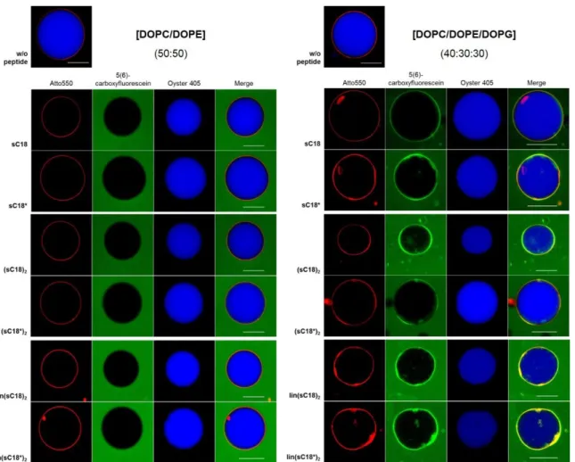
All peptides used in this work and their sequences are listed in Table 4.
Table 4. Sequences and references of investigated peptides. All peptides are amidated at the C-terminus and N-terminally labeled with 5(6)-carboxyfluorescein B: L-propargylglycine, X: L-(Ɛ-azido)-lysine; ___: amino acids involved in cyclization.
Peptide Sequence Reference
sC18 GLRKRLRKFRNKIKEK [68, 78, 130]
sC18* GLRKRLRKFRNK [77]
(sC18)
2GLRKRLRKFRNKIKEK GLRKRLRKFRNKIKEK
[68, 78]
lin(sC18)
2GLRKRLRKFRNKIKEKGLRKRLRKFRNKIKEK
[129], this work (sC18*)
2GLRKRLRKFRNK GLRKRLRKFRNK
lin(sC18*)
2GLRKRLRKFRNKGLRKRLRKFRNK
cyc1 BLRXRLRKFRNK [77]
cyc2 BLRKRLRXFRNK [77]
cyc3 BLRKRLRKFRNX [77]
Tylotoin KCVRQNNKRVCK [126]
rTylotoin RNCVKKNCRVKQ
this work Tylotoin-sC18 KCVRQNNKRVCKGLRKRLRKFRNKIKEK
sC18-Tylotoin GLRKRLRKFRNKIKEKKCVRQNNKRVCK
Tylotoin-sC18* KCVRQNNKRVCKGLRKRLRKFRNK
sC18*-Tylotoin GLRKRLRKFRNKKCVRQNNKRVCK
3.1.2 Lipids
All lipids used in this study are depicted in Table 5.
Table 5. Lipids used in this study were obtained as chloroform solution (10 mg/ml).
Lipid Company
1,2-dioleoyl-sn-glycero-3-[phospho-L-serine]
(DOPS)
Avanti Polar Lipids, Inc. (Alabaster, USA)
1,2-dioleoyl-sn-glycero-3-[phospho-rac-(1- glycerol)] (DOPG)
Avanti Polar Lipids, Inc. (Alabaster, USA)
1,2-dioleoyl-sn-glycero-3- phophoethanolamine (DOPE)
Avanti Polar Lipids, Inc. (Alabaster, USA)
1,2-dioleoyl-sn-glycero-3-phosphocholine (DOPC)
Avanti Polar Lipids, Inc. (Alabaster, USA)
Atto550 labeled DOPE Atto Tec (Siegen, Germany)
cholesterol (Chol) Sigma-Aldrich (Taufkirchen, Germany)
L
-α-phosphatidylinositol-4,5-bisphosphate (porcine brain, PIP
2)
Avanti Polar Lipids, Inc. (Alabaster, USA)
sphingomyelin (porcine brain, SM) Jena Bioscience (Jena, Germany)
3.2 Solid phase peptide synthesis (SPPS)
The peptides used in this thesis were synthesized via automated solid phase peptide synthesis as described below.
3.2.1 Automated solid phase peptide synthesis
Automated solid phase peptide synthesis offers a suitable strategy to produce chemical
engineered peptides simultaneously. The peptides used were synthesized on a multiple
Syro I peptide synthesizer following the Fmoc/t-Bu strategy. The synthesis runs in open
polypropylene reactor vessels (2 ml syringes) equipped with a fritted disc. The software
SyroXP calculates the amounts of amino acids (0.4 mol/l) and solvents required for the
complete synthesis run. In order to synthesize a peptide amide, Rink amide resin
(NovaBiochem, Bad Soden, Germany) was used. The Fmoc-protected Rink amide resins
(30 mg, 0.5 mmol peptide per gram resin; 15 µmol ≙ 1 eq.) were first swollen in DMF, then the Fmoc protecting groups were cleaved with 40% piperidine in DMF and following another deprotection step/cleavage with 20% piperidine in DMF. After four washing steps using DMF, the N
α-Fmoc protected amino acids were introduced in 8-fold excess by using a double coupling procedure and in situ activation with Oxyma and DIC. In a last step, when the defaulted amino acid sequence of the peptides was completed, the N-terminal Fmoc- protecting group was removed under previously mentioned conditions and the resin was washed with DMF. A reaction scheme of the automated solid phase peptide synthesis is illustrated in Figure 11.
Figure 11. Schematic overview of general steps in solid phase peptide synthesis. R: different side-chains of amino acids. PG: protecting groups of amino acid side chains.
3.2.2 5(6)-carboxyfluorescein (CF) – labeling on resin
To allow microscopic and flow cytometric analysis, the peptides were N-terminally labeled with 5(6)-carboxyfluorescein. The labeling procedure was performed while the peptides were still bound to the resin with fully protected side chains. Thus, the resin was swollen in 1 ml DMF for at least 15 min. After the solvent was removed, 3 eq. of CF and HATU were dissolved in 300 µl DMF and added to the swollen resin. Subsequently, 3 eq. of DIPEA were added directly to the syringe and the reaction was carried out under vigorous shaking for 2 × 2 h at RT or overnight with 5 eq. of Oxyma and DIC. After the incubation time, the resin has been washed five times with DMF, CH
2Cl
2, MeOH and Et
2O and finally dried for 10 min in a Speed-Vac. To monitor whether the coupling was successful, a Kaiser test was performed. CF-polymers, which might have formed, were cleaved by treatment with 20%
piperidine for 45 min.
3.2.3 Kaiser test [88]
In Fmoc solid phase peptide synthesis, monitoring the completeness of the coupling step is mandatory. Therefore, the Kaiser test is used to control the presence or absence of free amino groups based on the reaction of ninhydrin with amines. Therefore, a few resin beads were placed in a 1.5 ml reaction vessel and one drop of each solution (I, II and III; Table 6) was added consecutively. The samples were incubated at 95 °C for 5 min. A blue coloring of the resin beads and the solutions indicates free amino groups and hence an incomplete coupling, whereas a yellowish color represents no free amino groups and therefore a negative result. As a positive control the primary amine ethanolamine was chosen.
Table 6. Compositions of the solutions required for the Kaiser test.
Solution Composition
Solution I 1.0 g ninhydrin in 20 ml EtOH Solution II 80.0 g phenol in 20 ml EtOH
Solution III 0.4 ml KCN solution in 20 ml piperidine 3.2.4 Sample cleavage
To verify the success of the coupling reaction, it is advisable to perform a sample cleavage.
Therefore, 2.5 µl of triisopropylsilane (TIS) and 2.5 µl of water, working as scavengers, were
added to a small amount of resin beads. In the case of Tylotoin conjugates containing
cysteine within the sequence, 7 µl of thioanisole and 3 µl of 1,2-ethandithiol (EDT) must be
used as scavengers. Afterwards, the mixture was filled up to 100 µl with TFA and the
reaction was carried out under shaking conditions for 3 h at RT. After the incubation time,
1 ml of ice-cold diethyl ether was added to the mixture to precipitate the peptide and was left for 20 min or overnight at -20 °C. Afterwards, the mixture was washed with 1 ml diethyl ether and centrifuged (4 °C, 4 min at 10,000 x g) for at least five times until no scavenger smell was perceived. The resulting pellet was finally dried for 10 min in the Speed-Vac and dissolved in 100 µl H
2O/t-BuOH (3:1, v/v). Finally, the sample was analyzed by HPLC-ESI- MS.
3.2.5 Full cleavage of peptide
The peptide amides were cleaved with a mixture of TFA/TIS/water (95:2.5:2.5, v/v/v) except for Tylotoin conjugates, which were cleaved by a mixture of TFA/thioanisole/EDT (90:7:3, v/v/v) within 3 h as previously described. The peptides were precipitated from cold diethyl ether (10 ml) at -20 °C and collected by six centrifugation steps (4 °C, 4 min at 5,000 x g) and lyophilized from water/t-BuOH (LyoVac GT2, Leybold, Germany). A sample of the peptide solution was analyzed by HPLC-ESI-MS.
3.3 Peptide analysis
3.3.1 Analytical HPLC-MS
A high-performance liquid chromatography electrospray ionization mass spectrometry (HPLC-ESI-MS) method was used to evaluate the purity and the identity of the peptides.
Therefore, the peptide solution was diluted with a starting gradient of 10% ACN in water with 0.1% formic acid (FA). 10 µl of the solution was injected and separated by an ACN gradient which was raised from 10 to 60% by the HPLC. The flow rate was set to 0.6 ml/min.
The analytics were performed on an Elite LaChrom VWR-Hitachi system using a Chromolith Performance column (RP-18, 130 Å, 2 µm, 100 × 4.60 mm, Merck). One part of the eluate was passed to the electrospray ionization source whereby ions were generated and their m/z ratio was determined.
3.3.2 Preparative RP-HPLC
Purification of the peptides was achieved by preparative RP-HPLC on a Phenomenex Jupiter column (Proteo, 90Å, 4 µm, 250 × 15.00 mm) using a linear gradient of 10 – 60% B in A (A = 0.1% TFA in water; B = 0.08% TFA in ACN) over 45 min and a flow rate of 6 ml/min.
The peptides were detected at 220 nm and the fractions were collected separately.
Afterwards, the peptide solution was removed from ACN using either the Speed-Vac or the
XcelVap™ and then lyophilized overnight.
3.3.3 Circular dichroism (CD) spectroscopy
The CD spectra were recorded from 260 nm to 180 nm in 0.5 nm intervals at 20 °C using a JASCO J-715 spectropolarimeter in a N
2atmosphere. Peptide solutions were dissolved in 10 mM potassium phosphate buffer (pH 7.0) containing either 0 or 50% (v/v) trifluoroethanol (TFE) to a final peptide concentration of 20 µM. The CD spectra were measured using a 1 mm quartz cell and each measurement was repeated three times. Instrument parameters were set as follows: sensitivity, 100 mdeg; scan mode, continuous; scan speed, 50 nm/min;
response time, 2 s and bandwidth, 1.0 nm. All CD spectra were smoothed and corrected by subtraction of the CD spectrum form the respective buffer to eliminate potential measurement errors caused by the solvents. The ellipticity was expressed as molar ellipticity [Θ] in
𝑑𝑒𝑔×𝑐𝑚2𝑑𝑚𝑜𝑙
.
3.4 Peptide-lipid interactions
3.4.1 Preparation of large unilamellar vesicles (LUVs)
For the preparation of different composed LUVs, several lipids were required, which are dissolved in CHCl
3. To remove residual solvent, the lipid mixture was placed in a round- bottomed flask under vacuum at 37 °C for at least 1 h to remove residual solvent. The dried lipid film was either hydrated with HKS-glucose buffer (for flow cytometric studies and CF- release experiments) or with 25 mM phosphate buffer, pH 7.4 (for CD spectroscopic studies) at 45 °C for 5 min to form liposomes with a final concentration of 2 mM (CD spectroscopic analysis), 4 mM (flow cytometric studies) or 8 mM (CF-release experiments).
To form LUVs, the resulting multilamellar vesicle (MLV) dispersion was subsequently run through 10 freeze/thawing cycles and passed 21 times through a mini-extruder equipped with a 0.4 µm polycarbonate track-etch membrane (Avanti Polar Lipids, Inc.). Liposome preparations were analyzed by dynamic light scattering indicating a range of 250 – 400 nm in diameter.
HKS-glucose buffer
HEPES 25 mM (pH 7.4)
KCl 150 mM
Sucrose 10%
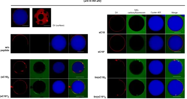
Table 7. All lipid compositions used during the thesis. For peptide-induced CF-leakage, LUVs entrapping 100 mM CF were prepared. The vesicle membranes were labeled with 0.2 mol% Atto550-DOPE for flow cytometric studies.
LUV compositions
Lipids [mol%]
zwitterionic anionic
DOPC DOPE DOPG Sphingomyelin Cholesterol
I 50 50 - - -
II 40 30 30 - -
III 25 - 5 50 20
IV 37.5 37.5 5 - 20
V 22.5 22.5 5 50 -
3.4.2 Preparation of giant unilamellar vesicles (GUVs)
Giant unilamellar vesicles were generated as previously described [76, 148]. Briefly, super low melting agarose (1%, w/v) was coated on a clean glass slide and dried on a hot plate (∼50 °C) for 30 min. Afterwards, 10 µl of the respective lipid solution was spread on the agarose film and dried in vacuo for at least 1 h to remove residual chloroform. To visualize the membranes, the lipid solution was prior doped with 0.2 mol% Atto550-DOPE. Then, a seal ring was placed onto the lipid coated areas on the slide to obtain two sealed chambers.
For the preparation of GUVs encapsulating the blue dye Oyster 405 (Luminaris GmbH, Münster, Germany), dextran buffer containing additionally 5 μM of Oyster 405 (300 μl each) was added to the hybrid film. The glass slide was then left in the dark for 2 h to allow hydration and swelling of the lipids. To harvest the GUV suspension, the glass slide was gently tilted in all directions to detach the liposomes from the surface. The giant liposomes were then stored in LoBind tubes (1.5 ml, Eppendorf, Hamburg, Germany) at RT and were used within three days.
Dextran buffer
HEPES 10 mM (pH 7.4)
KCl 50 mM
NaCl 50 mM
Dextran 1 mg/ml (from Leuconostoc spp., 6 kDa)
Table 8. All lipid compositions used during the thesis. The vesicle membranes were labeled with 0.2 mol% Atto550-DOPE or with 0.2 mol% DiI to visualize the loosely packed liquid-disordered lipid phase (Ld).
GUV compositions
Lipids [mol%]
zwitterionic anionic
DOPC DOPE DOPG Sphingomyelin Cholesterol
I 50 50 - - -
II 40 30 30 - -
III 25 - 5 50 20
IV 37.5 37.5 5 - 20
V 22.5 22.5 5 50 -
3.4.3 Preparation of giant plasma membrane vesicles (GPMVs)
Cells (HEK-293 800,000; MCF-7 250,000) were seeded in a 6-well culture plate (Sarstedt, Nümbrecht, Germany) and were grown to 70 – 80% confluency in appropriate growth medium supplemented with 4 mM
L-glutamine and 10% fetal bovine serum (FBS). Giant plasma membrane vesicles (GMPVs) were prepared as described earlier by R. E. Scott, 1976 [162] with minor modifications. First, the cells were washed twice with GPMV buffer.
To induce the formation of cell-free vesicles, 1 ml of freshly prepared GPMV buffer supplemented with paraformaldehyde (PFA) and dithiothreitol (DTT) at final concentrations of 25 mM and 2 mM respectively were added and the cells were incubated for 2 h at 37°C while slowly shaken (60 rpm, Certomat H/SII, Braun Biotech). After incubation, the GPMV- rich cellular supernatant was transferred into a 15 ml conical tube, where the GPMVs were allowed to settle down on ice for 30 min. Afterwards, 20 – 50% of the total volume were transferred from the bottom of the conical tube into a 1.5 ml Eppendorf tube. For microscopic studies, 0.5 µg/ml DiI stain (final concentration, in EtOH) was added and incubated for 15 min at 37 °C. After isolation, GPMVs were stored at 4 °C for 1 – 2 days without visible degradation.
GPMV buffer
HEPES 10 mM (pH 7.4)
NaCl 150 mM
CaCl
22 mM
3.4.4 Secondary structure via CD spectroscopy
In the presence of LUVs, the peptide to lipid molar ratio in the cuvette was set to 1/50 and the instrument settings were chosen as described in section 3.3.3. All CD spectra were averaged over three scans, smoothed and the background was subtracted from the signal, respectively. The ellipticity was expressed as molar ellipticity [Θ] in
𝑑𝑒𝑔×𝑐𝑚2𝑑𝑚𝑜𝑙