Radial optic neurotomy as a treatment for anterior
ischemic optic neuropathy secondary to optic disc drusen
Abstract
Importance: Radial optic neurotomy (RON) was first described by Opremcak as a treatment for patients with central retinal vein occlusion
Isabel Pinxten
1Peter Stalmans
1(CRVO). The most common cause of visual loss in patients with optic disc drusen is nonarteritic anterior ischemic optic neuropathy (NAION).
The pathogenesis of nonarteritic anterior ischemic optic neuropathy 1 University Hospitals Leuven, Belgium
associated with optic disc drusen is assumed to be similar to the com- partment-like syndrome described by Opremcak in the case of central retinal vein occlusion.
Observation: An 82-year-old male with bilateral optic disc drusen presented with bilateral visual loss and severe visual field defects con- sistent with nonarteritic anterior ischemic optic neuropathy. A radial optic neurotomy was performed to treat the most affected eye. Post- operatively, significant and persistent improvement of visual acuity and improved automated perimetry were observed in the operated eye.
Conclusion:Optic nerve head decompression by radial optic neurotomy could be a treatment option in patients with nonarteritic anterior ischemic optic neuropathy associated with optic disc drusen and severe visual field defects.
Keywords:radial optic neurotomy, optic disc drusen, anterior ischemic optic neuropathy
Introduction
Optic disc drusen are calcific deposits in the optic nerve head formed secondary to alterations in axoplasmic transport and axonal degeneration. Optic disc drusen are found in 0.34% to 2.4% of people and are more prevalent in Caucasians and women. Most patients have well-pre- served visual function. However, with age, visual field defects (mainly arcuate) are often observed and can progress unnoticed by patients [1]. Severely impaired visual function in optic disc drusen is rare. The most common cause of visual loss is ischemic optic neuropathy due to vascular occlusion. The pathogenesis is presum- ably a small optic disc size in eyes with optic disc drusen in combination with an even smaller optic nerve canal leading to mechanical distortion of blood vessels in the laminar and prelaminar region [2]. These mechanical ef- fects are thought to cause compression of the optic nerve fibers and predispose to a relative vascular insufficiency in the optic nerve head, leading to the development of ischemic optic neuropathy in eyes with optic disc drusen [3]. Radial optic neurotomy (RON) was first described by Opremcak et al. as a treatment for patients with central retinal vein occlusion (CRVO) and was later reported by several other authors [4], [5], [6], [7]. CRVO was hypothes- ized to be a compartment-like syndrome caused by an increase in pressure within the scleral outlet at the optic disc. This scleral outlet compartment consists of the posterior scleral ring with the cribriform plate, the optic
nerve and the central retinal vessels. Increased pressure within this space may compress the lumen of the central retinal vein and result in CRVO. RON is used to decom- press the scleral outlet via an internal, vitreoretinal ap- proach, resulting in the reperfusion of the retina [4]. As optic disc drusen may also cause an increase in pressure within this space and because there is no alternative treatment option available, we considered radial optic neurotomy as an approach in a case of optic disc drusen associated with nonarteritic anterior optic neuropathy (NAION).
Case description
An 82-year-old man presented with acute bilateral visual loss over five days. He had a two-year history of optic disc drusen associated with atypical visual field defects in both eyes. Over the last five weeks he had noticed a slight deterioration of vision in both eyes. The patient had been using Betoptic®(0.5% betaxolol-hydrochloride) drops once a day in both eyes for several years. Examination revealed that visual acuity in the right eye had diminished from LogMAR 0.3 two years earlier to LogMAR 0.5 at presenta- tion. In the left eye, visual acuity had diminished from LogMAR 0.2 to LogMAR 0.7. Pupillary reflexes were nor- mal. The Ishihara color vision test was severely disturbed in both eyes. Applanation tonometry measurements were 12 mmHg in the right eye and 13 mmHg in the left eye.
1/4 GMS Ophthalmology Cases 2014, Vol. 4, ISSN 2193-1496
Case Report
OPEN ACCESS
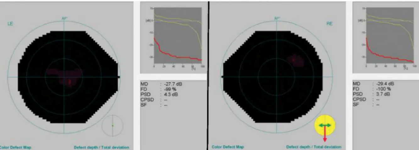
Figure 1: Automated perimetry at presentation demonstrating severely disturbed central visual fields (RE: right eye, LE: left eye).
Ophthalmoscopy revealed pale optic discs with optic disc drusen. There was no clinical evidence of disc swelling or embedded blood vessels at the disc, and no retinal abnormalities were found. Although Goldmann perimetry revealed no changes in previously existing peripheral visual field defects, automated perimetry (Hum- phrey C10-2) demonstrated severely disturbed central visual fields in both eyes (Figure 1). The erythrocyte sedimentation rate was 38 mm/hr and was considered slightly elevated when adjusted for age. Because a bilat- eral arteritic AION could not be ruled out, the patient re- ceived 1500 mg methylprednisolone-sodium-succinate intravenously per day for three days. Ophthalmological examination remained unchanged after three days. A biopsy from the temporal artery was taken, which was negative. Computed tomography of the brain confirmed signs of optic disc drusen in both eyes. Magnetic reson- ance imaging of the brain and electroencephalography were performed but could not identify any neurological cause of visual loss. Ophthalmological examination was repeated three weeks later. The patient complained of further deterioration of vision. Visual acuity in the right eye remained unchanged. In the left, eye visual acuity had diminished to counting fingers. Ophthalmoscopy re- vealed no changes. Automated perimetry remained al- most unchanged. An AION secondary to optic disc drusen was considered. To maximize perfusion pressure in the eyes, Betoptic® drops were replaced with Cosopt® (dorzolamide hydrochloride-timolol maleate) drops twice a day. Acetylsalicylic acid 80 mg once a day was pre- scribed. A check-up was scheduled after 10 days. In case of no improvement or further deterioration, an optic neurotomy would be considered in the most affected eye.
After 10 days, the patient reported further deterioration of vision. Automated perimetry revealed no changes and remained severely disturbed. To stabilize the condition and because no other treatment option was available, a 23-gauge vitrectomy with phacoemulsification and optic neurotomy was performed on the left eye under retrobul- bar anesthesia. Two optic neurotomies were performed as described by Opremcak et al. [4]. The incisions were made radial to the optic disc and parallel to the nerve
fiber pattern at the nasal edge of the optic disc using a 23-gauge neurotomy knife with sharp and blunt edges at either side of the tip (DORC, product number 1293.S06).
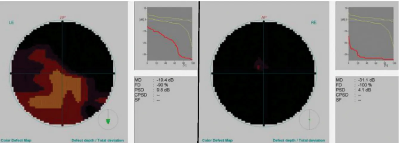
The tip of the blade was placed at the edge of the optic disc with the sharp edge aiming peripherally and directed posteriorly into the optic nerve just beyond the 1 cm marker at the tip of the knife (Figure 2). Two days after surgery, visual acuity had improved significantly to Log- MAR 0.2 in the left eye. Automated perimetry was re- peated 11 days after surgery and revealed a marked im- provement (Figure 3). The improvement of visual acuity and automated perimetry remained persistent. Nine months after surgery, visual acuity in the left eye was slightly better than LogMAR 0.2, and there was a further improvement of the central visual field. The final ophthal- mological check-up was performed 18 months after sur- gery. Automated perimetry remained stable at this point.
Figure 2: Optic disc left eye after optic neurotomy
2/4 GMS Ophthalmology Cases 2014, Vol. 4, ISSN 2193-1496
Pinxten et al.: Radial optic neurotomy as a treatment for anterior ...
Figure 3: Eleven days after surgery, automated perimetry revealed a marked improvement in the left eye (RE: right eye, LE: left eye).
Discussion
NAION is a common cause of acute visual loss. Most commonly NAION is due to transient nonperfusion or hy- poperfusion of the optic nerve head circulation, which leads to infarction in predisposed patients [8]. As men- tioned earlier, optic disc drusen represent a form of disc anomaly in which the nerve head is crowded with a small scleral canal and a mechanical distortion of blood vessels in the laminar and prelaminar region, predisposing these patients to NAION [2]. Purvin et al. studied the natural outcome of patients with NAION associated with optic disc drusen and noted that these patients were generally younger than patients with standard NAION and had more favorable visual outcomes [9]. However, 66% of eyes ex- hibited no either change or improvement of only one line or more compared to the initial examination, and 33%
exhibited worsening of visual acuity by one or more lines.
These observations illustrate that the natural outcome of NAION associated with optic disc drusen is not favor- able in most patients. Moreover, an established treatment for optic disc drusen is not yet available. Vascular com- plications, such as venous and arterial occlusions, asso- ciated with optic disc drusen are advised to be treated as in cases without drusen [1]. Although various medical and surgical interventions have been used to treat NAION in cases without drusen, none have been proven effective [10], [11], [12], [13]. Because the pathogenesis of NAION associated with optic disc drusen is assumed to be similar to the pathogenesis of CRVO, as stated by Opremcak et al., we considered optic neurotomy as a treatment in our patient [4]. However, experience with the surgical treat- ment of optic disc drusen is rare. Jirásková and Rozsíval published a case series of optic nerve sheath decompres- sion in patients with optic disc drusen and impaired visual fields and reported significant improvements in visual function [14]. However, this surgical technique is relatively invasive. Nentwich et al. presented a case series of six patients with visual field defects associated with optic nerve drusen [15]. All patients were treated with RON. As in our patient, the authors observed long-lasting improvements in visual field for the majority of patients.
In cases without postoperative visual field improvement, further progression of visual field constriction was preven- ted, and the preoperative visual acuity was maintained.
In conclusion, lacking an alternative treatment, optic nerve head decompression by RON could be considered as a treatment option in patients with NAION associated with optic disc drusen and severe visual field defects. As the experience with surgical treatment is limited, particu- larly RON for optic disc drusen, more studies on this topic are necessary.
Notes
Data declaration
The authors confirm that they had full access to all the data in the article and take responsibility for the integrity of the data.
Competing interests
Isabel Pinxten declares that she has no competing in- terests. Peter Stalmans received lecture fees from Alcon, DORC International, by Dutch Ophthalmic, USA and Fluoron as well as grant supports from Bausch & Lomb and Thrombogenics.
References
1. Auw-Haedrich C, Staubach F, Witschel H. Optic disk drusen. Surv Ophthalmol. 2002 Nov-Dec;47(6):515-32. DOI: 10.1016/S0039- 6257(02)00357-0
2. Jonas JB, Gusek GC, Naumann GO. Anterior ischemic optic neuropathy: nonarteritic form in small and giant cell arteritis in normal sized optic discs. Int Ophthalmol. 1988;12(2):119-25.
DOI: 10.1007/BF00137137
3. Gittinger JW Jr,Lessell S, Bondar RL. Ischemic optic neuropathy associated with optic disc drusen. J Clin Neuroophthalmol. 1984 Jun;4(2):79-84.
3/4 GMS Ophthalmology Cases 2014, Vol. 4, ISSN 2193-1496
Pinxten et al.: Radial optic neurotomy as a treatment for anterior ...
4. Opremcak EM, Bruce RA, Lomeo MD, Ridenour CD, Letson AD, Rehmar AJ. Radial optic neurotomy for central retinal vein occlusion: a retrospective pilot study of 11 consecutive cases.
Retina (Philadelphia, Pa). 2001;21(5):408-15. DOI:
10.1097/00006982-200110000-00002
5. Ramezani AR. Radial optic neurotomy for central retinal vein occlusion. J Ophthalmic Vis Res. 2009 Apr;4(2):115-21.
6. Verdaguer Agustí P, Nadal Reus J. Evolución clínica de la neurotomía óptica radial a largo plazo [Long-term clinical outcome of radial optic neurotomy]. Arch Soc Esp Oftalmol. 2010 Nov;85(11):370-5. DOI: 10.1016/j.oftal.2010.08.017 7. Aggermann T, Brunner S, Krebs I, Haas P, Womastek I, Brannath
W, Binder S; ROVO Study Group. A prospective, randomised, multicenter trial for surgical treatment of central retinal vein occlusion: results of the Radial Optic Neurotomy for Central Vein Occlusion (ROVO) study group. Graefes Arch Clin Exp Ophthalmol.
2013 Apr;251(4):1065-72. DOI: 10.1007/s00417-012-2134- 1
8. Hayreh SS. Management of ischemic optic neuropathies. Indian J Ophthalmol. 2011 Mar-Apr;59(2):123-36. DOI: 10.4103/0301- 4738.77024
9. Purvin V, King R, Kawasaki A, Yee R. Anterior ischemic optic neuropathy in eyes with optic disc drusen. Arch Ophthalmol.
2004 Jan;122(1):48-53. DOI: 10.1001/archopht.122.1.48 10. The Ischemic Optic Neuropathy Decompression Trial Research
Group. Optic nerve decompression surgery for nonarteritic anterior ischemic optic neuropathy (NAION) is not effective and may be harmful. JAMA. 1995 Feb;273(8):625-32. DOI:
10.1001/jama.1995.03520320035038
11. Newman NJ. The ischemic optic neuropathy decompression trial.
Arch Ophthalmol. 2007 Nov;125(11):1568-70. DOI:
10.1001/archopht.125.11.1568
12. Atkins EJ, Bruce BB, Newman NJ, Biousse V. Treatment of nonarteritic anterior ischemic optic neuropathy. Surv Ophthalmol.
2010 Jan-Feb;55(1):47-63. DOI:
10.1016/j.survophthal.2009.06.008
13. Dickersin K, Manheimer E, Li T. Surgery for nonarteritic anterior ischemic optic neuropathy. Cochrane Database Syst Rev. 2012 Jan 18;1:CD001538. DOI: 10.1002/14651858.CD001538.pub3 14. Jirásková N, Rozsíval P. Výsledky 62 dekompresí obalů zrakového nervu [Results of 62 optic nerve sheath decompressions]. Cesk Slov Oftalmol. 1999 May;55(3):136-44.
15. Nentwich MM, Remy M, Haritoglou C, Kampik A. Radial optic neurotomy to treat patients with visual field defects associated with optic nerve drusen. Retina (Philadelphia, Pa). 2011 Mar;31(3):612-5. DOI: 10.1097/IAE.0b013e318209b748
Corresponding author:
Isabel Pinxten, M.D.
University Hospitals Leuven, Herestraat 49, 3000 Leuven, Belgium, Phone: +3216332385, Fax: +3216332367 isabelpinxten@hotmail.com
Please cite as
Pinxten I, Stalmans P. Radial optic neurotomy as a treatment for anterior ischemic optic neuropathy secondary to optic disc drusen. GMS Ophthalmol Cases. 2014;4:Doc05.
DOI: 10.3205/oc000018, URN: urn:nbn:de:0183-oc0000180
This article is freely available from
http://www.egms.de/en/journals/oc/2014-4/oc000018.shtml Published:2014-07-08
Copyright
©2014 Pinxten et al. This is an Open Access article distributed under the terms of the Creative Commons Attribution License
(http://creativecommons.org/licenses/by-nc-nd/3.0/deed.en). You are free: to Share — to copy, distribute and transmit the work, provided the original author and source are credited.
4/4 GMS Ophthalmology Cases 2014, Vol. 4, ISSN 2193-1496
Pinxten et al.: Radial optic neurotomy as a treatment for anterior ...