Serous retinal detachment after trabeculectomy in angle recession glaucoma
Abstract
An 18-year-old male with 360 degree angle recession after blunt trauma in his right eye developed uncontrolled intraocular pressure (IOP) despite
Avik Kumar Roy
1Debananda Padhy
1four antiglaucoma medications (AGM) with advancing disc damage. He underwent trabeculectomy with intraoperative mitomycin-c (MMC) ap-
plication. There was an intraoperative vitreous prolapse which was 1 Glaucoma Service, LV Prasad Eye Institute, Patia,
Bhubaneswar, India managed accordingly. On post-surgery day 1, he had shallow choroidal
detachment superiorly with non-recordable IOP. This was deteriorated 1 week postoperatively as choroidal detachment proceeded to serous retinal detachment. He was started with systemic steroid in addition to topical route. The serous effusions subsided within 2 weeks time. At the last follow up at 3 months, he was enjoying good visual acuity, deep anterior chamber, diffuse bleb, an IOP in low teens off any AGM and attached retina. This case highlights the rare occurrence of serous ret- inal detachment after surgical management of angle recession glau- coma.
Keywords:angle recession, serous retinal detachment, complication of trabeculectomy
Introduction
Complications are not uncommon after trabeculectomy as the integrity of the globe is permanently distorted.
Hypotony and shallow anterior chamber (AC) are particu- larly troublesome as they are harbinger of more sinister complications like corneal decompensation, cataract development, ostium closure by formation of peripheral anterior synechia and choroidal hemorrhage [1]. This case depicts the rare occurrence of serous retinal detach- ment resulting from hypotony following trabeculectomy in a case of angle recession glaucoma.
Case description
An 18-year-old male was presented to the emergency clinic with a sudden decrease in vision following a blunt trauma with tennis ball 3 days before. His best corrected visual acuity on presentation was hand movement with perception of rays accurate in all quadrants in the right eye. The intraocular pressure (IOP) was digitally high with corneal edema and hyphema in the anterior chamber.
He was managed conservatively with topical steroid eye drops, cycloplegics and 2 anti-glaucoma medications namely brimonidine 0.2% and timolol 0.5% twice daily.
The hyphema and vitreous hemorrhage settled down in another 3 weeks time when the visual acuity returned to 20/60, angle showed 4-quadrants angle recession, disc showed 0.7 cupping with inferior rim thinning. However the IOP was still 34 mm Hg – hence prostaglandin ana- logue bimatoprost 0.01% at bed time was added. The
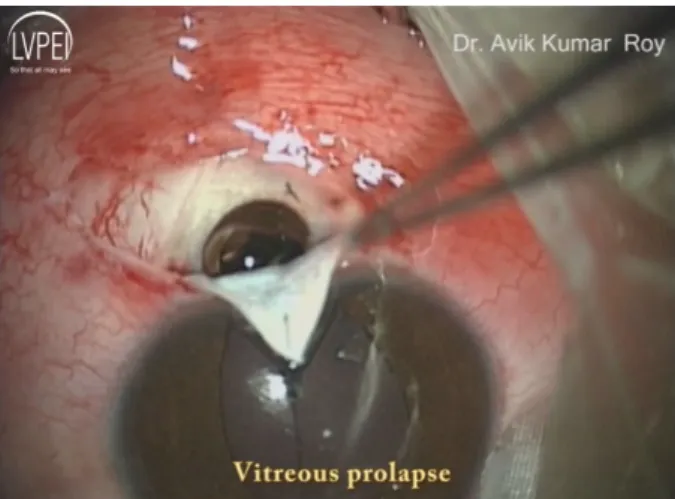
IOP did not show substantial improvement over the next 3 weeks, so the patient was advised for filtering surgery under guarded visual prognosis. Intraoperatively there was vitreous prolapse during deep block excision (Figure 1), probably from pre-existing zonular weakness and phacodonesis. This was managed with anterior vit- rectomy.
Figure 1: Intraoperative microphotograph showing vitreous prolapse during deep block excision of trabeculectomy
On postoperative day one, the visual acuity was 20/125 but the IOP was non-recordable. There was serous choroidal detachment which proceeded to frank serous retinal detachment superiorly (Figure 2), with visual acuity
1/3 GMS Ophthalmology Cases 2015, Vol. 5, ISSN 2193-1496
Case Report
OPEN ACCESS
dropping to counting finger at 2 meters and IOP being still non-recordable. He was treated with topical predniso- lone drop at 2-hourly intervals plus oral prednisolone at 1 mg/kg/day dosage. The effusion settled in 2 weeks time after which the steroids were gradually tapered.
Gradually the vision and IOP got improved. At the last follow up visit after 3 months, the best corrected visual acuity and IOP were 20/25 and 12 mm Hg off any med- ication, respectively (Figure 3, Figure 4).
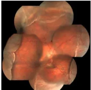
Figure 2: Fundus photograph showing combined choroidal detachment and serous retinal detachment superiorly
Figure 3: Final slit lamp photograph showing well formed anterior chamber
Figure 4: Final slit lamp photograph showing well formed bleb
Discussion
The occurrence of transient choroidal detachment after trabeculectomy is relatively common. However most of these remain subtle and resolve spontaneously with conservative management [2]. Ocular hypotony alters pressure relationships that normally prevent fluid accu- mulating in the supra-choroidal space. The risk is in- creased when there is ocular inflammation – by causing abnormal vascular permeability. The additional compo- nent of cyclitis and intraocular penetration of mitomycin- c (MMC) leading to aqueous hyposecretion also contrib- utes to ocular hypotony. Eyes with thickened sclera causing reduced transscleral outflow and eyes with ab- normally high hydrostatic pressure as with raised episcler- al pressure in Sturge-Weber syndrome (SWS) are more prone to this complication. Interestingly, there are only two cases of serous retinal detachment following trabec- ulectomy reported up till now – the first one in a 10-year- old girl with SWS [3]. The second case pertains to a 35- year-old gentleman who developed an isolated serous retinal detachment two days after glaucoma filtering surgery for his traumatic glaucoma [4]. Management of hypotony is mostly cause-specific. We hypothesized that increased inflammation from intraoperative vitreous loss could have caused the serous effusion in our case. Hence we treated the patient with full steroid therapy and the detachments resolved in 2 weeks time.
To the best of our knowledge, the complication of com- bined serous retinal detachment and choroidal detach- ment after trabeculectomy in angle recession glaucoma has not been previously reported.
Notes
Competing interests
The authors declare that they have no competing in- terests.
References
1. Azuara-Blanco A, Katz LJ. Dysfunctional Filtering Blebs. Surv Ophthalmol. 1998;43(2):93-126. DOI: 10.1016/S0039- 6257(98)00025-3
2. Johnstone MA. Hypotony: What is it? How should we manage it?
J Glaucoma. 2000 Apr;9(2):131-3. DOI: 10.1097/00061198- 200004000-00001
3. Simmons RJ, Krupin T, Hitchings R. Serous Retinal Detachment After Glaucoma Filtration Surgery in Sturge-Weber Syndrome. J Glaucoma. 1992 Apr;1(1):58-62. DOI: 10.1097/00061198- 199204000-00013
4. Aydin A, Cakir A, Unal MH, Ersanli D. Central serous retinal detachment following glaucoma filtration surgery. J Fr Ophtalmol.
2012 Sep;35(7):529.e1-5. DOI: 10.1016/j.jfo.2011.10.010
2/3 GMS Ophthalmology Cases 2015, Vol. 5, ISSN 2193-1496
Roy et al.: Serous retinal detachment after trabeculectomy in ...
Corresponding author:
Avik Kumar Roy, MS
Glaucoma Service, LV Prasad Eye Institute, Patia, Bhubaneswar 751024, Odisha, India
royavikkumar@gmail.com
Please cite as
Roy AK, Padhy D. Serous retinal detachment after trabeculectomy in angle recession glaucoma. GMS Ophthalmol Cases. 2015;5:Doc15.
DOI: 10.3205/oc000037, URN: urn:nbn:de:0183-oc0000372
This article is freely available from
http://www.egms.de/en/journals/oc/2015-5/oc000037.shtml Published:2015-12-28
Copyright
©2015 Roy et al. This is an Open Access article distributed under the terms of the Creative Commons Attribution 4.0 License. See license information at http://creativecommons.org/licenses/by/4.0/.
3/3 GMS Ophthalmology Cases 2015, Vol. 5, ISSN 2193-1496
Roy et al.: Serous retinal detachment after trabeculectomy in ...