Post-translational lysine-acetylation of Ran and its regulation by Sirtuin
deacetylases
Inaugural-Dissertation zur
Erlangung des Doktorgrades
der Mathematisch-Naturwissenschaftlichen Fakult¨at der Universit¨at zu K¨oln
vorgelegt von Philipp Knyphausen
aus Eckernf¨orde Hundt Druck GmbH, K¨oln
2016
Post-translational lysine-acetylation of Ran and its regulation by Sirtuin
deacetylases
Inaugural-Dissertation zur
Erlangung des Doktorgrades
der Mathematisch-Naturwissenschaftlichen Fakult¨at der Universit¨at zu K¨oln
vorgelegt von
Philipp Knyphausen
aus Eckernf¨orde
Berichterstatter: Dr. Michael Lammers (Betreuer)
(Gutachter) Prof. Dr. Kay Hofmann
Tag der m¨undlichen Pr¨ufung: 20. Januar 2016
Zusammenfassung
Durch die Fortschritte in der Hochdurchsatz-Massenspektrometrie hat sich ge- zeigt, dass posttranslationale N-(ε)-Lysin-Acetylierung bei Tausenden von Pro- teinen vorkommt. So modifizierte Proteine finden sich beim Menschen und an- deren Organismen in allen Zellkompartimenten und sind in vielen F¨ allen an es- sentiellen zellul¨ aren Prozessen beteiligt. Viele Aspekte posttranslationaler Lysin- Acetylierung sind jedoch nur unvollst¨ andig verstanden, einschließlich ihrer Regulie- rung durch Lysin-Acetyltransferasen und Lysin-Deacetylasen (KDACs). In dieser Arbeit wurde untersucht, welchen Einfluss diese Modifikation auf die Funktion des kleinen GTP-bindenden Proteins Ran hat, dem in der Zelle unter anderem eine zentrale Rolle bei der Regulation des Kerntransports zukommt. Hierzu wurde mit Hilfe eines erweiterten genetischen Codes stellenspezifisch acetyliertes Ran in E.
coli hergestellt.
Untersucht wurden zun¨ achst f¨ unf zuvor identifizierte Ran-Acetylierungsstellen hin- sichtlich ihrer Auswirkungen auf die intrinsische GTP-Hydrolyse Rate von Ran, die Bildung von Exportkomplexen (anhand des Exportrezeptors CRM1 und des Ex- portsubstrats Spn1) und die Interaktion von Ran mit RanBP1 und dem GTPase- aktivierenden Protein RanGAP. Insgesamt waren sowohl bei der intrinsischen als auch der RanGAP-stimulierten GTP-Hydrolyse nur schwache Effekte zu messen.
Dahingegen sorgte die Acetylierung von Ran am Lysin 159 (K159) f¨ ur eine deut- lich gesenkte Affinit¨ at von Ran zu RanBP1, wenn Ran im aktiven Zustand vor- lag. Dar¨ uberhinaus war eine st¨ arkere Bindung von Spn1 an einen Komplex aus CRM1·Ran zu beobachten, wenn Ran an den Stellen K37, K99 oder K159 acety- liert war. Anhand dieser Ergebnisse l¨ asst sich schließen, dass wesentliche Funktio- nen des Proteins Ran durch Acetylierung beeinflusst werden.
Ein in vitro Screen wurde durchgef¨ uhrt, um potenzielle KDACs f¨ ur Ran zu identi-
fizieren. NAD
+-abh¨ angige KDACs der Sirtuin-Klasse zeigten Aktivit¨ at gegen¨ uber
zwei Acetylierungsstellen von Ran, K37 und K71. Die Spezifit¨ at der SIRTs wur-
de daraufhin anhand einer weiteren acetylierten Variante von Ran (RanAcK38)
analysiert. Da bei RanAcK38 im Vergleich zu RanAcK37 eine deutlich langsamere
Deacetylierungsrate zu beobachten war, wurde als n¨ achstes di-acetyliertes Ran-
AcK37/38 getestet. Die Deacetylierungsrate von di-acetylierten Ran war erstaun-
licherweise vergleichbar mit derjenigen von RanAcK37. Deacetylierungsexperimen-
te unter single turnover -Bedingungen ergaben, dass die Deacetylierung im Ran-
AcK37/38-Hintergrund als erstes an der Stelle K38 erfolgen muss. Die F¨ ahigkeit
von Sirtuinen zwei benachbarte AcKs zu deacetylieren wurde schließlich anhand
zweier weiterer Proteine untersucht, von denen bekannt war, dass sie unter an-
derem di-acetyliert vorkommen. Dabei handelte es sich um das Tumorsuppressor-
Protein p53 und Phosphoenolpyruvatcarboxykinase 1 (PEPCK1). Es stellte sich
heraus, dass p53 an zwei Di-Acetylierungsstellen (K372/372 und K381/382) durch
Sirtuin 1 und 2 deacetyliert wird. Entgegen der Erwartungen war bei PEPCK1
keine Deacetylierung durch Sirtuine festzustellen. Diese Ergebnisse lassen einige
bedeutende Schlussfolgerungen f¨ur die Substratspezifit¨at von Sirtuinen zu.
“Now my own suspicion is that the Universe is not only queerer than we suppose, but queerer than we can suppose.”
John Burdon Sanderson Haldane, Biologist
Abstract
Through recent advances in high-throughput mass spectrometry it has become evident that post-translational N-(e)-lysine-acetylation is a modification found on thousands of proteins of all cellular compartments and all essential physiological processes. Many aspects in the biology of lysine-acetylation are poorly under- stood, including its regulation by lysine-acetyltransferases and lysine-deacetylases (KDACs). Here, the role of this modification was investigated for the small GTP- binding protein Ran, which, inter alia, is essential for the regulation of nucleocy- toplasmic transport. To this end, site-specifically acetylated Ran was produced in E. coli by genetic code expansion.
For five previously identified sites, Ran acetylation was tested regarding its impact on the intrinsic GTP hydrolysis rate, the assembly of export complexes (modeled in vitro with the export receptor CRM1 and the export substrate Spn1) and the interaction of Ran with its GTPase activation protein RanGAP and RanBP1.
Overall, mild e↵ects of Ran acetylation were observed for intrinsic and RanGAP- stimulated GTP hydrolysis rates. The interaction of active Ran with RanBP1 was negatively influenced by Ran acetylation at K159. Moreover, CRM1 bound to Ran acetylated at K37, K99 or K159 interacted more strongly with Spn1. Thus, lysine-acetylation interferes with essential aspects of Ran function.
An in vitro screen was performed to identify potential Ran KDACs. The NAD
+-
dependent KDACs of the Sirtuin class showed activity towards two acetylation
sites of Ran, K37 and K71. The specificity of Sirtuins was further analyzed based
on an additional Ran acetylation site, K38. Since deacetylation of RanAcK38
was much slower compared to RanAcK37, di-acetylated RanAcK37/38 was tested
next. The deacetylation rate of di-acetylated Ran was comparable to that of
RanAcK37. Deacetylation experiments under single turnover conditions revealed
that deacetylation occurs first at the K38 site in the di-acetylated RanAcK37/38
background. The ability of Sirtuins to deacetylate two adjacent AcKs was further
investigated based on two proteins, which had previously been found to be di-
acetylated and targeted by Sirtuins, namely the tumor suppressor protein p53 and
phosphoenolpyruvate carboxykinase 1 (PEPCK1). p53 was readily deacetylated
at two di-acetylation sites (K372/372 and K381/382), whereas PEPCK1 was not
deacetylated in vitro. Taken together, these results have important implications
for the substrate specificity of Sirtuins.
Contents
Abstract i
List of Figures vii
List of Tables ix
1 Introduction 1
1.1 The small GTP-binding protein Ran . . . . 1
1.1.1 Localization of Ras proteins . . . . 3
1.1.2 Nucleotide exchange and hydrolysis . . . . 5
1.1.3 Nucleocytoplasmic transport . . . . 8
1.1.4 Mitotic spindle assembly . . . 12
1.1.5 Nuclear envelope formation . . . 13
1.2 Lysine-acetylation . . . 14
1.2.1 The writers: Lysine-acetyltransferases (KATs) . . . 16
1.2.2 The erasers: Lysine-deacetylases (KDACs) . . . 17
1.2.3 The readers: Bromodomain containing proteins . . . 21
1.2.4 The canonical roles of lysine-acetylation . . . 23
1.2.5 The acetylome: Novel roles for lysine-acetylation? . . . 25
1.2.6 Lysine-acetylation of Ran . . . 27
1.2.7 The genetic code expansion concept (GCEC) . . . 28
1.3 Aim of the thesis . . . 30
2 Material and Methods 31 2.1 Materials . . . 31
2.1.1 Chemicals, kits and enzymes . . . 31
2.1.2 Primers . . . 31
2.1.3 Vectors and constructs . . . 33
2.1.4 Crystallization screens . . . 35
2.1.5 Bu↵ers . . . 36
2.1.6 Media, Antibiotics and inhibitors . . . 38
2.2 Molecular biology techniques . . . 38
2.2.1 Purification of DNA . . . 38
2.2.2 Polymerase chain reaction (PCR) . . . 38
2.2.3 Restriction enzyme-based cloning . . . 39
iii
Contents iv
2.2.4 Circular polymerase extension cloning (CPEC) . . . 39
2.2.5 Site-directed mutagenesis . . . 40
2.2.6 Transformation of E. coli . . . 40
2.3 Biochemical methods . . . 40
2.3.1 Expression of recombinant proteins . . . 40
2.3.2 Lysis of cells . . . 41
2.3.3 Purification of GST-tagged proteins . . . 41
2.3.4 Purification of His
6-tagged proteins . . . 42
2.3.5 Determination of protein concentration . . . 42
2.3.6 SDS-polyacrylamide gel electrophoresis (SDS-PAGE) . . . . 43
2.3.7 Western blotting and immunodetection . . . 43
2.3.8 Generation of the RanAcK37-specific antibody . . . 44
2.3.9 Exchange of Ran-bound nucleotides . . . 45
2.3.10 High pressure liquid chromatography (HPLC) . . . 45
2.3.11 Activity assay for deacetylases . . . 46
2.3.12 KDAC-screen . . . 46
2.3.13 Deacetylase assays . . . 47
2.3.14 PEPCK1 activity assay . . . 47
2.4 Cell culture . . . 48
2.4.1 Cultivation of cell lines . . . 48
2.4.2 Transfection . . . 48
2.4.3 Ni
2+-NTA pull-down . . . 49
2.5 Biophysical methods . . . 50
2.5.1 Isothermal titration calorimetry (ITC) . . . 50
2.5.2 Mass spectrometry (MS) . . . 50
2.6 Crystallographic methods . . . 52
2.6.1 Crystallization . . . 52
2.6.2 Preparation of crystals for data collection . . . 52
2.6.3 Data collection and processing . . . 53
3 Results 55 3.1 Ran acetylation: E↵ects and regulation . . . 55
3.1.1 Purification of acetylated Ran . . . 55
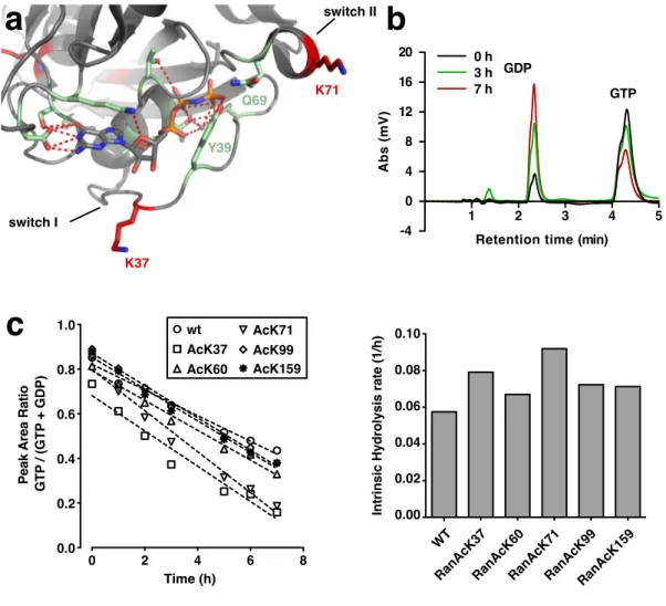
3.1.2 E↵ect of Ran acetylation on intrinsic GTP hydrolysis . . . . 58
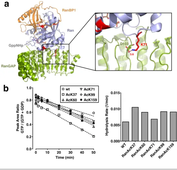
3.1.3 GAP-catalyzed nucleotide hydrolysis and binding to Ran- GAP is not a↵ected by Ran acetylation . . . 59
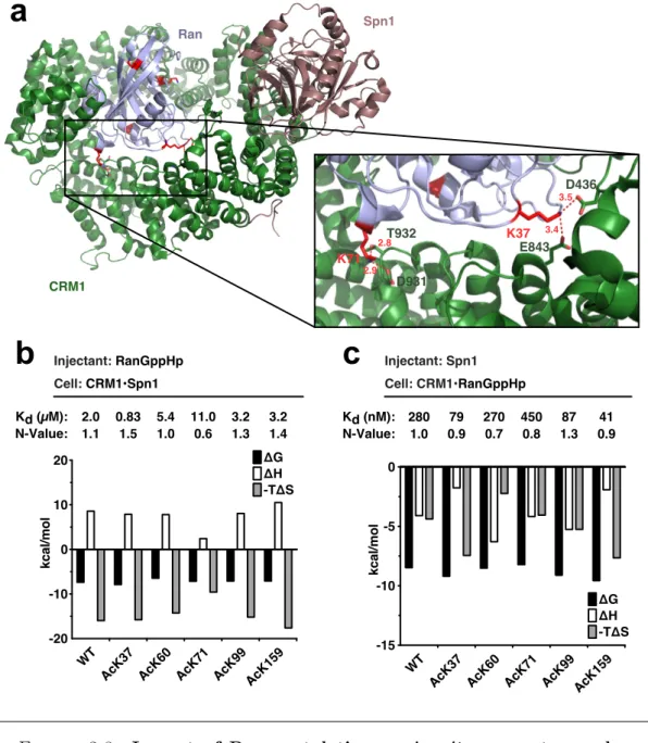
3.1.4 Impact of Ran acetylation on the interaction with RanBP1 . 64 3.1.5 Ran acetylation interferes with export complex formation . . 65
3.1.6 in vivo acetylation of Ran by KAT overexpression . . . 68
3.1.7 An in vitro KDAC screen identifies SIRT1-3 as Ran deacety- lases . . . 71
3.2 Analysis of di-deacetylation by Sirtuins . . . 76
3.2.1 SIRT2 deacetylates Ran at two adjacent lysines . . . 76
3.2.2 Mass spectrometric analysis of Ran di-deacetylation . . . 77
Contents v
3.2.3 Mutational analysis of the Ran di-deacetylation site . . . 80
3.2.4 Structural insights into di-deacetylation . . . 81
3.2.5 SIRT1 and SIRT3 are able to di-deacetylate Ran . . . 87
3.2.6 PEPCK1 is not deacetylated by SIRT2 in vitro . . . 88
3.2.7 Deacetylation of p53-AcK381/382 . . . 91
3.2.8 Deacetylation of p53-AcK372/373 . . . 93
4 Discussion 99 4.1 Incorporation of acetyl-lysine with the GCEC . . . 99
4.2 Ran acetylation in regulation of export complex formation and release100 4.3 Regulation of Ran acetylation . . . 102
4.4 Implications for the substrate specificity of classical KDACs . . . . 107
4.5 Implications for the substrate specificity of Sirtuins . . . 109
4.6 On the role of di-acetylation . . . 114
4.7 Conclusions and Outlook . . . 114
A Appendix 117
Bibliography 125
Abbreviations 125
Acknowledgements 167
List of Figures
1.1 Structural comparison of GDP- and GTP-bound GNBPs . . . . 3
1.2 The nucleotide exchange cycle . . . . 5
1.3 Mechanisms of GTPase activation by GAPs . . . . 8
1.4 The nuclear pore complex (NPC) . . . . 9
1.5 Ran-dependent nuclear import and export . . . 11
1.6 Roles of Ran during mitotic spindle assembly . . . 13
1.7 Molecular e↵ects of acetylation of lysine . . . 15
1.8 Chemistry and structure of Sirtuin deacetylases . . . 19
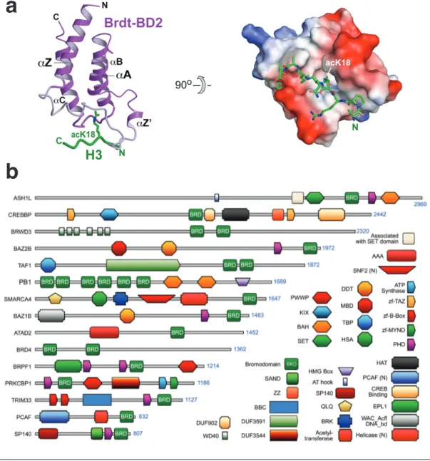
1.9 Bromodomains are readers of acetylated lysine residues . . . 22
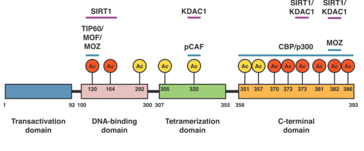
1.10 Domain structure and acetylation sites of p53 . . . 25
1.11 The genetic code expansion concept (GCEC) . . . 29
2.1 Scheme of PEPCK1 activity assay . . . 48
3.1 Exemplary purification of acetylated Ran (RanAcK71) . . . 56
3.2 Purification of acetylated Ran and Ran interaction partners . . . . 57
3.3 E↵ect of Ran acetylation on intrinsic GTP hydrolysis . . . 60
3.4 E↵ect of Ran acetylation on RanGAP-stimulated hydrolysis . . . . 61
3.5 Interaction of RanGAP with RanGppNHp · RanBP1 . . . 63
3.6 Interaction of RanBP1 with acetylated Ran . . . 65
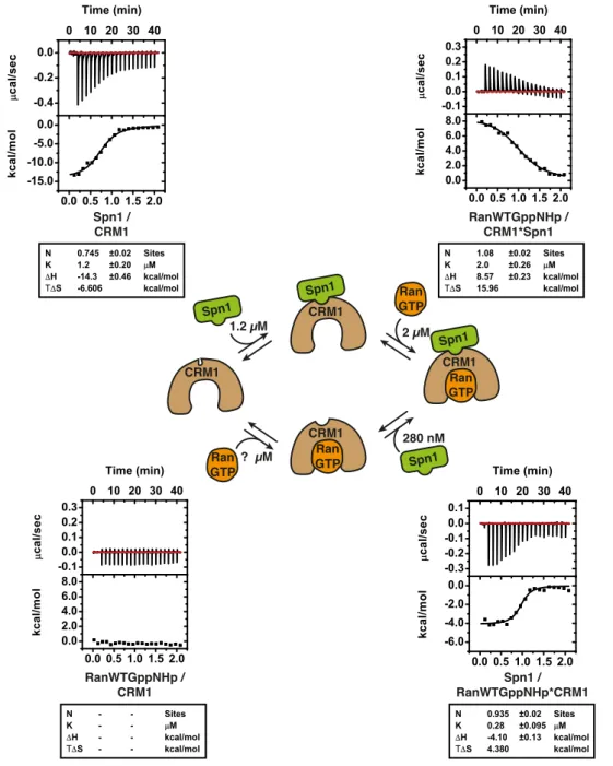
3.7 ITC-based in vitro export complex formation assay . . . 67
3.8 Impact of Ran acetylation on in vitro export complex formation . . 69
3.9 MS based detection of acetylated Ran in HEK293T cells . . . 71
3.10 Activities of recombinant KDACs used for in vitro deacetylase screen 72 3.11 In vitro deacetylase screen for recombinant Ran . . . 73
3.12 Deacetylation of RanAcK37 and -AcK71 by SIRT1-3 . . . 74
3.13 Deacetylation of RanGDP and -GppNHp by SIRT2 . . . 75
3.14 SIRT2 can deacetylate Ran at two neighboring lysines . . . 78
3.15 Sequential deacetylation of RanAcK37/38 . . . 80
3.16 Mutational analysis of RanAcK38 deacetylation . . . 82
3.17 Interaction of Ran-derived N-(e)-trifluoroacetyl-L-lysine peptides with SIRT2 . . . 83
3.18 Crystallisation of SIRT2 with a RanTFAcK37 13-mer . . . 84
3.19 Structure of SIRT2 with RanTFAcK37 13-mer peptide . . . 86
3.20 Molecular interactions between SIRT2 and the RanTFAcK37 13- mer peptide . . . 87
vii
List of Figures viii
3.21 Di-deacetylation by SIRT1 and SIRT3 . . . 88
3.22 Analysis of PEPCK1 deacetylation . . . 95
3.23 Deacetylation of p53 by SIRT1 . . . 96
3.24 Dynamics of p53 deacetylation at the di-acetylation site K381/382 . 97 3.25 Deacetylation of p53 at the di-acetylation site K372/K373 . . . 98
4.1 Regulation of Ran functions by lysine-acetylation . . . 103
4.2 Sequence context of acetylation sites tested for deacetylation . . . . 111
4.3 Structural context of sites tested for deacetylation . . . 113
A.1 Maps of vectors used for GCEC . . . 118
A.2 ITC measurements of RanGppNHp · RanBP1 and RanGAP . . . 119
A.3 ITC measurements of RanGDP and RanBP1 . . . 120
A.4 ITC measurements of RanGppNHp and RanBP1 . . . 121
A.5 ITC measurements of RanGppNHp and CRM1 · Spn1 . . . 122
A.6 ITC measurements of RanGppNHp · CRM1 and Spn1 . . . 123
A.7 Ramachandran plots by residue type . . . 124
List of Tables
2.1 Cloning primers . . . 32
2.2 Primers for site-directed mutagenesis . . . 33
2.3 Constructs for recombinant protein expression . . . 35
2.4 Crystallization screens . . . 35
2.7 Antibodies . . . 44
2.8 Recombinant KDACs used for the screen . . . 47
3.1 Data collection, refinement and structure validation of the SIRT2(50- 356) · RanTFAcK37-13-mer-peptide structure (molecular replacement). 85 4.1 Lysine-acetylation sites of Ras superfamily members. . . 105
4.2 Comparison of Ran acetylation sites with preferred substrate se- quences of classical KDACs. . . 108
ix
1 Introduction
The acquisition of a bacterial endosymbiont by an archeon about two billion years ago marks the beginning of eukaryotic evolution (reviewed in Koonin, 2015; Spang et al., 2015). The energetic gain associated with this symbiosis is thought to have allowed for the seemingly unconstrained genome size and morphological complexity seen in eukaryota (Lane and Martin, 2010). To account for the diverse novel tasks in the evolving eukaryotic cell, many protein families have undergone massive functional diversification, one of which is the Ras superfamily, which is essential for the regulation of cell proliferation, cellular adhesion, the cytoskeleton, vesicular transport and membrane trafficking (Jkely, 2003). Ran (Ras-related nuclear) has taken a central role in establishing the nuclear envelope and the regulation of nuclear transport and mitosis, all three being hallmarks of the eukaryotic domain of life. In this introduction, an overview is presented of the mechanisms of these Ran- directed processes. Furthermore, lysine-acetylation as a conserved and reversible post-translational modification is introduced, which will lead to the question how it might regulate Ran function, a question that will be of central relevance in this thesis.
1.1 The small GTP-binding protein Ran
Ran is a member of the Ras superfamily, the founding member of which is Ras (Rat sarcoma). Ras was initially discovered as the factor conferring oncogenicity to two murine viruses, the Kirsten and the Harvey murine sarcoma virus, hence the names K-Ras and H-Ras, respectively (Harvey, 1964; Kirsten et al., 1970). It later turned out that specific point mutations in the Ras sequences lead to this oncogenicity and that their wildtype counterparts are present in rat and human genomes (Capon et al., 1983; Chang et al., 1982; Ellis et al., 1981; Tsuchida et al.,
1
Introduction 2 1982). Ras has since been regarded as a proto-oncogene, having important cellular functions in the non-mutated form but also becoming an oncogenic factor upon mutation of certain amino acid residues.
As stated above, the Ras superfamily has expanded in the early eukaryotic evolu- tion and members of the five major branches (Ras, Rho, Rab, Ran and Arf) can thus be found in all eukaryotes unless they have been lost secondarily (Colicelli, 2004). About 167 members of the Ras superfamily are present in humans. Inter- estingly and in contrast to the many paralogs of other Ras superfamily members, only one Ran gene is found in mammals and a few often almost identical ones in plants (Rojas et al., 2012). In addition, Ran is one of the most conserved proteins of nucleated cells (Ach and Gruissem, 1994; Bush and Cardelli, 1993; Chen et al., 1994).
All Ras superfamily members have in common that they are relatively small pro- teins (20-30 kDa), able to bind guanosine di- or triphosphates (GDP and GTP, respectively) with high affinity and to adopt two three-dimensional conformations, depending on which nucleotide is bound. This behavior is referred two as a binary
‘molecular switch’ and makes this protein family particularly well-suited to act in cellular signaling pathways (Vetter and Wittinghofer, 2001). This is reflected by the diverse cellular processes that are regulated by these small guanine nucleotide binding proteins (GNBPs). In addition to the mere binding of GDP or GTP, Ras superfamily members are intrinsically able to hydrolyze bound GTP, although this activity is generally very low and di↵ers substantially between di↵erent groups. In particular, the GTPase activity of the Arf members Sar1, SRb and Arf are hardly detectable (Bi et al., 2002; Legate and Andrews, 2003; Randazzo and Kahn, 1994).
The shared features of small GNBPs and other GTP binding proteins can be
directly attributed to their shared central domain, the G-domain, a fold consist-
ing of a twisted beta sheet with six parallel and anti-parallel beta strands, four
alpha helices packed on both sides of the beta sheet and nine connecting loops
(de Vos et al., 1988; Pai et al., 1989). This approximately 20-kDa domain pos-
sesses characteristic consensus elements, so called G boxes, which on the one hand
are required for nucleotide binding (and hydrolysis) and on the other hand me-
diate the switch-like behavior: G1, GxxxxGK(S/T); G2, x(P/A)T(I//V/L); G3,
DxxGQ; G4, (T/N)KxD; and G5, (C/S)A(K/L)(S/T) (Bourne et al., 1991; Dever
et al., 1987). While the G4-5 confer specificity for the guanine base over adenine,
Introduction 3 the G1 or P-loop makes contacts with the b- and g-phosphates of GTP and coor- dinates the Mg
2+-ion, which is needed for high affinity nucleotide binding (Saraste et al., 1990). Interestingly, the G2 and G3, besides also being involved in binding of the Mg
2+-ion, contact the g-phosphate, which is only present in GTP. Thus, upon GTP hydrolysis these interactions are released, which results in a change in conformation and is the explanation for the switch-like behavior of small GNBPs.
Due to this behavior, G2 and G3 are called switch I and switch II, respectively and their GTP-bound conformation has been compared to a loaded spring (Milburn et al., 1990; Vetter and Wittinghofer, 2001). As shown in Fig. 1.1, the switch I and II regions adopt a flexible conformation in the GDP-bound state compared to the rigid conformation seen in the GTP-bound state.
Figure 1.1: Structural comparison of GDP- and GTP-bound GNBPs.
Selected Ras-related proteins in GTP- or GDP-form are shown as superimposed ribbon representations. The switch I and II regions are shown in green and turquoise, respectively. Characteristic elements of Rho, Arf and Ran are in- dicated (red: C-terminus of Ran, magenta: Rho insert, blue: Arf N-terminal
helix) (taken from Vetter and Wittinghofer, 2001).
1.1.1 Localization of Ras proteins
Another feature of many Ras superfamily members is their post-translational mod-
ification by lipids, which anchor them to cellular membranes. In many cases this
reflects their roles in membrane-associated processes. For instance, members of
Introduction 4 the Rab and Arf families are important for vesicle formation and transport and the Rho family for the regulation of cell shape changes. However, the type of modifi- cation di↵ers between the subfamilies. Ras and Rho family members are modified at a cysteine residue in their C-termini by farnesyltransferase or geranylgeranyl- transferase type 1 (GGTase 1), which recognize a C-terminal CAAX motif (C:
Cys, A: aliphatic, X: any amino acid) (Anderegg et al., 1988; Casey et al., 1989;
Clarke et al., 1988; Katayama et al., 1991; Maltese et al., 1990). In addition to the farnesyl- or geranylgeranyl-modifications, a nearby palmitoylation or polybasic patch can further strengthen their membrane attachment and direct their sub- cellular localization through interactions with distinct membrane compartments of di↵erent lipid compositions (Apolloni et al., 2000; Choy et al., 1999; Hancock et al., 1990; Rocks et al., 2005; Roy et al., 2005). Similarly, Rab family proteins are geranylgeranylated at their cysteine-containing C-termini by the action of RabG- GTase (Jiang et al., 1993; Khosravi-Far et al., 1991). However, in most cases, two prenyl-groups are attached to Rab proteins. This is not specified by a CAAX motif but by an interaction between a conserved surface feature of Rab proteins with the Rab escort protein-1 (REP-1), which in turn interacts with RabGGTase (Andres et al., 1993; Pylypenko et al., 2003). REP-1 can accommodate both hydrophobic prenyl-groups (one in its interior and the other more solvent exposed) and remains bound to Rab proteins after prenylation until delivery to their target membrane (Pylypenko et al., 2006). Besides REP-1, there are also other proteins that can bind prenylated Rho or Rab proteins and regulate their delivery to or retrieval from membranes. These are subsumed under the term GDP-dissociation inhibitor (GDI) and, as the name suggests, preferentially bind to GDP-bound small GNBPs and prevent their activation (Gosser et al., 1997; Longenecker et al., 1999; Sasaki et al., 1990). Another factor that solubilizes a variety of farnesylated proteins, inter alia Ras subfamily members, is phosphodiesterase 6 d subunit (PDE-d). In this case however, the binding occurs regardless of the GNBP’s nucleotide state (Chandra et al., 2012; Nancy et al., 2002). Members of the Arf family are often an- chored to the membrane by a myristoyl-group, which is linked to their N-terminus.
In addition to the myristoyl-anchor, an amphipathic N-terminal helix, which is re-
leased from an intramolecular sequestration upon GTP-loading, can insert itself
into the membrane (Antonny et al., 1997). In fact, some Arf family members,
like for instance Sar1, localize to membranes solely based on the action of this
helix (Bielli et al., 2005). Ran is one of only a few Ras superfamily members that
does not localize to membranes but is instead predominantly found in the nucleus
Introduction 5 during interphase (Bischo↵ and Ponstingl, 1991b). It is furthermore not lipidated (i.e. prenylated or modified by fatty acid esterification) and has no poly-basic patch.
1.1.2 Nucleotide exchange and hydrolysis
Through their dynamic switch regions, GNBPs have the ability to bind to di↵erent interaction partners, depending on which nucleotide is bound. Most interaction partners bind with high affinity to the more rigid conformation of the switch re- gions of GTP-bound GNBPs and are activated upon binding. These so-called e↵ectors can subsequently exert their down-stream signaling functions or directly mediate e↵ects such as actin nucleation. Nevertheless, there are also many pro- teins known to interact with the GDP-bound state of GNBPs, which likewise play important roles. The fact that GNBPs usually show slow rates of intrinsic nu- cleotide exchange and GTP hydrolysis (Bischo↵ et al., 1990; Klebe et al., 1995), make GNBPs appear not well-suited for dynamic signaling processes. However, both the nucleotide exchange and the GTPase rates can be accelerated over several orders of magnitude by guanine nucleotide exchange factors (GEFs) and GTPase activating proteins (GAPs), which are usually specific for individual GNBPs (re- viewed in Bos et al., 2007) (Fig. 1.2).
Figure 1.2: The nucleotide exchange cycle. Overview of the nucleotide exchange cycle of GNBPs (G: small GNBP, GAP: GTPase activating protein, GEF: guanine nucleotide exchange factor, GDI: GDP dissociation inhibitor)
(taken from Bos et al., 2007).
Introduction 6 The affinity of GNBPs for nucleotides typically lies in the picomolar range and, as a consequence, the dissociation rate of the nucleotide is very slow (John et al., 1990; Klebe et al., 1995). The binding of a GEF to its cognate GNBP promotes the dissociation of the bound nucleotide and thus allows for a new nucleotide molecule to bind. For the latter step to take place, the incoming nucleotide has to displace the GEF, which remains bound to the GNBP after nucleotide release. Since the GEF-GNBP complex is highly stable (as is the nucleotide-GNBP complex), the new nucleotide has to modify the affinity of the GEF for the GNBP, ultimately leading to the release of the GEF. Thus, the exchange reaction relies on the recip- rocal negative influence on the affinity of either GEF or nucleotide for the GNBP and occurs in successive reversible steps (Vetter and Wittinghofer, 2001).
The modulation of the nucleotide affinity by GEFs is achieved by a similar mech- anism even though GEFs are structurally unrelated for di↵erent Ras superfamily branches (Boriack-Sjodin et al., 1998; Renault et al., 2001; Worthylake et al., 2000). The high affinity for the nucleotide is to a large extent a result of the inter- actions of the phosphates with the Mg
2+-ion and the P-loop. GEFs use a so-called
‘push-and-pull’ mechanism to interfere with these interactions: The GEF pushes out the Mg
2+-ion by relocating residues of the P-loop and the switch II of the GNBP or, instead for the latter, by introducing own elements into the nucleotide binding pocket. In addition, the switch I is pulled out of its normal position, which further reduces nucleotide affinity (Vetter and Wittinghofer, 2001). In gen- eral, GEFs promote nucleotide exchange of GNBPs irrespective of the nucleotide state of the GNBP (GTP or GDP) (Haney and Broach, 1994; Lenzen et al., 1998).
However, because the concentration of GTP in the cell is about 10-fold higher than that of GDP, the accelerated nucleotide exchange by GEFs e↵ectively lead to GTP-loading of the respective GNBP. Nevertheless, depending on the physi- ological state of the cell the GTP:GDP ratio can change substantially and lead to a shift in favor of GDP- or GTP-loading, which, at least in yeast, can have a profound influence on intracellular signaling processes (Rudoni et al., 2001; Sagot et al., 2005). The specific GEF for Ran is RCC1 (regulator of chromatin conden- sation), which enhances the nucleotide exchange rate of Ran by about 10
5-fold (Bischo↵ and Ponstingl, 1991a,b; Klebe et al., 1995).
As mentioned above, the GTPase activity of Ras superfamily proteins is generally
very low although intrinsically the catalytic machinery is present. The mechanism
by which GAPs stimulate the hydrolysis activity involves the stabilization of the
Introduction 7 intrinsically mobile catalytic center of GNBPs and, in most cases, the introduction of catalytic residues. However, like GEFs, GAPs are structurally unrelated for dis- tinct GNBPs and thus di↵erences in the molecular details of GTPase activation are found. In principle, GTP-hydrolysis by GNBPs most likely occurs through a sub- strate assisted catalysis mechanism. This means that GTP itself serves as a base to abstract a proton from a water molecule. The resulting OH then performs a nucleophilic attack on the g-phosphate leading to an inversion at the g-phosphorus atom (Schweins et al., 1995). The reaction probably happens in a single step, a so-called in-line transfer (Feuerstein et al., 1989). For Ras, it was shown that the reaction is catalyzed by stabilization of the transition state by the critical residue glutamine-61, the mutation of which renders Ras unable to hydrolyze GTP (Priv et al., 1992). This glutamine is also critical for the action of RasGAP, which sta- bilizes it and makes it able to orient the water molecule for nucleophilic attack of the g-phosphate. Moreover, it inserts an arginine into the phosphate-binding site, which neutralizes negative charges of the b and g-phosphate and thereby stabilizes the transition state (Sche↵zek et al., 1997). A similar mechanism is also observed for Rho and Cdc42 and, with some variations, also for Rab and Sar1 (Bi et al., 2002; Nassar et al., 1998; Pan et al., 2006; Rittinger et al., 1997). The mechanism of Ran GTPase activation through RanGAP is di↵erent in that RanGAP does not introduce any catalytic residue into the GTP binding pocket. Instead, RanGAP uses an Asp to correct the, in this case, improperly positioned catalytic glutamine (Gln69). The role of the arginine provided in trans by other GAPs is taken over by a tyrosine 39 of Ran that forms hydrogen bonds to the g-phosphate as well as the Gln69 side chain (Seewald et al., 2002) (see Fig. 1.3).
The location of RCC1 and RanGAP in the cell provides clues for the distribution of RanGDP and RanGTP in the cell. RCC1 associates with chromatin through- out the cell cycle and is thus found in the nucleus during interphase (Ohtsubo et al., 1989). Binding of Ran to RCC1 allosterically promotes the interaction of RCC1 with chromatin, which in turn stimulates nucleotide exchange (Chen et al., 2007; Li et al., 2003a). Thus, RanGTP is generated close to chromatin. By con- trast, RanGAP is located in the cytoplasm during interphase, which leads to the conversion of RanGTP into RanGDP in the cytoplasm (Hopper et al., 1990).
This di↵erential distribution of RanGTP and GDP is key for nucleocytoplasmic
transport since the stability of import and export complexes is directly regulated
by RanGTP. Moreover, it is important for the role of Ran in mitotic spindle
Introduction 8
a
b
Ran/RanGAP Rho/RhoGAP
Ras/RasGAP
Figure 1.3: Mechanisms of GTPase activation by GAPs. (a) Depic- tion of two di↵erent mechanism of GTPase activation for Ran and Ras/Rho.
RanGAP induces GTP hydrolysis without introducing an arginine finger. (b) Ribbon representation of di↵erent Ras-like proteins (blue) with their cognate GAPs (red). The GNBPs are shown in the same orientation (taken from Bos
et al., 2007).
assembly and nuclear envelope formation. These three processes will be briefly introduced in the subsequent sections.
1.1.3 Nucleocytoplasmic transport
The nuclear compartment is the most distinctive feature of eukaryotes. It pro- vides a compartmentalization between the cytosol and the nucleoplasm, which is essential for a number of cellular processes and perhaps most importantly to spatially separate transcription and translation, preventing the translation of un- spliced mRNAs (Cavalier-Smith, 1991; Martin and Koonin, 2006). The nuclear envelope consists of two parallel membranes and forms a continuous lumen with the endoplasmic reticulum (ER). The two membranes are pierced with nuclear pore complexes (NPCs) that allow the unaided passage of molecules with a diameter of
⇠ 5 nm, which corresponds to a molecular weight of ⇠ 30 kDa (Mohr et al., 2009).
Larger macromolecules are not able to pass NPCs by passive di↵usion or are at
Introduction 9 least significantly delayed and their transport thus relies on nuclear transport re- ceptors. Depending on the transport direction they facilitate, these are subdivided in importins and exportins. NPCs are large (125 MDa) protein complexes that, in vertebrates, comprise about 30 di↵erent protein species, each multiply represented to form a hollow cylinder with a central pore. The permeability barrier consists of disordered FG-repeat domains (FG: phenylalanine-glycine) that extent into the central pore and form a dense network of filaments. Transport receptors are able to traverse the barrier formed by NPCs by interacting with the FG-repeats while other proteins are rejected (reviewed in Gruenwald et al., 2011) (see Fig. 1.4).
Figure 1.4: The nuclear pore complex (NPC). (Left) Schematic model of the NPC. NTF: nuclear transport factor, ONM/INM: outer/inner nuclear membrane, Nup: nuclear pore protein (taken from Strambio-De-Castillia et al., 2010). (Right) Electron microscopic pictures of the NPC with top picture show- ing the cytoplasmic side and bottom picture showing the nucleoplasmic side
(taken from Allen et al., 2000).
Importins, such as the prototypical importin-b, bind nuclear import signals (NLS)
of import cargo (via the adapter protein importin-a) in the cytoplasm where
the level of RanGTP is low (Goerlich et al., 1995, 1994). Once the import
receptor · cargo complex enters the nucleoplasm, it encounters the high concen-
tration of RanGTP. Upon binding of RanGTP, the complex disassembles, leading
to the release of the cargo (Moroianu et al., 1996). The importin remains bound
Introduction 10 to RanGTP until it exits the nucleus where RanGAP can stimulate GTP hydrol- ysis of Ran. Similarly, exportins, such as CRM1 (chromosomal maintenance 1), that reside predominantly in the nucleus bind to RanGTP. However, in contrast to importins, the interaction of exportins with RanGTP allows the simultaneous binding to cargo molecules that carry nuclear export signals (NES) (Fukuda et al., 1997; Ossareh-Nazari et al., 1997; Stade et al., 1997). Upon binding to its export receptor, the cargo molecule can translocate into the cytoplasm where the ex- port receptor · cargo complex is disassembled and RanGTP converts into RanGDP through the action of RanGAP. Thus, the di↵erential distribution of RanGDP and RanGTP is instrumental for the facilitated transport of macromolecules through the nuclear pore. The fact that each transport event ultimately involves the hy- drolysis of a GTP-molecule also satisfies the energetic prerequisites posed by the second law of thermodynamics (Nachury and Weis, 1999). Ran itself is concen- trated in the nucleus during interphase. However, for stoichiometric reasons, the import of Ran cannot be mediated by bona fide importins (Ribbeck et al., 1998).
Instead, Ran is imported specifically by NTF2 (nuclear transport factor 2), which binds Ran only in its GDP-form and drastically accelerates its transport through the NPCs (Ribbeck et al., 1998; Smith et al., 1998). The complex of RanGDP and NTF2 is then disassembled in the nucleus by a yet unknown process (Yamada et al., 2004).
Importins and exportins require an additional factor for the dissociation from Ran, which in mammals is either RanBP1 or RanBP2 (Ran binding protein 1 and 2, respectively). This is due to the fact that Ran is inaccessible to RanGAP when bound to importins/exportins and thus GTP hydrolysis cannot be stimulated.
Through the binding of RanBP1 or -2 to exportin/importin-RanGTP complexes RanGAP can efficiently induce GTP hydrolysis and the complex can be disassem- bled (Lounsbury and Macara, 1997; Maurer et al., 2001; Yaseen and Blobel, 1999).
RanBP1 is a ⇠ 23 kDa protein with a single Ran binding domain (RanBD) that is essential for its high affinity for RanGTP (Bischo↵ et al., 1995; Vetter et al., 1999).
RanBP2 is a much larger multidomain protein (358 kDa) anchored to NPCs, which
not only possesses four RanBDs but also catalyzes the transfer of SUMO1 (small
ubiquitin like modifier) and interacts with SUMO-modified proteins. Interest-
ingly, a major target of the SUMOylation activity of RanBP2 is RanGAP, which
remains bound to RanBP2 after SUMO-transfer (Mahajan et al., 1997; Matunis
et al., 1996; Pichler et al., 2002; Zhu et al., 2006). Thus, the RanBP2 · RanGAP
Introduction 11 complex combines transport receptor disassembly and GAP activity, both pro- cesses occurring immediately at the cytoplasmic side of NPCs (for an overview of nucleocytoplasmic transport see Fig. 1.5).
NES
Ran GTP
CRM1 Ran GTP Ran
GTP
CRM1 Ran GTP NES CRM1
NES CRM1
Ran GTP
Ran GTP
Cytoplasm
Nucleoplasm
NES CRM1
Ran RCC1 GTP
Importin β
NTF2
NTF2 Ran GDP
Ran GDP Ran
GDP
Ran GDP
Ran Importin α GDP
NLS
Importin β Importin α
NLS
Importin β Importin β
Importin α NLS Importin α
NLS
Ran BP1 Ran
BP2 Ran GAP Ran
BP2 Ran GAP
Ran GAP
Ran GAP
Figure 1.5: Ran-dependent nuclear import and export. Ran gradient:
RanGDP is imported by its cognate transport factor NTF2. RCC1 is chro- matin associated and catalyzes the conversion of RanGDP to RanGTP, leading to high RanGTP concentrations in the nucleus. Import: Importin-a and -b form a complex in the cytoplasm, which then recognizes a substrate carrying a nuclear localization signal (NLS). The import complex is disassembled after passage through the nuclear pore upon binding of RanGTP. The RanGTP- importin-b complex is disassembled by the action of RanBP2/SUMO-RanGAP at the cytoplasmic periphery of the nuclear pore or, alternatively, by soluble RanGAP and RanBP1 (not shown), both leading to the conversion of RanGTP to RanGDP. Export: Export substrates, carrying a nuclear export signal (NES), and RanGTP cooperatively bind to CRM1 to form an export complex. As for importin-b, this complex is then disassembled by RanBP2/SUMO-RanGAP or by soluble RanGAP and RanBP1. For clarity reasons, the re-import of CRM1
is not shown (NPC model adapted from Katta et al., 2014).
Introduction 12
1.1.4 Mitotic spindle assembly
In addition to its role in nucleocytoplasmic transport, Ran plays a central role in the assembly of the bipolar mitotic spindle during cell division. The mitotic spindle is a highly organized microtubule structure responsible for the equal dis- tribution of chromatids to each daughter cell and starts to form after nuclear envelope breakdown. Interestingly, many factors involved in nucleocytoplasmic transport are also important during mitotic spindle assembly. RCC1 remains bound to chromatin during mitosis and, thus, catalyzes the localized nucleotide exchange of GDP to GTP on Ran (Carazo-Salas et al., 1999). The activity of soluble RCC1 is controlled by RanBP1 during mitosis through the formation of an inhibitory complex comprising RanBP1, Ran and RCC1 (Zhang et al., 2014a).
The localized source of RanGTP is crucial for the spatially controlled release of inhibitory complexes between importin-a/-b and NLS-containing spindle assem- bly factors (SAFs) in the vicinity of the chromatin (Kalab et al., 1999; Nachury et al., 2001; Zhang et al., 1999). One important SAF is TPX2 (Targeting protein for Xklp2), which, after it is released from importin-a /-b inhibition, interacts with Aurora A kinase and keeps it in an active state (Gruss et al., 2001). Au- rora A kinase then phosphorylates microtubule nucleation and stabilization factors around the chromatin (Bayliss et al., 2003; Eyers et al., 2003; Scrofani et al., 2015;
Tsai et al., 2003). Thus, the RanGTP gradient is e↵ectively translated into a phosphorylation gradient. The microtubule nucleation-promoting environment in proximity to the chromosomes is further refined by the CPC (chromosomal passen- ger complex): It is located to the centromeric regions of each chromosome where its kinase subunit Aurora B promotes spindle assembly through phosphorylation of microtubule destabilizing factors (Sampath et al., 2004). Recent studies suggest that the newly formed microtubules then serve as a starting point for microtubule amplification by Augmin, a process that is also stimulated by RanGTP (Petry et al., 2013). Eventually, microtubules are captured by kinetochore proteins and polymerization pushes their (-)-ends towards the spindle poles. The protection of these (-)-ends is mediated by MCRS1 (microspherule protein 1), which, again, is positively regulated by RanGTP (Meunier and Vernos, 2011).
Interestingly, other components of nucleocytoplasmic transport are involved in
the formation of the kinetochores. The nucleoporin complex Nup-107-160 (Orjalo
et al., 2006) and a complex of RanGAP, RanBP2 and CRM1 are both found at
kinetochores and are important for attachment, polymerization and stability of
Introduction 13 kinetochore fibers to kinetochores (Arnaoutov et al., 2005; Arnaoutov and Dasso, 2005). Moreover, CRM1 appears to play a role in the proper centromeric localiza- tion of the CPC (Knauer et al., 2006) and to antagonize the negative regulation of the kinetochore localization of RanGAP by importin-b (Roscioli et al., 2012) (Fig. 1.6).
Kinetochore Microtubule
RanGTP
RanGDP TPX2
Aur. A
α α
γ-TuRC
β β
RCC1RCC1 RCC1RCC1
RanGTP
TPX2 RanGTP-dependent MT nucleation
around the chromosomes K-fiber formation
a b
Kinetochore Microtubule γ-TuRC
Augmin Augmin KMN
KMN
?
RanGTP RanBP1CRM1
RanGAP CRM1
RanBP1 RanGAP
Figure 1.6: Roles of Ran during mitotic spindle assembly. (a) RanGTP mediates the release of spindle assembly factors like TPX2 from inhibitory complexes with importin-a/b. TPX2 can then bind and activate Aurora A kinase, which in turn promotes the microtuble nucleation activity of the g- Tubulin ring complex (g-TuRC). RanGTP is also required for the localization of CRM1 · RanGAP · RanGTP to kinetochores. (b) Together with other pro- tein complexes, CRM1 · RanGAP · RanGTP regulate the stability and formation of kinetochore fibers. These are amplified by the action of Augmin, which is
positively regulated by RanGTP (adapted from Scrofani et al., 2015).
1.1.5 Nuclear envelope formation
In contrast to fungi and many unicellular (‘lower’) eukaryotes, in which the nuclear envelope (NE) remains intact during mitosis, in plants and metazoans it is usually completely disassembled during mitosis, a situation referred to as ‘open mitosis’
(reviewed in Boettcher and Barral, 2013). Reassembly of the NE is temporally
controlled by the dephosphorylation of integral inner nuclear membrane proteins
(for instance Lamin B receptor), which can then reassociate with chromatin (Fois-
ner and Gerace, 1993; Pfaller et al., 1991; Tseng and Chen, 2011). In addition,
the RanGTP gradient is again used by cells to guide the coating of post-mitotic
Introduction 14 chromatin by ER sheets (Anderson and Hetzer, 2007; Lu et al., 2011; Zhang and Clarke, 2000). Generation of RanGTP is also required for the reassembly of NPCs after mitosis (Askjaer et al., 2002; Rotem et al., 2009; Zhang et al., 2002). The fact that NE and NPC structures are formed around RanGTP-coated beads con- vincingly demonstrated the involvement of Ran in both NE and NPC formation (Zhang and Clarke, 2000). These functions of Ran appear to be mediated by the release of NPC components and membrane vesicles on the surface of chromatin, which were previously bound to Importin-b (Anderson and Hetzer, 2007; Askjaer et al., 2002; Rotem et al., 2009; Zhang et al., 2002). However, the precise roles of Ran during NE and NPC formation are not fully understood and it is not clear how they are integrated with other pathways involved.
1.2 Lysine-acetylation
Acetylation is a very common protein modification in eukaryotes. However, two forms of acetylation exist, one that occurs at the N-(a)- and the other at the N- (e)-amino group. Approximately 85% of eukaryotic proteins are co-translationally acetylated at their N-(a)-termini (Van Damme et al., 2011), which is catalyzed by N-terminal acetyltransferases (NATs). Despite the fact that many di↵erent roles for N-terminal acetylation have been found, it remains largely enigmatic why it is so widespread (Hollebeke et al., 2012). It has been shown to influence the fate of a protein in di↵erent and, in some cases, contradictory ways. These include protein stability (Hershko et al., 1984; Hwang et al., 2010), localization (Forte et al., 2011) and protein synthesis (Kamita et al., 2011). The role of acetylation at the e-amino group of lysines has been studied only for relatively few cases while for a majority of proteins its role is poorly understood, both functionally and on the level of regulation. Importantly and in contrast to N-(a)-terminal acetylation, N-(e)-acetylation is highly reversible and is thus potentially involved in many dynamic signaling processes in the cell.
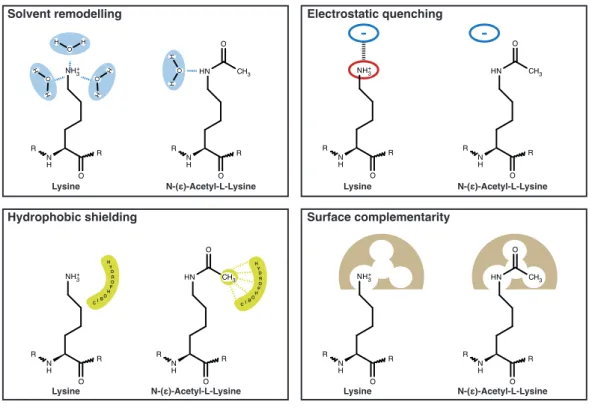
In theory but also from the known examples, it is clear that lysine-acetylation can impact the fate of a protein in several di↵erent ways, many of them being a direct consequence of the di↵erent chemical properties of acetyl-lysine compared to lysine (for details on immediate molecular e↵ects of lysine-acetylation see Fig.
1.7). For instance, lysine-acetylation of a particular protein can (positively or
negatively) influence its affinity to another macromolecule, co-factor or substrate.
Introduction 15 More indirect e↵ects of lysine-acetylation include the crosstalk with other post- translational modifications such as ubiquitination or the alteration of subcellular localization of a protein by changing the properties of a specific localization signal (Li et al., 2012). As will be discussed in the following second part of the intro- duction, post-translational lysine-acetylation has captured the attention of many researchers in recent years. This is mainly due to the identification of many novel acetylation sites throughout the proteome, which was possible through the huge technological advances in high-throughput mass spectrometry (MS). Given that the functional consequences of most of these newly discovered acetylation events is not known, many questions in the acetylation research field remain unanswered and are difficult to address in a high-throughput manner. The next sections will cover the most important proteins subjected to this modification and will give an overview about its regulation.
NH3+
N H R R
O
HN
N H R R
O CH3 O O
H H
O H H O
H
H HH O
NH3+
N H R R
O
HN
N H R R
O CH3
- -
ONH3+
N H R R
O
HN
N H R R
O CH3 O
H Y D R O P H BO CI
H Y D R O P H BO CI
NH3+
N H R R
O
HN
N H R R
O CH3 O
Solvent remodelling Electrostatic quenching
Hydrophobic shielding Surface complementarity
Lysine N-(ε)-Acetyl-L-Lysine Lysine N-(ε)-Acetyl-L-Lysine
Lysine N-(ε)-Acetyl-L-Lysine Lysine N-(ε)-Acetyl-L-Lysine
Figure 1.7: Molecular e↵ects of acetylation of lysine. Illustration of immediate molecular e↵ects of lysine-acetylation (taken from PhD thesis of S.
de Boor, 2015).
Introduction 16
1.2.1 The writers: Lysine-acetyltransferases (KATs)
Lysine-acetyltransferases (KATs) catalyze the transfer of the acetyl-group from acetyl-Coenzyme A (acetyl-CoA) to an N-(e)-amino group of a lysine side chain.
KAT activity was first demonstrated in an enzyme from Tetrahymena thermophila that efficiently acetylates lysines in histones and was thus termed histone acetyl- transferase type A (HAT A) (Brownell and Allis, 1995; Brownell et al., 1996).
HAT A turned out to be a close ortholog of Gcn5 (general control nonrepressed 5), which had previously been identified as a transcriptional regulator and thus the discovery of Gcn5-KAT activity immediately provided a link between histone acetylation and transcriptional regulation. To date, 21 proteins with KAT activity are known to be present in humans (EC 2.3.1.48), the substrates of which are not limited to histones (hence the term KAT instead of HAT) (Glozak et al., 2005; Lee et al., 2007). Based on structure and sequence characteristics, the di↵erent KATs have been assigned to one of five subfamilies: HAT1, Gcn5/pCAF, MYST (MOZ, Ybf2, Sas2, and Tip60), p300/CBP, or Rtt109. All KATs discovered until now, share a conserved fold comprising a three-stranded b-sheet and an a-helix, which is crucial for binding of acetyl-Coenzyme A and substrate coordination. How- ever, despite this similarity, the sequence homology between the KAT subfamilies is remarkably low and di↵erent catalytic mechanisms can be observed between KAT subfamilies (Friedmann and Marmorstein, 2013). Importantly, the N- and C-terminal extensions around the core region and also other protein domains (such as bromodomains; see below) are required for their substrate specificity and activ- ity (Polesskaya and Harel-Bellan, 2001). Moreover, KAT activity may be modu- lated by their intracellular localization, embedding into multi-protein-complexes or post-translational modifications, including auto-acetylation (Creaven et al., 1999;
Lee et al., 2007; Poveda et al., 2004; Santos-Rosa et al., 2003). Despite significant
advances in the identification of KAT targets it remains largely an open question
how KAT specificity is achieved. It seems however that due to the low sequence
conservation many more KAT genes remain to be discovered in the human genome
(Yuan and Marmorstein, 2013). This view was recently substantiated in a study by
Montgomery et al. (2014), in which several proteins with previously unrecognized
KAT activity were identified.
Introduction 17
1.2.2 The erasers: Lysine-deacetylases (KDACs)
Enzymes that catalyze the removal of an acetyl-group from a lysine residue have initially been discovered for histones and hence been termed histone deacetylases (HDACs). To account for their emerging role in deacetylation of non-histone pro- teins, they are now more generally referred to as lysine-deacetylases (KDACs). To date, two groups of KDACs have been identified, which use remarkably di↵erent reaction mechanisms and co-factors. One group comprises the so-called classical KDACs, which catalyze the removal of the acetyl-moiety via a Zn
+2-dependent hydrolysis reaction. Based on their sequence similarity to yeast homologs and their domain organization, these enzymes have initially been subdivided into dif- ferent classes (class I: KDAC1, -2, -3 and -8; class IIa: KDAC4, -5, -7 and -9;
class IIb: KDAC6 and -10; class IV: KDAC11) (Dokmanovic et al., 2007; Marks et al., 2001). The class I KDACs KDAC1 and KDAC2 are almost identical and can partially compensate for each other (Lagger et al., 2002). Mild phenotypes are observed for most lineage specific knockouts of either KDAC1 or KDAC2 but deletion of both has dramatic e↵ects, leading to blockade of G1-to-S-phase tran- sition (Yamaguchi et al., 2010). Nevertheless, full-body knockout of either one results in perinatal lethality in mice as does the full-body knockout of KDAC3 and -8 (Bhaskara et al., 2008; Haberland et al., 2009a; Montgomery et al., 2007).
The e↵ects class I KDAC ablation can in most cases be attributed to their role in
regulating transcription. However, they are also involved in splicing, DNA repair,
replication, mitosis and meiosis. They are part of large multi-protein complexes
such as CoREST and are themselves able to homo- or heterodimerize (Moser
et al., 2014, reviewed in). Class IIa KDACs are di↵erent from the other classical
KDACs in that they posses an N-terminal domain that is an important site for
post-translational phosphorylation and is required to establish interactions with
transcription factors. Phosphorylation leads to nuclear export and thus prevents
repression of their target genes (McKinsey et al., 2000). The expression of class IIa
KDACs are often tissue-specific, which is reflected in their tissue-specific functions
(Chang et al., 2006; Dequiedt et al., 2003; McKinsey et al., 2000). Relatively little
is known about the function of class IIb member KDAC10 and class IV member
KDAC11, although the latter has recently gained attraction for its role in sup-
pression of an anti-tumor immune response (Sahakian et al., 2015). In contrast,
the second class IIb member, KDAC6, is well described for its role in a-tubulin
deacetylation (see section 1.2.4). In general, KDAC6 appears to have the most
Introduction 18 non-histone targets among the classical KDACs (Bertos et al., 2004; Verdel et al., 2000; Zhang et al., 2003). Interestingly, KDAC6 possesses a zinc-finger domain in its C-terminus through which it can bind to ubiquitin and thus establishes a link between ubiquitination and acetylation (Boyault et al., 2006; Hook et al., 2002).
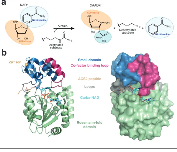
Class III KDACs are unrelated to the classical KDACs and are referred to as Sirtuins based on their homology to the founding member, the yeast Silent infor- mation regulator 2 (Sir2). Sir2 is critical for the silencing of mating type loci in yeast, which is mediated by deacetylation of critical lysines of histones H3 and H4 (Imai et al., 2000). For deacetylation, Sirtuins require nicotinamide adenine dinu- cleotide (NAD
+) as a co-factor, which is broken down to the to reaction products nicotinamide (NAM) and 2’- or 3-’O-acetyl-ADP-ribose (OAADPr) (Tanny and Moazed, 2001) (Fig. 1.8a). Interestingly, NAM, is an inhibitor of Sirtuins and, thus, NAM turnover by PNC1 (pyrazinamidase/nicotinamidase 1) modulates Sir- tuin activity in the cell (Avalos et al., 2005; Gallo et al., 2004). In addition, the second reaction product, OAADPr is an important signaling molecule and sub- strate for downstream enzymatic processes (Liou et al., 2005; Tong and Denu, 2010). Given their NAD
+-dependence, the activity of Sirtuins is intimately linked to the NADH/NAD
+ratio and thus the metabolic state of the cell. In fact, it is becoming increasingly clear that age-dependent decline of NAD
+-levels, which is caused by increased activity of PARPs (poly-ADP-ribose-proteins) in response to DNA damage (PARP1 also uses NAD
+as a substrate), leads to a concomitant decline of Sirtuin activity (Bai et al., 2011; Mohamed et al., 2014; Pillai et al., 2005).
Seven Sirtuins are present in human, of which SIRT3, -4 and -5 are predominantly found in mitochondria, SIRT6 and -7 are mostly nuclear and SIRT1 and -2 shuttle between the cytoplasm and the nucleus (Michishita et al., 2005). They all share a conserved domain structure consisting of a small domain that binds a Zn
2+ion and a second domain in a Rossmann-fold, which is characteristic for NAD
+- binding proteins. The two domains are connected by several loops, which form an extended cleft between both domains. Acetyl-lysine and NAD
+enter this cleft from opposing sides and meet in a tunnel where they contact the catalytic residues of the enzyme (Sanders et al., 2010) (Fig. 1.8b).
Sirtuins are in fact not restricted to acetyl-lysine as a substrate but can catalyze
the removal of other lysine-acylations such as crotonylation (SIRT1, -2, -3; Brooks
and Gu, 2011), malonylation and succinylation (SIRT5; Du et al., 2011; Peng et al.,
Introduction 19
a
b
Rossmann-fold domain ACS2 peptide
Carba-NAD Loops Small domain Zn2+ ion
Co-factor binding loop