N-terminal domain of the centromeric histone variant Cse4 in Saccharomyces cerevisiae
Dissertation
zur Erlangung des akademischen Grades doctor rerum naturalium
(Dr. rer. nat.)
im Fach Biologie
eingereicht an der
Lebenswissenschaftlichen Fakultät der Humboldt-Universität zu Berlin
von
Juliane Pöpsel
Präsident der Humboldt-Universität zu Berlin Prof. Dr. Jan-Hendrik Olbertz
Dekan der Lebenswissenschaftlichen Fakultät Prof. Dr. Richard Lucius
Gutachter/innen:
1. Prof. Dr. Ann Ehrenhofer-Murray 2. Prof. Dr. Peter Bayer 3. Prof. Dr. Harald Saumweber
Tag der mündlichen Prüfung:
07.07.2015
Abstract
The centromeric regions of eukaryotic chromosomes are essential for proper chromosome segregation during mitosis. The nucleosomal composition in these regions differs from canonical nucleosomes in that histone H3 is replaced by the conserved centromeric variant CENP-A (Cse4 in Saccharomyces cerevisiae), which is the most prominent epigenetic feature of centromeres. The CENP-A N-terminal domain is larger than canonical histone tails as the centromeric histone variants of higher eukaryotes. A functionally critical part of this flexible, positively charged histone tail is the essential N-terminal domain (END). Interestingly, arginine 37 (R37) residing in this region was previously found to be methylated and to act as an epigenetic regulator of kinetochore recruitment and chromosome segregation. Lysine 49 (K49) was defined as an acetylation site, but this modification remained to be characterized in detail.
In this study, it was shown that Cse4K49 acetylation depends on the histone acetyltransferase (HAT) Gcn5, and that in this context the enzyme acts within the SAGA/SLIK complex, whereas an involvement of the smaller ADA complex was not observed. Furthermore, a cell-cycle dependence of the K49 acetylation was found, with acetylation levels increasing in early S-phase and a decreasing in late S-phase, while R37 methylation persisted throughout the entire S-phase. Furthermore, direct protein-protein interactions were investigated via peptide pull-down experiments of Cse4 with kinetochore components. Ctf19 was found to bind both acetylated and unacetylated Cse4K49 with a preference for unacetylated Cse4. The analysis of secondary structural elements of the Cse4 N-terminus via CD-spectroscopy showed that the N-terminus is an intrinsically unstructured domain, which may fold upon binding to interaction partners like Ctf19.
Taken together, the findings presented here show that the transcriptional co-activator complex SAGA functions at centromeric regions in S. cerevisiae, and that the Cse4K49 acetylation (Cse4K49ac) has an influence on the recruitment of the kinetochore subunit Ctf19. This suggests an epigenetic regulatory mechanism in chromosome segregation involving a specific acetylation on the N-terminus of Cse4.
Zusammenfassung
Die zentromerischen Regionen eukaryotischer Chromosomen sind notwendig für die Segregation der Schwesternchromatiden während der mitotischen und der meiotischen Zellteilung. In den Nukleosomen in diesen Regionen ist das kanonische Histon H3 durch die in allen Eukaryoten hochkonservierte zentromerische Variante CENP-A (Cse4 in Saccharomyces cerevisiae) ersetzt. Indem CENP-A die Assemblierung einzelner Untereinheiten des Kinetochors vermittelt, markiert es die zentromerische Chromosomenregion und ist damit essentiell für die korrekte Segregation der Chromosomen während der Zellteilung. Kürzlich wurde gezeigt, dass Cse4 posttranslational modifiziert wird. Von Bedeutung für diese Arbeit sind die in einer essentiellen Domäne des N-terminus liegende Methylierung von Arginin 37 (R37) und die Acetylierung von Lysin 49 (K49), die durch phänotypische Suppression eine genetische Interaktion und eine antagonistische Funktion bei der epigenetischen Regulation der Assemblierung der Kinetochoruntereinheiten zeigen.
In dieser Arbeit wurde gezeigt, dass die Acetylierung an Cse4K49 von der Histonacetyltransferase (HAT) Gcn5 abhängt, und dass dieses Enzym hierbei Komponenten des SAGA-Komplexes, aber nicht des ADA-Komplexes benötigt.
Außerdem konnte in dieser Arbeit nachgewiesen werden, dass die Acetylierung von K49 in der frühen S-Phase zunimmt und am Ende der S-Phase abnimmt. Die Methylierung von R37 hingegen ist während der gesamten S-Phase erhöht. Es konnte weiterhin eine biochemische Interaktion der N-terminalen Domäne von Cse4 mit der COMA-Untereinheit Ctf19, der zentralen Region des Kinetochors, nachgewiesen werden, möglicherweise mit einer Präferenz zu einer nicht acetylierten Form von Cse4K49. Schließlich wurde per CD-Spektroskopie gefunden, dass die N- terminale Domäne von Cse4 intrinsisch unstrukturiert ist.
Zusammenfassend konnte in dieser Arbeit gezeigt werden, dass der SAGA Komplex eine Funktion an der zentromerischen Region in S. cerevisiae aufweist. Des Weiteren ist die Acetylierung an Cse4K49 (Cse4K49ac) in der Rekrutierung der Kinetochoruntereinheit Ctf19 involviert, und legt damit einen neuen epigenetischen Regulationsmechanismus während der Chromosomensegregation nahe.
Table of Contents
Abstract ... II Zusammenfassung ... III Table of Contents ... IV List of Tables ... VII List of Figures ... VIII Abbreviations ... X
1. Introduction ... 1
1.1. Eukaryotic chromatin organization ... 1
1.2. Classes of chromatin ... 6
1.3. Centromeric chromatin structure ... 8
1.4. Nucleosome structure at the centromeres ... 11
1.5. Kinetochores: Structure and function in chromosome segregation ... 12
1.5.1. Inner kinetochore components ... 13
1.5.2. Linker kinetochore components ... 14
1.5.3. Outer kinetochore components ... 15
1.6. Posttranslational modification of histones ... 16
1.6.1. Acetylation of histones ... 17
1.6.2. Epigenetic regulation of chromosome segregation ... 18
1.7. The centromeric histone variant CENP-ACse4 ... 19
1.7.1. Posttranslational modifications on Cse4 ... 20
1.8. Histone-modifying enzymes ... 21
1.8.1. Histone acetyltransferases (HATs) ... 22
1.8.2. Gcn5-related N-acetyltransferases (GNATs) ... 23
1.9. The Gcn5 containing complexes SAGA/ADA ... 24
1.10. Aim of this work ... 27
2. Material and Methods ... 29
2.1. Escherichia coli strains ... 29
2.2. Escherichia coli media and growth conditions ... 29
2.3. Saccharomyces cerevisiae strains ... 29
2.4. Saccharomyces cerevisiae media and growth conditions………...35
2.5. Construction of Saccharomyces cerevisiae strains ... 35
2.5.1. Crossing, sporulation and dissection of asci ... 35
2.5.2. DNA techniques in Saccharomyces cerevisiae ... 36
2.5.3. Preparation of Saccharomyces cerevisiae lysates ... 36
2.6. Molecular cloning ... 37
2.7. Purification of recombinant proteins ... 38
2.7.1. Recombinant protein expression ... 38
2.7.2. Preparation of E. coli cell lysates………...………39
2.7.3. Affinity purification of GST- and 6x His-tagged proteins ... 39
2.7.4. Gel Filtration ... 40
2.8. Immunoprecipitation ... 40
2.8.1. Synchronization of Saccharomyces cerevisiae cells ... 41
2.8.2. FACS analysis ... 42
2.9. Protein- protein interaction assays ... 42
2.9.1. Peptide pull-down assay ... 42
2.9.2. Pull-down assay ... 43
2.9.3. Yeast two-hybrid assay ... 43
2.10. SDS-PAGE and immunoblotting ... 43
2.11. Dot-blot analysis ... 45
2.12. Affinity purification of polyclonal antibodies ... 45
2.12.1. Peptide-coupling to sulfolink beads ... 45
2.12.2. Peptide-based affinity purification of polyclonal antibodies ... 46
2.13. Analytical purification of Cse4 ... 47
2.14. CD spectroscopy ... 48
3. Results ... 49
3.1. Identification of the histone acetyltransferase (HAT) responsible for the acetylation on K49 of CENP-ACse4 ... 49
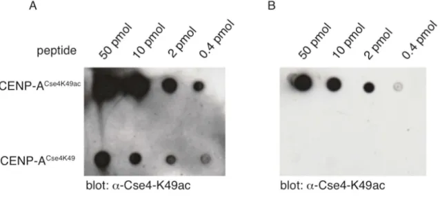
3.1.1. Purification of polyclonal antibodies against acetylated Cse4K49 ... 50
3.1.2. Cse4K49ac depends on the histone acetyltransferase Gcn5 ... 51
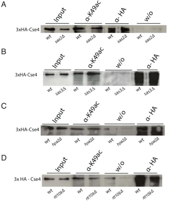
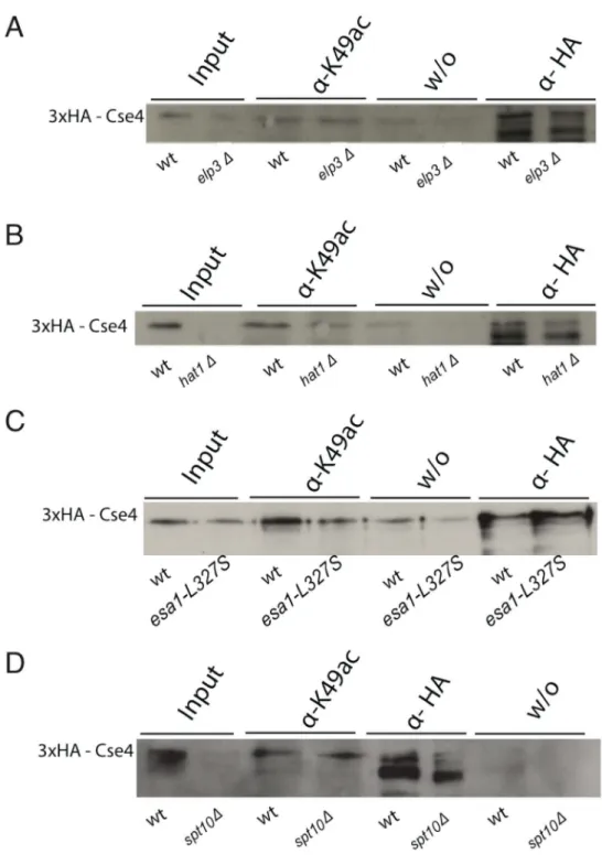
3.1.3. Deletion of other HAT genes did not decrease the acetylation on Cse4K49... 52
3.1.4. Detection of the Cse4K49 acetylation with antibodies in yeast ... 55
3.1.5. Definition of the HAT complex, in which Gcn5 acts in the K49 acetylation ... 57
3.1.6. Genetic analysis of SAGA mutants combined with cbf1∆ and cse4R37A ... 60
3.1.7. Genetic analysis of HAT mutants combined with cbf1∆ cse4R37A ... 61
3.1.8. Effect of SAS2 overexpression on the acetylation status of Cse4K49 ... 64
3.2. Cell-cycle dependence of posttranslational modifications on Cse4 ... 65
3.2.1. Cell-cycle study of the suppressor triple mutant cbf1∆ cse4R37AK49R ... 65
3.2.2. Study of the cell-cycle dependence of Cse4K49 acetylation ... 66
3.3. Investigation of R37 methylation and K49 acetylation on Cse4 during the cell cycle ... 67
3.3.1. Immunoprecipitation of methylated Cse4R37 ... 68
3.3.2. Fluctuation of K49 acetylation and R37 methylation on Cse4 during S-phase .. 68
3.4. Protein-protein interaction studies of Cse4 with COMA kinetochore components ... 70
3.4.1. Purification of the N-terminal domain of Cse4 ... 72
3.4.2. Affinity purification and elution of 6x His-Ctf1935-363 ... 73
3.4.3. Interaction of 6x His-Ctf1935-363 and GST-Cse414-134 ... 74
3.4.4. Peptide pull-down of 6xHis-Ctf1935-363 ... 75
3.4.5. Peptide pull-down of Okp1 and Okp1R164C ... 77
3.5. CD-spectroscopy of GST-Cse414-134 ... 78
3.6. Interactions of Cse4 N-termini with each other ... 81
4. Discussion ... 83
4.1. The acetylation of Cse4K49 depends on Gcn5 ... 84
4.1.1. Which complex containing Gcn5 participates in Cse4K49 acetylation? ... 86
4.2. Phenotypical characterization of HAT and SAGA mutants ... 88
4.2.1. Deletion of HAT genes did not suppress the phenotype of cbf1Δ cse4R37A .... 88
4.2.2. Deletion of GCN5, ADA2 and SPT7 did not suppress the phenotype of cbf1Δ cse4R37A ... 89
4.3. The dynamics of Cse4K49 acetylation levels in the cell cycle ... 90
4.3.1. Biochemical interactions of Ctf19, Okp1/Okp1R164C and the N-terminus of Cse4 ... 93
4.4. A crosstalk between the PTMs on Cse4 ... 94
4.4.1. Secondary structure determinations of the N-terminal domain of Cse4 ... 95
4.5. Conclusions ... 96
5. References ... 97
6. Appendix ... i
6.1 Quantification of immunoprecipitation experiments ... i
6.2 FACS profiles of cell-cycle arrested yeast cells ... v
List of Tables
Table 1: E. coli strains used in this study ... 29
Table 2: Saccharomyces cerevisiae strains used in this study ... 30
Table 3: Plasmids used in this study ... 32
Table 4: Oligonucleotides used in this study ... 33
Table 5: Antibodies used in this study ... 44
Table 6: Peptides used in this study ... 46
List of Figures
Figure 1: High-resolution crystal structure of the nucleosomal core particle ... 2
Figure 2: Histone fold architecture ... 3
Figure 3: Chromatin organization in eukaryotes. ... 5
Figure 4: Centromeric regions of eukaryotic organisms. ... 10
Figure 5: Centromeric nucleosome structures in the budding yeast ... 12
Figure 6: Model of the Saccharomyces cerevisiae kinetochore. ... 16
Figure 7: Sequence alignment of CENP-A species and topological view of CENP-A from budding yeast ... 21
Figure 8: Modified cloning site of the pET41b+ vector. ... 38
Figure 9: Peptide-based affinity purification of an α-Cse4K49ac specific antibody ... 50
Figure 10: Identification of Gcn5 as the histone acetyltransferase that acetylates Cse4K49 ... 51
Figure 11: No influence of other histone acetyltransferases on Cse4K49 acetylation. ... 53
Figure 12: No influence of other histone acetyltransferases on Cse4K49 acetylation. ... 54
Figure 13: Acetylation on Cse4K49 was not detected in histone extracts ... 56
Figure 14: Decreased acetylation of Cse4K49 in SAGA mutants. ... 59
Figure 15: Phenotypical characterization of spt7 in combination with cbf1 or the point mutation cse4R37A in the essential N-terminal domain. ... 60
Figure 16: Genetic analysis of HAT mutants in combination with cbf1 and the point mutation in the essential N-terminal domain of Cse4 (cse4R37A). ... 62
Figure 17: Genetic analysis of HAT mutants in combination with cbf1 and the point mutation in the essential N-terminal domain of Cse4 (cse4R37A). ... 63
Figure 18: Influence of Sas2 overexpression on Cse4. ... 64
Figure 19: cse4K49R suppresses the G2/M-phase delay in cbf1 cse4R37A ... 66
Figure 20: Cell-cycle dependence of Cse4K49 acetylation... 67
Figure 21: Immunoprecipitation of methylated Cse4R37 ... 68
Figure 22: Acetylation of Cse4K49 was decreased whereas methylation of Cse4R37 remained at a constant level at the end of S-phase ... 69
Figure 23: Quantification of alterations of the posttranslational modifications Cse4K49 acetylation and Cse4R37 methylation in S-phase progression ... 70
Figure 24: Affinity purification of the fusion protein GST-Cse414-134. ... 72
Figure 25: Expression and affinity purification of 6xHis-Ctf1935-363. ... 73
Figure 26: Elution steps of 6xHis-Ctf1935-363. ... 74
Figure 27: Interaction of 6xHis-Ctf1935-363 and GST-Cse414-134 in pull-down assay ... 75
Figure 28: Ctf19 binding to Cse4K49ac and Cse4K49 peptides ... 76
Figure 29: Peptide pull-down of Okp1 and Okp1R164C. ... 78
Figure 30: Size exclusion chromatography of Cse414-134. ... 79
Figure 31: Far-UV spectra of Cse414-134 and GST ... 81
Figure 32: Yeast-two-hybrid assay of N-terminal CENP-ACse4 ... 82
Figure 33: Identification of Gcn5 as the histone acetyltransferase that acetylates Cse4K49 ... i
Figure 35: No influence of other histone acetyltransferases on Cse4K49ac. ... ii
Figure 36: Decreased acetylation of Cse4K49 in SAGA mutants. ... iii
Figure 36: Immunoprecipitation of methylated Cse4R37 ... iv
Figure 37: Cell-cycle dependence of Cse4K49 acetylation... iv
Figure 38: Yeast cells in S-phase progression ... v
Figure 39: FACS analysis of arrested cells. ... vi
Abbreviations
5-FOA 5-fluoro-orotic acid
aa amino acid
ac acetylation (eg. Cse4 K49 = Cse4 K49ac)
ACN acetonitrile
ADA adaptor
AT-rich adenine thymine-rich
ATP adenosine triphosphate
bp base pair
CATD centromere associated targeting domain Cbf1 centromere binding factor 1
CDEI/II/III centromere DNA element I/II/III CenH3 centromeric histone H3
CENP-A centromere protein A Co-IP co-immunoprecipitation
COMA Ctf19/Okp1/Mcm21/Ame1
Cse4 chromosome segregation 4
DNA deoxyribonucleic acid
DTT di-thiothreitol
END essential N-terminal domain in Cse4 FACS fluorescence associated cell sorting
FPLC Fast Performance Liquid Chromatography Gcn5 general control nonderepressible 5
HAT histone acetyltransferase HDAC histone deacetylase
HJURP Holliday Junction Recognition Protein HML homothallic mating left
HMR homothallic mating right HP1 Heterochromatin Protein 1
HPLC High Performance Liquid Chromatography
HU hydroxyurea
IP immunoprecipitation
LB Luria-Bertani
Mbp mega base pair
me methylation (eg. Cse4R37 = Cse4R37me)
me3 trimethylation
The nomenclature of yeast (Saccharomyces cerevisiae) proteins was taken from the Saccharomyces cerevisiae genome database (SGD). Amino acids were given in the single-letter code, e.g. K = lysine, R = arginine.
MNase micrococcal nuclease
MS mass spectrometry
MTF methyltransferase
nm nano meter
NP-40 Nonidet P-40
OD optical densitiy
ORF open reading frame
otr outer repeats
PBS phosphate buffer saline PCR polymerase chain reaction
pmol pico mole
PTM posttranslational modification PVDF polyvinylidene fluoride
RNA ribonucleic acid
rpm rounds per minute
RT room temperature
SAC spindle assembly checkpoint SAGA Spt–Ada–Gcn5 acetyltransferase
SAM S-adenosyl-L-methionine
SDS-PAGE sodium dodecyl sulfate polyacrylamide gel electrophoresis
SLIK SAGA-like
TE Tris-HCL/EDTA buffer
TFA trifluoroacetic acid
ts temperature sensitive
wt wild-type
YM yeast minimal medium
YPD yeast peptone medium
1. Introduction
1.1. Eukaryotic chromatin organization
The DNA of every cell in a eukaryotic organism comprises all the genetic information it requires to perform essential processes. It is organized in the form of genes, which are defined as the coding regions for proteins and the associated regulatory sequences, such as the promoter regions. It is compacted with histones and non- histone proteins to form chromosomes. The entirety of the DNA of one organism is referred to as the genome, and the genome of human somatic cells, the diploid chromosome set, consists of 46 chromosomes, two of which are the sex chromosomes. The DNA is packaged tightly in the nucleus, if one considers that all of the DNA molecules in one human cell consist of 3.27 Gbp, i.e. 3.27 billion base pairs, but fits into a nucleus with 1-5 μm diameter. Unexpectedly, this enormous amount of DNA only comprises about 30,000 – 40,000 genes, which code for about 100,000 proteins. This implies that most of the human genome is composed of non- coding regions (Lander et al., 2001).
Among eukaryotes, vertebrates and higher plants have the largest genomes and numbers of chromosomes per cell. Interestingly, the human genome contains only twice as many protein coding genes than that of the fruit fly Drosophila melanogaster, suggesting that more than just the DNA sequence is needed for generating the variety of information that results in a complex eukaryotic organism (reviewed in Jenuwein and Allis, 2001). The DNA is associated in a nucleoprotein complex with non-histone proteins and histones, referred to as chromatin, which provides the compaction of the DNA. DNA packaging is not static but highly dynamic and thus contributes to gene regulation by regulating the accessibility of the genetic information. This additional level of genetic information and regulation, termed
“epigenetic”, is defined as heritable changes in phenotype that are not due to changes in the DNA sequence itself.
In comparison to higher eukaryotes, the budding yeast Saccharomyces cerevisiae possesses a relatively small genome of only 12.1 Mbp of DNA per cell, carrying about 6.300 genes. On its 16 chromosomes, most of the DNA codes for proteins and
therefore, a comparably small amount of the DNA of this unicellular organism comprises non-coding regions. This simplicity makes the yeast a tractable model system for genetic and biochemical studies. The budding yeast genome was the first eukaryotic genome to be completely sequenced in 1996 (Goffeau et al., 1996).
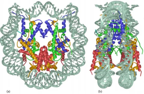
Each cell has to accomplish a DNA condensation process that demands high accuracy and fidelity. The basic level of condensation is conferred by the canonical nucleosome, a structure of 145-147 bp of the DNA double helix wrapped around one core particle 1.7 times in a left-handed manner. The core particle is composed of histone proteins, with two copies each of H2A, H2B, H3 (in human H3.1) and H4. The structure of this nucleosome core particle was first solved at 2.8 Å resolution by Luger et al., 1997 (Figure 1).
Figure 1: High-resolution crystal structure of the nucleosomal core particle
Core particle structure viewed in two orientations. (a) View down the superhelical axis; (b) 90° rotation of the y-axis to highlight the flat, disc-like shape of the nucleosome. H3 and H4 make up two heterodimers and are shown in blue and green, respectively. H2A and H2B, which also form two heterodimers, are shown in yellow and red, respectively. The DNA double helix is depicted in grey.
Taken from (Luger, 2001).
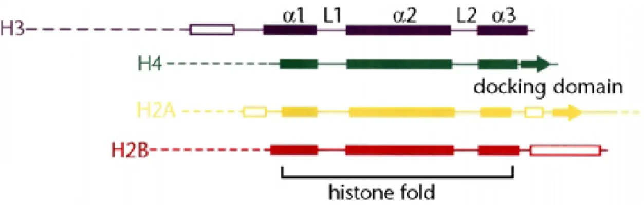
The topology of the nucleosome core particle reflects its function. For example, the histone fold domains (HFDs) allow the association of the eight histones and a left- handed, approximately two fold winding of the DNA around the histone hetero- octamer to constitute an overall globular structure. HFDs are characterized by one
central α-helix flanked by two shorter α-helices. Additionally, there are neighboring β- strands N-terminal to the HFD in H3, at the C-terminal region of H2B and on both sides flanking the globular domain in H2A, shown in Figure 2 (Arents and Moudrianakis, 1995; Luger et al., 1997).
The flexible, less structured histone tails mediate physical interactions with associated proteins and DNA. They can be posttranslationally modified in many ways, which drives the dynamics of chromatin condensation, active transcription, gene silencing and chromosome segregation. Histones can be exchanged, and the newly synthesized and reassembled histones may carry modifications that are distinct from the displaced histone. In other cases, histone variants are recruited to chromatin to replace canonical histones. The concerted placement and the combination of canonical histones and histone variants as well as their covalent modification patterns reflect the epigenetic chromatin status of a cell (reviewed in Jenuwein and Allis, 2001; Kouzarides, 2007).
Figure 2: Histone fold architecture
Domain organization of the four canonical histones H3, H4, H2A and H2B. α-helices of the histone fold domains are shown as solid boxes, loops between the helical regions are termed L1 and L2; α-helices and β-strands of the histone fold extensions are shown as open boxes and tails of the histones are shown as dotted lines. Adapted from Luger, 2001.
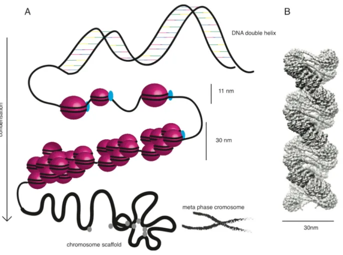
The first level of DNA compaction are the nucleosomes, which are arranged in a beads-like substructure and are 11 nm in width, shown in Figure 3A (Wang et al., 2008). With the assistance of the highly basic linker histone H1, the next higher order of chromatin structure, the 30 nm fiber, is established. Histone H1, which exhibits no structural homology with core histones, associates at the dyad axis of the nucleosome core engaging a short linker DNA sequence (Allan et al., 1980; Bednar et al., 1998; Richmond and Davey, 2003; Khorasanizadeh, 2004). Histone H1
proteins are conserved among eukaryotes structurally, but not in their amino acid sequence (Maeder and Bohm, 1991). The yeast H1, termed Hho1, and the human H1 are both non-essential proteins, indicating that besides the association of H1 to the nucleosomes, additional processes must be crucial for chromatin condensation.
For example, physical interactions of the N-terminal domains of the four core histones could drive the condensation process, while H1 stabilizes higher order chromatin structures (Shen, 1995). Whereas H1 of most species assembles approximately in a 1:1 stoichiometry with nucleosomes, Hho1 is found in yeast chromatin at an average frequency of every 37th nucleosome only and its distribution is restricted to distinct genomic (Patterton et al., 1998; Freidkin and Katcoff, 2001).
This observation supports the notion that Hho1 association is auxiliary, but not essential for higher chromatin compaction, or that Hho1 serves a specific purpose yet to be characterized. Experimental data showed that 30 nm chromatin fibers could be assembled in vitro in the presence and in the absence of this linker histone H1, but the reconstituted fibers differed in their quaternary structures (Schalch et al., 2005;
Huynh et al., 2005; Pepenella et al., 2014).
The structure of the next higher level of compaction, the 30 nm fiber, has long been a matter of debate in the chromatin field, and different models have been proposed.
The two prevailing models are the one-start stack solenoid model, and the two-start stack zigzag model, which comprise two subclasses of chromatin structures (Finch and Klug, 1976).
The fiber of the first group is composed of a one-start helical stack of nucleosomes, is shaped like a helical ribbon, and the compaction of this model is higher than in the second group, the cross-linker model (Felsenfeld and McGhee, 1986). The latter group is organized as two-start helix. During the reconstitution process, this structure was generated using long stretches of linker DNA exceeding 217 base pairs, which span the fiber crosswise (Robinson and Rhodes, 2006). With respect to the latter model, the structure prediction depends considerably on the DNA linker lengths that lead to different structures of the 30 nm fiber (Bednar et al., 1998; Wu et al., 2007).
A recently published cryogenic electron microscopy (cryo-EM) structure demonstrated repeating tetranucleosomal units, which coil in a left-handed twist.
Here, the nucleosomes zigzag back and forth, with the linker Histone H1 and straight
linker DNA between the core nucleosomes. The whole structure is essentially compatible with the two-start zigzag model (Song et al., 2014; Figure 3 B).
Figure 3: Chromatin organization in eukaryotes.
A: The figure schematically illustrates the successive chromatin condensation levels in eukaryotes.
The basis is build by DNA (black lines) and histone octamers (pink bubbles), which assemble into nucleosomes and make up the 11 nm fiber together with histone H1 (light blue). This structure is further organized into the 30 nm fiber. Subsequently and with the help of non-histone proteins, which form the chromosome scaffold, the condensation to higher organized chromatin is achieved. The metaphase chromosome (Modified from http://dellairelab.medicine.dal.ca/research.html) marks the final state of condensation. B: 30nm chromatin fiber model, cryo-EM reconstruction of in vitro reconstituted chromatin. Taken from (Song et al., 2014).
Higher order folding occurs in the presence of the two major chromosome scaffold components, topoisomerase II and condensin, and is defined as the further compaction of 30 nm fiber loops which associate on the scaffold. However, whether or not the 30 nm fiber structure persists upon higher order chromatin folding in vivo has been a matter of controversy (Maeshima et al., 2010; Nishino et al., 2012).
Recent studies have provided new insights into the mode of condensation in mitosis.
Condensin shows ATPase activity and binds directly to DNA molecules. It actively
supercoils DNA in vitro and is required for establishing the mitotic chromosome structure and for the process of chromosome segregation in vivo (Kimura et al., 1999; Hagstrom et al., 2002). Our understanding of higher order chromatin compaction is still lacking detail as compared to the first levels of condensation. The loops of the chromosome scaffold appear to be regularly folded. The localization of genomic loci and the distances between those loci are highly variable in the nucleus and also vary among organisms. The folding processes of interphase and mitotic chromosomes are being investigated in the physical field of complex polymer systems. Along with optical as well as electron microscopic techniques, these approaches promise substantial insights into the mechanisms of chromosome packaging in the future (Berger et al., 2008; Trask et al., 1993; Fudenberg and Mirny, 2012).
1.2. Classes of chromatin
Eukaryotic chromatin can acquire various degrees of compaction, which are generally distinguished into two forms: the closed, highly compacted heterochromatin, and the euchromatic regions (Heitz, 1928; Brown, 1966). The latter can be defined as open, decondensed chromatin marked by a high rate of gene transcription. One mark of active transcription in human cells is the occurrence of the histone variant H3.3, which replaces the replicative H3.1 variant at these sites (Ahmad and Henikoff, 2002). The distribution of functional, protein-coding genes is unequal in favor of the euchromatic regions of the chromosome, shown by the fact that 2.9 Gbp (more than 90% of the whole genome sequence length) of chromatin appears to be euchromatic (Lander, E.S., International Human Genome Sequencing Consortium, 2001).
Two forms of heterochromatin can be distinguished. One is referred to as constitutive, which means that it serves structural purposes important for genome integrity, and it is mostly found at the telomeric or pericentromeric regions of a chromosome (Trojer and Reinberg, 2007; Grewal and Jia, 2007). The second form, termed facultative heterochromatin, is marked by direct gene silencing and the possible switch between active and inactive chromatin. This reversible process is strongly regulated by several chromatin remodeling complexes. Their activity is, in
turn, tightly regulated by the absence or presence of posttranslational modifications (PTMs) of histones mediated by chromatin modifying enzymes, and also by the occurrence of histone variants. For example, it has been shown that H3K4 methylation characterizes actively transcribed gene regions, whereas H3K9 methylation marks heterochromatin (Barski et al., 2007). Upon the latter modification, which functions as a tether, heterochromatin protein 1 (HP1) is recruited as an adaptor for various effector proteins, such as histone modifiers, cohesin and transcription silencing factors (Grewal and Jia, 2007; Trojer and Reinberg, 2007).
A well-known example for the extensively regulated transcriptional silencing in facultative heterochromatin is the inactivation of the mammalian female X- chromosome, a process known as dosage compensation. Here, in each cell of the female organism, one of the two X-chromosomes is silenced in a cascade of heterochromatin establishment and maintenance processes. It is coated by the noncoding RNA Xist, which induces hypoacetylation on histones and, among others, H3K27 methylation (Chow et al., 2005). The subsequent condensation process is necessary to avoid a double X-chromosomal transcription level in females compared to males. Additionally, the inactive X-chromosome is marked by a histone variant;
macro-H2A replaces H2A and participates in the maintenance of X inactivation (Costanzi and Pehrson, 1998; Avner and Heard, 2001).
Histone deacetylation of H4K16 earmarks silent chromatin and is mediated by a histone deacetylase (HDAC) complex, which contains the silent information regulator 2, Sir2, in yeast or its homologue SIRT3 in humans (Gottschling et al., 1990; Imai et al., 2000; Pikaart et al., 1998). In facultative heterochromatic structures, H4K16 acetylation has been shown to counteract hypercondensation, and is mediated in S.
cerevisiae by a specific histone acetyl transferase, Sas2, which acts in the SAS-I complex (Corona et al., 2002; Sutton et al., 2003; Meijsing and Ehrenhofer-Murray, 2001). The interaction between the deacetylated, positively charged H4 tail and the acidic patch of the H2A-H2B dimer of the neighboring nucleosome contributes to the condensation process. These electrostatic interactions are disturbed upon the acetylation of H4K16 (Shogren-Knaak et al., 2006; Gordon et al., 2005; Robinson et al., 2008). This is in agreement with the findings that Saccharomyces cerevisiae cells lacking the H4K16 HAT Sas2 show increased silencing at subtelomeric regions (Suka et al., 2002).
1.3. Centromeric chromatin structure
The composition and function of centromeric chromatin differs from euchromatic or heterochromatic chromatin regions in diverse ways. Due to the fact that low transcription levels are present within these regions, it can be defined as a particular form of heterochromatin. It is demarcated as the essential locus at which kinetochores assemble to build the platform for spindle microtubule attachment to ultimately segregate the highly condensed sister chromatids during mitosis. In humans, centromeric chromatin is embedded in regions of pericentromeric heterochromatin and exhibits a distinct pattern of epigenetic marks in the form of histone variants and posttranslational modifications (Sullivan and Karpen, 2004;
Allshire and Karpen, 2008).
The histone variant CENP-A (Centromere Protein-A) replaces histone H3 (H3.1 in human) on centromeres and thereby epigenetically marks centromeres in all eukaryotic organisms (Yoda et al., 2000; Palmer et al., 1987; Earnshaw and Rothfield, 1985). It is responsible for the localization of almost all centromere components, basically the kinetochore subunits (Foltz et al., 2006; Liu et al., 2006).
In contrast to the assembly of human H3.1 and H3.3 into canonical nucleosomes, which involves the participation of CAF-1 (Chromatin Assembly Factor-1) and the Histone Regulator A, HIRA, the specific histone chaperone HJURP (Holliday junction recognition protein) is necessary for incorporation of CENP-A into centromeric nucleosomes in early G1 phase (Verreault et al., 1996; Foltz et al., 2009; Bodor et al., 2013; Pchelintsev et al., 2013). Despite only minor homology in their amino acid sequence, the mammalian chaperone HJURP and its fungal orthologue Scm3 (Suppressor of chromosomal missegregation) display corresponding chaperone-like roles for centromere specific nucleosome incorporation of newly synthesized CENP- A histones (Dunleavy et al., 2009; Mizuguchi et al., 2007; Stoler et al., 2007; Williams et al., 2009).
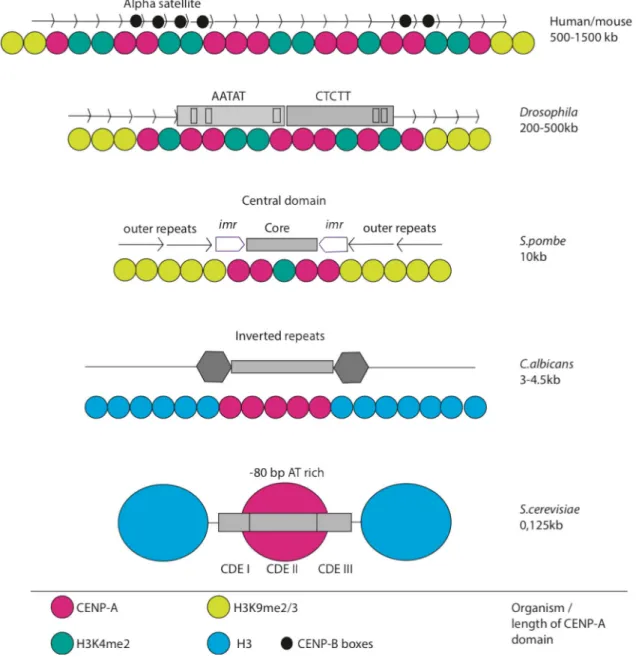
In the large centromeric regions of mammalian and Drosophila cells, the replacement of the canonical histone H3 occurs partially, whereas in Candida albicans and in the point centromeres of budding yeast, nucleosomes exclusively carrying CENP-A are found as shown in Figure 4 (Blower et al., 2002). Distinct posttranslational modification patterns mark the centromeric sites in chromatin. In contrast to
heterochromatic regions, where H3K9 di- or trimethylation occurs, this PTM is lacking on centromeric heterochromatin, whereas H3K4 dimethylation, a mark of euchromatin, is also found in human and Drosophila cells as a feature of the centromeres (Figure 4; Blower et al., 2002; Santos-Rosa et al., 2002). An example of a mitotic feature that marks mitosis but does not mark centromeres is Aurora B mediated H3S10 phosphorylation. This PTM has been shown to correlate with mitosis in all eukaryotes. It recruits the histone deacetylase Hst2, which in turn deacetylates H4K16 and permits chromatin hypercondensation during chromosome segregation in mitosis (Wilkins et al., 2014).
The centromeric regions of eukaryotes differ in size, reaching from single nucleosomes in the point centromeres of budding yeasts to large regional centromeres in higher eukaryotes (Henikoff et al., 2001). In the fission yeast S.
pombe, the centromeric regions encompass 40 - 110 kb. Heterochromatic regions with methylated H3K9 containing nucleosomes, termed outer repeats (otr), flank the innermost repeats (imr) and the central, non-repetitive core region (cnt). Several copies of the centromeric histone H3 variant CENP-ACnp1 are placed at the core domain and, together with the imr regions, they are crucial for kinetochore assembly (Figure 4, mid panel; Pidoux and Allshire, 2004; Allshire and Karpen, 2008).
The regional centromeres found in humans are particularly complex. The DNA sequence possesses homogeneous arrays of chromosome-specific α-satellite repeats, which are 171 bp in length and are arranged in tandem over a genomic region of about 1500 kb (Figure 4 upper panel). The DNA binding protein CENP-B binds specifically to a 17 bp motif, termed CENP-B box and promotes a higher order centromere chromatin packaging. These structures form the basis for kinetochore and microtubule attachment during mitosis and meiosis (Figure 4 upper panel; Choo, 2001; Schueler and Sullivan, 2006; Allshire and Karpen, 2008).
A special centromere was found in the budding yeast S. cerevisiae, which possesses a DNA sequence-dependent point centromere, with a single microtubule attaching to one kinetochore. This strict sequence dependency is unusual and, to our knowledge, occurs only in these unicellular organisms. There are 125 bp of DNA wrapped around the centromeric nucleosome-like structure, and the sequence is divided into three consensus elements, CDEI, II and III (centromere DNA element). CDEI consists of the 8 bp motif 5`- RTCACRTG -3` and has been shown to connect the centromeric
nucleosome to the kinetochore via binding of the inner kinetochore component Cbf1 (Cai and Davis, 1989). CDEII is a 78-86 bp AT-rich element, and deletion or shortening of this element results in strong defects in the segregation of sister chromatids. The 25 bp CDEIII element binds directly to the inner kinetochore complex CBF3, a four subunit, sequence-specific DNA binding protein complex, which is crucial for centromere activity, shown in Figure 4, lower panel (Baker et al., 1989; Fitzgerald-Hayes, 1987).
Figure 4: Centromeric regions of eukaryotic organisms.
The centromere diversity among different model organisms is depicted. The yeast Saccharomyces cerevisiae contains one CENP-A nucleosome, a `point centromere`, Candida albicans and Schizosaccharomyces pombe have small regional centromeres. Higher eukaryotes such as insects like Drosophila melanogaster up to mammals like Homo sapiens show huge and complex centromeric regions. Modified from (Allshire and Karpen, 2008).
1.4. Nucleosome structure at the centromeres
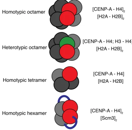
The physical arrangement of centromeric nucleosomes in general has been a subject of controversies for a long time. Several models have been proposed for the nucleosome composition at centromeric regions. The conventional view shows CENP-A nucleosomes in a canonical-like, octameric structure with two copies of H2A-H2Band CENP-A-H4dimers each and the DNA wrapped around the core in a left-handed manner. This model has been supported by in vitro reconstitution studies in budding yeast with its CENP-A homologue CENP-ACse4 as well as by crystal structure analyses using human CENP-A (Figure 5; Camahort et al., 2009; Sekulic et al., 2010) Consistent with these findings, quantitative fluorescence microscopy studies suggested a CENP-A copy number of two per cell (Joglekar et al., 2006;
Aravamudhan et al., 2013).
The second proposed centromeric nucleosome structure represents a so-called hemisome, containing a single copy of each histone, and the DNA twisted around each hemisome in a non-canonical, positively supercoiled, right-handed manner. The DNA fragment that is wrapped around the tetramer is about 120 bp in length. This model was recently postulated for the CENP-ACid-H4-H2A-H2B tetrameric nucleosomes in Drosophila (Figure 5; Furuyama and Henikoff, 2009; Dalal et al., 2007).
Interestingly, an additional model different from the classical four-histone composition has emerged. In this model, instead of the two copies of H2A-H2B dimers, the CENP-A specific chaperone HJURP/Scm3 associates with CENP-ACse4-H4 to form a nucleosome-like structure, indicated in Figure 5 (Mizuguchi et al., 2007; Stoler et al., 2007). The DNA helix is wrapped around this heterohexamer in a conventional, left- handed manner. This model was further supported by crystallographic data of a reconstituted histone-chaperone/histone complex (Zhou et al., 2011). However, recently published live-cell imaging data suggest that nucleosome formation, turnover of CENP-ACse4 and its association with Scm3 are highly dynamic and cell-cycle dependent processes (Wisniewski et al., 2014). According to this study, an Scm3 dimer from a cytoplasmatic pool binds a newly synthesized CENP-ACse4 dimer and assembles with H4 to form a heterohexamer. Subsequently, these Cse4-H4 tetramers are introduced into transient octameric nucleosomes, with Scm3 as an
attached molecule. These results combine and expand the octameric and the heterohexameric models and could explain controversial nucleosome structure predictions of former studies (Wisniewski et al., 2014).
Figure 5: Centromeric nucleosome structures in the budding yeast
In the upper panels, the proposed homotypic and heterotypic octameric structures, similar to that of canonical nucleosomes, are shown. Below, the hemisome structure, which consists of one tetramer of the four histones H2A, H2B, CENP-A and H4, is shown. The lower panel depicts a homotypic hexamer consisting of two Cse4-H4 dimers and an Scm3 homodimer. Taken from (Allshire and Karpen, 2008).
1.5. Kinetochores: Structure and function in chromosome segregation
Amphitelic sister chromatid segregation during mitosis and meiosis depends on the assembly of functional kinetochores on the centromeric regions of each chromosome. Kinetochores are large, stretched multiprotein structures, which are highly conserved throughout all eukaryotes and exhibit a number of functions; one is the specific recognition of centromeric chromatin components, such as the histone variant CENP-A and in budding yeast centromere specific DNA elements (CDEI-III), and the attachment of the centromeric chromatin. Inner kinetochore proteins mediate
these events (Westermann et al., 2007). A second feature is the orientation of the sister chromatids towards the plus ends of the spindle microtubules, which depends on the proper arrangement of the kinetochores. The third function is to connect the centromeres to the microtubules, which generate the force and tension required for the separation of the sister chromatids during anaphase (Figure 6; Hauf and Watanabe, 2004; Santaguida and Musacchio, 2009). An additional function that comes into operation if tension is absent in case of monotelic or syntelic attachment of the kinetochore-microtubule structures, is the induction of a mitotic delay by recruiting signaling proteins of the spindle assembly checkpoint (SAC) to the kinetochore involving the protein kinase Ipl1, i.e. Aurora B (Biggins and Murray, 2001; Burke and Stukenberg, 2008). Besides other factors controlling SAC activation and mitotic delay, such as deficiencies in the spindle pole bodies or the presence of dicentric chromosomes, a mutation of the inner kinetochore subunit Ctf13 has been connected to the recruitment of SAC proteins Bub1 (BubR1) and Bub3, Mad1 and 2 and concomitant mitotic checkpoint activation (Hardwick et al., 1996; Wang and Burke, 1995). Moreover, the large KMN cluster, which is composed of the three sub complexes KNL1, MIS12 and NDC80 and part of the outer kinetochore region, functions in chromosome segregation as an anchor for microtubules and provides a scaffold to generate spindle checkpoint signals (Foley and Kapoor, 2013).
The kinetochore of the budding yeast is composed of about 60 different protein subunits, which are organized in clusters, according to their distance to the point centromere. Using fluorescence labeled kinetochore subunits and high resolution imaging techniques for arrangement and distance analyses, a symmetric organization of the kinetochore around the cylindrical microtubule lattice was predicted and the whole budding yeast kinetochore complex was calculated to be approximately 68 nm in length (Joglekar et al., 2009; Santaguida and Musacchio, 2009; Figure 6).
1.5.1. Inner kinetochore components
The inner components of the yeast kinetochore are responsible for the assembly of the middle and outer kinetochore subunits during mitosis (Westermann and Schleiffer, 2013). The non-conserved CBF3 complex, which is composed of the four
essential subunits Ctf13, Ndc10, Cep3 and Skp1, binds to the CDEIII DNA element (Figure 6). The zinc-finger subunit of Cep3, but also Ndc10 has been shown to be crucial for DNA binding and both bind directly to the CDEIII element (Espelin et al., 1997). Ctf13 is activated upon folding by the molecular chaperone Hsp90 and phosphorylation by Sgt1 kinase. Activated Ctf13 binds Skp1, and subsequently the final complex with Ndc10 and Cep3 is formed, followed by CDEIII binding. In cooperation with CENP-ACse4, Mif2 (CENP-C in human) and Cbf1 are then recruited to centromeres (Bansal et al., 2004; Kitagawa et al., 1999; Lingelbach and Kaplan, 2004). Cbf1, which is a helix-turn-helix DNA binding protein, interacts with CDEI. It functions as a homodimer and is found only in budding yeasts (Baker et al., 1989;
Cai and Davis, 1989). The deletion of CBF1 results in a growth phenotype and the cells are auxotrophic for methionine. The latter phenotype points at a transcriptional regulation function of Cbf1, which binds to several genomic sites including the promoters of methionine biosynthesis genes (Cai and Davis, 1990; Kent et al., 1994).
While Cbf1 has originally been described as a transcription factor, its function has also been linked to kinetochore assembly and chromosome segregation (Masison and Baker, 1992). The exact function of this unique protein in yeast is not well understood, but interestingly, when combined with the non-methylated CENP- ACse4R37A mutant, a strong growth phenotype was observed, indicating a kinetochore-establishing function of Cbf1 (Samel et al., 2012; Figure 6).
1.5.2. Linker kinetochore components
Linker components of the kinetochore are responsible for connecting the inner layer to the outer kinetochore proteins, and they are recruited after the centromeric DNA has been replicated and the inner kinetochore has been assembled to the point centromere in yeast. Most of the middle kinetochore components are highly conserved from unicellular eukaryotes to vertebrates (Westermann and Schleiffer, 2013). One large cluster is the CTF19 complex containing the sub complex COMA (Ctf19-Okp1-Mcm21-Ame1, Figure 6). Okp1, Mcm21 and Ame1 co-purified with Ctf19 and have been functionally linked to centromeres by genetic and biochemical studies. In particular, COMA function has been described in a hierarchical cascade as a recruitment factor of other linker components, such as MIND (Mtw1, Nnf1, Nsl1, Dsn1), Ctf3 and Chl4/Iml3. MIND as well as COMA mutants show unstable spindle
and monopolar attachment phenotypes, and consistent with the activation of the spindle checkpoint, cells show a mitotic delay (De Wulf et al., 2003; Foley and Kapoor, 2013; Ortiz et al., 1999; Scharfenberger et al., 2003; Stoler et al., 1995).
1.5.3. Outer kinetochore components
The attachment to the plus ends of the spindle microtubule is mediated by the outer kinetochore (Foley and Kapoor, 2013). Using fluorescence imaging of labeled Knl1 in C. elegans embryos, it was shown that the KNL1 complex acts downstream of CENP-A and is responsible for the assembly of the kinetochore-microtubule interface (Desai et al., 2003). The stretched NDC80 complex is an Spc24 and 25, Nuf2 and Ndc80 containing heterotetramer that binds directly to plus-ends of microtubules (Janke et al., 2001). Using quantitative fluorescence microscopy, the number of NDC80 complexes was determined. Together with the whole KMN network, approximately eight NDC80 complexes associate around one microtubule, resulting in a conical shape (De Wulf et al., 2003; Joglekar et al., 2006; Wigge and Kilmartin, 2001). The MTW1 and SPC105 complexes are associated with spindle pole bodies (SPBs), shown in a co-localization study of outer kinetochore proteins (Nekrasov et al., 2003). The outermost kinetochore region is marked by a large complex termed DAM1. It is most likely a ring, and approximately 16 heterodecameric complexes reaching around one microtubule have been predicted (Joglekar et al, 2006;
(Westermann et al., 2006; Figure 6). It has been shown that high tension applied to Dam1 stabilizes the microtubule plus-ends, but its function does not seem to be essential, as Dam1 is not evolutionarily conserved in higher eukaryotes and not essential in fission yeast (Figure 6; Franck et al., 2007; Gachet et al., 2008; Sanchez- Perez et al., 2005).
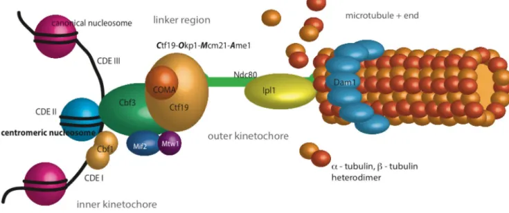
Figure 6: Model of the Saccharomyces cerevisiae kinetochore.
All presently known subcomplexes of the yeast kinetochore are arranged according to their distance from the centromeric nucleosome on the left (blue) to the attachment to the microtubules on the right (orange). The composition of the single subcomplexes is depicted, not the numbers of the protein complexes per centromere. Based on (Westermann et al., 2007; Santaguida and Musacchio, 2009).
1.6. Posttranslational modification of histones
The mostly accessible amino acid side chains in the amino-termini of histone are prominent targets for enzymatically mediated, covalent posttranslational modifications. The two main groups that can be distinguished are small chemical modifications, including phosphorylation, methylation and acetylation on one hand, and the attachment of large peptides such as ubiquityl- and sumoyl- moieties to histones on the other hand. Furthermore, there are much less frequent PTMs, for instance ADP ribosylation, proline isomerization and arginine deamination.
Acetylation, ubiquitylation and sumoylation occur on lysine residues only.
Phosphorylation is restricted to serine and threonine residues, and mono- or dimethylation occurs on arginine or lysine residues whereas trimethylation is exclusively found on lysine residues. Asymmetric or symmetric dimethylation is only found on arginines (Kouzarides, 2007). The large quantity of PTMs that have been discovered so far and their innumerable combinations have led to the idea that PTMs and their patterns epigenetically control biological functions like gene transcription
activation and repression, DNA replication and chromosome segregation (Bannister and Kouzarides, 2011; Kouzarides, 2007). At least three mechanistic models have been proposed to categorize the large number of PTMs. The first concept comprises cis-modifying effects by chromatin modifications such as histone phosphorylation or acetylation and refers to the direct structural changes imposed on chromatin conformation. The second group of PTM-induced alterations refers to inhibitory effects on chromatin binding factors. One example is H3S10 phosphorylation, which is mediated by Aurora B. Upon phosphorylation, the binding of HP1 to the nearest trimethylated H3K9 is sterically blocked during heterochromatin formation, keeping the chromatin accessible to the transcription machinery. In the third model, PTMs are involved in mediating chromatin binding factor specificity. For example, the methyllysine-binding domain of HP1, the chromodomain, binds specifically to trimethylated H3K9. Both latter models are trans-acting mechanisms (Fischle et al., 2005; Kouzarides 2007).
1.6.1. Acetylation of histones
Histone acetylation typically occurs at multiple lysine residues and is mediated by a variety of histone acetyltransferase complexes (Brown et al., 2000; see chapter 1.8.1). One study showed that the modification patterns are likely to act downstream in the regulation of co-expressed genes (Kurdistani et al., 2004). In another study, a cumulative effect depending on the amount of acetylation on one histone tail was described (Dion et al., 2005). In most of the cases, histone acetylation is associated with transcriptional activation. The attachment of this small chemical group goes along with a partial neutralization of the positively charged histone tail, reducing the affinity of histones to the negatively charged DNA backbone. Consequently, this modification tends to prevent inter- and intranucleosomal interactions with DNA molecules and is therefore likely to play a role in open chromatin formation.
Acetylation seems to correlate with transcription, as indicated by many studies including early Chromatin immunoprecipitation (ChIP) experiments in chicken erythrocytes, demonstrating that the active β-globin gene associates with high levels of histone acetylation (Hebbes et al., 1994). Interestingly, histone acetylation is not equally distributed on active genes, but is enriched at promoter regions, where it acts as a recruitment factor for bromodomain containing proteins (see chapter 1.8.2).
Studies showed that the SWI/SNF histone-remodeling complex binds to acetylated histones, which leads to nucleosome displacement at the respective promoter regions (Workman, 2006).
1.6.2. Epigenetic regulation of chromosome segregation
The assumption that chromosome segregation is in part epigenetically regulated has been a matter of debate over many years. Given that histone variants participate in the epigenetic regulation of many cellular processes, it is reasonable that the centromeric histone variant CENP-A is a major subject of epigenetic studies.
Surprisingly little is known about the specific function of PTMs on the N- and C- terminal regions of centromeric histones as compared to the abundant knowledge of modification patterns on canonical histones. Sequence differences to canonical H3 suggest distinct modification patterns on the unique N-terminal regions and, consequently, a distinct function of those domains in kinetochore assembly and chromosome segregation (Figure 7A).
In human cells, interspersed H3 blocks mediate the specificity of CENP-A deposition.
Following replication of centromeric chromatin, a distinct code of PTMs triggers the recruitment of factors that are required for the replacement of H3 with CENP-A or the deposition of new CENP-A nucleosomes into nucleosomal gaps. H3K4 dimethylation characterizes the canonical histone H3 in the centromeric region in human cells and thereby contributes to centromere specificity (Allshire and Karpen, 2008; Sullivan and Karpen, 2004). Furthermore, genetic studies in fission yeast revealed that mutant alleles of the essential histone deacetylase Clr6 (Cryptic loci regulator 6) disturb epigenetically maintained repression at centromeres and cause chromosome missegregation. These experiments connected the deacetylation of the N-terminal histone tails to centromere function (Grewal et al., 1998).
Recently, it was shown that histone phosphorylation marks the majority of CENP-A nucleosomes in human cells and has a direct influence in chromosome segregation during mitosis. Serines 16 and 18 in prenucleosomal CENP-A N-terminal regions become phosphorylated and form a salt-bridged secondary structure, which leads to intramolecular associations in the CENP-A histones (Bailey et al., 2013). Using phospho-mimetic CENP-A nucleosome arrays in analytical ultracentrifugation
experiments, it was demonstrated that phosphorylation results in larger intranucleosome associations and counteracts the hyper-oligomerized state exhibited by unmodified CENP-A nucleosome arrays. These analyses have shown that two modifications in the N-terminal domain of CENP-A affect the physical and biochemical properties of the chromatin fiber at the centromere during chromosome segregation (Bailey et al., 2013).
1.7. The centromeric histone variant CENP-A
Cse4In the budding yeast, the gene for the centromeric histone variant CENP-A is termed CSE4, which stands for Chromosome segregation protein 4. This histone is a 27 kDa protein and was first described in a genetic screen to identify strains with defects in chromosome segregation, as an essential gene that is needed for cell division (Stoler et al., 1995; Chen et al., 2000; Meluh et al., 1998; Baker et al., 1998). Although centromeric histone H3 variants are highly conserved in eukaryotes (CENP-A in humans, CENP-ACid in Drosophila, CENP-ACnp1 in fission yeast), similarities in amino acid sequence are restricted to the histone fold domain in CENP-ACse4, which shares about 64% sequence identity with canonical histone H3 (Figure 7A); (Wells and McBride, 1989). While all histones exhibit largely unstructured N-terminal regions, the N-terminal tail of CENP-ACse4 is highly unique in that it is almost as large as the histone fold domain, i.e. about 130 aa (Stoler et al., 1995; Meluh et al., 1998). This N- terminus is unusually long as compared to other N-termini, e.g. of human and fission yeast CENP-A, which are only 45 and 20 amino acids in length, respectively.
The N-terminal region of the Drosophila CENP-ACid is also extended and 120 amino acids in length (Allshire and Karpen, 2008). Interestingly, there is no sequence homology of CENP-ACid and CENP-ACse4 tails, suggesting a strong evolutionary fluctuations along with species-specific CENP-A interacting proteins (Figure 7A).
Overexpression or mislocalization of CENP-A has been connected to strong segregation defects in Drosophila and aneuploidy and human cells, which is a hallmark of cancer development (Tomonaga et al., 2003; Heun et al., 2006; Moreno- Moreno et al., 2006).
CENP-A as well as its homologues and H3 are structurally distinct, but both assemble with H4 to build functional heterotetramers. What differentiates CENP-A
from H3 is the centromeric targeting domain CATD, which is located in the α1 helix, loop 1 and α2 helix of the histone fold domain. This motif is 15 aa in length and contains essential hydrophobic amino acids. This domain has been shown to mediate the assembly of rigid and more compact CENP-A-H4 tetramers, which lead to differently arranged higher order centromeric heterochromatic structures. It is also required for the localization of CENP-A-H4 tetramers to centromeres (Black et al., 2004). Recombinant H3 constructs, which were provided with CATD, could be targeted and assembled to centromeres properly. Whereas CENP-A depletion is lethal, this synthetic hybrid could rescue CENP-A function (Black et al., 2007).
Whereas the histone fold domain (HFD) is required for correct centromere targeting, this is not the case for the N-terminus. However, the stretched tail of CENP-ACse4 contains the essential N-terminal domain (END), which extends from aa 28-60 and its deletion has been shown to be lethal in budding yeast (Figure 7B; Chen et al., 2000;
Morey et al., 2004).
1.7.1. Posttranslational modifications on Cse4
As outlined above, the N-terminal domains of canonical histones and the histone variants are subject to posttranslational modifications. Recently, the modification pattern of the budding yeast CENP-ACse4 was resolved by mass spectrometric analysis, showing phosphorylation sites on serines 22, 33, 40 and 105, one methylation site on R37 and one acetylation site on K49, which are all positioned at the N-terminus of CENP-ACse4. Except for serines 22 and 105, all of them are located in the essential N-terminal domain (Samel et al., 2012; Boeckmann et al., 2013).
CENP-A histones are necessary for the recruitment and assembly of the kinetochore, and PTMs on their tails may be critical for chromosome segregation (Choy et al., 2012). Although little is known about the involvement of PTMs on CENP-ACse4 in pathways that are crucial for the segregation process, our group showed by genetic interaction studies in yeast that R37 methylation is critical for the recruitment of many kinetochore proteins, e.g. the COMA subunits Ctf19, Ame1, Okp1 and Mcm21 of the linker layer complex CTF19. Mutant alleles of these kinetochore components showed synthetic lethality or synthetic growth defects in combination with cse4R37A.
Furthermore, this point mutation caused temperature sensitivity in cells that lack Cbf1
and G2/M phase delay at restrictive temperatures, indicating an epigenetic regulatory function of Cse4R37 modifications in kinetochore assembly and cell-cycle progression (Samel et al., 2012).
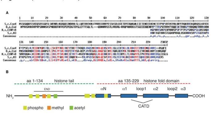
Figure 7: Sequence alignment of CENP-A species and topological view of CENP-A from budding yeast
A: Amino acid sequence alignment of budding yeast (upper panel), Drosophila and human CENP-A proteins, compared to budding yeast canonical histone H3 and the consensus sequence (lower panels). Alignment was generated with Multalin (http://multalin.toulouse.inra.fr/multalin). B:
Topological organization of the S. cerevisiae CENP-A molecule. -helices are shown as blue boxes, CATD indicated the centromeric targeting domain. Phosphorylation sites of serines, methylation of R37 and acetylation of K49 are depicted as yellow, orange and green boxes, respectively. END indicates the essential N-terminal domain. Modified from (Luger et al., 2001; Allshire and Karpen, 2008).
1.8. Histone-modifying enzymes
Several histone-modifying enzyme families are known, including histone acetyltransferases (HATs, see chapter 1.8.1.), arginine and lysine specific methyltransferases, kinases, and ubiquitin conjugating enzymes.
In humans, arginine monomethylation, asymmetric and symmetric dimethylation are catalyzed by specific S-adenosyl-L-methionine dependent protein arginine methyltransferases (PRMTs). These enzymes are classified into type I-III enzymes,
of which type III PRMTs catalyze monomethylations. Type I and II catalyze a monomethylated intermediate, followed by type I mediated asymmetric dimethylation or type II mediated symmetric dimethylation (Bedford and Clarke, 2009; Di Lorenzo and Bedford, 2011). In yeast, three arginine methyltransferases Hmt1, Rmt2 and Hsl7 have been described (Henry and Silver, 1996; Lee et al., 2000a; Niewmierzycka and Clarke, 1999).
Lysine methylation is mediated by members of the large family of KMTs, which share the conserved SET domain harboring the catalytic activity and mediate mono-, di- and trimethylation (Zhang et al., 2012). These enzymes are highly specific, for example the yeast H3K79me2/3 specific methyltransferases Dot1, which has been functionally linked to telomeric silencing (Singer et al., 1998).
Histone deacetylases (HDACs) and arginine and lysine specific histone demethylases (HDMs) remove the methyl and acetyl moieties and convert histones into the initial unmodified state. According to the dynamics of chromatin condensation and decondensation, the outcome of all these modification processes have to be precisely timed to ensure gene transcription regulation, proper DNA replication, sister chromatid separation, DNA damage response and other essential cellular events and to prevent the cell from malignant developments (Butler et al., 2012; Marmorstein and Trievel, 2009).
1.8.1. Histone acetyltransferases (HATs)
Histone acetyltransferases (HATs) are the best described histone modifying enzymes. Such enzymes posttranslationally modify lysines of histone tails by transferring an acetyl group from acetyl-CoA to an ε-amino moiety of the target lysine side chain (Brownell and Allis, 1996). Almost 20 years ago, it was shown that the enzymes p55 as well as the human and yeast Gcn5 proteins have HAT activity, and the list of histone acetyltransferases has been extended since then (Brownell and Allis, 1995; Brownell et al., 1996; Kuo et al., 1996; Wang et al., 1997). Many of the HATs are involved in central processes, like transcriptional activation (Gcn5, Esa1, Sas3 and more), dosage compensation (MOF), DNA repair (Tip60), or histone deposition (Hat1) (Kimura et al., 2005; Marmorstein and Trievel, 2009).
Until today, four HAT families are known, which are grouped according to their amino acid sequence homology and their catalytic mechanisms (reviewed in Marmorstein 2001). The first are the GNAT family (Gcn5-related N-acetyltransferases) and their close relatives from the p300/CBP (CREB binding protein; for both see chapter 1.8.2.). Members of the MYST (MOZ, Ybf2/Sas3, Sas2, Tip60) family share the chromodomain and zinc-finger domains as common features. Esa1 acts in the NuA4 complex, which is essential in yeast and specific for the acetylation of H4K8, K12 and K16, but is also able to acetylate the N-terminal tail of H3, and the H2A variant Htz1 on K14 in yeast (Allard et al., 1999; Berndsen and Denu, 2008; Brown et al., 2000).
Another MYST complex is SAS-I (something about silencing). It has been demonstrated that its catalytic subunit Sas2 acetylates H4K16 with high specificity, and the deletion of SAS2 causes silencing phenotypes (Meijsing and Ehrenhofer- Murray, 2001; Osada et al., 2001). Only the MYST and the PCAF/Gcn5 histone acetyltransferases are conserved from yeast to human, and the p300/CBP is conserved among metazoans. The fourth group contains the fungal specific HAT Rtt109, which is specific for H3K56 acetylation and was named for its function as a regulator of Ty1 transposition gene product 109, (Driscoll et al., 2007; Han et al., 2007; Schneider et al., 2006).
1.8.2. Gcn5-related N-acetyltransferases (GNATs)
The first member of the protein superfamily of Gcn5-related N-acetyltransferases was defined in tetrahymena as p55 histone acetyltransferase, which directly linked histone acetylation of chromatin templates to gene activation (Brownell et al., 1996).
Gcn5 (general control nonderepressible 5) was originally described as a co-factor of transcriptional activators, and has been shown to acetylate N-termini of H3 with high affinity, and to a lesser extent, N-termini of H4 (Georgakopoulos and Thireos, 1992;
Goodman and Smolik, 2000; Turner, 1991).
Further members of the GNAT protein family are the human P/CAF (p300/CBP associated factor), as well as Hpa2, Hat1 and Elp3, which were first described in S.
cerevisiae. Most of the enzymes share a bromodomain with the members of the p300/CBP family, which is a highly conserved structural feature composed of a bundle of four α-helices that recognizes acetylated lysine residues and tethers HATs