Mechanistic studies on the regulation of Ran- function by post-translational lysine-acetylation
Inaugural-Dissertation zur
Erlangung des Doktorgrades
der Mathematisch-Naturwissenschaftlichen Fakultät der Universität zu Köln
vorgelegt von
Susanne de Boor (geb. Haase) aus Rostock
Köln, 2015
Berichterstatter: Dr. Michael Lammers (Betreuer) Prof. Dr. Kay Hofmann
Tag der letzten mündlichen Prüfung: 02.06.2015
Das kleine G-Protein Ran ist an elementaren zellulären Prozessen wie dem nukleozytoplasmatischen Transport sowie dem Aufbau der Kernhülle und der mitotischen Spindeln beteiligt. Die zwei nukleotidabhängigen Zustände, GTP- und GDP-gebunden, zeichnen sich durch unterschiedliche Konformationen zweier regulatorischer Schleifen (Switch I und Switch II) aus, welche maßgeblich die Effektor-Bindung beeinflussen. Der Übergang zwischen den zwei Zuständen wird durch regulatorische Proteine unterstützt, welche die Hydrolyse und den Austausch des Nukleotides beschleunigen. Die unterschiedliche zelluläre Lokalisation dieser Proteine führt zur Ausbildung eines RanGDP/GTP-Gradienten. Eine umfangreiche massenspektrometrische Studie im Jahr 2009 identifizierte fünf Acetylierungsstellen in humanem Ran, welche mittlerweile auch durch Folgestudien in anderen Organismen bestätigt wurden. Ziel dieser Arbeit war es, die Auswirkungen der Ran-Acetylierung auf die Funktion von Ran zu untersuchen. Des Weiteren sollten Einblicke in die Stöchiometrie und Regulation dieser post-translationalen Modifikation von Ran gewonnen werden um eine Einschätzung der physiologische Relevanz zu ermöglichen.
Im Zuge der Arbeit wurde mittels Erweiterung des genetischen Codes von E.coli stellen-spezifisch acetyliertes Ran (an den Lysinen 37, 60, 71, 99 und 159) hergestellt. Die Proteine wurden gereinigt und hinsichtlich ihrer intrinsischen Eigenschaften sowie der Interaktion mit dem Nukleotid-Austauschfaktor RCC1, dem wichtigsten Import-Rezeptor Importin-β und dem Ran-bindenden Protein NTF2 untersucht. Die umfangreiche in vitro Charakterisierung zeigt, dass die Acetylierung einzelner Lysine die RCC1-Interaktion und Nukleotid- Austauschrate, sowie die Ran-Lokalisierung und die Bildung von Import- Komplexen beeinflusst. Ran-Acetylierung an den Lysinen 37, 99 oder 159 hat eine höhere Importin-β-Affinität zur Folge. Acetyliertes Lysin 71 hat einen dominant negativen Effekt bezüglich RCC1 welcher mit dem der T24N-Mutante vergleichbar ist: die Bindungsaffinität ist erhöht, während die Nukleotid- Austauschrate herabgesetzt ist. Weiterhin verhindert diese Acetylierung die Interaktion mit NTF2, was wiederum die Ausbildung des Ran-Gradienten bzw.
die Lokalisation von Ran beeinträchtigt. Für Lysin 99-acetyliertes Ran war ein
Die durchgeführten Untersuchungen zur Regulation der Ran-Acetylierung führten zur Identifikation Ran-spezifischer Acetyltransferasen und Deacetylasen in vitro und in vivo. Sirt1, -2 und -3 wirken als Deacetylasen auf Acetyl-Lysin 37, wohingegen Acetylierung an Lysin 71 spezifisch von Sirt2 entfernt wird.
Immunoblotting und massenspektrometrische Untersuchungen identifizierten
Tip60, p300 und CBP als Ran-spezifische Acetyltransferasen und die Lysine 37,
134 und 142 als hauptsächliche Acetyl-Akzeptoren. Zusammengefasst deutet die
vorliegende Arbeit auf ein hohes regulatorische Potential für die Acetylierung
von Ran in Abhängigkeit vom zellulären Kontext hin.
Carl Edward Sagan, Astronomer
The small GTP-binding protein Ran regulates fundamental cellular processes such as nucleocytoplasmic transport, nuclear envelope formation and mitotic spindle assembly. The two nucleotide states of Ran, GTP- and GDP-bound, are charac- terized by distinct conformations of two regulatory regions (switch I and switch II) which determine effector binding. The interconversion of these two states is facilitated by regulatory proteins (RanGAP, RCC1). The differential localiza- tion of these proteins leads to the formation of a Ran∗GDP/GTP gradient. A mass-spectrometrical screen of the acetylome of human cell lines in 2009 iden- tified five acetylation sites in Ran, which were confirmed by subsequent screens in different organisms. The aim of this thesis was to study the impact of Ran- acetylation on Ran-function. Furthermore, the abundance and regulation of this post-translational modification was investigated to judge the physiological rele- vance of Ran-acetylation.
In this study, recombinant Ran was site-specifically acetylated at lysines 37, 60, 71, 99 and 159 using the genetic-code expansion concept in E.coli. The proteins were purified and characterized regarding their intrinsic properties and the interaction with the nucleotide exchange factor RCC1, the major import receptor Importin-β and the Ran-shuttling protein NTF2 (nuclear transport factor 2). The compre- hensive in vitro characterization revealed that acetylation of single lysines impacts on RCC1-interaction and -catalysis, Ran-localization and import-complex forma- tion. Ran acetylated at lysines 37, 99 or 159 exhibits a higher binding affinity for Importin-β. Acetylation of lysine 71 (in switch II) has a dominant negative effect on RCC1 analogous to the T24N-mutant of Ran. Furthermore, it abolishes NTF2-interaction, which consequently affects Ran-localization and the formation of the Ran-gradient. Moreover, Ran acetylated at lysine 99 resembles a loss-of- function mutant regarding RCC1, lowering affinity and nucleotide exchange rates.
Regulatory enzymes for Ran-specific de-/acetylation were identified in vitro and
in vivo. Sirt1, -2 and -3 are deacetylating enzymes for lysine 37 of Ran, whereas
lysine 71 is specifically deacetylated by Sirt2. The enzymatic acetylation of Ran
by the acetyltransferases Tip60, p300 and CBP was shown by immunoblotting
and mass-spectrometrical analysis and lysines 37, 134 and 142 were identified as
the major acetyl-acceptors of Ran. Taken together, these results suggest a strong
regulatory potential of Ran-acetylation depending on the cellular context.
Contents
Abstract iv
List of Figures ix
List of Tables xi
1 Introduction 1
1.1 The Ras superfamily . . . . 1
1.1.1 The G-domain . . . . 3
1.1.2 Regulatory cycle . . . . 5
1.2 The small GTPase Ran . . . . 8
1.2.1 Nucleocytoplasmic transport . . . . 8
1.2.2 Mitotic spindle assembly . . . 11
1.2.3 Nuclear envelope formation . . . 13
1.2.4 Ran-interacting proteins . . . 13
1.3 Protein lysine acetylation . . . 19
1.3.1 Characteristics and regulation of lysine acetylation . . . 20
1.3.2 Lysine acetylation as an emerging PTM . . . 24
1.3.3 Tools to study lysine acetylation . . . 28
1.3.4 Genetic code expansion concept . . . 33
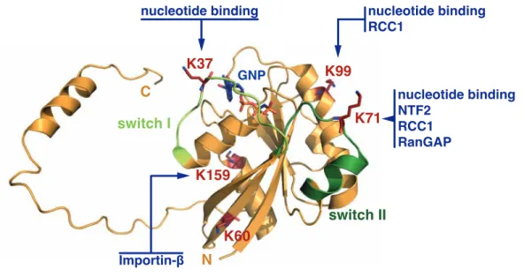
1.4 Acetylation of Ran . . . 35
1.5 Aim of this thesis . . . 37
2 Material and Methods 39 2.1 Materials . . . 39
2.1.1 Primer . . . 39
2.1.2 Vectors . . . 40
2.1.3 Buffers and solutions . . . 41
2.2 Biomolecular methods . . . 43
2.2.1 Cloning . . . 43
2.2.2 Purification of DNA . . . 43
2.2.3 Transformation . . . 44
2.3 Biochemical methods . . . 44
2.3.1 Preparative expression of recombinant proteins . . . 44
2.3.2 Lysis of cells . . . 44
v
2.3.3 Purification of recombinant proteins . . . 45
2.3.4 Analytical size-exclusion chromatography . . . 46
2.3.5 Determination of protein concentration . . . 46
2.3.6 SDS-Polyacrylamide gelelectrophoresis (SDS-PAGE) . . . 47
2.3.7 Western blotting and immunodetection . . . 47
2.3.8 Nucleotide exchange on Ran protein . . . 48
2.3.9 High pressure liquid chromatography (HPLC) . . . 48
2.3.10 Activity Test for Deacetylases . . . 49
2.3.11 KDAC-Screen . . . 49
2.3.12 Deacetylase (KDAC)-Assays . . . 50
2.3.13 Acetyltransferase (KAT)-Assays . . . 50
2.3.14 Pull-down of endogenous Ran . . . 51
2.4 Cell culture . . . 52
2.4.1 Cultivation of cell lines . . . 52
2.4.2 Transfection . . . 52
2.4.3 Immunocytochemistry . . . 52
2.4.4 Pull-down of His
6-tagged Ran (in vivo KAT assay) . . . 52
2.5 Biophysical methods . . . 53
2.5.1 Mass spectrometry . . . 53
2.5.2 Isothermal titration calorimetry (ITC) . . . 55
2.5.3 Fluorescence spectroscopy . . . 56
2.5.4 Data visualization . . . 59
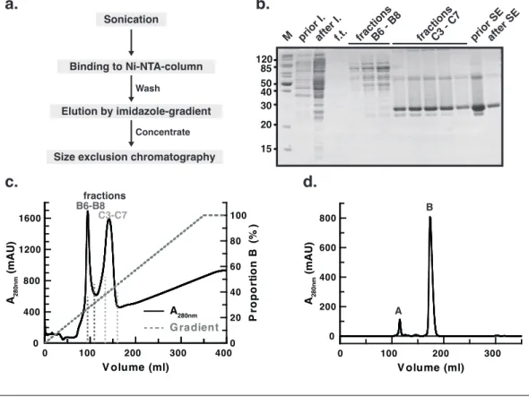
3 Experimental results 61 3.1 Protein purification and identification . . . 61
3.1.1 Purification of acetylated Ran . . . 62
3.1.2 Quality control of acetylated Ran . . . 63
3.1.3 Purification of Ran-interaction partners . . . 65
3.1.4 Nucleotide exchange on Ran . . . 66
3.2 Biophysical studies on the effect of Ran acetylation . . . 68
3.2.1 Effect of Ran-acetylation on NTF2 interaction . . . 68
3.2.2 Effect of Ran-acetylation on RCC1-interaction and nucleotide exchange . . . 73
3.2.3 Effect of Ran-acetylation on Importin-β interaction . . . 78
3.3 Regulation of Ran acetylation . . . 82
3.3.1 Ran acetylation in vivo . . . 82
3.3.2 Regulation by specific deacetylases . . . 83
3.3.3 Regulation by acetyl-transferases . . . 88
4 Discussion 95 4.1 Summary of results . . . 95
4.2 Implications for the biological relevance of Ran acetylation . . . 97
4.3 General implications for the investigation of protein acetylation . . 104
4.4 Conclusion and outlook . . . 107
A Appendix 109
Bibliography 121
Abbreviations 121
Acknowledgements 149
List of Figures
1.1 Structure of the G-domain of GNBPs . . . . 4
1.2 Regulatory cycle of GNBPs of the Ras superfamily . . . . 5
1.3 Molecular basis for GAP-accelerated GTP-hydrolysis . . . . 6
1.4 Schematic presentation of nucleotide binding and GEF-mechanism . 7 1.5 The nucleocytoplasmic transport cycle . . . 10
1.6 Ran is involved in mitotic spindle formation . . . 11
1.7 Crystal structure and mechanism of action of RCC1 . . . 14
1.8 Crystal structre of Importin-β . . . 17
1.9 Impact of lysine acetylation . . . 19
1.10 Regulation of lysine acetylation . . . 21
1.11 KATs utilize a direct attack mechanism of acetylation . . . 23
1.12 Structure and acetyl-lysine recognition of the PCAF bromodomain . 24 1.13 Functional roles of lysine acetylation . . . 26
1.14 MS-based global analysis of lysine acetylation . . . 29
1.15 Comparison of quantification strategies . . . 30
1.16 Chemical ligation for the modification of C- or N-terminal domains 32 1.17 Acetyl-lysine-analog by alkylation of cysteines . . . 32
1.18 Genetic-code expansion concept . . . 34
1.19 Ran acetylation sites and possible effects . . . 36
2.1 Structure of 2
‘/3
‘-O-(N-Methyl-anthraniloyl)-GppNHp . . . 56
3.1 Purification of Ran AcKs . . . 63
3.2 Incorporation of acetyl-lysine into recombinant Ran . . . 64
3.3 Purification of Ran-binding proteins . . . 65
3.4 Nucleotide exchange on Ran . . . 67
3.5 ITC of NTF2 and Ran wt/AcK71/K71Q . . . 69
3.6 Analytical size-exclusion chromatography of Ran-NTF2-complexes . 70 3.7 Subcellular localization of acetylation mimetics . . . 71
3.8 Ran in comlex with NTF2 . . . 72
3.9 Signature plots of the interaction of Ran and RCC1 . . . 74
3.10 RCC1-catalyzed nucleotide exchange measured by stopped-flow . . 76
3.11 Ran in comlex with RCC1 . . . 77
3.12 Thermodynamic characterization of the Importin-β - Ran interaction 79 3.13 Association of Importin-β and Ran measured by stopped-flow . . . 80
3.14 Verification of enzymatic activity for all KDACs . . . 83
ix
3.15 Deacetylation of recombinant Ran AcKs by classical KDACs and
Sirtuins . . . 84
3.16 Ran is deacetylated by sirtuins 1, 2 and 3 . . . 85
3.17 Dependence of Sirt2 deacetylation on complex-formation and nucleotide- state . . . 86
3.18 Sirt2 distinguishes between Ran AcK37 and AcK38 . . . 88
3.19 Acetylation of Ran by CBP in vitro . . . 89
3.20 MS/MS spectrum of the AcK142-peptide . . . 90
3.21 Mass-spectrometrical analysis of Ran-acetylation in vitro . . . 91
3.22 Ran-acetylation by KAT-overexpression in vivo . . . 93
4.1 Ran acetylation sites detected in different MS-based studies . . . . 99
4.2 The acetylation pattern of Ran is tissue-dependent (in mouse/rat samples) . . . 103
4.3 Sirtuin activity on published substrates as detected by a microarray screen . . . 105
A.1 tRNA-synthetase/tRNA-pair used for GCEC . . . 110
A.2 ESI-MS spectra of Ran wt and all AcKs . . . 111
A.3 Interaction of NTF2 and Ran wt/AcKs . . . 112
A.4 Interaction of RCC1 and Ran∗GDP wt/AcKs . . . 113
A.5 Interaction of RCC1 and Ran∗GppNHp wt/AcKs . . . 114
A.6 RCC1-catalyzed nucleotide exchange on Ran . . . 115
A.7 Interaction of Importin-β and Ran wt/AcKs . . . 116
A.8 Association of Importin-β and Ran wt/AcKs . . . 117
A.9 Association of Importin-β and Ran wt/AcKs . . . 118
A.10 MS/MS spectra . . . 119
List of Tables
1.1 Subfamilies of the Ras superfamily (after (Rojas et al., 2012)) . . . 2
1.2 Motifs of the G-domain . . . . 4
1.3 Mitotic targets of RanGTP in spindle assembly . . . 12
1.4 Sirtuins . . . 21
2.1 Cloning primer . . . 39
2.2 Quick change primer . . . 40
2.4 Purification strategies for recombinant proteins . . . 45
2.5 Antibodies . . . 48
2.6 Human KDACs . . . 50
3.1 Parameters for the purification of Ran-binding proteins . . . 66
3.2 Characterization of the Ran-NTF2 interaction by ITC . . . 69
3.3 Cell lines and conditions (all ± Inhibitors) used for the pull-down with RCC1 . . . 83
xi
Chapter 1 Introduction
1.1 The Ras superfamily
Cellular signal transduction enables prokaryotic and eukaryotic cells to react to ex- tracellular stimuli- a prerequisite for survival. G-proteins (guanine nucleotide bind- ing proteins, GNBPs) are key players in signal transduction. Based on sequence- and structure-similarities, GNBPs are divided into two major classes: the TRAFAC (translation factors) and the SIMIBI (signal recognition particle, MinD and BioD) class. More than twenty families are organized in both classes together, which are further subdivided into 57 subfamilies (Leipe et al., 2002). TRAFAC-GNBPs are involved in signal transduction, translation, cell migration and intracellular transport processes. The translation factor superfamily (e.g. EF-Tu), the Ras su- perfamily (e.g. Ras, Rho, Ran) and the myosin-kinesin superfamily (e.g. myosin) are well known examples. SIMIBI proteins are involved in protein targeting and -localization and their biological function is associated with homo- or heterodimer- ization. The signal recognition-associated GTPase superfamily (SRP and its re- ceptor) is one example for the SIMIBI class. Based on their biological function GNBPs can be divided into:
1. α-subunits of heterotrimeric G-proteins (G
αs) 2. Translation factors (e.g. EF-Tu, IF-2)
3. Ras superfamily (e.g. Ras, Rho, Ran) 4. SRP and SRP-receptor
5. Large GNBPs (e.g. Dynamin, GBP)
1
Table 1.1: Subfamilies of the Ras superfamily (after (Rojas et al., 2012))
Ras Rho Rab Arf Ran
Members 39 22 65 30 1
Function differentiation cytoskeleton vesicle transport vesicle transport nucleocytoplasmic transport
proliferation cell growth mitotic spindle formation
apoptosis nuclear envelope formation
Examples (H/K/N)-Ras Rho(A/B/C) Rab1A Arf1-6 Ran
Rap1A Rnd(1/2/3) Rab1B Arl1-10
Rap1B Rac(1/2) Rab2
RalA CDC42 Rab3A
Rit1/2 TC10 Rab3B
Rhes Rif Rab10
The Ras superfamily comprises more than 150 human small GNBPs with a molec- ular weight of 20 to 25 kDa. The prototypical member of the superfamily, Ras (rat sarcoma), was first identified in 1979 as the product of a gene later characterized as a proto-oncogene (Chien et al., 1979; Langbeheim et al., 1980; Shih et al., 1979).
20 - 30 % of all human tumors carry mutations in the RAS proto-oncogene (Prior et al., 2012). Based on functional- and sequence similarity the Ras superfamily is further subdivided into five main-families: Ras, Rho, Rab, Arf and Ran (Ro- jas et al., 2012). Ras family proteins play important roles in cell differentiation, -proliferation and apoptosis. Rho-proteins (ras-homologous) are involved in cy- toskeleton organization, cell growth and regulation of gene expression. Members of the Rab (ras-like proteins from brain) and Arf (ADP-ribosylation factor) fam- ilies regulate the generation and transport of vesicles. Ran (ras-related nuclear) is crucial for nucleocytoplasmic transport, mitosis and formation of the nuclear envelope.
The importance of G-proteins in signal transduction is based on the ability to adopt two different states: the GTP-bound ”on”-state and the GDP-bound ”off”- state. The two states differ in the conformation of the highly conserved core domain of the G-proteins, the G-domain. Downstream targets, so called effector proteins, are only able to bind to the GTP-bound conformation, defining this as the active state. The fact that both conformational states are interconvertible by nucleotide exchange, predestines G-proteins to act as molecular switches. The hydrolysis of GTP to GDP results in the inactivation of the G-protein, which terminates the signal transduction and prevents permanent ”on”-signaling.
With the exception of the Ran and Rad subfamilies, all small G-proteins are
post-translationally modified by lipidation. This lipid anchor is crucial for tar-
geting specific subcellular membranes and, consequently, for regulation of the bi-
ological activity of the GNBPs. Ras- and Rho-proteins are farnesylated and/or
geranylgeranylated by specific farnesyltransferses and geranylgeranylases, respec- tively (Lowy and Willumsen, 1989). The lipid-anchor is attached to the C- terminal CaaX-consensus sequence (C: Cysteine, a: aliphatic, X: any amino acid) by thioetherbond-formation with the cysteine side chain (Hancock et al., 1989).
Following lipidation, the farnesyltransferase catalyzes the proteolytic cleavage af- ter the cysteine and methylation thereof (Dai et al., 1998; Schmidt et al., 1998).
For N- and H-Ras additional palmitoylation was reported. In that case, one or two cysteines N-terminal to the CaaX-Box are modified by a reversible thioesterbond.
A constant cycle of palmitoylation/depalmitoylation drives the rapid exchange of Ras between the plasma membrane and the golgi (Rocks et al., 2005). K-Ras, in contrast, is not palmitoylated. Instead, a polybasic domain consisting of multi- ple lysines targets K-Ras to the plasma membrane independent of the secretory pathway (Hancock et al., 1990). Furthermore, Rab-proteins are geranylgerany- lated and Arf-proteins were found to be N-terminally myristoylated. For lipi- dated Rho- and Rab-proteins additional regulatory proteins were identified. The guanine-nucleotide dissociation inhibitors (GDIs) bind to the switch regions of the GDP-state and accommodate the lipid-anchor in a hydrophobic pocket (Figure 1.2). Thereby the activation by GEF-action is prevented and the protein is sol- ubilized from biomembranes. Moreover, PDEδ (Phosphodiesterase 6 subunit δ) solubilizes a broad spectrum of farnesylated cargoes (Chandra et al., 2012; Ismail et al., 2011).
1.1.1 The G-domain
The G-domain of Ras, with 166 residues and a mass of approximately 20 kDa,
can be regarded as the minimal signaling unit in all G-proteins (Vetter and Wit-
tinghofer, 2001). It is characterized by a mixed six-stranded β-sheet flanked by five
helices (Figure 1.1). This α, β protein fold is typical for nucleotide binding pro-
teins, although variations in the number of sheets and helices are possible. Four to
five conserved sequence elements (Table 1.2), all located in loop domains, are lined
up along the nucleotide binding pocket and mediate the high-affinity binding of
guanine nucleotides (Figure 1.4.a) (Bourne et al., 1991; John et al., 1990; Schmidt
et al., 1996; Via et al., 2000). The P-loop contacts the β- and γ-phosphate of the
nucleotide and, hence, makes the major contribution to high-affinity nucleotide
binding (Saraste et al., 1990). Mutations of S/T to N result in dominant negative
small GTPases with a drastically reduced nucleotide affinity (Farnsworth and Feig,
a. b.
Figure 1.1: Structure of the G-domain of GNBPs. Superimposition of several GNBPs of the Ras superfamily on the G-domain in the GTP- (a) and GDP-conformation (b). The switch regions adopt a rigid conformation in the GTP-form and exhibit a high degree of exibility in the GDP-form. Extra elements in the structures of Rho (insert helix, magenta), Ran (C-terminal extension, red) and Arf (N-terminal helix, blue) are highlighted. Taken from
(Vetter and Wittinghofer, 2001)
1991; Feig, 1999; John et al., 1993; Klebe et al., 1995a). The switch I and II re- gions primarily contact the -phosphate of the bound nucleotide. Removal of this phosphate during GTP-hydrolysis leads to conformational changes in both loops and consequently controls e ector binding (Spoerner et al., 2001; Wittinghofer and Pal, 1991). The bifurcated H bond between an Asp side chain in G4 and the nucleotide base as well as a main chain interaction between an invariant Ala in G5 (SAK motif) and the guanine oxygen are the determinants of the speci city for guanine.
Table 1.2: Motifs of the G-domain
Motif Sequence Function Ras Ran
G1 , P-loop GxxxxGK(S/T) - contacts - and -Phosphate 10GAGGVGKS17 17GDGGTGKT24 - coordinates Mg2+
G2, switch I T - hydrogen bond to -Phosphate 30DEYDPTIE37 32TGEFEKKYVATLG44 - hydrogen bond to Mg2+
G3, switch II DxxG - contacts -Phosphate (G) 57DTAGQEEYSAM67 65DTAGQEKFGGLRDG78 - coordinates Mg2+(D)
G4 NKxD - contacts guanine base (speci city) 116NKCD119 122NKVD125 G5 SAK - contacts guanine base (speci city) 145SAK147 150SAK152
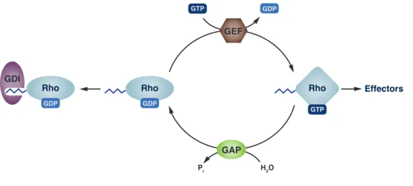
1.1.2 Regulatory cycle
The existence of two nucleotide-dependent interconvertible conformational states is the basis for the relevance of small GNBPs as molecular switches. The GTP- bound state adopts a rigid conformation (Figure 1.1.a) which is recognized by effector proteins and activates downstream signaling cascades. Hydrolysis of the bound GTP releases switch I and switch II from this conformation, since the interactions to the γ-phosphate are lost. In the GDP-bound state the switch regions are highly flexible and, thus, not recognized by effector proteins or, if so, only with very low affinity. This ”inactive” protein state is, however, interacting with regulatory proteins such as exchange factors. This cycle of activation and inactivation is depicted in scheme 1.2.
GAP GEF
GTP
Rho
GDP
Rho
GTP GDP
H2O Pi
Effectors Rho
GDP
GDI
Figure 1.2: Regulatory cycle of GNBPs of the Ras superfamily.
Small G-proteins (e.g. Rho) are regulated by guanine-nucleotide exchange factors (GEF) and GTPase-activating proteins (GAPs). Lipidated Rho- and Rab-proteins are, furthermore, regulated by guanine-nucleotide dissociation in-
hibitors (GDIs).
The basic machinery for interconversion of both states, by nucleotide hydrolysis and nucleotide exchange, is provided by the G-protein itself. However, intrinsic rates for GTP-hydrolysis as well as nucleotide exchange are too slow to be of physiological relevance. Hence, both processes are accelerated 10
5-fold by GTPase- activating proteins (GAPs) and guanine-nucleotide exchange factors (GEFs).
GAPs of small GNBPs are not conserved and they often approach the catalytic site
from different directions. Furthermore, different catalytic mechanisms are utilized
to accelerate GTP-hydrolysis (Scheffzek et al., 1997; Seewald et al., 2002). In most
cases, the insertion of a positively charged residue by the GAP (either arginine
. . .
Figure 1.3: Molecular basis for GAP-accelerated GTP-hydrolysis.
(a)Insertion of an arginine of RasGAP into the active site of Ras to position the catalytic glutamine. (b) RanGAP provides an asparagine to disrupt an inhibitory state created by tyrosine 39. Modified after (Rehmann and Bos,
2004)
or asparagine) stabilizes the catalytic machinery and lowers the activating energy of the catalytic step. Ras- and Rho proteins contain a conserved glutamine in switch II (Glu61 Ras/Rac/CDC42, Glu63 Rho) which positions a catalytic water molecule for nucleophilic attack on the the γ-phosphate of GTP (Resat et al., 2001). Ras- and RhoGAPs insert an arginine residue into the active site, the so called arginine-finger, to position the catalytic glutamine and to neutralize the developing negative charge in the hydrolysis step (Figure 1.3.a) (Ahmadian et al., 1997).
RanGAP provides an asparagine instead of the arginine-finger to accelerate the
intrinsic GTP-hydrolysis. Furthermore, tyrosine 39 of Ran holds the catalytic
glutamine (Glu69) and the water in an anti-catalytic position. Asparagine 131
of RanGAP displaces Tyr39 and allows for rapid hydrolysis by disruption of the
inhibitory state (Figure 1.3.b) (Brucker et al., 2010). The GTP-hydrolysis of Rap
is based on, yet, another mechanism, independent of a catalytic glutamine and the
arginine-finger. In fact, a threonine residue without a detectable role in catalysis is
found in the position of the catalytic glutamine. However, the missing glutamine
in Rap is replaced by an asparagine in trans, the Asn-thumb, which is inserted
into the active site by RapGAP and occupies exactly the same position as the
catalytic glutamine in the other G-proteins (Scrima et al., 2008).
b.
a.
Figure 1.4: Schematic presentation of nucleotide binding and GEF-mechanism.(a) Schematic presentation of molecular contacts between GppNHp (a GTP-analogon) and residues of Ras (Wittinghofer and Waldmann, 2000). (b) Schematic diagram of the push-and-pull mechanism for GEF-induced nucleotide dissociation. Insertion of GEF-residues into/close to the magnesium- binding site and the P-loop disturbs nucleotide binding and reduces the nu- cleotide affinity. Switch I is pushed out and switch II pulled towards the nu-
cleotide binding site (Vetter and Wittinghofer, 2001).
G-proteins bind GDP/GTP with low nanomolar to picomolar affinities and con-
sequently exhibit very slow intrinsic nucleotide dissociation rates. GEFs accel-
erate these rates by several orders of magnitude by reducing the affinity for the
nucleotide. The catalytic domains of various GEFs and also the specific bind-
ing sites differ throughout the Ras superfamily. However, GEF-binding always
induces conformational changes in the switch regions and the P-loop, which steri-
cally perturb the magnesium binding site (Lenzen et al., 1998). This mechanism is
referred to as ”push-and-pull mechanism”, since switch I is pushed out of its nor-
mal position, whereas switch II is pulled toward the nucleotide binding site (Figure
1.4.b)(Vetter et al., 1999). Insertion of GEF-residues close to or into the P-loop
and Mg
2+-binding area disturbs the nucleotide binding and drastically reduces
the nucleotide affinity. The fact that the P-loop lysine (Lys16 in Ras), formerly
contacting negative charges of the phosphates, is reoriented and interacts with
either the invariant Asp (Asp
57in Ras) or the conserved Glu (Glu
62in Ras) of
switch II, furthermore favors nucleotide release (Gasper et al., 2008). The stable
nucleotide-free complex of GTPase and GEF is dissociated by subsequent rebind-
ing of nucleotide. Noteworthy, the GEF-activity does not influence the binding
selectivity regarding GDP or GTP. Rebinding of GTP, instead of GDP, is simply
favored by a higher cellular GTP-concentration.
1.2 The small GTPase Ran
Ran was originally isolated with a Ras-like sequence from a human teratocar- cinoma cell line (Drivas et al., 1990). Ran is a highly abundant and strongly conserved small GNBP, which is ubiquitously expressed in all eukaryotic cells (Ren et al., 1993). During interphase it shows predominantly nuclear localization, whereas it is widely distributed throughout the cell during mitosis. As a family member of the Ras superfamily, Ran features the characteristic G-domain and passes through the regulatory cycle (Scheffzek et al., 1995). In addition to the G- domain, Ran exhibits a C-terminal extension consisting of an unstructured linker followed by a 16 residue α-helix. In the GDP-bound state this extension contacts the protein core opposite of the switch I. In the GTP-bound state the unstructured C-terminus is extending away from the core, exposing the switch region for effec- tor recognition and -binding. In contrast to the majority of small GTPases, Ran does not bind to biomembranes and, thus, is not post-translationally modified by prenylation. Ran carries an acidic C-terminus (-DEDDDL) which stabilizes the GDP-bound state and mediates interactions with RCC1, RanGAP, and RanBP1 (Richards et al., 1995).
Ran is involved in a variety of essential cellular processes. It is a key player in the transport of proteins in and out of the nucleus and also RNA is exported from the nucleus in a Ran-dependent process (K¨ ohler and Hurt, 2007; Weis, 2002). Ran, furthermore, controls the cell cycle in yeast, regulates several mitotic processes and is involved in nuclear envelope formation (reviewed in (Clarke and Zhang, 2008)).
Most of the functions of Ran are based on a gradient of Ran∗GDP/Ran∗GTP which is established by differential localization of the two regulating proteins Ran- GAP (cytosolic) and RCC1 (nuclear).
1.2.1 Nucleocytoplasmic transport
The nucleus of eukaryotic cells is delimited by the nuclear envelope, consisting of
an inner nuclear membrane and an outer nuclear membrane. The outer nuclear
membrane is continuous with the endoplasmatic reticulum. The inner nuclear
membrane is lined by the nuclear lamina, a fibrillar network out of intermediate
filaments and membrane associated proteins. The exchange of proteins, RNA and
small molecules between the cytosolic and the nuclear compartment is a prereq- uisite for survival of every eukaryotic cell. The translocation processes of these molecules are regulated with varying stringency. To this end, the nuclear envelope is perforated by nuclear pores - channels connecting inner and outer membrane and harboring the nuclear pore complexes (NPCs). NPCs are large multimeric protein assemblies out of 30 - 60 different proteins (nucleoporins) reaching a size of around 120 MDa in mammalian cells (Grossman et al., 2012; Reichelt et al., 1990; Rout and Aitchison, 2000). Phenylalanine-Glycine-(FG)-rich repeats (consensus sequence:
FG, FxFG or GLFG) of nuclear pore proteins form a three-dimensional meshwork with hydrogel-like properties, which enables passive diffusion of molecules smaller than 25 - 40 kDa (Frey and G¨ orlich, 2009; Frey et al., 2006). Larger molecules either possess the intrinsic capacity to interact with the NPC or the transloca- tion is facilitated by association with specific transport receptors. These import- (importins) and export-receptors (exportins) recognize specific nuclear localiza- tion signals (NLS) and nuclear export signals (NES), respectively. From in vitro studies and single-molecule imaging experiments, an import rate capacity of 1000 molecules per second and NPC was estimated (Ribbeck and G¨ orlich, 2001; Yang et al., 2004). Only facilitated, energy-dependent transport permits such an ef- ficient translocation. However, the translocation process itself does not require energy. Rather, the energy consumption enables a directed transport against a concentration gradient. And in fact, transport-associated GTP-hydrolysis takes place after translocation trough the nuclear pore.
The cycling of Ran between its GDP- and GTP-bound state is crucial for nucle- ocytoplasmic transport (reviewed in (Cautain et al., 2014)). The spatial separa- tion of Ran∗GTP-generation in the nucleus and GTP-hydrolysis in the cytosol generates the Ran∗GDP/GTP-gradient, which provides directionality to the nu- cleocytoplasmic transport (Kal´ ab et al., 2006). RCC1, the GEF of Ran, binds to chromatin and catalyzes the nucleotide exchange to generate Ran∗GTP in the nucleus. The mechanism of action of Ran∗GTP with transport receptors is differ- ent for import- and export processes (Figure 1.5). In the case of protein import, binding of Ran∗GTP dissociates import-complexes in the nucleus (G¨ orlich et al., 1996). These complexes are formed in the cytosol and consist of a cargo molecule, an adaptor protein (e.g. Importin-α) and the import-receptor (e.g. Importin-β).
The cargo protein carries a specific nuclear localization signal (one or two short
stretches of positively charges amino acids) which is recognized and bound by
Importin-α. Importin-β, the actual transport receptor, binds to the complex and
Figure 1.5: The nucleocytoplasmic transport cycle.Schematic presen- tation of the Ran-mediated nucleocytoplasmic transport cycle. Ran∗GTP is generated by the RanGEF (RCC1) at the chromatin, whereas hydrolysis of Ran∗GTP is accelerated by RanGAP at the cytosolic side of the nuclear pore complex. Protein export by export receptors (Exp.) as well as the classical import pathway, involving Importin-α (Imp-α) and Importin-α, are depicted.
facilitates translocation trough the NPC. The binding of Ran∗GTP to Importin- β in the nucleoplasm results in the dissociation of the import-complex and the release of the cargo molecule. Subsequently, the Importin-β-Ran∗GTP-complex passes the NPC and dissociates as a consequence of RanGAP-catalyzed GTP- hydrolysis in the cytosol.
In contrast to import processes, nuclear Ran∗GTP-binding to an exportin (e.g.
Crm1) initiates export-complex formation by enhancing cargo binding. The ternary
complex is exported through the NPC and dissociates upon GTP-hydrolysis by
RanGAP. The cytosolic Ran-binding proteins (RanBPs) RanBP1 and RanBP2
co-stimulate the GAP-reaction. Both transport cycles, import and export, are
closed by re-import of Ran∗GDP into the nucleus. This process is mediated by
the nuclear transport factor 2 (NTF2), which binds specifically to GDP-bound Ran.
1.2.2 Mitotic spindle assembly
Several observations suggested a participation of Ran and Ran-interacting pro- teins in mitosis. Expression of dominant mutants of Ran disrupted cell cycle pro- gression and cell cycle defects were also observed when other components of the Ran-system were perturbed (Ren et al., 1994; Sazer and Nurse, 1994). Premature chromatin condensation after loss of RCC1 in a temperature-sensitive hamster cell line (tsBN2), furthermore, suggested a pivotal role for RCC1 during mitosis (Nishi- moto et al., 1978; Uchida et al., 1990). Ran influences many different processes during mitosis. It promotes the nucleation of microtubuli at the centrosomes and plays a role in chromatin-directed nucleation (Carazo-Salas et al., 1999; Kalab et al., 1999; Ohba et al., 1999; Zhang et al., 1999). Moreover, Ran affects mi- totic motor proteins involved in spindle assembly, regulates the microtubulin-flux during mitosis and stabilizes microtubuli (Carazo-Salas et al., 2001; Wilde et al., 2001).
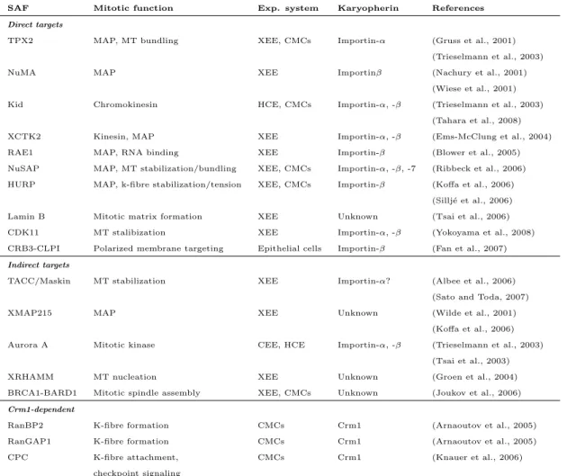
In most cases, Ran acts by releasing mitotic spindle assembly factors (SAFs) from inhibitory complexes with Importin-α and/or Importin-β. Since Ran∗GTP is the active species, able to bind to importins, the detachment of SAFs is linked to the Ran∗GDP/GTP-gradient and, consequently, to Ran∗GTP-generation by RCC1
a. b.
Figure 1.6: Ran is involved in mitotic spindle formation. (a) Re- lease of spindle assembly factors (SAFs) from inhibitory complexes is coupled to Ran∗GTP-generation by RCC1. (b) Localization of SAFs via their NES by a Crm1-Ran∗GTP-dependent pathway. (modified after (Clarke and Zhang,
2008))
close to the chromatin. For example, the inactivation of TPX2 (target protein for Xklp2) by association of Importin-α is abolished by Ran∗GTP-mediated complex dissociation. TPX2 is a microtubuli-associated protein targeting motor proteins to the minus-ends of microtubuli (Wittmann et al., 2000). It, furthermore, activates the Aurora A kinase which is crucial for proper centrosome separation after the mitotic spindle has been formed (Eyers and Maller, 2004). On the other hand, NuMA (nuclear mitotic apparatus protein) is released from an inhibitory complex with Importin-β to regulate microtubuli organization in the spindle.
Table 1.3: Mitotic targets of RanGTP in spindle assembly
SAF Mitotic function Exp. system Karyopherin References
Direct targets
TPX2 MAP, MT bundling XEE, CMCs Importin-α (Gruss et al., 2001) (Trieselmann et al., 2003)
NuMA MAP XEE Importinβ (Nachury et al., 2001)
(Wiese et al., 2001) Kid Chromokinesin HCE, CMCs Importin-α, -β (Trieselmann et al., 2003)
(Tahara et al., 2008)
XCTK2 Kinesin, MAP XEE Importin-α, -β (Ems-McClung et al., 2004)
RAE1 MAP, RNA binding XEE Importin-β (Blower et al., 2005)
NuSAP MAP, MT stabilization/bundling XEE, CMCs Importin-α, -β, -7 (Ribbeck et al., 2006) HURP MAP, k-fibre stabilization/tension XEE, CMCs Importin-β (Koffa et al., 2006)
(Sillj´e et al., 2006) Lamin B Mitotic matrix formation XEE Unknown (Tsai et al., 2006) CDK11 MT stalibization XEE Importin-α, -β (Yokoyama et al., 2008) CRB3-CLPI Polarized membrane targeting Epithelial cells Importin-β (Fan et al., 2007) Indirect targets
TACC/Maskin MT stabilization XEE Importin-α? (Albee et al., 2006) (Sato and Toda, 2007)
XMAP215 MAP XEE Unknown (Wilde et al., 2001)
(Koffa et al., 2006) Aurora A Mitotic kinase CEE, HCE Importin-α, -β (Trieselmann et al., 2003)
(Tsai et al., 2003)
XRHAMM MT nucleation XEE Unknown (Groen et al., 2004)
BRCA1-BARD1 Mitotic spindle assembly XEE, CMCs Unknown (Joukov et al., 2006) Crm1-dependent
RanBP2 K-fibre formation CMCs Crm1 (Arnaoutov et al., 2005)
RanGAP1 K-fibre formation CMCs Crm1 (Arnaoutov et al., 2005)
CPC K-fibre attachment, CMCs Crm1 (Knauer et al., 2006)
checkpoint signaling
Abbreviations: XEE (Xenopus laevis egg extract), CMC (cultured mammalian cells), HCE (human cell extract), MT (microtubuli), MAP (microtubuli-associated protein), K-fibre (kinetochore-fibre), Kid (kinesin-like DNA binding protein), XCTK2 (Xenopus carboxy-terminal kinesin 2), RAE1 (ribonucleic acid export 1), NuSAP (nucleolar- and spindle-associated protein), HURP (hepatoma upregulated protein), CDK11 (cyclin-dependent kinase-11), CRB3-CLPI (Crumbs homolog 3 ending in CLPI), TACC (transforming acidic coiled-coil-containing protein 1), XMAP215 (X microtubuli-associated protein 215), XRHAMM (Xenopus receptor for hyaluronic acid-mediated motility), BRCA-1 (breast cancer type-1 susceptibility protein), BARD-1 (BRCA-associated RING domain protein), CPC (chromosome passenger complex); Table modified after (Clarke and Zhang, 2008)
Apart from the Importin-dependent recruitment of factors, some proteins are also
localized via their NES by a Crm1-Ran∗GTP-dependent pathway, analogous to
the protein export during interphase. Another way of regulatory influence of Ran
on mitotic processes, especially mitotic spindle assembly, involves the APC/C (anaphase-promoting complex). APC/C promotes the ubiquitination and subse- quent degradation of several SAFs, namely BARD1, HMMMR, HURP and NuSAP (Song and Rape, 2010). Importin-β was shown to inhibit the APC/C-dependent ubiquitination of these factors. Thus, the interplay of APC/C and the Importin-β- Ran∗GTP pathway regulates mitotic spindle assembly by controlling the amount of SAFs. Table 1.2.2 lists mitotic targets of Ran∗GTP in the process of spindle assembly.
1.2.3 Nuclear envelope formation
Besides its importance for nucleocytoplasmic transport and regulation of mitotic spindle assembly, Ran is required for nuclear envelope reformation at the end of mitosis and for post-mitotic NPC assembly (Hetzer et al., 2000; Ryan et al., 2003;
Zhang and Clarke, 2000). Bead-immobilized Ran was able to induce the formation of nuclear envelope-like structures in cell-free Xenopus egg extracts. Surprisingly, these structures contained NPCs. Furthermore, Hetzer et al. showed the ne- cessity of RCC1 and Ran for the early stages of nuclear envelope formation. The molecular mechanisms of post-mitotic NPC-assembly are not yet fully understood, however, the involvement of Ran, Importin-β and the Nup107-Nup160-complex is clear (Harel et al., 2003; Walther et al., 2003). While an excess of Importin-β inhibits nuclear membrane fusion and NPC-assembly, Ran∗GTP stimulates these processes. Most likely, Importin-β acts as a negative regulator by refraining the nucleoporins Nup107-Nup160 from the chromatin surface. In analogy to the releas- ing effect on SAFs, Ran∗GTP overcomes this effect of Importin-β by dissociating the inhibitory complexes.
1.2.4 Ran-interacting proteins
Guanine-nucleotide exchange factor (GEF)
RCC1 (regulator of chromosome condensation 1) localizes to chromatin and is the
only GEF acting on the small GNBP Ran (Bischoff and Ponstingl, 1991a,b; Oht-
subo et al., 1989). The yeast protein Mog1 stabilizes the nucleotide-free conforma-
tion of Ran, however, true GEF-activity could not be shown, therefore Mog1 was
postulated to be a nucleotide-release factor (Steggerda and Paschal, 2000, 2001).
The exible N-terminal region (NTR) of RCC1 is responsible for chromatin binding (Moore et al., 2002). Deletion thereof disperses RCC1 from mitotic chromosomes, resulting in spindle abnormalities due to the disturbance of the Ran GTP localiza- tion. The NTR, furthermore, carries a bipartite NLS recognized by Importin-/ 3 and is subject to post-translational modi cation. The N-terminal methionine is removed and serine two is mono-, di- or tri-methylated (Chen et al., 2007a). This methylation is required for chromatin interaction but is not regulated during the cell cycle. In contrast to the stable methylation, phosphorylation at serine two and eleven by CDK1/cyclin B1 is restricted to mitosis. Phosphorylation disrupts the binding of Importin-/ 3 to the N-terminal NLS and thereby preserves the mobility of RCC1 during metaphase (Hutchins et al., 2004; Li and Zheng, 2004).
Ran
Chromatin
90°
a. b.
Ran•GDPT Ran•GDPT•RCC1L Ran•GDPL•RCC1T
Ran•RCC1T Ran•GTPL•RCC1T Ran•GTPT•RCC1L
Ran•GTPT
RCC1
GDP GTP
RCC1
ternary complex
ternary complex
Figure 1.7: Crystal structure and mechanism of action of RCC1.
(a) Cartoon-presentation of the -propeller-fold of RCC1 from two di erent angles (PDB:1I2M). Secondary structure elements are colored in grey (sheets), green (helices) and orange (loops) and the Ran- (yellow) and chromatin-binding site (light-blue) are indicated. (b) Multi-step mechanism of RCC1-catalyzed nucleotide exchange on Ran. The observed ternary complex comprises two intermediates, which change from tight (T) to loose (L) binding of RCC1 or
nucleotide and vice versa.
Alternative splicing of the single RCC1-gene in animal cells results in three RCC1 isoforms (RCC1α/ β/ γ) which exhibit different expression levels in specific tis- sues (Hood and Clarke, 2007). The β- and γ-isoforms contain short inserts in the NTR, resulting in altered chromatin binding properties, molecular interactions and slightly different regulation by phosphorylation.
The crystal structure of RCC1 revealed a seven bladed β-propeller with seven 51 - 68 residue repeats (Figure 1.7.a) (Renault et al., 1998). Ran-interaction buries a large solvent-accessible surface area but otherwise entails only minor structural alterations (Renault et al., 2001). Ran contacts all seven propeller blades of RCC1.
The binding-interface is relatively flat, except for a protruding β-sheet in blade three (residues 146 - 153 of RCC1), the so called β-wedge. Structural changes in the Ran structure upon binding of RCC1 involve switch II, the helices α3 and α4, as well as the region around the NKxD base-binding motif. Ran, furthermore, adopts the GDP-conformation - with an extra β-strand in the switch I region.
The C-terminal extension is not visible in the complex-structure, indicating a high degree of flexibility.
RCC1 has the highest binding affinity for nucleotide-free Ran but binds to both
nucleotide-states of Ran with similar (lower) affinity (Ran∗GDP 1.3·10
5M
−1; Ran∗GTP
1.8·10
5M
−1)(Klebe et al., 1995a). In the course of RCC1-catalyzed nucleotide ex-
change a ternary complex Ran∗GXP∗RCC1 is formed, which indicates that GXP
and RCC1 claim spatially distinct or partially overlapping binding sites. A simple
mechanism of facilitated dissociation is accepted for the reaction meaning that
the high-affinity binding is the product of several low-affinity interactions (Klebe
et al., 1995a; Prinz and Striessnig, 1993). In other words, the low-affinity binding
of RCC1 to Ran∗GXP is followed by a competition between RCC1 and GXP for
weak interactions and results in a number of small conformational changes. As
a result of this process, a ternary complex with low nucleotide affinity and high
RCC1 affinity is formed, which allows fast dissociation of GXP and formation of
a tight binary Ran∗RCC1 complex. The rebinding of nucleotide, in turn, dis-
places RCC1 in an analogous manner - via the formation of a ternary complex
(Figure 1.7). Notably, RCC1-action does not influence the binding characteris-
tics (affinities or on/off-rates) of GDP or GTP on Ran. The net generation of
Ran∗GTP is solely due to a 10-fold higher concentration of GTP over GDP in
the cell. The RCC1-mediated catalysis of nucleotide exchange on Ran results in
a 10
5-fold increased nucleotide exchange rate (from 1.8x10
−5s
−1to 2.1 s
−1)(Klebe et al., 1995a).
Ran GTPase activation protein (RanGAP)
The Ran GTPase-activating protein (RanGAP) was first reported as rna1, a mu- tant defective in mRNA processing and -transport in S.cerevisiae (Hartwell, 1967;
Hopper et al., 1978). RanGAP is exclusively cytosolic during interphase, however, a fraction is tethered to the cytosolic face of the NPC. The localization to the nuclear pore is mediated by SUMO-1-modification at the C-terminus and binding to the Ran binding protein 2 (RanBP2) and the E2-conjugating enzyme Ubc9 (Mahajan et al., 1997; Matunis et al., 1996, 1998; Werner et al., 2012).RanGAP stimulates the GTP-hydrolysis on Ran 10
5-fold from 1.5x10
−5s
−1to 5.0 s
−1(Klebe et al., 1995a). RanBPs are able to co-stimulate the GAP-reaction (Bischoff et al., 1995; Villa Braslavsky et al., 2000). RanGAP-catalyzed hydrolysis either takes place in the cytosol or on the cytoplasmic fibrils of the NPC and is independent of the translocation process trough the nuclear pore (Ribbeck and G¨ orlich, 2001). In contrast to RasGAPs and RhoGAPs, RanGAP does not utilize an arginine finger to complement and stabilize the catalytic machinery of the G-protein (Seewald et al., 2002). In fact, the catalytic machinery is exclusively provided by Ran it- self. However, RanGAP assists in positioning the catalytic glutamine Q69 and by shielding the active site from the solvent. Please refer to section 4.2 for mechanistic details.
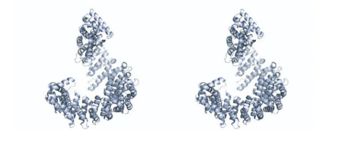
Importin-β
Importin-β belongs to the karyopherin-β superfamily, which is also named Importin- β superfamily after the first receptor identified. The karyopherins are subdivided into importins and exportins to specify the direction of transport. The superfam- ily counts 14 members in yeast and more than 20 in humans. Proteins carrying an NLS or NES are bound by the respective karyopherin and transported through the NPC. A small number of proteins is able to pass the NPC directly (e.g. β-catenin) and also mRNAs use a different pathway (K¨ ohler and Hurt, 2007).
Importin-β is the most prominent member of the family, which is also reflected by
a high intracellular concentration of approximately 3 µM (Kutay et al., 1997). Im-
port cargo recognition and protein import is mediated by adaptor proteins, which
bind the cargo-NLS and Importin-β simultaneously. Up to eleven adaptor pro-
teins are known so far and each heterodimer exhibits a different cargo specificity
(Fried and Kutay, 2003; J¨ akel et al., 2002). In the classical import pathway the
NLS of cytoplasmic cargoes is rst recognized and bound by Importin- , which then associates with Importin- via its N-terminal Importin--binding (IBB) do- main. The ternary complex (NLS/Importin- /) translocates trough the NPC and dissociates upon Ran GTP binding to Importin- in the nucleus.
Importin- is the typical structure of the superfamily (Figure 1.8). Two anti- parallel -helices joined by a turn form the basic HEAT-repeat (Huntingtin, elon- gation factor 3, protein phosphatase 2A, and the yeast kinase TOR1) element. The overall protein consists of 19 such HEAT-repeats forming a short superhelix with an N-terminal Ran-binding domain (Cingolani et al., 1999; Vetter et al., 1999).
Comparison of the seven available crystal structures of Importin- complexes (Importin- , 1QGK; Ran, 1IBR; FxFG-Nucleoporin, 1F59; SREBP-2, 1UKL;
Nup1P, 1BPT; Snurportin, 2P8Q; Snail1, 3W5K) reveals individual binding sites and modes of interaction for every interaction partner. The Importin exhibits a high degree of exibility, creating binding sites by opening up repeats or wrapping the superhelix around the binding partner (Stewart, 2003).
The passage through the nuclear pore is regulated by the NPC, which prevents passive di usion of proteins larger than 40 kDa. The central tunnel of the NPC is lined by FG (phenylalanine-glycine)-rich repeat-containing nucleoporins. Mem- bers of the Importin- superfamily interact with these FG-repeats and facilitate translocation without direct energy consumption. One transport model suggests a gradient of increasing a nity through the pore, with the highest a nity of the transport complex for the last nucleoporin (Ben-Efraim and Gerace, 2001; Py- htila and Rexach, 2003). The identi cation of two FG-repeat-binding regions on
Figure 1.8: Crystal structre of Importin-.Stereo-view of the -helical
structure of Importin-, forming a superhelix.
Importin-β initiated speculations about a multistep translocation process (Bed- nenko et al., 2003). The importin could step from one nucleoporin to the adjacent one by alternating detachment of one FG-binding site.
Apart from nucleocytoplasmic transport processes, Importin-β is involved in mi- totic spindle assembly and nuclear envelope formation (refer to subsections 1.2.2, 1.2.3). Furthermore, Importinβ/α were reported to be part of the second, delayed signaling wave after axonal injury (Guzik and Goldstein, 2004; Hanz et al., 2003).
Jakel et al. also assigned a cytoplasmic chaperone function to the Importin-β/-7- complex (J¨ akel et al., 2002) which, by shielding basic domains, prevents aggrega- tion of certain proteins (e.g. histone 1) in the cytosol.
Nuclear transport factor 2 (NTF2)
The Ran-binding protein NTF2 was identified as an essential protein in yeast (Corbett and Silver, 1996; Nehrbass and Blobel, 1996; Paschal et al., 1996). It is a homodimer able to bind to Ran and FG-repeat-containing nucleoporins (Isgro and Schulten, 2007). Since NTF2 transports Ran∗GDP back into the nucleus, it is crucial for Ran-dependent nucleocytoplasmic transport (Ribbeck et al., 1998;
Smith et al., 1998). Mutants defective in Ran-binding disrupt protein-transport and proper cell-cycle progression (Clarkson et al., 1997; Stewart et al., 1998).
The crystal structure of NTF2 shows a cone-shaped fold forming a distinctive hydrophobic cavity surrounded by negatively charged residues (Bullock et al., 1996). This specific fold is common to the members of the NTF2-like superfamily.
Due to the fact that this fold can accommodate many different sequences, this superfamily comprises proteins without a common sequence motif and with or without enzymatic activity. Examples for the NTF2-like superfamily are SnoaL polyketide cyclase, scytalone dehydratase and Mba1 (multi-copy Bypass of AFG3).
NTF2 specifically binds Ran∗GDP and the interaction primarily involves the
switch II region (residues 65 - 78 of Ran), to a lesser extend switch I (39 - 43)(Stewart
et al., 1998). The binding to the switch region explains the nucleotide-specificity
of NTF2. The aromatic ring of Phe72 of Ran is buried in the hydrophobic cavity
of NTF2. This corresponds to 22% of the buried binding surface and is a major
contribution to complex formation. Furthermore, three salt bridges have been re-
ported to be essential for the interaction: Glu42
N T F2- Arg76
Ranand D92/94
N T F2-
Lys71
Ran. Mutation of these residues results in the loss of interaction between
NTF2 and Ran (Kent et al., 1999; Stewart et al., 1998).
1.3 Protein lysine acetylation
Post-translational modification of proteins is a diverse and powerful regulatory tool of living cells. The spectrum includes anorganic (e.g. phosphorylation) and organic functional groups (e.g. acylation, glycosylation), proteins (e.g. ubiquity- lation, SUMOylation), lipids (e.g. prenylation) and alterations of bonds or con- formations (e.g. disulfide bonds, peptidy-prolyl-cis/trans-isomerization). Protein acetylation is a kind of acylation taking place at the -amino group of lysines or at the α-amino group of the N-terminal amino acid. Most eukaryotic proteins are co-translationally acetylated at the N-terminus by a family of N-terminal acetyl- transferases (NATs) (Arnesen et al., 2009; Brown and Roberts, 1976). In contrast to lysine acetylation, N-terminal acetylation is an irreversible PTM. It is involved in the regulation of protein degradation via the N-end rule (Scott, 2011), protein targeting (Hwang 2010), metabolism and apoptotic processes (Hwang et al., 2010;
Scott et al., 2011).
NH3+
NH R R
O
HN
NH R R
O CH3 O H O
H
O H O H
H
H HH O
NH3+
NH R R
O
HN
NH R R
O CH3
- -
ONH3+
NH R R
O
HN
NH R R
O CH3 O
HYD RO HP BO CI
HY DR PO OH IB C
NH3+
NH R R
O
HN
NH R R
O CH3 O
Solvent remodelling Electrostatic quenching
Hydrophobic shielding Surface complementarity
a. b.
c. d.
Lysine N-(ε)-Acetyl-L-Lysine Lysine N-(ε)-Acetyl-L-Lysine
Lysine N-(ε)-Acetyl-L-Lysine Lysine N-(ε)-Acetyl-L-Lysine
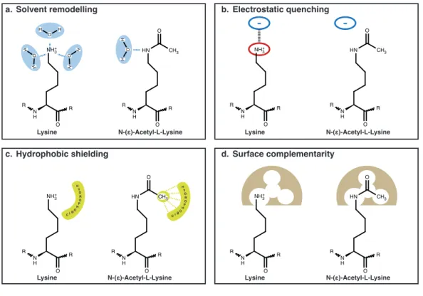
Figure 1.9: Impact of lysine acetylation.Comparison of chemical charac- teristics of lysine and acetyl-lysine regarding (a) solvent remodeling, (b) elec- trostatic and (c) hydrophobic interactions and (d) surface complementarity.
Reversible acetylation at the -amino group of lysines was first identified on his-
tones by Allfrey et al. in 1964 (Allfrey et al., 1964). It took more than twenty
years until the first non-histone target of acetylation was discovered: the cytosolic α-tubulin (L’Hernault and Rosenbaum, 1983). The tumor suppressor p53 and the HIV transcriptional regulator Tat followed another decade later (Gu and Roeder, 1997; Ott et al., 1999). By now the existence of lysine acetylation in all organ- isms, from bacteria to humans, is established knowledge (Kim and Yang, 2011;
Soufi et al., 2012).
The molecular effects of this relatively small modification on the lysine side chain are diverse and provide a broad spectrum of regulatory modes of action (Figure 1.9). The effect of electrostatic quenching might be the most dominant effect.
Acetylation of the lysine side chain neutralizes the prominent positive charge, which can entail drastic consequences for protein structure or protein-protein in- teractions, e.g. by disrupting an essential salt bridge. The altered spacial de- mand of acetyl-lysine is, furthermore, relevant for the surface complementarity of two interaction partners, e.g. enzymes and substrate. The modification can either create or disrupt a complementary interaction surface and consequently fa- cilitate/strengthen or disrupt/weaken binding, respectively. The acetylation also changes chemical characteristics of the side chain and thereby affects the hydropho- bicity and the solvent-interaction (solvent remodeling) of the lysine residue.
1.3.1 Characteristics and regulation of lysine acetylation
Lysine acetylation is a reversible post-translational modification (PTM). The dy- namics of this system originate from the existence of enzymes which generate (writers) and remove (erasers) the modification in a regulated manner. The recog- nition of the lysine acetylation by specific proteins (readers) allows for the signaling function in several cellular processes.
Lysine deacetylases
The removal of the acetyl-moiety by hydrolysis is catalyzed by lysine deacety-
lases (KDACs). There are 18 human KDACs, subdivided into four classes (KDAC
I - IV): classes I, II and IV utilize a Zn
2+-dependent mechanism, whereas KDAC
class III uses nicotinamide adenine dinucleotide (NAD
+) as a cofactor. Enzymes
of KDAC class III are referred to as sirtuins- named after the yeast homolog Sir2
(silent information regulator), which was originally identified as a regulator of
transcriptional silencing of mating-type loci, telomeres and ribosomal DNA (Shore
et al., 1984). Based on sequence-based phylogenetic analysis mammalian sirtuins
NH3+
NH R R
O
HN
NH R R
O CH3 O H3C SCoA
O
CoAS-
OH- CH3COO-
L-Lysine N-(ε)-Acetyl-Lysine
KATs
KDACs
Sirtuins
NAD+ 2‘- or 3‘-O-Acetyl-ribose
+ Nicotinamide
reader (BRD)
Figure 1.10: Regulation of lysine acetylation.Lysine-acetylation at the -amino group is a reversible post-translational modi cation which is regulated by acetyltransferases (KATs) and deacetylases (KDACs and sirtuins). Proteins with a bromodomain (BRD) can speci cally recognize and bind acetyl-lysine.
can be further subdivided into four classes (I - IV)(Table 1.4). Despite their function as transcriptional regulators, sirtuins are also regarded as energy sen- sors, since their NAD
+-dependence establishes a direct link to the cell metabolism (Imai et al., 2000). The activation of sirtuins was reported to be bene cial for metabolism-related diseases (e.g. diabetes 2, obesity), neurodegenerative diseases (Alzheimers, Parkinsons) and the stimulation of mitochondrial activity (reviewed in (Houtkooper et al., 2012)). Of the seven human sirtuins only three (sirtuins 1, 2 and 3) exhibit a decent deacetylase activity, whereas the other members of
Table 1.4: Sirtuins
Sirtuin Class Localization Activity Target Refs.
Sirt1 I Nucleus, Deacetylation FOXO1, p53, (Daitoku et al., 2011; Lee and Gu, 2013) cytosol Notch, NF- B, (Guarani et al., 2011; Kauppinen et al., 2013)
SREBP-1c (Ponugoti et al., 2010)
Sirt2 I Cytosol Deacetylation Tubulin, PEPCK, (Jiang et al., 2011; North et al., 2003) FOXO1, PAR3 (Beirowski et al., 2011; Daitoku et al., 2011) Sirt3 I Mitochindria Deacetylation GDH (Lombard et al., 2007)
SOD2, IDH2 (Qiu et al., 2010; Someya et al., 2010) Sirt4 II Mitochondria ADP-ribosylation GDH (Haigis et al., 2006)
Sirt5 III Mitochondria Deglutarylation, (Park et al., 2013) demalonylation,
desuccinylation
Sirt6 IV Nucleus Deacetylation, H3K9, H3K56 (Pan et al., 2011) demyristoylation
Sirt7 IV Nucleolus Deacetylation H3K18 (Barber et al., 2012)